Embed Size (px)
Citation preview

A critical view on hypoxia training:
horse versus human
Word count: 18532
Ward Moerman
Student number:01205631
Supervisor: Prof. dr. Catherine Delesalle
Supervisor: Drs. Berit Boshuizen
A dissertation submitted to Ghent University in partial fulfilment of the requirements for the degree of
Master of Veterinary Medicine
Academic year: 2017 - 2018

Ghent University, its employees and/or students, give no warranty that the information provided in this
thesis is accurate or exhaustive, nor that the content of this thesis will not constitute or result in any
infringement of third-party rights.
Ghent University, its employees and/or students do not accept any liability or responsibility for any use
which may be made of the content or information given in the thesis, nor for any reliance which may be
placed on any advice or information provided in this thesis.

3
Preface
I would like to express my gratitude to my promotor and co-promotor, who were very helpful by giving
feedback and with finding articles when asked. I also want to thank my family, who supported me
throughout the years and during the writing of this thesis.
.

Tabel of Content
Abbreviations ........................................................................................................................................................... 5
Summary ................................................................................................................................................................. 6
Introduction .............................................................................................................................................................. 7
Background ......................................................................................................................................................... 7
The mechanism of hypoxia training ..................................................................................................................... 7
Definition of the problem ...................................................................................................................................... 8
Goal of the study ................................................................................................................................................. 8
1 Principles of oxygen transport ......................................................................................................................... 9
1.1 Oxygen uptake ....................................................................................................................................... 9
1.1.1 Oxygen uptake in the alveoli .............................................................................................................. 9
1.1.2 Partial pressure ................................................................................................................................ 10
1.1.3 Solubility .......................................................................................................................................... 10
1.2 Oxygen transport .................................................................................................................................. 12
1.2.1 Hemoglobin ...................................................................................................................................... 12
1.2.2 Oxygen-hemoglobin affinity ............................................................................................................. 12
1.2.3 Influencing factors of the oxygen-hemoglobin affinity ...................................................................... 13
2 Oxygen transport in horses ............................................................................................................................ 16
2.1 Differences with humans ...................................................................................................................... 16
2.1.1 Hemoglobin affinity .......................................................................................................................... 16
2.1.2 The storage type spleen of the horse .............................................................................................. 16
2.2 Oxygen delivery during exercise .......................................................................................................... 17
2.2.1 The role of oxygen during perfomance ............................................................................................ 17
2.2.2 The respiratory system, a limiting factor for athletic performance in horses. ................................... 18
3 Hypoxia training in humans ........................................................................................................................... 20
3.1 The theory of altitude training ............................................................................................................... 20
3.2 Strategies ............................................................................................................................................. 21
3.2.1 Live high-train high LHTH ................................................................................................................ 21
3.2.2 Live high-train low LHTL .................................................................................................................. 21
3.2.3 Live-high, train-high and low ............................................................................................................ 22
3.2.4 Live low-train high ............................................................................................................................ 23
3.2.5 Sprint interval training in hypoxia ..................................................................................................... 23
4 Hypoxia training in horses ............................................................................................................................. 24
4.1 Introduction .......................................................................................................................................... 24
4.2 Types of hypoxia training ..................................................................................................................... 25
4.2.1 Live high train high ........................................................................................................................... 25
4.2.2 Live low-train high ............................................................................................................................ 29
5 Discussion ..................................................................................................................................................... 32
6 Conclusion ..................................................................................................................................................... 35
7 Bibliography ................................................................................................................................................... 36

5
Abbreviations
EPO: Erythropoietin
FiO2: Fraction of inspired oxygen
Hb: Hemoglobin
IET: Incremental Exercise Test
PaCO2: Arterial partial pressure of carbon dioxide
PaO2: Arterial partial pressure of oxygen
PCO2: Partial pressure of carbon dioxide
PCV: Packed Cell Volume
PO2: Partial pressure of oxygen
SET: Standardized Exercise Test
2,3-DPG: 2,3-Diphosphoglycerate

6
Summary
Hypoxia training is a well-known training strategy to enhance performance in human athletes. The
paradigm of hypoxia training is that hypoxia is induced by decreasing the partial pressure of oxygen in
the air. This can be done either due to the decreased atmosphere pressure at high altitude, or due to
an artificially decreased fractional concentration of oxygen in the air. This decreased partial pressure
induces hematological and non-hematological adaptations, resulting in a more efficient oxygen supply
to the tissues. This increased efficiency of oxygen supply enhances performance when competing in
normoxia. Hypoxia training is becoming more popular in the equine world. This raises the question
whether horses could benefit from hypoxia training. Although this is a well-known and popular strategy
in humans, there is no consensus in human science about the effectiveness of hypoxia training in all its
forms. Scientific literature, discussing hypoxia training in horses, is scarce. Little research has been
done in the past, however, the topic is becoming more popular in recent years. A major obstacle for
researchers, evaluating the hematological responses to hypoxia training in horses, is the storage of
erythrocytes in the spleen of the horse. Small effects have been detected in horses. However, the effects
are unconvincing due to equivocal results and a low number of horses, used in the studies.
Nevertheless, the small effects that were detected indicate that hypoxia training might induce changes
and even enhance performance in horses. Therefore, more research is needed with larger groups to
clarify the effects of hypoxia training.
Hypoxie training is een training strategie die wereldwijd wordt toegepast door atleten. Het paradigma
van hypoxie training is dat hypoxie wordt geïnduceerd door een lage partiële druk van zuurstof in de
lucht. Deze lage partiële druk kan bekomen worden door een verlaagde atmosferische druk, zoals op
hoge hoogte, of door het artificieel verlagen van de fractionele concentratie van zuurstof in de lucht.
Deze lage partiële druk kan hematologische en niet-hematologische veranderingen teweegbrengen die
zorgen voor een efficiëntere zuurstoftoevoer naar de weefsels. Deze efficiëntere zuurstoftoevoer werkt
prestatie verbeterend bij een competitie op lage hoogte, waar de partiële druk van zuurstof hoger ligt.
Hypoxie training wordt populairder in de paardenwereld. Hierdoor is er meer belang voor het al dan niet
werken van hypoxie bij paarden. Hoewel dit een gekende strategie is bij mensen, is er nog altijd geen
consensus in de humane wetenschap over de effectiviteit van hypoxie training in al zijn vormen. Er is
weinig wetenschappelijke literatuur gepubliceerd over hypoxie training bij paarden. Wel wordt de laatste
jaren meer wetenschappelijk onderzoek gedaan op dit onderwerp. De stapelmilt van het paard is een
groot struikelblok bij het onderzoeken van hematologische veranderingen onder invloed van hypoxie.
Er werden reeds kleine effecten gevonden bij paarden. Jammer genoeg zijn de resultaten niet
overtuigend door het verschil in resultaten tussen de studies en het lage aantal paarden dat gebruikt
werd in de studies. Desondanks suggereren de kleine verschillen die gemeten werden dat hypoxie
training effecten zou teweegbrengen en prestaties zou verbeteren. Om dit met zekerheid te kunnen
zeggen is er meer onderzoek nodig met een groter aantal paarden.

7
Introduction
Background This thesis discusses hypoxia training in both horses and humans. The goal of this study is to gain
insight in the applicability of hypoxia training to obtain better performances in horses. Hypoxia training
as a performance enhancing tool is a well-known training strategy for human athletes. It was shown that
altitude training could enhance the performance of an athlete up to 3 percent (Levine and Stray-
Gundersen., 1997; Saugy et al., 2016) although it could not be confirmed by some studies (Fudge et al.,
2012; Lundby et al., 2012; Siebenmann et al., 2012; Saugy et al., 2016). However, in equine sports
these methods have not been used frequently and there is little scientific research on this topic. If
hypoxia training of horses can lead to better sport performance, it could be a very interesting addition to
their training program, in particular in top sports where the difference between winning or losing is very
small.
The mechanism of hypoxia training The concept of hypoxia- or altitude training is to use the variety of adaptations to hypoxia to improve the
performance of the athlete (Wickler and Anderson; 2000; Fudge et al., 2012; Lundby et al., 2012;
Siebenmann et al., 2012; Hespel, 2014; Saugy et al., 2016). Hypoxia can be induced by decreasing the
partial pressure of oxygen in the inspired air (Klein, 2013).
The uptake of oxygen in blood can be separated in two segments. A small amount of oxygen dissolves
in blood plasma as a result of Henri’s law but the major part of oxygen uptake in blood can be attributed
to the chemical binding to hemoglobin in red blood cells (van Oosterom and Oostendorp, 2008). The
maximal oxygen capacity of blood is mostly determined by the maximal saturation of hemoglobin
(Ainsworth, 2004; Kingston, 2008; van Oosterom and Oostendorp, 2008; Klein, 2013). Oxygen
saturation in blood is determined by the partial pressure of oxygen. This relationship of oxygen saturation
and partial pressure can be shown in the oxyhemoglobin dissociation curve. (van Oosterom and
Oostendorp, 2008; Klein, 2013)
Partial pressure of oxygen in a dry gas mixture is determined by two factors. The first factor is the fraction
of oxygen in the gas mixture. The atmosphere contains 21 percent oxygen, these fractions are equal at
sea-level and at high altitude (Klein, 2013). Therefore, this is not the mechanism of classical high-altitude
training. However, some hypoxia training devices decrease the fraction of oxygen by increasing the
amount of nitrogen dioxide in the inspired air (Hespel, 2014). The second factor that determines the
partial pressure, is the barometric pressure. The barometric pressure is the result of the density of
molecules in the air. If the barometric pressure is low, the oxygen molecules are less densely packed in
the air which results in a low partial pressure of oxygen (Klein, 2013). The barometric pressure
decreases with increasing altitude. This mechanism is used when training at high altitude.
The most important adaptation of the body to high altitude is the increase of erythropoietin production
by the kidneys. Erythropoietin stimulates the red blood cell production which results in an increased
packed cell volume. Other adaptations are a higher blood volume, improved oxygen diffusion between
alveoli and blood, increased muscle myoglobin content, increased capillarity in muscles and a higher
number of mitochondria and mitochondrial enzymes in myocytes (Hespel, 2014). All of these
adaptations result in an increased oxygen transport in the (human) body. Improved oxygen transport
results in better performance due to an increased stamina. Endurance athletes benefit the most from
these adaptations, since they mainly rely on aerobic muscle metabolism (Hespel, 2014).
However, the classical high-altitude training seems to be outdated. Research has shown that this kind
of training, where the athlete trains and lives at high altitude is not the most effective way to improve the
performance. Recent studies revealed that the method of ‘living high-training low’ is much more
effective, since the method of ‘living high-training high’ reduces the quality of the training (Stray-
Gundersen et al., 2014). At high altitude there is a lower arterial oxygen pressure and the maximal heart
rate decreases. This decreases oxygen transport which results in a lower quality of training. The athlete
is not able to train at the same intensity as he would be training at sea-level and the lower quality of
training results in a detraining-effect (Levine and Stray-Gundersen; 1997). Therefore, the living high-
training low concept is now a widespread method amongst human athletes all over the world.

8
Definition of the problem A possible explanation for the lack of interest in hypoxia training of horses is the practical implication of
altitude training with equine athletes. First and foremost, there are not many race tracks or equine
training centers at altitude. Secondly, the adaptations are temporary so the horses should revisit these
centers after a certain period to maintain the benefits.
Since the discovery of the ‘living high-training low’ method there has been a revolution in hypoxia masks,
hypoxia tents and hypoxia rooms for human athletes (Brocherie et al., 2017). This has made hypoxia
training an option for many athletes in addition to their normal training regime. Furthermore, there are
hypoxia masks and hypoxic rooms available for horses now as well. It is of course imperative to provide
scientific evidence of the effectiveness of these tools.
There are several difficulties when studying hypoxia training in horses. In contrast to humans, the horse
has a spleen which contains a storage of red blood cells. These cells are released during stress or
exercise by a splenic contraction, which can double the amount of circulating red blood cells. The
amount of red blood cells that is released by the spleen shows a high variation. The amount of release
depends on the sympathetic activity. Also, the splenic storage capacity varies between horse breed and
age. Because of the sympathetic influence on the splenic release, it is difficult to interpret hematocrit
levels of horses “at rest”. The level of excitation may vary between different horses at rest. The horse
could show an increased excitability as it gets fitter what results in a higher resting hematocrit. (Wickler
and Anderson, 2000; Greene et al., 2006; Kingston, 2008; Mckeever et al., 2011)
Therefore, it is difficult to use resting hematocrit levels to follow up the effect of altitude training on
hematologic parameters and performance capacity. Researchers have been trying to avoid this difficulty
by using heart rates and lactate levels to approximate the same metabolic effort between the various
performance tests (Wickler and Anderson, 2000).
Another difficulty in studying hypoxia training in horses is the measurement of fitness of the horse. The
standard assessment of fitness, more specifically aerobic capacity, in human athletes is to measure the
maximal oxygen consumption or VO2max. For horses there is no accurate system to measure the
maximal oxygen consumption on the race tracks. However, it can be measured using a high-speed
treadmill. But, apart from the more standardized environment and easy access for various
measurements, the treadmill may not replicate the physiologic responses to field exercise (Evans,
2008).
Goal of the study The goal of this study is to describe the effect of hypoxia training on the performance of sports horses.
The hematological and muscle adaptations of horses subjected to altitude training will be described and
compared to the effects found in human athletes. Practical implications such as the minimum altitudes
and duration of exposure to hypoxic conditions required to show a significant improvement of
performance will be studied in this thesis. However, not much research has been published on these
topics about horses. Therefore, extrapolating from human research will be necessary. I want to provide
a critical view on the research that has been performed on hypoxia training. I find it is very important to
be critical on these studies and not to accept the outcome of a few studies as the definite truth. Finally,
I will give my opinion on how further research should be performed and how it would improve the
knowledge on hypoxia training in the horse.

9
LITERATURE REVIEW
1 Principles of oxygen transport
1.1 Oxygen uptake
1.1.1 Oxygen uptake in the alveoli
The uptake of oxygen in the alveoli occurs by diffusion through the respiratory membrane (Figure 1)
The rate of diffusion is affected by the concentration gradient, surface area, membrane permeability and
membrane thickness (Silverthorn and Johnson, 2010; Robinson, 2013). This can be described in Ficks
law: rate of diffusion = (surface area x concentration gradient x membrane permeability)/ membrane
thickness. In general, the surface area, membrane permeability and membrane thickness are more or
less constants in the body. These are optimized to get a high diffusion rate. Therefore, the concentration
gradient is the most important force that drives the diffusion of gases, mostly oxygen and carbon dioxide,
in the alveoli (Silverthorn and Johnson, 2010). The concentration gradient of a gas is affected by the
partial pressure and the solubility of the gas (Germann and Stanfield, 2001).
Figure 11
1 http://droualb.faculty.mjc.edu/Course%20Materials/Physiology%20101/Chapter%20Notes/Fall%202007/chapter_17%20Fall%202007%20Phy%20101.htm last consulted at 20/5/2018

10
1.1.2 Partial pressure
Air is a mixture of gases. The total pressure of air, being 760 mm Hg at sea level with zero humidity, is
determined by the sum of the pressures of the individual gases in the air (Germann and Stanfield, 2001;
Robinson, 2013). This individual pressure of a gas is called the partial pressure. The partial pressure is
determined by the percentage of the gas in relative to the total quantity of the gas mixture, the fractional
concentration, and the total pressure of the gas mixture (Germann and Stanfield, 2001; Robinson, 2013).
One can calculate the partial pressure of oxygen in the air at sea level and zero humidity by multiplying
the fractional concentration, i.e. 21%, and the total air pressure, i.e. 760 mm Hg. 0,21x760 mm Hg= 160
mm Hg (Germann and Stanfield, 2001; Robinson, 2013).
When air arrives in the alveoli, it has a humidity of 100 % at a temperature of 37 °C. Therefore, the
partial pressure of water is 47 mm Hg. Because of this increase of partial pressure of water, the partial
pressures of the other gases decrease. So, the PO2 in the alveoli goes down to 152 mm Hg. The actual
PO2 in the alveoli is around 100mm Hg. This is because there is constant exchange between blood and
the alveoli and because the air entering the alveoli is a mixture of fresh air and less oxygenated air that
was in the conducting zone (Germann and Stanfield, 2001; Robinson, 2013). At high altitudes,
atmosphere pressures are low, resulting in a decreased partial pressure of oxygen. Conversely, the
partial pressure of oxygen can be artificially reduced by fractional concentration of oxygen. This can be
done by extracting oxygen from the air, or by adding another gas to the air, such as nitrogen (Germann
and Stanfield, 2001; Gore et al., 2007; Robinson, 2013).
1.1.3 Solubility
Oxygen and carbon dioxide are exchanged between the air in the alveoli and the blood in the capillaries.
When a gas and a liquid are in contact with each other, gas will dissolve in the liquid or will leave the
liquid to find an equilibrium. At this equilibrium, the gas molecules in the liquid and the ones in the air
have the same partial pressure. Germann and Stanfield (2001) give the example of opening a bottle of
soda, where the gas leaves the soda when opening because the pressure of the air is much lower than
the pressure used when bottling the soda. The molecules dissolve in the liquid at the same rate as the
molecules that move from the liquid to the gaseous state. Although the partial pressures are equal, that
does not mean that the concentrations are equal (Germann and Stanfield, 2001). This is because some
gases dissolve more easily than others in a given liquid. This relation between concentration, solubility
and partial pressure can be explained with Henry’s Law: c=kP where c is the molar concentration of the
dissolved gas, P is the partial pressure of the gas and k is Henry’s Law constant (experimentally
determined). k is a constant, so the relationship between the concentrations of a gas at two different
pressures can be described as c1/P1=c2/P2. Therefore, the concentration of a gas in a liquid is directly
related to the partial pressure of that gas. Thus, the concentration gradient that determines the rate of
diffusion according to Fick’s Law can be replaced by the partial pressure gradient (Germann and
Stanfield, 2001).
The exchange of gases in the alveoli and in the tissues, is driven by the partial pressure gradient of this
particular gas (Figure 2). Each gas will diffuse from an area of high partial pressure (or high
concentration) to an area with a low partial pressure (low concentration). The PO2 in the alveoli is around
100mm Hg. Blood in the pulmonary arteries has a PO2 of around 40 mm Hg. So, when blood passes
the alveoli, oxygen diffuses down its partial pressure gradient to the blood. Therefore, the PO2 of blood
in the pulmonary veins is around 100 mm Hg. The opposite occurs with carbon dioxide. PCO2 in the
alveoli is 40mm Hg and the PCO2 in the blood entering the pulmonary capillaries is 46 mm Hg. As a
result, CO2 diffuses from the blood to the alveoli down its partial pressure gradient (Germann and
Stanfield, 2001).

11
Figure 22
The equilibration of blood and alveolar air takes around 0,25 seconds (with normal respiratory
membrane thickness). By that time, blood has traveled only one-third of the length of a capillary,
providing a margin of safety. For example, when exercising, the blood flow is much faster, so there is
less time to equilibrate. Even if the blood flows three times faster, there is still full equilibration (Germann
and Stanfield, 2001; Robinson, 2013).
2 http://droualb.faculty.mjc.edu/Course%20Materials/Physiology%20101/Chapter%20Notes/Fall%202007/chapter_17%20Fall%202007%20Phy%20101.htm last consulted at 20/5/2018
12
1.2 Oxygen transport
1.2.1 Hemoglobin
The driving force that makes oxygen move from the alveolar space to the blood is the pressure gradient
(Rhoades et al., 1995). Despite the fact that there is full equilibration between the alveolar PO2 and the
blood PO2, there is as little as 3ml oxygen dissolved in 1 liter of blood. Germann and Stanfield (2001)
state that cardiac output should be around 83l/min to provide the tissues of sufficient amounts of oxygen
where a normal cardiac output of a human is approximately 5l/min. Arterial blood contains around 200
ml of oxygen per liter of blood. That means that only 1,5 percent (3ml/L) of the transported oxygen is
dissolved in blood. The other 98,5 percent is bound to hemoglobin that is found in the cytoplasm of
erythrocytes. So, the body is dependent on oxygen that is bound to hemoglobin (Germann and Stanfield,
2001; Silverthorn and Johnson, 2010; Robinson, 2013).
Hemoglobin is a protein that is made of four protein subunits which are all bound to a heme group.
These heme groups are porphyrin molecules that contain an iron molecule. These irons are the biding
sites for oxygen. Each group can bind one oxygen molecule, so one molecule of hemoglobin can carry
four molecules of oxygen. Oxygen is bound reversibly to hemoglobin. When the complex is bound to
oxygen, it is called oxyhemoglobin. A hemoglobin complex without oxygen bound on it is called
deoxyhemoglobin. When all binding sites are occupied with oxygen, the hemoglobin is saturated
(Germann and Stanfield, 2001; Silverthorn and Johnson, 2010; Robinson, 2013).
1.2.2 Oxygen-hemoglobin affinity
The amount of oxygen that binds to hemoglobin depends on the PO2 in the surrounding fluid, i.e. the
PO2 of the plasma that is surrounding the erythrocytes. The reaction of oxygen with hemoglobin can be
written as Hb+ O2 Hb∙O2, where Hb is deoxyhemoglobin, and Hb∙O2 is oxyhemoglobin. The law of
mass action states that an increase in the concentration of the reactans, drives the reaction to the right.
Conversely, a decrease of reactans drives the reaction to the left. As the reaction follows the law of
mass action, there is more oxyhemoglobin formed when the oxygen concentration increases. As
mentioned above, there is a direct correlation between concentration of oxygen and PO2. As the PO2 in
the pulmonary capillaries is high, there is a high amount of oxyhemoglobin formed so there is more
saturation of hemoglobin (Germann and Stanfield, 2001; Silverthorn and Johnson, 2010; Robinson,
2013).
This correlation between hemoglobin saturation and oxygen can be displayed in the hemoglobin-oxygen
dissociation curve (Figure 3). Although saturation levels of hemoglobin increase when the PO2 rises,
there is no linear relationship between the two. When PO2 rises linear, saturation increases in a
sigmoidal way. The reason why the saturation does not rise in a linear way, is because the affinity of
hemoglobin for oxygen depends on how many oxygen molecules are bound to the hemoglobin. When
an oxygen molecule binds to one of the subunits of hemoglobin, there is a conformational change in the
hemoglobin molecule that increases the affinity of hemoglobin to oxygen. Therefore, it increases the
likelihood that other oxygen molecules will bind with hemoglobin (Germann and Stanfield, 2001). At very
low PO2 levels, there are almost no oxygen molecules bound to hemoglobin. When the PO2 increases,
more hemoglobin molecules have at least one oxygen molecule bound to itself. This causes an increase
in affinity for other oxygen molecules resulting in a steep part of the hemoglobin-oxygen dissociation
curve. This steep section of the curve can be observed at PO2 values between 15 mm Hg and 60 mm
Hg (Germann and Stanfield, 2001).

13
The physiological PO2 in systemic veins which is around 40 mm Hg lies in this section of the curve at
this level, saturation is around 75 percent. Once the PO2 is higher than 60 mm Hg, the slope of the curve
flattens because there are fewer binding sites available. Around 80 mm Hg, the slope becomes almost
flat. At normal alveolar and arterial PO2 (100 mm Hg), hemoglobin is 98 percent saturated (Silverthorn
and Johnson, 2010). This means that, as blood flows through the pulmonary capillaries, the hemoglobin
picks up nearly the maximum amount of oxygen that it can carry. We can also conclude that, as the
saturation of systemic veins lies around 75 percent, that respiring tissues in the body take up about 25
percent of oxygen, leaving a large reserve of oxygen to supply the needs (Germann and Stanfield,
2001).
Fig.3, from: (van Oosterom and Oostendorp, 2008)
1.2.3 Influencing factors of the oxygen-hemoglobin affinity
There are factors that affect the affinity for of hemoglobin for oxygen (Figure 4). All factors that change
the conformation of the hemoglobin protein can affect its ability to bind oxygen. A decrease in affinity
indicates that a higher PO2 is required to achieve any given level of saturation (Germann and Stanfield,
2001). This also means that oxygen is unloaded more easily from hemoglobin and therefore making it
more available for the respiring tissues (Silverthorn and Johnson, 2010). A Decrease causes the curve
to shift rightward. An increase in affinity causes the curve to shift leftwards meaning that oxygen is
loaded more easily onto hemoglobin (Germann and Stanfield, 2001).
Increased temperature, increased PCO2 or decreased pH all work to improve oxygen unloading from
hemoglobin in the respiring tissues, they decrease the affinity of hemoglobin for oxygen by shifting the
hemoglobin-oxygen dissociation curve to the right. The opposite is true for decreased temperature,
decreased PCO2 and increased pH (Germann and Stanfield, 2001; Silverthorn and Johnson, 2010;
Robinson, 2013; Hubbel and Muir, 2014).

14
.
Fig. 4, from: (Hubbel and Muir, 2014)
1.2.3.1 Temperature:
As an increased temperature affects the tertiary structure of proteins, it also changes the structure of
hemoglobin. Therefore, in tissues with high metabolic activity, the temperature rises. Thus, there is a
decreased affinity between oxygen and hemoglobin, increasing the unloading of oxygen. More oxygen
is unloaded in highly active tissues. Conversely, as blood flows through the lungs, the temperature
drops, resulting in a increase in affinity which means more oxygen loading (Germann and Stanfield,
2001; Silverthorn and Johnson, 2010; Robinson, 2013; Hubbel and Muir, 2014).
.
1.2.3.2 Ph, the Bohr effect
When oxygen binds to hemoglobin, certain amino acids in the protein release hydrogen ions. Hb+O2
Hb∙O2 + H+
Considering the law of mass action, if there is an increase in concentration of hydrogen ions, the reaction
is pushed to the left. This means that more oxygen is dissociated from hemoglobin, even when PO2 is
constant (Germann and Stanfield, 2001). The Bohr effect is important because hydrogen ions tend to
increase in active tissue, which demands more oxygen (Germann and Stanfield, 2001; Silverthorn and
Johnson, 2010; Robinson, 2013; Hubbel and Muir, 2014).
.
1.2.3.3 PCO2 The carbamino effect
Carbon dioxide reacts reversibly with amino groups in hemoglobin, forming carbaminohemoglobin.
Hb+CO2 HbCO2. Again, the law of mass action states that when the concentration of CO2 increases,
the reaction will be pushed to the right, forming more carbaminohemoglobin. Increase of CO2
concentration occurs when there is high metabolic activity in the tissues. The binding of carbon dioxide
to hemoglobin changes the conformation of hemoglobin, reducing the affinity for oxygen. This is called

15
the carbamino effect (Germann and Stanfield, 2001; Silverthorn and Johnson, 2010; Robinson, 2013;
Hubbel and Muir, 2014).
.
1.2.3.4 2,3-diphosphoglycerate
2,3-DPG is produced in erythrocytes from an intermediate of the glycolysis pathway. When there are
high concentrations of oxyhemoglobin in the erythrocytes, the production of 2,3-DPG is suppressed by
inhibiting the enzyme that forms 2,3-DPG. However, when oxyhemoglobin levels are low, 2,3-DPG
synthesis increases. This occurs with chronic hypoxia due to high altitudes and anemia. 2,3-DPG
decreases the affinity of hemoglobin for oxygen. Thus, decreasing the affinity of hemoglobin for oxygen
enhances the unloading of oxygen, which is needed in tissues that suffer from chronic hypoxia (Wickler
and Anderson, 2000; Germann and Stanfield, 2001; Greene et al., 2006; Silverthorn and Johnson, 2010;
Robinson, 2013; Hubbel and Muir, 2014).
16
2 Oxygen transport in horses
2.1 Differences with humans
2.1.1 Hemoglobin affinity
The general principles of oxygen exchange and oxygen transport are the same for horses as for other
mammalians. Although there are differences. The composition of the globin proteins in hemoglobin
differs between species. These globins are composed of particular types and sequences of amino acids.
The type and sequence of the amino acids define the different types of hemoglobin. The composition of
globin is critical in the oxygen binding (Robinson, 2013).
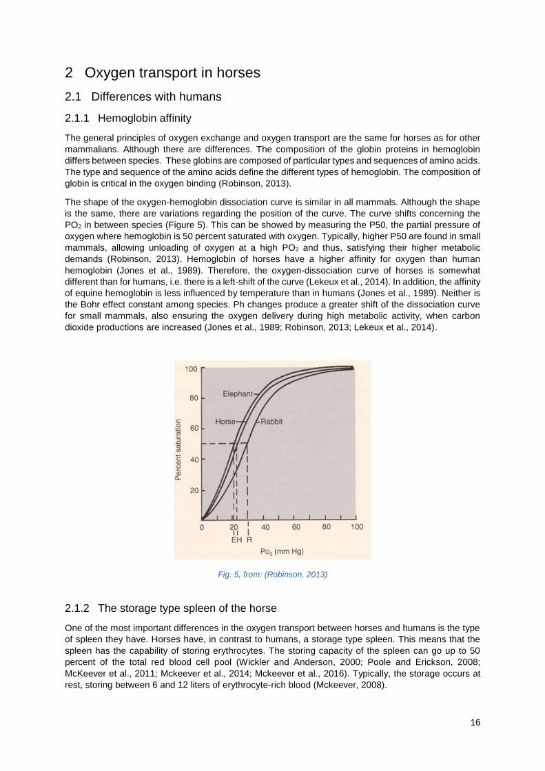
The shape of the oxygen-hemoglobin dissociation curve is similar in all mammals. Although the shape
is the same, there are variations regarding the position of the curve. The curve shifts concerning the
PO2 in between species (Figure 5). This can be showed by measuring the P50, the partial pressure of
oxygen where hemoglobin is 50 percent saturated with oxygen. Typically, higher P50 are found in small
mammals, allowing unloading of oxygen at a high PO2 and thus, satisfying their higher metabolic
demands (Robinson, 2013). Hemoglobin of horses have a higher affinity for oxygen than human
hemoglobin (Jones et al., 1989). Therefore, the oxygen-dissociation curve of horses is somewhat
different than for humans, i.e. there is a left-shift of the curve (Lekeux et al., 2014). In addition, the affinity
of equine hemoglobin is less influenced by temperature than in humans (Jones et al., 1989). Neither is
the Bohr effect constant among species. Ph changes produce a greater shift of the dissociation curve
for small mammals, also ensuring the oxygen delivery during high metabolic activity, when carbon
dioxide productions are increased (Jones et al., 1989; Robinson, 2013; Lekeux et al., 2014).
Fig. 5, from: (Robinson, 2013)
2.1.2 The storage type spleen of the horse
One of the most important differences in the oxygen transport between horses and humans is the type
of spleen they have. Horses have, in contrast to humans, a storage type spleen. This means that the
spleen has the capability of storing erythrocytes. The storing capacity of the spleen can go up to 50
percent of the total red blood cell pool (Wickler and Anderson, 2000; Poole and Erickson, 2008;
McKeever et al., 2011; Mckeever et al., 2014; Mckeever et al., 2016). Typically, the storage occurs at
rest, storing between 6 and 12 liters of erythrocyte-rich blood (Mckeever, 2008).

17
Exercise induces splenic contraction which results the release of erythrocytes in the blood. This
mobilization of erythrocytes increases the oxygen transport capacity. The splenic contraction is
mediated by catecholamines and occurs very rapidly during exercise, with the splenic content being
mixed with the central circulation within 1 to 1,5 minutes of exercise (Mckeever, 2008). The level of the
catecholamine response is determined by the intensity and duration of exercise. This means that the
level of the release is determined by the intensity and duration of exercise. However, there are variations
concerning the capacity of the spleen in association with breed and age of the horse. Draught horses
tend to have a lower relative splenic weight compared to Thorougbred horses. Furthermore, the splenic
capacity changes with increasing age (from one to three years old) (Kingston,2008). This mechanism
results in an increase in oxygen transport capacity, an important factor in the high aerobic capacity of
the horse (Kingston, 2008). This has been shown in studies where horses had a reduced exercise
capacity after being splenectomized (Kingston, 2008).
2.2 Oxygen delivery during exercise
2.2.1 The role of oxygen during perfomance
Maximal oxygen uptake, maximal oxygen consumption or aerobic capacity (VO2max) is used within
humans as an indicator of performance. It is the maximal amount of oxygen that is consumed during an
incremental exercise test. The aerobic capacity is limited by oxygen supply because the mitochondrial
oxidative enzyme capacity that uses oxygen exceeds the oxygen delivery capacity of the
cardiorespiratory system. Therefore, the maximal oxygen uptake will be increased by enhancing the
oxygen delivery to the muscles (Poole and Erickson, 2008). The pathway of oxygen, starting in the
atmosphere and ending in the muscle mitochondria, takes a set of steps that adapt themselves to the
increased demand for oxygen. To achieve high performance, there must be a close coordination
between respiratory, cardiovascular and muscle systems. This close synergy between these systems
endeavor to deliver oxygen as efficient as possible to the muscle mitochondria (Poole and Erickson,
2008).
The components that determine the oxygen delivery can be written down in the formula: oxygen
delivery=heart rate x stroke volume x concentration of hemoglobin x oxygen-binding capacity of
hemoglobin x % of oxygen binding sites filled+ oxygen dissolved in plasma. This formula can be
simplified to: Oxygen delivery= Cardiac output x arterial oxygen content. (Poole and Erickson, 2008).
The cardiovascular system adapts by increasing the cardiac output and redistributing the circulation
both in muscles and in the lungs (Poole and Erickson, 2008). As mentioned above, the horse is a natural
blood doper. During exercise the spleen releases erythrocytes almost doubling the amount of red blood
cells in the blood (Poole and Erickson, 2008).
Delivery in the tissues depends on the release of oxygen from erythrocytes. The extraction of oxygen
from the blood can increase by four times during exercise. This increase is achieved by a rightward shift
of the oxygen-hemoglobin dissociation curve. This shift is a result of the acidosis (Bohr-effect),
hypercarbia (carbamino-effect) and hyperthermia in the muscle environment. There is also an increase
in 2,3-DPG during exercise because the low PO2 in the muscle environment stimulates glycolysis in the
erythrocytes (Kingston, 2008). The increased extraction of oxygen helps the blood to transport more
carbon dioxide. This is because more extraction means more deoxyhemoglobin, meaning more
formation of carbaminohemoglobin (Kingston, 2008).
It is clear that there is a strong correlation between the total amount of erythrocytes present in the horse
and performance (Levine and Stray-Gundersen, 1997; Mckeever, 2008; Fudge et al., 2012; McGowan
and Hodgson, 2014; Mckeever et al., 2016). However, to have optimal oxygen delivery to the tissues it
is not only the number of erythrocytes that is the important factor. Next to red cell volume, plasma volume
is also part in the optimization of oxygen delivery. Oxygen uptake and delivery are dependent on the
optimal number of red blood cells but also on the optimal volume to insure cardiac filling pressure. Red
cell volume and plasma volume also affect the blood flow during exercise. Too many red blood cells and
not enough plasma can cause changes in blood viscosity which can change blood flow and therefore
oxygen delivery (Mckeever, 2008).

18
2.2.2 The respiratory system, a limiting factor for athletic performance in horses.
Oxygen transport and exchange are key factors during exercise. The metabolic demands of the horse
can rise more than 30-fold relative to resting conditions during high-intensity exercise (Ainsworth, 2008).
Oxygen consumption, VO2, in rest lies around 4-5ml/kg/min. The average maximum oxygen
consumption in standardbreds performing incremental exercise tests lies around 138ml/kg/min, whereas
in Thouroughbreds the maximal oxygen consumption lies around 142ml/kg/min with individual records
going up to 190ml/kg/min (Ainsworth, 2008). As exercise intensity increases, the output of the respiratory
and cardiovascular systems increases to meet the high metabolic demands (Ainsworth, 2008). Several
researchers found that the respiratory system could be a limiting factor for maximal performance in
horses whereas in other mammalians the limiting factor is the cardiovascular or musculoskeletal system.
More specifically in humans, it is the cardiovascular system that is the primary limiting factor (Poole and
Erickson, 2008; Franklin et al., 2012; Lekeux et al., 2014). Poole and Erickson (2008) state that horses
have been subjected to selective breeding for several thousands of years based upon athletic
performances. They states that this has produced a disproportionate increase in the horse’s heart size
and pumping capacity compared to the capacity of the lungs. In other words, the cardiovascular and
musculoskeletal systems are able to transport and utilize more oxygen than the respiratory system can
provide (Poole and Erickson, 2008; Franklin et al., 2012).
2.2.2.1 Exercise-induced arterial hypoxemia
Athletic horses experience arterial hypoxemia and desaturation during maximal exercise, developing a
significant alveolar to capillary oxygen pressure gradient (Poole and Erickson, 2008). Therefore, arterial
pressures of oxygen can drop below the normal PO2 of 60 mmHg. On the other hand, PCO2 can rise
above 60 mm Hg. This phenomenon does not seem to occur in ponies or other domestic species
(Franklin et al. 2012). In man, alveolar hyperventilation drives arterial PCO2 below resting levels and
alveolar PO2 elevates. Whereas in horses, alveolar PO2 may fall during maximal exercise (Poole and
Erickson, 2008). However, not all horses seem to be disadvantaged with this decrease in PaO2, in
particular those with a low maximal oxygen uptake (VO2max). Exercise-induced arterial hypoxemia does
not occur in the average human. Though, it occurs in elite athletes presumably due to the superior
cardiovascular and musculoskeletal systems in highly trained athletes and the fact that the respiratory
system is unable to respond to the high oxygen demands during high-intensity exercise (Franklin et al.,
2012). However, the hypoxemia is not as substantial as in horses. The arterial hypoxemia is the result
of deficient gas exchange. There is an increase in difference between alveolar and arterial PO2. In
humans, the hypoxemia can be attributed to ventilation-to-perfusion (Va/Q) mismatching, exercise
diffusion limitation during maximal intensity and intra-pulmonary shunting of blood. Whereas in horses,
ventilation-to-perfusion mismatch is presumed to be not as significant for the EIAH. Intra-pulmonary
shunting has, in contrast to humans where it can contribute to EIAH, not been demonstrated in horses
(Poole and Erickson, 2008; Franklin et al., 2012; Lekeux et al., 2014).
2.2.2.2 The limiting factors
2.2.2.2.1 Erythrocyte transit time
In the horse, diffusion limitation appears to be the major cause. As mentioned above, the equilibration
of blood and alveolar air takes around 0,25 seconds for humans. By that time, blood has traveled only
one-third of the length of a pulmonary capillary, providing a margin of safety. In exercise, the transit time
can be reduced significantly. Studies in ponies showed that transit times during exercise were 0,35
seconds (Poole and Erickson, 2008; Lekeux et al., 2014).These values are not exactly known for horses
but it is predicted that, considering the high cardiac output, the time would be even shorter, creating an
incomplete equilibration between the arterial and alveolar PO2. The average transit time for a red blood

19
cell within the pulmonary capillary in a horse at rest is estimated between 0,75 and 1 second. This time
is presumed to be three to four times longer than necessary to equilibrate (Poole and Erickson, 2008).
The cardiac output of a horse can increase up to 13-fold during maximal exercise. This increase induces
a small increase in capillary volume but the main result is the decrease in capillary transit time. Transit
times during exercise are estimated from 0,3 seconds to 0,5 seconds. Poole and Erickson (2008) state
that this times probably are an overestimation, bearing in mind that elite race horses can reach extreme
cardiac output values around 400 L/min. They also explain that there will be a population of cells that
have a shorter transit time and therefore be undersaturated. Because of the shape of the dissociation
curve, it is not possible for the normally saturated cells to compensate for these undersaturated cells.
This mixing of normoxemic and hypoxemic blood results in arterial hypoxemia (Poole and Erickson,
2008).
2.2.2.2.2 Oxygen-hemoglobin dissociation curve right-shift
The equilibration is also affected by the rightward-shift of the oxygen-hemoglobin dissociation curve.
This reduced oxygen-hemoglobin affinity is caused by elevated blood temperatures, acidosis and arterial
hypercapnia. Another factor is called alveolar hypoventilation: The arterial hypercapnia (exceeding 65
mmHg) causes an alveolar PO2 fall from 100mm Hg to 90mm Hg (Poole and Erickson, 2008; Franklin
et al., 2012).
2.2.2.2.3 Blood-gas barrier
Another factor that can affect the equilibration is the thickening of the blood-gas barrier (such as
interstitial edema during exercise). Evidence to support this theory is conflicting, both in man as in horse
(Poole and Erickson, 2008).
2.2.2.2.4 Respiratory frequency
Exercise induces the ventilatory pump to increase its minute ventilation (tidal volume x respiratory
frequency). At rest, the average minute ventilation of a well-trained horse is about 80 L/min. During
heavy exercise, the minute ventilation may reach values of 1800L/min (Lekeux, 2014). Respiration and
locomotion are imperatively connected to each other in galloping horses. Average step and respiratory
frequencies lie around 110 to 130 per minute, whereas at rest frequencies are around 10-15 (Franklin
et al., 2012; Lekeux et al., 2014). As the respiration is obligatory synchronous with stride frequency, it
was thought that this might be responsible for hypoventilation of the alveoli due to shortened inspiratory
and expiratory times. At maximal stride frequency, the increase of minute ventilation depends on the
increase in tidal volume which is not tightly coupled to stride frequency (Franklin et al., 2012; Lekeux et
al., 2014). Franklin et al. (2012) suggests that “during strenuous exercise, further increase of the tidal
volume would result in an increase in the work of breathing. There would be a critical level of ventilation
above which any further increase in oxygen uptake would be consumed by the respiratory muscles
resulting in a metabolic cost.”

20
3 Hypoxia training in humans
Altitude training or hypoxia training is a training strategy that is commonly known worldwide. Many elite
athletes use it to improve their sports performance. The fact that it is used all over the world suggests
that this strategy is accepted as a performance enhancer. However, it is not generally accepted in the
scientific world. As there is little clear and rigorous scientific evidence for the benefits of altitude training,
the concept stays disputable.
3.1 The theory of altitude training
The practice of increasing the hematocrit by blood doping or erythropoietin (EPO) injections has been
illegally used in sports and has been examined by scientists since the late 1960’s. There is scientific
evidence that red cell mass and blood volume affects the exercise capacity. Researchers found that
VO2max increased by 5 to even 9 percent by transfusion of homologous red blood cells (Eichner, 2007).
Besides blood transfusion, the administration of erythropoietin has been analyzed by scientists.
Administration of EPO increased the hematocrit and increased the VO2 max by 7 percent. This benefit
lasted up to three weeks after the last administration of EPO (Eichner, 2007). As these methods are
banned, athletes and coaches search for a legal way to increase their hematocrit, i.e. high- altitude
training.
As told in the previous chapter, the partial pressure of oxygen is determined by the barometric pressure
and the fraction of oxygen in the gas mixture. At higher altitudes, the barometric pressure decreases.
Therefore, the PO2 decreases. Gore et al. (2007) define the different altitudes as follows: sea level= 0-
1000m, Low altitude= 1000-2000m, moderate altitude= 2000-3000, high altitude= 3000-5000m and
extreme altitude= 5000-8848m. The altitudes that are traditionally used for hypoxia training are between
1500 meters and 3000 meters i.e. low to moderate altitude (Gore et al., 2007). As the PO2 decreases in
the alveoli, the PO2 will also be lower in blood after equilibration. Due to the decreased PO2 in blood,
the saturation of hemoglobin will be lower. This results in a decrease in oxygen transport. The body will
be stressed by the hypoxia resulting from the high altitude. The hypoxia is thought to trigger the
erythropoietin production in the kidneys. This increase in EPO stimulates the production of erythrocytes
in the bone marrow (Gore et al., 2006). This means an increased oxygen-carrying capacity (Levine and
Stray-Gundersen; 1997). As described in chapter 2, the aerobic capacity VO2max can be increased by
enhancing the oxygen delivery to the muscles. Thus, when returning to sea-level, the performance is
improved. Also in competitions at high altitude the athlete would be in advantage because of the
adaptations. This is considered to be the main mechanism. Although, there might be a number of
physiological adaptations. Oxygen extraction and substrate utilization in the skeletal muscles might be
ameliorated by biochemical and structural adaptations (Levine and Stray-Gundersen; 1997). Besides
the increase in erythrocyte production and muscle efficiency, there are other factors that have been
proposed such as angiogenesis and improved buffering capacity (Constantini et al., 2017).
Levine and Stray-Gundersen (1997) found that living at an altitude of 2500 meters for four weeks
stimulated the erythropoietin production which increased the red blood cell mass volume by ten percent.
This ten percent is comparable to the percentages found in the studies which analyzed the effect of
erythrocyte infusion. They conclude that the “endogeneous erythrocyte infusion” is at least partially
responsible for the improvement in maximal aerobic power, given the significant but loose correlation
they found between the increase in VO2max, the increase in red blood cell mass and hemoglobin
concentration. As already mentioned before, the scientific evidence is controversial (Gore et al., 2007).
Hahn et al. (2001) state in their study that most of the controlled studies that have been done fail to
observe a positive effect. However, at high level competitions a small improvement of performance can
mean a huge difference in the results. Gore et al. (2007) mentions that an improvement of only 0,5
percent is needed for top athletes to increase their chance of winning medals at an international
competition. As most studies are done with less than twenty athletes, there is a small chance to detect
such low magnitude changes (Gore et al., 2007).

21
3.2 Strategies
3.2.1 Live high-train high LHTH
This is the classical altitude training and the first strategy that was used. At the Olympic games in 1968
in Mexico City (2225m), East African athletes, who won of most of the endurance foot footraces, were
said to have an advantage to other athletes because of their training habits. They used to live and train
at moderate altitudes and therefore they were acclimatized to the high altitude (Eichner, 2007; Lundby
et al., 2012). Apart from many anecdotal reports of elite athletes who used this method during training
periods, there are little good controlled studies that investigates the effects of LHTL. Moreover, the
studies that are done include small numbers of subjects (Lundby et al., 2012).
In the 1970’s there was a study published that according to Lundby et al. (2012) is “the seemingly best
controlled, but largely ignored study” on this topic. The study (Mellerowicz et al., 1970) included 22
subjects with a moderate aerobic capacity to either an altitude training of four weeks at 2020 meters or
a sea-level training. The running performance and VO2max of the altitude group were considerably
increased compared to the sea-level group. This effect endured up to two weeks after the end of the
high-altitude training. However, the altitude group could be influenced by a placebo effect, whereas the
sea-level group could have suffered from a nocebo effect. This is because it is not known if the subjects
had notion of the hypothesis of high altitude training effects before they started with the study. Five years
later, another study that was performed on LHTH, showed no significant increase of VO2max by high-
altitude training compared to the controlled group. The main issue here was that the high-altitude group
and the control sea-level group trained at the same relative intensity and therefore at a lower intensity
(Lundby et al., 2012). This study therefore raised the discussion whether the hypoxia due to high-altitude
has an effect on training intensity and as a result performance. This discussion led to the development
of the Live High-Train Low method (Lundby et al., 2012).
The conclusion on whether the LHTH method is effective in increasing performance or not, is arguable.
Lundby et al. (2012) conclude that there is a possibility that LHTH increases sea-level performance in
some, but not all individuals. Another conclusion that is made, is that the minimum altitude should be
set at 2000m. Studies that had been made on LHTH where the altitude of training was below 1900m,
had no increase in performance. While studies of training between 2100 and 2650m showed an increase
in performance. Thus, to obtain the potential benefits, athletes should live and train above 2000m. Lastly,
the duration of the time spend at high altitude should not be less than three to four weeks (Lundby et
al., 2012).
3.2.2 Live high-train low LHTL
A question that has been raised concerning the live high-train high method, is that the hypoxia could
limit the intensity of the training. This theory was proposed to the fact that athletes that had been training
for the same relative intensity as the control group at sea-level, suffered from a detraining effect (Levine
and Stray-Gundersen, 1997). This was thought because of a decrease in absolute intensity due to the
high altitude. Because of the lower PO2 at high altitudes, athletes are forced to decrease their intensity.
This means that athletes who decrease their training intensity can suffer from detraining effects. Hahn
et al. (2001) found that cyclist self-selected lower workload during high-intensity intervals at moderate
altitude. This choice seemed to be influenced by physiological and perceptual feedback. The main
element that influences this physiological and perceptual feedback is thought to be the lower arterial
oxygen content (Hahn et al., 2001). Training, in contrast to living, at moderate altitude could drop the
hemoglobin saturation levels down to 80 percent during base training. For every 100 m above 1500 m,
there is a decrease of 1 percent in maximal aerobic power. Training at lower intensities result in reduced
oxygen requirements and therefore lower rates of oxygen flux to the mitochondria resulting in detraining
(Constantini et al., 2017). Furthermore, well-trained athletes might have a stronger reduction in aerobic

22
power because of the hypoxia, even at lower altitudes. To maintain competitive performance, it is crucial
to keep training velocity and oxygen flux. This concept is also used when athletes decrease training
volume but maintain or increase intensity (Levine and Stray-Gundersen, 1997; Hahn et al., 2001).
It seems that the increase in red blood cell mass and increase in VO2max was offset by a reduction in
training velocity and oxygen flux. This eventually leads to no improvement in performance. While some
researchers saw an increase of VO2max, others saw a stagnation of VO2max, which they assumed to
be due to the detraining effect (Levine and Stray-Gundersen, 1997; Hahn et al., 2001; Constantini et
al.,2017). Levine and Stray-Gundersen (1997) suggested the live high-train low method as a solution
for this problem. They compared LHTL and LHTH. As mentioned before they saw an increase in VO2max
in both groups but the performance only improved in the LHTL group.
The concept with LHTL is that by living and sleeping at high altitude, athletes benefit from the
adaptations. But by training at sea-level, they avoid the problems of training at high altitude (Lundby et
al., 2012). For many athletes it is not realistic to live at high altitudes and train at sea-level. A way to
surpass this problem is to use “nitrogen housing”. This method has been popular amongst athletes the
last years because it is easy to work with. It is a closed room or tent that are flushed with N2 or were a
device has been placed that extract oxygen out of the air to lower the PO2. As explained in chapter 1,
the PO2 depends on the atmospheric pressure and the fraction of oxygen. By these two methods the
fraction of oxygen decreases whilst the atmospheric pressure remains the same. Therefore, it is called
normobaric hypoxia. Several studies showed that there is no difference in the physiological response
between normobaric and hypobaric hypoxia (Saugy et al., 2016). The method of normobaric hypoxia
opens up new strategies. Saugy et al. (2016) suggest that “it would be interesting to adjust the hypoxic
dose by modifying the time spent in the room or the altitude setting to the physiological responses and
training levels”. However, the disadvantages of the “altitude tents” are that sleeping and living in those
conditions are not very comfortable. It is also difficult to acquire the minimum hypoxic dose that is
required to trigger the system for adaptation. Researchers (Schmidt and Prommer, 2008) suggested
that more than 14 hours a day may be necessary to have a significant increase in red blood cells and
total hemoglobin mass. An advantage to normobaric hypoxia is that hypobaric hypoxia induces more
altered breathing patterns and episodes of apnea during sleep (Constantini et al., 2017). There are no
differences in the levels of blood oxygen saturation between altitude rooms and natural altitudes (Hahn
et al., 2001). Athletes who sleep at simulated or natural altitudes show a gradual increase of blood
oxygen saturation over the first nights. This indicates that acclimatization starts quickly even when the
rest of the time is spent in normoxia although the first increase of saturation is due to hyperventilation
(Hahn et al., 2001; Constantini et al., 2017).
It is clear that the LHTL induces an increase in EPO of which the levels can rise up to 80 percent above
the baseline (Hahn et al, 2001). However, the increase in EPO does not directly imply an improvement
of performance. Even hematological changes, i.e. increase in erythrocytes are not equivocal. As some
researchers found a mean increase of 5 to 9 percent in red cell volume and 8 percent increase of
hemoglobin concentration, others found no significant increase (Hahn et al., 2001). Several reasons are
proposed for the lack of response, including the time spent at high altitude or the techniques used to
analyze red cell volume (Hahn et al., 2001). Although LHTL is not in all studies related with significant
increases in hemoglobin mass, red cell volume or VO2max, there are non-significant improvements of
performance found of magnitudes of 1-2,5 percent(Hahn et al., 2001).
3.2.3 Live-high, train-high and low
Interestingly, the reduction of workload due to the physiological and perceptual stress at moderate
altitude does not occur at every intensity of exercise. There is a threshold that must be exceeded before
the intensity of the training is compromised. In other words, high altitude training below this threshold
also improves performance (Hahn et al, 2001; Constantini et al., 2017). Endurance athletes train for
most of the time below this threshold, only a few times a week they train at high intensity. At this moment
they should train at low altitude. This model is seen as a variation of the LHTL model. Therefore, it is
often referred to as LHTL in literature (Constantini et al.,2017).

23
3.2.4 Live low-train high
Another, even more discussed protocol is the live low-train high protocol, LLTH. The theory behind this
method is that training in hypoxia, the partial pressure of oxygen in muscle tissue will decrease more
than in normoxia. Because of the lower partial pressures, the training stimulus will be greater than in
normoxia and therefore the training response will be increased resulting in better performances. Hypoxia
induces rapid cellular responses via hypoxia inducible factor (HIF). Despite the rapid response, these
studies provide no clear evidence for improvement in performance (Lundby et al., 2012). Lundby et al.
(2012) say that “in contrast to LHTH and LHTL, it seems safe to conclude that LLTH does not increase
exercise performance at sea level in endurance athletes any more than simply training at sea level.”
This statement has been confirmed with a recent study of the same research group. Although there
were indications that LLTH could improve performance at high altitude (Robach et al., 2014).
3.2.5 Sprint interval training in hypoxia
This protocol is a new variation on the live low-train high method. As said before, hypoxia decreases the
VO2max. Therefore, athletes are not able to train at the same absolute intensity in hypoxia which could
be the reason that there are no effects seen, regardless of the greater peripheral adaptations. Short
sprint exercises might overcome this problem because this type of exercise might not be strongly
influenced by the reduction of VO2max. High-intensity training (but not sprint training) at high altitude
has been tested several times with disappointing outcomes (Lundby and Robach, 2016). The
conclusions about the effect of sprint training on endurance performance were doubtful. The question
raised whether it could increase the ability to perform repeated sprints instead of increasing endurance.
Also, here the outcomes were equivocal. The overall results on sprint interval training in hypoxia are
very conflicting, going from great effects up to 55 percent improvement of performance going to zero
effects. As Lundby and Robach (2016) conclude: “Based on the available literature we are of the opinion
that hypoxic sprint interval training cannot be recommended.” Thus, more studies are needed in this
area.

24
4 Hypoxia training in horses
4.1 Introduction
As hypoxia training is widely used in human athletes, the question rises whether horses could benefit
from this type of training as well. Despite the controversy about the effectiveness of hypoxia training on
equine performance, the subject has gained popularity amongst scientists and horse trainers. It is only
in recent years that more studies have been published on this topic. The traditional method of living at
high altitudes was not very practical since there are few training facilities at high altitude in the world.
The fact that hypoxia training is becoming more popular could be due to the development of hypoxic
chambers that makes hypoxic training more accessible for horses.
The research in the field of hypoxia training of horses has to deal with some difficulties. One of the
effects of altitude training shown in human athletes is the increase of red blood cells. When studying
horses, a major problem is that the horse has a red blood cell storage in the spleen. The spleen can
store six to twelve liters of red cell-rich blood. The blood, stored in the spleen has a hematocrit of 65-
75% (Mckeever, 2008). Catecholamines induce a splenic contraction. Sympathetic activity constricts
blood vessels of the spleen and constricts the muscular capsule of the spleen, resulting in an increase
of red blood cells in the circulation (Robinson, 2013). The splenic contraction can double the amount of
circulating red blood cells. (Wickler and Anderson, 2000; Poole and Erickson, 2008; McKeever et al.,
2011; Mckeever et al., 2014; Mckeever et al., 2016). Noradrenaline is released locally in the sympathic
nerves as a stress response. While noradrenaline and adrenaline are released systemically by the
adrenal medulla as a stress response. The local catecholamine release increases with intensity and
duration of exercise, while plasma catecholamine concentrations increase in a curvilinear way with
increasing intensity and are not always apparent below 50% of the maximal aerobic capacity (Mckeever
et al, 2014). The problem is that the splenic contraction cannot be controlled in a clinical setting. This
implicates that studying the hematocrit response to altitude training in horses is a challenging process
as even “resting values” cannot be trusted. If the horse is at rest there can be an invisible psychological
influence on stress levels. It is difficult to measure the level of stress of the horse which means that
resting values can differ from one moment to another (Wickler and Anderson, 2000). Systemical cortisol
and adrenaline levels and salivary cortisol levels could be used to monitor the stress level in horses but
are expensive and can complicate the studies. (Peeters et al., 2011; Ayala et al., 2012; Mckeever et al.,
2014).
This lack of control over the splenic contraction was one of the major limitations of two studies,
performed in the late sixties, on effect of high-altitude in horses (De Aluja et al., 1968; Collins et al.,
1969; Wickler and Greene, 2003). Recent studies used different techniques to bypass this obstacle. The
two main techniques that are used, are exercise and administration of adrenaline or an alfa-adrenergic
agonist drug.
Taking blood samples at maximal intensity, mostly at the end or after an incremental exercise test, can
be used to determine the hematocrit after the release of the splenic content. Splenic contraction is in
function of the stress-level of a horse. This stress can be either psychological stress or physical stress.
As Wickler and Anderson (2000) state: “Exercise increases sympathetic activity in horses and thus
increases hematocrit.” Hence, when a horse is exercising at maximal intensity, it is assumed that the
splenic contraction is at a maximal level. Therefore, one can measure hematocrit in a horse when it is
performing a maximal intensity exercise (Wickler and Anderson, 2000; Mckeever et al., 2011). Similar
to the maximal intensity strategy, one can try to approximate the level of intensity by monitoring the heart
rate during the exercise tests (Wickler and Anderson, 2000; Mckeever et al., 2011).
However, caution is needed when determining the hematocrit during or after exercise. The hematocrit
measured during exercise is an overestimation caused by dynamic fluid shifts that are induced by
exercise (Mckeever, 2008). These dynamic fluid shifts are linked to exercise intensity. This is why
problems can emerge when using different exercise intensities to measure hematocrits that are used to
compare between treatment groups or that are used to compare before and after training (Mckeever,
2008).

25
Another technique is to give the horse an infusion of epinephrine (adrenaline) or an alfa-adrenergic
agonist drug such as phenylephrine. The epinephrine induces a splenic contraction and therefore a
release of red blood cells in the circulation (Greene et al., 2006). To ensure total mobilization of red
blood cells Greene et al. (2006) used in their study a 2ml loading dose, followed by
1,3micrograms/kg/min of 1:1000 epinephrine. However, the administration of epinephrine or an alfa-
adrenergic agonist can be dangerous for the horse. Epinephrine can cause side-effects such as
tachycardia, profuse sweating and facial twitching (Dineau et al., 2013; Mckeever et al., 2014).
Phenylephrine, a specific alfa-1 adrenergic agonist, can cause bradycardia, hypertension, second-
degree atrioventricular blocks, premature ventricular contractions and severe hemorrhage that can lead
to death. These severe hemorrhages are more likely to happen in horses older than 15 years old
(Frederick et al., 2010).
The last method to bypass the splenic contraction in hypoxia studies, is to use splenectomize horses.
This simple, but rather invasive strategy avoids the problem of the storage of erythrocytes in the spleen.
Mckeever et al. (2016) used splenectomized horses to test the effect of erythropoietin administration on
the systemic hematocrit and oxygen transport. The horses used in that study had been splenectomized
for a minimum of 1 year. To this date, no studies on the effects of high-altitude have been published
with splenectomized horses.
4.2 Types of hypoxia training
4.2.1 Live high train high
As in humans, the first type of hypoxia training used in scientific research was the live high-train high
method. Horses were transported to a training center at high altitude and lived and trained at this altitude.
The articles that have been published on live high-train high in horses are accomplished at high altitude
(3000-5000m) according to the classification of Gore et al. (2007), as described in chapter 4, hypoxia
training in humans. In five articles, horses were trained at an altitude of 3800 meters above sea-level
(Greene et al.,1999; Greene and Wickler, 2000; Wickler and Anderson, 2000; Greene et al., 2006;
Mckeever et al., 2011).
The articles of Greene et al. (1999), Greene and Wickler (2000), Wickler and Anderson (2000) and
Mckeever et al. (2011) , are from the same experiment, but discuss different parameters found in the
test. The parameters that were discussed in these studies were respiratory gases and acid-base balance
in arterial and venous blood, pulmonary artery pressures, metabolic capacity in muscle tissue, packed
cell volume, total blood volume, red cell volume, plasma volume, mean cell volume, mean corpuscular
hemoglobin, mean corpuscular hemoglobin concentration, 2,3-diphosphoglycerate, lactic acid, heart
rate, speed on the track, and plasma erythropoietin. For these articles, six horses, of which one pony,
were transported to a high-altitude training facility for 9 full days (The Barcroft Facility of the University
of California White Mountain Research Station in the White Mountain range.) Before going to the high-
altitude facility, the horses were trained for at least four months. During the training months, their physical
condition was monitored by performing standardized exercise tests on a track twice a week. The horses
underwent a test on the treadmill to determine their maximal heart rate and maximal exercise-induced
hematocrit. This test was performed twice before going to high altitude, at one and two weeks before
the transport (Greene et al.,1999; Greene and Wickler, 2000; Wickler and Anderson, 2000; Mckeever
et al., 2011). The standardized exercise test on the treadmill was performed at low altitude, at one or
two days before transport to high altitude. There were no SET’s performed at high-altitude due to
logistical reasons. Standardized exercise tests on a track were done at one or two days before transport
to high altitude. The track-tests at high altitude were also done at day two, four and eight of exposure to
hypoxia. Blood samples were taken at each of the standardized exercise tests. Resting blood samples
were taken 2 days before transport. At the test site, resting blood samples were taken at day two, four
and eight of exposure to hypoxia. Resting blood samples were taken for two days after altitude
exposure. At all phases of the SET on track and on the treadmill, blood samples for analyzing PCV and
lactic acid. At the end of both SET’s were taken to analyze red blood cell count, hemoglobin

26
concentrations and DPG concentrations. (Greene et al.,1999; Greene and Wickler, 2000; Wickler and
Anderson, 2000; Mckeever et al., 2011).
Horses showed an increase in PCV and red blood cell count at rest, the first days of the altitude
exposure. The initial resting PCV was 33,8 ± 1,9%, whereas at altitude it increased to 44,1 ± 2,7%. It
must be mentioned that the increase was not consistent during the stay at high altitude. The average
resting PCV remained at a value of 46,1 ± 4,5% for at least two days after return to low altitude. (Wickler
and Anderson, 2000; Wickler and Greene, 2003; Greene et al., 2006) However, this increase of
erythrocytes at rest cannot be seen as an acclimatization response. As mentioned before, the number
of erythrocytes present in circulation at rest, is not a reliable parameter due to variable splenic storage.
It is possible that this increase is a result of stress due to the transport and to the new environment,
coupled with dehydration (Wickler and Anderson, 2000; Wickler and Greene, 2003). In addition, these
elevated values were seen after just two days at high-altitude. These increases could not be the result
of erythropoietin-induced erythropoiesis in this short amount of time (Mckeever et al., 2011). An
increase in red blood cells due to erythropoiesis takes multiple and/or sustained elevations in EPO,
which are beyond the exposure time in this study (Mckeever, 2011). The PCV did not change during the
standardized exercise test on a track at altitude in comparison to low altitude. What did change after
nine days of altitude acclimatization, was the PCV during the maximal exercise on a treadmill. It
increased from 50,5±0,8% pre-altitude to 53,1±0,3% post-altitude. As described before, these values
are more reliable for estimating the hematocrit because maximal effort induces maximal splenic
contraction (Wickler and Anderson, 2000; Wickler and Greene, 2003).
2,3-DPG, an affinity modulator for hemoglobin to oxygen, increases (19,4 ± 1,7 to 29,4 ± 0,37µmol/g;
P=0,041) significantly after exposure to high altitude. In this study, DPG/Hb concentrations at rest were
not different from the samples taken during exercise. But the overall concentrations increased (P=0,03)
during the training period at high altitude and stayed elevated for at least two days after return to low
altitude (Wickler and Anderson, 2000; Wickler and Greene, 2003). An increase of 2,3-DPG results in a
decrease in affinity from hemoglobin to oxygen, as mentioned in chapter 1. This means that oxygen
delivery to the tissues is improved. This could be a compensatory mechanism to the hypoxia at high
altitude to ensure adequate oxygen delivery. Although the ameliorated delivery could be offset by PH
changes due to hyperventilation that occurs at high altitude (Wickler and Anderson, 2000). However,
the fact that 2,3-DPG stayed elevated for at least two days after return to low altitude, could be
interesting for athletic horses during exercise at sea-level. An increased oxygen delivery can result in
better performance. Despite the increased post-altitude values of 2,3-DPG, the researcher found no
effect on exercise. The significance of an increased 2,3-DPG in horses can be questioned because it
may be limited by the oxygen loading at the lungs (Wickler and Anderson, 2000).
Findings concerning the total blood volume are equivocal. Over the duration of a nine-day high-altitude
exposure, total blood volume increased (P=0,008) by 19% (from 76,2 ± 4,2 ml/kg to 91,4 ± 4,8 ml/kg)
(Wickler and Anderson, 2000). The increase in total blood volume was a result of an increase of the red
blood cell volume and an increase in plasma volume. (Wickler and Anderson, 2000; Wickler and Greene,
2003) However, the increased total blood volume, found in this study, are in the range of normal
increases correlated with training, as training has a positive effect on red blood cell volume (Wickler and
Anderson, 2000; McGowan and Hodgson, 2014). Although the total blood volume increased, it did not
reach the levels of total blood volume in athletic horses (Wickler and Anderson, 2000). An increase of
red blood cell volume can result in better performance as there is a correlation between red blood cell
volume and performance.
However, it has to be mentioned that total blood volume was calculated by using plasma volume and
hematocrit at maximal exercise. To determine plasma volume, the researchers used the Evans blue dye
method after maximal exercise. They injected the dye after the galloping phase on the treadmill. 15
minutes later, samples were taken to determine plasma volume. Caution is needed when interpreting
these values. In the time between the injection of the dye and the sampling, the horse should be at rest,
i.e. the cardiovascular system has to be in a steady-state. Any disturbance of the steady-state can affect
the distribution of the dye. This is because albumin, the protein on which the Evans Blue dye binds to,
can shift out of the vascular compartment due to elevated hydrostatic pressures. With that, there could
be a decrease in plasma volume after exercise due to a shift of water out of the vascular compartment

27
due to the elevated hydrostatic pressures, giving an artificially high concentration of dye. (Mckeever,
2008) It is reported that plasma volume can decrease by 15% to 20% after three 1-minutes steps of an
incremental exercise test (Mckeever, 2008).
The study showed no differences in average mean corpuscular volume (38,9 ± 1,2 to 37,5 ± 1,6fl ;
P=0,781) and mean corpuscular hemoglobin (14,4 ± 0,5 to 13,6 ± 1,1 pg ; P=0,564) between pre-and
postaltitude samples. However, the mean corpuscular hemoglobin concentration decreased (P=0,005)
during the exposure to high altitude (37,0 ± 0,1 to 35,5 ± 0,4 g/dl). A decrease in MCHC means that the
concentration of hemoglobin in a dl of erythrocytes was decreased. These findings are not entirely in
line with changes that may occur with intense exercise which are: small decreases in MCV and increases
in MCH and MCHC (Mcgowan and Hodgson, 2014).
There was an increase of lactic acid on day two of exposure but the subsequent values were not different
from low altitude. Also, the lactic acid values sampled after the SET’s on track at high altitude were not
different from the values sampled at the SET’s on track at low altitude. Maximal lactic acid concentrations
during the SET’s on the treadmill did not change with hypoxia exposure. This elevated lactic acid on day
two is probably not a reaction to the hypoxia but it is more likely a result of stress due to the new
environment and the transport. (Wickler and Anderson, 2000) Lactic acid tend to be lower at 15min after
exercise in the post- altitude period (4,90 ± 0,32 vs. 3,24 ± 0,31mM; P = 0,003) suggesting a better
recovery. (Wickler and Anderson, 2000) Moreover, heart rate recovery was faster after the high-altitude
exposure (91 vs 57s P=0,04). These two effects on recovery can be seen as a positive effect of hypoxia
training.
During the stay at high-altitude, the horses experienced a hypoxia-hypocapnia and a respiratory
alkalosis. Arterial and venous blood samples taken at rest and post-exercise, showed a decrease of
Pa02 by 42% and a decrease of PaCO2 by 41%. After eight days of exposure to high altitude PCO2
increased to 82% of the initial values. No arterial samples were taken at day eight at altitude but as the
PCO2 mirrored the PaCO2, the researchers concluded that the PaCO2 was also increased to 80% of the
initial values. The decreased PaCO2 can be attributed to hyperventilation that is a typical response to
high altitude, both in humans and horses (Greene et al., 1999). It has to be mentioned that respiratory
rate was not monitored during the study. What is remarkable is that the PCO2 increased after the initial
decrease during exposure whereas in humans it was documented that PCO2 continued to decrease
over a period of two weeks (Greene et al., 1999). Samples taken for respiratory gas data during exercise
carry some limitations. First of all, the samples taken were only jugular samples. In addition, the horses
had to stop exercising to take samples because there was no treadmill at the high-altitude facility.
(Greene et al, 1999) The hyperventilation and loss of CO2 caused an elevated PH, which was
conspicuous at day five of exposure. However, this respiratory alkalosis was not seen in the pony that
was participating in the test. The PH normalized at return to low altitude. (Greene et al., 1999) Arterial
HCO3- concentration decreased initially, but returned to normal values by day four of exposure to high
altitude.
The base excess did decrease with exposure but increased to normal values at time. After return to low
altitude, the post exercise base excess stayed low after return to low altitude. Strong ion difference
decreased during the time at high altitude and stayed depressed for at least two days after return to low
altitude. Based on these two findings, it appears that a metabolic acidosis has been induced as a result
of the respiratory alkalosis (Greene et al., 1999)
Pulmonary artery pressure was increased at the first day at high altitude (27,9 ± 2 to 45,4 ± 3 mmHg)
during the exposure, the average pressure was (36,3 ± 3 and 37,5 ± 3 mmHg). At twenty-four hours
post-altitude, the pulmonary artery pressure showed normal values (26,8 ± 0,8 mmHg). These increases
are consistent with values reported in humans at this altitude (Greene et al, 1999). The high elevation
of day 1 of exposure could be the result of stress due to transport and a new environment. (Greene et
al., 1999)
Mckeever et al. (2011) investigated the effect of hypoxia on plasma erythropoietin concentration [EPO].
They found that there was a quick response of [EPO] to exposure. The plasma concentration was
elevated almost four-fold on the first day of the high-altitude period. Although it increased rapidly, it did
not remain at high levels the following days at altitude. It decreased to normoxia levels the next day and

28
remained at these levels until at least two days after returning to sea-level. The researchers concluded
that it is the acute exposure to high altitude that increases plasma [EPO]. Exercise in combination with
high altitude does not potentiate the elevation of plasma [EPO]. (Mckeever et al., 2011) Studies
suggested that increases in red blood cells only appear after two to three weeks of repeated human
recombinant EPO, this conflicts with the findings in this study, where [EPO] was only elevated on the
first day after exposure (Mckeever et al., 2011).
PO2 is decreased when exposed to high altitude. This starts to increase approximately one week after
the start of the exposure. Horses also show hypocapnia (decreased PCO2) at high altitude. This can be
explained by the ventilatory responses at altitude, i.e. hyperventilation. The PCO2 gradually increases
with time at altitude. (Wickler and Anderson, 2000; Wickler and Greene, 2003; Greene et al., 2006).
At last, the researchers evaluated the effect of high altitude on the metabolic capacity of muscle tissue.
Samples were taken from the middle gluteal muscle before and after exposure. To evaluate the
metabolic capacity, they examined citrate synthetase (indicator of aerobic capacity), β-hydroxyacyl-
CoA-dehydrogenase (indicator of lipid metabolism), lactate dehydrogenase (indicator of anaerobic
capacity) and total protein activity. (Greene and Wickler, 2000) Studies had shown that there were
changes in tissue capillarity and oxidative capacity in muscles due to chronic hypoxia in rodents and
dogs, while effect of acute hypoxia is ambiguous. (Greene and Wickler, 2000). No significant differences
were found in activity of citrate synthase and β-hydroxyacyl-CoA-dehydrogenase after the period of
hypoxia. What did change significantly (P=0,010), was lactate dehydrogenase. It decreased from 725,4
± 43,4 pre-altitude, to 672,7 ± 51,5µmoles/g/min post-altitude. There was no significant difference
between total protein content pre- and post-altitude, however, there was a positive correlation found
between the total protein content and lactate dehydrogenase activity. The decrease of lactate
dehydrogenase, which is a glycolytic enzyme, suggests a reduction in glycolysis due to acclimatization.
This reduction in glycolysis, which can also be seen in humans, suggests that muscles are not becoming
more glycolytic at high altitude and indicates a tighter coupling between oxidative phosphorylation and
glycolytic flux (Greene and Wickler, 2000). The absence of changes in the mitochondrial enzymes, i.e.
citrate synthase and β-hydroxyacyl-CoA-dehydrogenase could be due to a too short stay at altitude.
(Greene and Wickler, 2000)
In a study that investigated the hematological and respiratory gas changes in horses and mules, the
animals stayed for thirteen days at high altitude (3800m). (Greene et al., 2006) This study compared
effects of exercise and altitude between horses and mules. The animals were trained for six weeks prior
to transport to high altitude. Two standardized exercise tests on a treadmill were performed at low
altitude before exposure to high altitude. At high altitude, the SET was performed on a graded dirt road
on day two, four and thirteen of exposure to hypoxia. Resting blood samples were taken at low altitude
for two days, and at high altitude at two, four and thirteen days of exposure to hypoxia. Blood samples
were taken during the SET’s at low altitude and at the SET’s at high altitude. (Greene et al., 2006)
Resting PCV of horses increased with altitude (P<0,01). The researchers found an exaggerated
increase in post-exercise PCV, which was probably due to altitude acclimatization. Also, red blood cell
counts increased with time of the exposure. These increases were, based on total protein
concentrations, not a result of plasma volume loss. Also 2,3DPG/gHb increased (P=0,001) in the horses
during the exposure until day 4. Blood lactate values varied during high altitude, having a peak at day
two, which can be the result of stress due to transport and a new environment.
Plasma volume was decreased after 14 days of exposure, however, Red cell volume increased by 7,9%.
As a result, total blood volume did not decrease in horses during the stay at high altitude. Plasma volume
was determined with an Evans Blue dye solution injection. It is not clear if the injection took place when
the horse was at rest, i.e. a cardiovascular steady-state. Total blood volumes were calculated with PCV
that was obtained after an epinephrine injection. (Greene et al., 2006) Care must be taken when this
epinephrine injection was performed at the time the researchers injected the Evans Blue dye solution.
This epinephrine injection can influence the results of plasma volume due to albumin shifts out of the
vascular compartment due to hydrostatic pressure. This can result in a decrease in plasma volume.
(Mckeever, 2008). However, the time of epinephrine injection and the Evans Blue dye solution injection
was not clear. Resting (P=0,0001) and exercising (P=0,0003) heart rates were elevated for 12 % at
altitude, suggesting higher metabolic demands. Findings on respiratory gases, and PH were similar to

29
the study of Greene et al. (1999). Hyperventilation led to hypocapnia and alkalosis during the entire
altitude stay. However, as also observed by Greene et al., (1999), there was an increase in PCO2 by
day 13 in exercise and at rest. Horses also experienced a hypoxia-induced-hypoxemia up to day 4. By
day 13 of exposure, PO2 returned to low altitude values (Greene et al., 2006). Exercise at altitude
increased the PO2 in horses. This is remarkable because this is different than what is reported in
humans. (Greene et al., 2006)
4.2.2 Live low-train high
Three recent studies have been performed on the live low-train high method with training in normobaric
hypoxia. This method has the advantage of being less complicated to use in non-experimental
conditions. Horses can do specific training on a treadmill in a hypoxic chamber or wearing a hypoxic
mask, while they live and complete the rest of their training program in normoxia. In the three studies,
the hypoxic air was accomplished by reducing the fraction of oxygen in the inspired air from 21% to
15%. This 15% FiO2 simulates the partial pressure of oxygen at an altitude of approximately 3000
meters above sea-level. (Davie et al., 2017)
Nagahisa et al. (2016) investigated in a cross-over study the effects of high intensity training in
normobaric hypoxia on skeletal muscle of eight trained thoroughbred horses (Figure 6). In their study,
they examined if chronic exercise in normobaric hypoxia could enhance activation of satellite cells,
angiogenesis and mitochondrial biogenesis (Nagahisa et al., 2016). The eight horses were separated
in two groups of four horses. They trained for four weeks, three times a week on a treadmill. One group
trained at normobaric hypoxia (with a fractional concentration of oxygen in the inspired air of 15%). The
second group trained in normobaric normoxia (FIO2 of 21%). After a detraining period of sixteen weeks,
the groups switched and underwent again a 4-week training period. Muscle samples were taken before
and after the 4-week training in combination with an incremental exercise test. Post-altitude samples
were taken immediately after, 4h, 24h, 3days and 7days after the incremental exercise test (Nagahisa
et al., 2016).
Fig. 6 (Nagahisa et al., 2016)
They found significant improvements of running distance, maximal oxygen uptake and capillary density
after hypoxic training over normoxic training (Nagahisa et al., 2016). They also found that hypoxia
training increased expression of myogenin, VEGF-A and HGF. According to the researchers, the
prolongation of the contribution of satellite cells to angiogenesis is one of the advantages of hypoxia
training.
The change in capillary density could be linked with the upregulation of VEGF-A and ANGPTI, which
are angiogenesis factors. The mRNA expression of VEGF-A was significantly enhanced by the hypoxia
training. In addition, the study showed that expression of VEGF-A mRNA was related to PGC-1α mRNA
expression. However, although the PGC-1α mRNA increased, the VEGF-A mRNA did not increase at
four hours after the IET. The attenuation of the exercise-induced upregulation of VEGF-A is assumed to
be a training adaptation (Nagahisa et al., 2016). It is thought that training in hypoxia could increase nitric
oxide production, which has anti-inflammatory effects and decreases IL-6 mRNA expression and nitric
oxide synthase. In this study, nitric oxide production was not determined but there was a decrease in IL-

30
6 mRNA expression. This could indicate that hypoxic training, indeed, enhances nitric oxide production
in comparison to normoxic training.
HIF-1α is an upregulation protein for VEGF-A that is regulated by oxygen concentration or nitric oxide.
Other factors that are associated with HIF-1α, were also examined. This includes HGF, which is involved
in the upregulation of HIF-1α mRNA, and IGFs and FGF-2, which are involved in the regulation of HIF-
1α. In this study, the HIF-1α mRNA changes were not similar to those of VEGF-A mRNA. This finding
could be a result of the regulation of the protein level, however, the changes in mRNA expression over
time were similar between HIF-1α and its receptor, KDR. It could be possible that mRNA expression of
both factors is regulated simultaneously (Nagahisa et al., 2016).
Satellite cells lie next to capillaries and are influenced by endothelial growth factors, including HGF,
VEGF, IGF-1 and FGF-2. These factors promote satellite cell proliferation. As satellite cells are activated
and differentiated, they release VEGF, ANGPT1, FGF-2 and HGF, which effects endothelial cells in a
proangiogenic way. This proangiogenic effect is most intense during differentiation (Nagahisa et al.,
2016). It is thought that satellite cells act as vehicles for muscle-regulated angiogenesis and have to be
activated for muscle repair and angiogenesis. In hypoxic conditions, satellite cells are activated by HIF-
1α while the differentiation is blocked. Muscle repair is supposed to be more quickly in transient hypoxia
than in normoxic conditions or chronic hypoxia (Nagahisa et al., 2016). This study showed an increase
of satellite cells in muscle fibers and an increase in capillary density after training in hypoxia. This was
not seen after training in normoxia. Nitric oxide is thought to stabilize and degrade HIF-1α. But nitric
oxide also regulates the release of HGF. As HGF activates satellite cells and upregulates HIF-1α mRNA,
the researchers think that if nitric oxide synthase and plasma nitrite/nitrate levels are increased by
hypoxic training, there will be a greater satellite cell activation and proliferation, resulting in more
angiogenesis (Nagahisa et al., 2016).
Injured muscle tissue, macrophages, phagocytose and necrotic tissue secrete FGF, IGF, HGF and IL-
6, which are growth factors for muscle progenitor cells. HGF and FGF-2 mRNA are thought to have an
important role in hypertrophy due to satellite cell activation. IGF-1 mRNA is seen as an important factor
for satellite cell regulations. The mRNA expressions of IGF-1, FGF-2, HIF-1α and IL-6 were decreased
at day three after the post-altitude IET. This suggest that the increased nitric oxide production could be
responsible for the decreased injury or inflammation (Nagahisa et al., 2016). It is thought that VEGF and
IGF-1 delivery for a longer period, has a greater impact on muscle repair and angiogenesis. This
suggests that if satellite cells were subjected and activated by hypoxia training for a longer period,
hypoxia training might contribute to increased angiogenesis (Nagahisa et al., 2016). This effect is
probably linked with activation and inhibition of differentiation of satellite cells by nitric oxide and HIF-
1α. It was found that myogenin mRNA expression, increases the expression of VEGF-A, ANGPT1, FGF-
2 and HGF. Myogenin mRNA expression takes place when the proangiogenic effects of differentiating
satellite cells are at its peak (Nagahisa et al., 2016).
Lastly, it has to be mentioned that the differences in running distance and maximal oxygen consumption
found in this study, could be influenced by the higher relative intensities used in hypoxic exercise than
in normoxic exercise. Therefore, the results should be interpreted with care. (Nagahisa et al., 2016;
Ohmura et al., 2017)
Davie et al. (2017) made a study on the transcriptional responses in horses to moderate intensity training
in hypoxia. Eight thoroughbred horses were divided into two groups, one normoxic control group and
one hypoxic group, that trained in a hypoxic chamber. They underwent the same training program for
six weeks. Before and after the six weeks of training, they did an incremental performance test on the
treadmill to determine if the training in hypoxia had an effect on performance capacity or not. Heart rate
and venous blood lactate were monitored throughout the exercise tests. For analysis of mRNA, muscle
biopsies were taken one day before the pre-training performance test and one day after the post-training
performance test. (Davie et al., 2017) They examined the samples for transcriptional changes in three
types of genes, i.e. genes linked to aerobic performance (VEGF, HIF-1, PPARγ, PGC-1α, COX1 and
COX4), genes linked to glucose metabolism (LDH, PFK, AK3 and PKm) and genes linked to oxidative
stress (SOD-2).

31
There were no significant differences found for most of the mRNAs, blood lactate and heart rate
response to an IET, between the normoxia group and the hypoxia group. However, there were changes
in expressions of mRNA from pre- to post-training period. The expressions of mRNA alone are not
sufficient to evaluate protein variations. However, it shows the impact of hypoxia on these areas, i.e. the
glycolytic pathway and aerobic metabolic system. It seems that stimulus in this study, was not sufficient
to induce more physiological responses in comparison to normoxic training. However, considering the
mixed results in literature, it is not clear if blood lactate and heart rate are sensitive indicators of the
changes resulting from hypoxia (Davie et al., 2017). Also, the small number of horses in this study could
be a limitation that affects the statistical power and could explain the outcome of this study.
To determine the effect of hypoxic training on maximal oxygen consumption of well-trained
thoroughbreds, Ohmura et al. (2017) trained five horses in normobaric hypoxia (15% fractional
concentration of oxygen in the inspired air.) for three weeks. Prior to this hypoxia training, they
underwent a supramaximal treadmill training of two sessions per week in which they did not improve
their maximal oxygen consumption. (Ohmura et al., 2017)
Total running distance increased after the hypoxia training period, however this difference was not
significant (P≤0,05). The researchers mention that the high variance in the distance-run data could have
resulted due to a low statistical power. This could be the reason why the results are not significant.
However, the increase of running distance, even if it is not significant, suggests that hypoxia training
could be beneficial for aerobic capacities in comparison with normoxia training.
The well-trained horses show an increase of maximal oxygen consumption after normobaric hypoxia
training. Remarkable is that, in the study of Ohmura et al. (2017), the horses had not increased their
maximal oxygen consumption during three weeks of supramaximal treadmill training in normoxia. In this
study, the mass specific (178 to 194 ml O2(STPD)/(min x kg)) and absolute maximal oxygen uptake (
86,6 to 93,6 l O2(STPD)/min) increased (P≤0,05) after the hypoxia training period (Ohmura et al., 2017).
It may be that hypoxia training generates a more severe hypoxemia, which may contribute to the
increasing maximal oxygen consumption.
Absolute exercise intensity was greater in normoxia because the total running distance, running speed
and run time were greater than in hypoxia. The relative intensity of normoxia and hypoxia were the
same. All horses ran to exhaustion although the run time in normoxia was longer. However, it was
difficult to compare running speed and distance because of the exercise protocols were not identical
(Ohmura et al., 2017).
Maximal oxygen consumption is increased by hypoxia training. However, the packed cell volume, a
factor that is thought to play a role in the increased performance, is not increased significantly (P≤0,05)
in this study. This suggests that non-hematological factors could play a role in the increase of maximal
oxygen consumption.
In this study, the end-run plasma lactate concentrations did not change after the hypoxia training period.
As end-run plasma lactate is related to net anaerobic capacity, it suggests that hypoxia training does
not increase net anaerobic power (Ohmura et al., 2017).
The researchers also examined the body weight of the horses, as it is reported that hypoxia training
could decrease body weight. In this study, body weight remained the same. This could be due to the
fact that the horses were only training in hypoxia, while they were living in normoxia, maintaining their
body weight (Ohmura et al., 2017).
32
5 Discussion
There is a strong belief in athletes, trainers and scientists that hypoxia training has a benefit on
performance. The great amount of research that has been done on this topic in human science, shows
the popularity of this training strategy (Lundby and Robach, 2016; Brocherie et al, 2017). Also, the use
of hypoxic chambers by human athletes is a common practice. There is only a small amount of literature
about hypoxia training in horses. (de Aluja et al., 1968; Collins et al., 1969; Greene et al.,1999; Greene
and Wickler, 2000; Wickler and Anderson, 2000; Greene et al., 2006; Mckeever et al., 2011; Nagahisa
et al., 2016; Ohmura et al., 2017; Davie et al.,2017) More recently, the topic has gained popularity
amongst scientists. This could be due to the increased use of hypoxic training for equine athletes and
the more advanced technology that eases the application of hypoxic training for horses and measure
the effects in horses. However, literature on hypoxia training in horses is scarce. In addition, all the
studies used different protocols, complicating the evaluation of hypoxia training in horses.
Hypoxia training for horses can be very interesting because of the respiratory system, which is the
limiting factor for athletic performance in horses, in contrast to other mammalians (Franklin et al., 2012;
Lekeux et al., 2014). The cardiovascular and musculoskeletal systems are able to transport and utilize
more oxygen than the respiratory system can provide (Franklin et al., 2012), this is why horses
experience an arterial hypoxemia during exercise. Therefore, the largest margin of profit considering
performance lies within the respiratory system because it is the limiting factor. If hypoxia training can
improve the supply of oxygen, it is likely that hypoxia training could improve equine performance.
The research on hypoxia training is complicated due to the numerous variables that influence the tests.
For example the level of training of the athletes, the height used, the training intensity, having a “good”
or a “bad day” at the moment of the tests, level of recuperation before the test et cetera. Another difficulty
to face with when studying hypoxia training in horses, is that you have different breeds of horses, which
can influence performances. Therefore it seems ideal to use the same type of horses, for example
thoroughbreds as done in the studies of Ohmura et al. (2017), Nagahisa et al. (2016) and Davie et al. (
2017). In addition, the level of training can also influence the outcome of these studies. Some research
groups measured differences in the level of arterial hypoxia after a training period, but these differences
still have to be separated from a normal training effect. This was correctly studied by Ohmura et al.
(2017), who trained horses at sealevel for a period until they did not improve anymore. After this period,
the horses had to perform the same training at altitude to see if they improved further using hypoxia
training. In addition, it is of great importance to work with a control group that trains in normoxia,
preferably with horses of the same training level to start with.
The number of horses used in the available studies is not very large, ranging from six to eight horses.
(Lundby and Robach, 2016; Brocherie et al, 2017). Also, the use of hypoxic chambers by human athletes
is a common practice. There is only a small amount of literature about hypoxia training in horses. (de
Aluja et al., 1968; Collins et al., 1969; Greene et al.,1999; Greene and Wickler, 2000; Wickler and
Anderson, 2000; Greene et al., 2006; Mckeever et al., 2011; Nagahisa et al., 2016; Ohmura et al., 2017;
Davie et al.,2017) This means that it is difficult to judge the results of these studies. Especially with the
low magnitudes that are seen in the results of this topic, studies can be too low in power to detect such
small differences due to the low number of horses used. As in studies on humans, the interpretation is
complicated because of the differences in experimental design. Each study has a different training
program for the horses. Changes in types of training, duration of hypoxia, recovery times et cetera, all
have an influence on the outcome of a study. The type of hypoxia training used is also a difficult issue.
In human research there is a lot discussion which type of hypoxia training is (the most) effective in
improving performance. The live high-train high is considered to be less effective than live high-train low
due to the detraining effect. Whereas, live low-train high is considered as ineffective by some
researchers. However, sprint trainings in hypoxia were introduced as a substitute for the live low-train
high method but there is no consensus on the effectiveness of these kind of training sessions. Because

33
of this discussion, it is difficult to choose the type that should be used to examine the effect on horses.
Another difficulty in evaluating the literature, is that each of the studies examines different factors. The
main question is whether hypoxia training improves performance or not. Therefore, it should be more
relevant to study the effects on performance. However, it can be more difficult to evaluate performance
than changes in factors in the body due to the large number of variables that can influence performance.
Evaluating the studies available on horses, we can conclude that there is a possibility that hypoxia
training can improve equine performance.
The goal of hypoxia training is to improve performance more than normoxia training does. The main
idea of hypoxia training is that the hypoxia triggers the body to adapt its system to these lower oxygen
levels in blood (Greene et al., 1999) and increases the efficacy of oxygen transport, elevating the
maximal oxygen uptake or aerobic capacity, the general indicator for performance. These adaptations
are assumed to take place in the pathway of oxygen. Ventilatory adaptations could elevate the uptake
at the level of the alveoli, changes in erythrocytes and hemoglobin could increase the oxygen-carrying
capacity and structural and biochemical changes in skeletal muscle could enhance oxygen extraction.
The effect on hematocrit/Packed cell volume or number of erythrocytes is one of the major factors that
is thought to increase with hypoxia training. It is known that elevated numbers of erythrocytes increases
maximal oxygen uptake and endurance performance in a direct proportion to a higher arterial oxygen
content in humans (Fudge et al., 2012). Levine and Stray-Gundersen found a correlation between the
elevated red blood cell mass volume, the hemoglobin concentration and the maximal oxygen uptake in
human athletes living at 2500 meters (Levine and Stray-Gundersen, 1997).
Total hemoglobin mass (and therefore red blood cell mass) correlates closely with maximal oxygen
uptake in humans (Mckeever et al., 2016). Elevated blood hemoglobin concentration, increases
endurance time and improves speed and mean power output, while it decreases blood lactate
concentrations during standardized exercise tests (McGowan and Hodgson; 2014). As mentioned
before, these factors are difficult to measure in horses due to the endogenous blood infusion by the
spleen. Although changes of PCV in horses at rest after hypoxia exposure were described, they are not
reliable. The level of splenic contraction cannot be measured. Therefore, one cannot know the amount
of blood that is still stored in the spleen. Epinephrine or an alfa-adrenergic agonist drug injection could
be a solution to this problem, despite the disadvantages and costs. An easy, alternative method is the
maximal exercise method, which imply maximal splenic contraction. Horses showed an increased PCV
after hypoxia training, both in living high-training high and living low-training high. Although the latter
was not significant (Ohmura et al., 2017). Even with these solutions, there are still factors that can
influence results. Dehydration, for example, can change the hematocrit. Another interesting fact, is that
storing blood samples could increase hematocrit and mean cell hemoglobin, which is associated with
enlargement of the erythrocytes, influencing the results (McGowan and Hodgson; 2014). The small
differences and many influencing factors suggest that more research is needed on this subject with a
greater number of horses.
The erythropoietin concentrations that were found in combination with hypoxia training in horses,
conflicts with the findings that a longer period of erythropoietin administration is needed to increase
packed cell volume. (Mckeever and Gordon, 2008; McGowan and Hodgson, 2014; Mckeever et al.,
2016) The EPO plasma concentration peaked at the first day of altitude, while quickly returning to normal
levels afterwards (Mckeever et al., 2011). This result suggest that it is not training in hypoxia, but the
hypoxia itself that increases the erythropoietin concentration. However, similar findings have been found
in humans (Gore et al., 2007). Interestingly, a theory suggests that the initial increase of erythropoietin
in blood is the result of the production exceeding the consumption in bone marrow. The fall that follows
the peak could be due to the accelerated erythropoiesis that consumes more erythropoietin. The higher
erythropoietin turnover leads to normal values of erythropoietin in the blood (Gore et al., 2007). We can
conclude that one study is not sufficient to make definitive conclusions. Since erythropoietin increases
the production of erythrocytes, there is a correlation with the number of erythrocytes in the blood and
erythropoietin. So, if PCV increases, it should be due to an increased erythropoietin concentration.
Therefore, more research has to be done to correlate hypoxia training with erythropoietin concentrations
and number of erythrocytes.

34
The increase of 2,3-DPG suggests an adaptation to deliver more easily oxygen to the tissues. This can
be seen as an improvement due to the hypoxia. However, it is not clear if this is significant in enhancing
performance.An increased capillary density in muscles due to hypoxia training also suggest more blood
flow and thus, an improved oxygen delivery to the muscles. Hypoxia could also reduce the inflammation
after exercise and increased muscle repair (Nagahisa et al., 2016). This could benefit recovery time,
which can be very important during competition and during training periods.
The effect on maximal oxygen uptake is the essence of the research on hypoxia training. Even if it is
not clear which factors are involved with the enhancement of performance, the most important question
is if hypoxia improves maximal oxygen uptake. In the two studies in horses that examined the maximal
oxygen uptake, it was increased with hypoxia training (Nagahisa et al., 2016; Ohmura et al., 2017). This
suggests that hypoxia training has an effect on performance.
Much more research has been done on hypoxia training in humans than has been done in horses.
However, caution is needed when extrapolating knowledge from humans to horses. Although there is a
large number of studies done in humans, there is no general consensus about the effects on hypoxia
training. Outcomes of studies done in all aspects of hypoxia training in humans are highly variable. Many
scientists are not convinced about the effects that are attributed to hypoxia training (Gore et al., 2007;
Lundby and Robach, 2016). One general difference between humans and horses in the research on
hypoxia training is, as explained before, the storage spleen of the horse. Horses are in se natural blood
dopers. This raises the question if horses still can increase their number of erythrocytes and benefit from
it, in contrast humans who are not blood dopers. Horses can have hematocrits of 50 to 60% during
exercise. It is not known if horses can tolerate elevated values of hematocrit due to hypoxia training. It
is possible that blood becomes too viscous and may lead to death (Mckeever and Gordon, 2008).
Horses differ from humans in the limiting factor for maximal performance. In horses the limiting factor is
the respiratory system, whereas in humans it is primarily the cardiovascular system that is the limiting
factor (Poole and Erickson, 2008; Franklin et al., 2012; Lekeux et al., 2014). This is why theoretically,
horses could benefit more from hypoxia training than humans. However, it is reported that human top
athletes can suffer from exercise-induced arterial hypoxemia due to the superior cardiovascular and
musculoskeletal systems of these highly trained athletes (Franklin et al., 2012). However, the hypoxemia
experienced in humans is much smaller than in horses (Franklin et al., 2012).
In horses, only two types of training have been tested scientifically. The live high-train high method and
the live low-train high method. In these two methods, effects were seen in both humans and horses.
Although effects were seen in the live high-train high, scientists think that hypoxia could limit the intensity
of the training, resulting in a detraining effect (Levine and Stray-Gundersen, 1997). This could also be
the case in horses. This could explain the more arguable effects seen in the studies done at high-altitude
(Greene et al.,1999; Greene and Wickler, 2000; Wickler and Anderson, 2000; Greene et al., 2006;
Mckeever et al., 2011). The other method used in horses, is the live low-train high method. However, in
human research, this method is questioned. A review article (Lundby et al., 2012) stated that it could be
concluded that this method is not effective in improving performance in comparison to normoxia training.
Although this method seems not to improve performance in humans, it could enhance performance of
horses (Nagahisa et al., 2016; Davie et al., 2017; Ohmura et al., 2017). There could also be a detraining
effect of the hypoxia in this method, however, to circumvent this detraining effect, sprint intervals could
be used as in humans. Nevertheless, this method is also questioned in humans and would be practically
more difficult to apply on horses. It would be interesting to see a study about the live high-train low
method, which is the most investigated method in humans and should have the largest effects.

35
6 Conclusion The question if hypoxia training works in horses, cannot be answered with a 100% certainty at this time.
However, improvements were measured after hypoxia training, even if they were not always significant.
It is certain that the effects that are derived from hypoxia training are small, but in top-level competition,
very small changes can make a big difference. Whether it is due to hematological effects, changes in
muscles, both or even other changes, is not clear. Because there are many indications that hypoxia
training works, it is worthwhile to keep searching for effects. Also in humans, there is no consensus
whether hypoxia training, in all its forms, improves performance or not. More and better research in the
human field is needed to come to a definitive conclusion. In the studies performed on horses, the number
of subjects that were used in the studies, were too small. This implies that it is difficult to detect small
differences. Therefore, it would be of great value to test hypoxia training on a larger scale. In addition,
a control group should be included. Normal training effects should be excluded and horses should be
monitored for a sufficient period prior and after hypoxia training. Ideally, top athlete horses should be
used. Improving performance is the ultimate goal of hypoxia training. Thus, the major parameter that
must be monitored is performance, preferably measured by maximal oxygen uptake. If there is
improvement in performance seen, one can try to search for the effects that lead to this improvement.
A large scale, controlled study, testing the effects of the live high-train low method could give a clear
view on the effects of hypoxia training in horses in the future.

36
7 Bibliography
Ainsworth, D.M., 2008. Lower airway function: responses to exercise and training in: Hinchcliff, K., Geor, R., Kaneps, A., Equine Exercise Physiology, Saunders Elsevier, Philadelphia, PA, USA, pp.193-209.
Ayala, I., Nieves, F.M., Gema, S., Candido, G.-P., Jose, G.C., Juan Carlos, I., 2012. Cortisol, adrenocorticotropic hormone, serotonin, adrenaline and noradrenaline serum concentrations in relation to disease and stress in the horse. Research in Veterinary Science, 93(1), 103-107.
Brocherie, F., Girard, O., Faiss, R., Millet, G.P., 2017. Effects of Repeated-Sprint Training in Hypoxia on Sea-Level Performance: A Meta-Analysis. Sports Medicine doi:10.1007/s40279-017-0685-3.
Collins, J.D., Farrelly, B.T., Byme, M.J., 1969. Changes in the number and quality of erythrocytes in horses transported to high altitudes. Irish Veterinary Journal, 23, 42–46.
Contstantini, K., Wilhite, D.P., Chapman, R.F., 2017. A clinician guide to altitude training for optimal endurance exercise performance at sea level. High altitude medicine and biology. 0(0) DOI: 10.1089/ham.2017.0020.
de Aluja, A.S., Gross, D.R., McCosker, P.J., Svendsen, J., 1968. Effect of altitude on horses. Veterinary Record, 82, 368–373.
Dineau,V., Depecker, M., Bizon-Mercier, C., Couroucé-Malblanc, A., 2013. Influence of detomidine and xylazine on spleen dimensions and on splenic response to epinephrine infusion in healthy adult horses. Veterinary Anesthesia and Analgesia, 40(4), 375-381.
Eichner, E.R., 2007. Blood doping: infusions, erythropoietin and artificial blood. Sports Medicine. 37(4-5), 389-391. Evans, D., 2008. Exercise testing in the field in: Hinchcliff, K., Geor, R., Kaneps, A., Equine Exercise Physiology, Saunders Elsevier, Philadelphia, PA, USA, pp.12-27.
Franklin, S.H., Van Erck-Westergren, E., Bayly, W.M., 2012. Respiratory responses to exercise in the horse. Equine Veterinary Journal. 44(6), 726-732. Frederick, J., Giguère, S., Butterworth, K., Pellegrini-Masini, A., Casas-Dolz, R., Turpin, M.M., 2010. Severe phenylephrine-associated hemorrhage in five aged horses. Journal of the American Veterinary Medical Association, 237(7), 830-834.
Fudge B.W., Pringle, J.S.M., Maxwell, N.S., Turner, G., Ingham, S.A., Jones, A.M., 2012. Altitude Training for Elite Endurance Performance: A 2012 update. Current Sports Medicine Reports 11, 148-154.
Germann, W.J., Stanfield, C.L., 2001. Principles of human physiology. Benjamin Cummings, San Francisco.
Gore, C.J, Clark, S.A., Saunders, P.U., 2007. Nonhematological mechanisms of improved sea-level performance after hypoxic exposure. Medicine and science in sports and exercise. 39(9), 1600-1609.
Greene,H.M., Hurson, M.J., Wickler, S.J., 2006. Haematological and respiratory gas changes in horses and mules exercised at altitude ( 3800 m). Equine Veterinary Journal 38, 551–556.
Hahn, A.G., Gore, C.J., Martin, D.T., Ashenden, M.J., Roberts, A.D., Logan, P.A., 2001. Comparative biochemistry and physiology. A 128, 777-789
Hainsworth,R., Drinkhill, M.J., 2007. Cardiovascular adjustments for life at high altitude. Respiratory Physiology and Neurobiology 158, 204-211.
Hespel, P., 2014. Fysiologische effecten van extreme hoogte in: Fysiologie en Biochemie van de inspanning, cursus Master in de Sportgeneeskunde, Leuven, Belgium, pp.125-146.
Ho, E.N.M., Chan, G.H.M., Wan, T.S.M., Curl, P., Riggs, C.M., Hurley, M.J., Sykes, D., 2015. Controlling the misuse of cobalt in horses. Drug Test. Analysis, 7, 21-30. Sports Medicine doi:10.1007/s40279-017-0685-3.

37
http://droualb.faculty.mjc.edu/Course%20Materials/Physiology%20101/Chapter%20Notes/Fall%202007/chapter_17%20Fall%202007%20Phy%20101.htm
Hubbell, J.A.E., Muir, W.W., 2014. Oxygenation, oxygen delivery and anaesthesia in the horse. Equine Veterinary Journal 47, 25-35.
Kingston, J.K., 2008. Hematologic and serum biochemical responses to exercise and training in: Hinchcliff, K., Geor, R., Kaneps, A., Equine Exercise Physiology, Saunders Elsevier, Philadelphia, PA, USA, pp.398-409.
Levine, B.D., Stray-Gundersen, J., 1997. “Living high-training low”: effect of moderate-altitude acclimatization with low-altitude training on performance. Journal of applied physiology 83, 102-112.
Lundby, C., Millet, G.P., Calbet, J.A., Bärtsch, P., Subudhi, A.W., 2012. Does ‘altitude training’ increase exercise performance in elite athletes? British Journal of Sports Medicine 46, 792-795.
Lundby, C., Robach, P., 2016. Does “altitude training” increase exercise performance in athletes? Experimental Physiology. 101(7), 783-788.
Mellerowicz, H., Meller, W., Woweries, J., 1970. Vergleichende untersuchungen über wirkungen von höhentraining auf die dauerleistung in meereshöhe. Sportarzt und Sportmedizin. 21 207–240
Persson, S.G.B., 1968. Blood volume, state of training and working capacity of race horses. Equine Veterinary Journal, 1(2), 52-62.
Rhoades, R.A., Tanner G.A., Wikoff, C., 1995. Medical physiology. Little, Brown, Boston, Mass.
Robach, P., Bonne, T., Flück, D., Bürgi, S., Toigo, M., Jacobs, R.A., Lundby, C., 2014. Hypoxic training: effect on mitochondrial function and aerobic performance in hypoxia. Medicine and Science in Sports and Exercise. 46(10), 1936-1945.
Robinson, N.E., 2013. Respiratory Function in: Klein, B.G., Cunningham’s textbook of Veterinary Physiology Fifth Edn., Saunders Elsevier, St. Louis, MO, USA, pp. 495-542.
Lekeux, P., Art, T., Hodgson, D.R., 2014. The respiratory system: anatomy, physiology, and adaptations to exercise and training in: Hodgson, D.R., Mckeever, K.H., McGowan, C.M., The athletic horse: principles and practice of equine sports medicine. Second Edn. Saunders Elsevier, St. Louis, MO, USA, pp. 125-154.
McGowan, C.M., Hodgson, D.R., 2014. Hematology and Biochemistry in: Hodgson, D.R., Mckeever, K.H., McGowan, C.M., The athletic horse: principles and practice of equine sports medicine. Second Edn. Saunders Elsevier, St. Louis, MO, USA, pp. 56-68.
Mckeever, K.H., 2008. Body Fluids and electrolytes: responses to exercise and training in: Hinchcliff, K., Geor, R., Kaneps, A., Equine Exercise Physiology, Saunders Elsevier, Philadelphia, PA, USA, pp. 328-349.
McKeever, K.H., Gordon, M.E., 2008. Endocrine alterations in the equine athlete in: Hinchcliff, K., Geor, R., Kaneps, A., Equine Exercise Physiology, Saunders Elsevier, Philadelphia, PA, USA, pp. 274-300.
McKeever, K., Wickler, S., Smith, T., & Poole, D., 2010. Effects of high altitude and exercise on plasma erythropoietin in equids. Comparative Exercise Physiology 7, 193-199.
Nakamoto, P.F., Ivamoto, R.K., Andrade, M.dosS., de Lira, C.A.B., Silva, B.M., da Silva, A.C., 2015. Effect of Intermittent Hypoxic Training Followed by Intermittent Hypoxic Exposure on Aerobic Capacity of Long Distance Runners. Journal of Strength and Conditioning Research 30, 1708-1720.
Mckeever, K.H., Arent, S.M., Davitt, P., 2014. Endocrine and immune responses to exercise and training in: Hodgson, D.R., Mckeever, K.H., McGowan, C.M., The athletic horse: principles and practice of equine sports medicine. Second Edn. Saunders Elsevier, St. Louis, MO, USA, pp. 88-107.
Orr, J.A., Bisgard, G.E., Forster, H.V., Buss, D.D., Dempsey, J.A., Will, J.A., 1975. Cerebrospinal fluid alkalosis during high-altitude sojourn in unanesthetized ponies. Respiratory Physiology, 25, 23-37.

38
Peeters, M., Sulon, J., Beckers, J.-F., Ledoux, D., Vandenheede., M., 2011. Comparison between blood serum and salivary cortisol concentrations in horses using an adrenocorticotropic hormone challenge. Equine Veterinary Journal, 43(4), 487-493.
Poole, D.C., Erickson, H.H., 2008. Cardiovascular function and oxygen transport: responses to exercise and training in: Hinchcliff, K., Geor, R., Kaneps, A., Equine Exercise Physiology, Saunders Elsevier, Philadelphia, PA, USA, pp. 212-240.
Saugy, J.J., Schmitt, L., Hauser, A., Constantin, G., Cejuela, R., Faiss, R., Wehrlin, J.P., Rosset, J., Robinson, N., Millet, G.P., 2016. Same Performance Changes after Live High-Train Low in Normobaric vs. Hypobaric Hypoxia. Frontiers in Physiology 7, 138.
Schmidt, W., Prommer, N., 2008. Effects of various training modalities on blood volume. Scandinavian journal of medicine and science in sports. 18(1), 57-69. Siebenmann, C., Robach, P., Jacobs, R.A., Rasmussen, P., Nordsborg, N., Diaz, V., Chirst, A., Vidiendal Olson, N., Maggiorini, M., Lundby, C., 2012. “Live high–train low” using normobaric hypoxia: a double-blinded, placebo-controlled study. Journal of Applied Physiology 112, 106-117.
Silverthorn, D.A., Johnson, B.R., 2010. Human physiology: an integrated approach. Fifth ed. Pearson/Benjamin Cummings, San Francisco.
Stephenson, R.B., 2013. Cardiovascular physiology in: Klein, B.G., Cunningham’s textbook of Veterinary Physiology Fifth Edn. Saunders Elsevier, St. Louis, MO, USA, pp. 158-262.
Stray-Gundersen, J., Chapman, R.F., Levine, B.D., 2001. “Living high-training low” altitude training improves sea level performance in male and female elite runners. Journal of Applied Physiology 91, 1113-1120.
van Oosterom, A., Oostendorp, T.F., 2008. Medische Fysica, Reed Business, Amsterdam, Nederland, pp. 17-21.
Wickler, S.J., Anderson, T.P., 2000. Hematological changes and athletic performance in horses in response to high altitude (3800 m). American Journal of Physiology – Regulatory, Integrative and Comparative Physiology 279, R1176-R1181
Wickler, S.J., Greene, H., 2003. The horse and high altitude. In: Clinical Techniques in Equine Practice 2, pp. 231-237