Embed Size (px)
Citation preview
A Consensus Model of Human Apolipoprotein A-I in its Monomeric and Lipid-free State
John T. Melchior1, Ryan G. Walker2, Allison L. Cooke1, Jamie Morris1, Mark Castleberry1, Thomas B. Thompson2, Martin K. Jones3, Hyun D. Song3, Kerry-Anne Rye4, Mike N. Oda5, Mary G. Sorci-Thomas6, Michael J. Thomas7, Jay W. Heinecke8, Xiaohu Mei9, David Atkinson9, Jere P. Segrest3,
Sissel Lund-Katz10, Michael C. Phillips10, and W. Sean Davidson1
Online Supplement
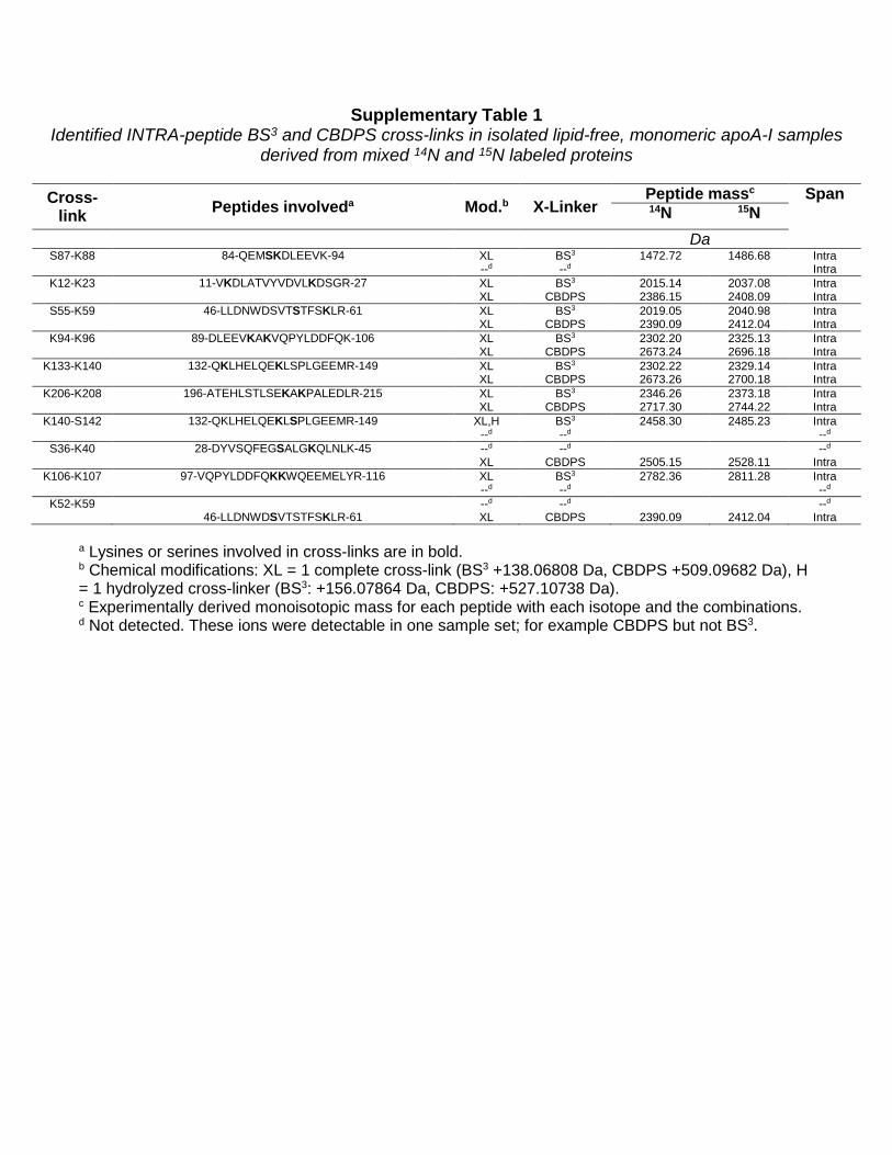
Supplementary Table 1 Identified INTRA-peptide BS3 and CBDPS cross-links in isolated lipid-free, monomeric apoA-I samples
derived from mixed 14N and 15N labeled proteins
Cross-link Peptides involveda Mod.b X-Linker
Peptide massc Span 14N 15N
Da
S87-K88 84-QEMSKDLEEVK-94 XL BS3 1472.72 1486.68 Intra --d --d Intra
K12-K23 11-VKDLATVYVDVLKDSGR-27 XL BS3 2015.14 2037.08 Intra XL CBDPS 2386.15 2408.09 Intra
S55-K59 46-LLDNWDSVTSTFSKLR-61 XL BS3 2019.05 2040.98 Intra XL CBDPS 2390.09 2412.04 Intra
K94-K96 89-DLEEVKAKVQPYLDDFQK-106 XL BS3 2302.20 2325.13 Intra XL CBDPS 2673.24 2696.18 Intra
K133-K140 132-QKLHELQEKLSPLGEEMR-149 XL BS3 2302.22 2329.14 Intra XL CBDPS 2673.26 2700.18 Intra
K206-K208 196-ATEHLSTLSEKAKPALEDLR-215 XL BS3 2346.26 2373.18 Intra XL CBDPS 2717.30 2744.22 Intra
K140-S142 132-QKLHELQEKLSPLGEEMR-149 XL,H BS3 2458.30 2485.23 Intra --d --d --d
S36-K40 28-DYVSQFEGSALGKQLNLK-45 --d --d --d XL CBDPS 2505.15 2528.11 Intra
K106-K107 97-VQPYLDDFQKKWQEEMELYR-116 XL BS3 2782.36 2811.28 Intra --d --d --d
K52-K59 --d --d --d 46-LLDNWDSVTSTFSKLR-61 XL CBDPS 2390.09 2412.04 Intra
a Lysines or serines involved in cross-links are in bold. b Chemical modifications: XL = 1 complete cross-link (BS3 +138.06808 Da, CBDPS +509.09682 Da), H = 1 hydrolyzed cross-linker (BS3: +156.07864 Da, CBDPS: +527.10738 Da). c Experimentally derived monoisotopic mass for each peptide with each isotope and the combinations. d Not detected. These ions were detectable in one sample set; for example CBDPS but not BS3.
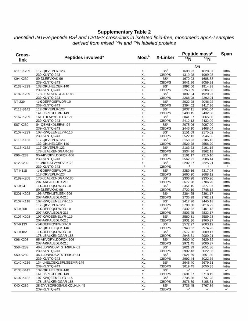
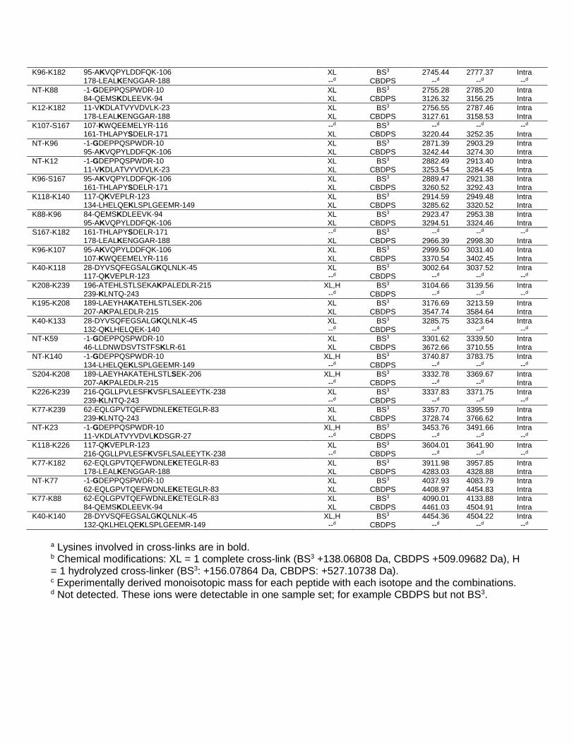
Supplementary Table 2 Identified INTER-peptide BS3 and CBDPS cross-links in isolated lipid-free, monomeric apoA-I samples
derived from mixed 14N and 15N labeled proteins
Cross-link Peptides involveda Mod.b X-Linker
Peptide massc Span 14N 15N
Da
K118-K239 117-QKVEPLR-123 XL BS3 1608.93 1628.87 Intra 239-KLNTQ-243 XL CBDPS 1319.98 1999.93 Intra K94-K239 89-DLEEVKAK-96 XL BS3 1670.93 1688.88 Intra 239-KLNTQ-243 XL CBDPS 2041.96 2059.91 Intra K133-K239 132-QKLHELQEK-140 XL BS3 1892.06 1914.99 Intra 239-KLNTQ-243 XL CBDPS 2263.09 2286.03 Intra K182-K239 178-LEALKENGGAR-188 XL BS3 1897.04 1920.97 Intra 239-KLNTQ-243 XL CBDPS 2268.08 2292.01 Intra NT-239 -1-GDEPPQSPWDR-10 XL BS3 2022.98 2046.92 Intra 239-KLNTQ-243 XL CBDPS 2394.02 2417.96 Intra K118-S142 117-QKVEPLR-123 XL BS3 2037.11 2061.04 Intra 141-LSPLGEEMR-149 XL CBDPS 2408.15 2432.08 Intra S167-K239 161-THLAPYSDELR-171 XL BS3 2041.07 2065.00 Intra 239-KLNTQ-243 XL CBDPS 2412.13 2432.09 Intra S87-K239 84-QEMSKDLEEVK-94 XL BS3 2075.06 2097.00 Intra 239-KLNTQ-243 XL CBDPS 2446.10 2468.04 Intra K107-K239 107-KWQEEMELYR-116 XL BS3 2151.09 2175.02 Intra 239-KLNTQ-243 XL CBDPS 2522.13 2546.06 Intra K118-K133 117-QKVEPLR-123 XL BS3 2158.23 2185.15 Intra 132-QKLHELQEK-140 XL CBDPS 2529.28 2556.20 Intra K118-K182 117-QKVEPLR-123 XL BS3 2163.23 2191.15 Intra 178-LEALKENGGAR-188 XL CBDPS 2534.26 2562.18 Intra K96-K239 95-AKVQPYLDDFQK-106 XL BS3 2191.17 2215.10 Intra 239-KLNTQ-243 XL CBDPS 2562.21 2586.14 Intra K12-K239 11-VKDLATVYVDVLK-23 XL BS3 2202.27 2225.21 Intra 239-KLNTQ-243 --d CBDPS --d --d --d NT-K118 -1-GDEPPQSPWDR-10 XL BS3 2289.16 2317.08 Intra 117-QKVEPLR-123 XL CBDPS 2660.20 2688.12 Intra K182-K208 178-LEALKENGGAR-188 XL BS3 2306.28 2335.20 Intra 207-AKPALEDLR-215 XL CBDPS 2677.28 2706.20 Intra NT-K94 -1-GDEPPQSPWDR-10 XL BS3 2351.15 2377.07 Intra 89-DLEEVKAK-96 XL CBDPS 2722.19 2748.13 Intra S201-K208 196-ATEHLSTLSEK-206 XL BS3 2364.25 2391.17 Intra 207-AKPALEDLR-215 XL CBDPS 2735.28 2762.21 Intra K107-K118 107-KWQEEMELYR-116 XL BS3 2417.26 2445.18 Intra 117-QKVEPLR-123 XL CBDPS 2788.30 2816.22 Intra NT-K208 -1-GDEPPQSPWDR-10 XL BS3 2432.22 2461.13 Intra 207-AKPALEDLR-215 XL CBDPS 2803.25 2832.17 Intra K107-K208 107-KWQEEMELYR-116 XL BS3 2560.31 2589.23 Intra 207-AKPALEDLR-215 XL CBDPS 2931.36 2960.27 Intra NT-K133 -1-GDEPPQSPWDR-10 XL BS3 2572.27 2603.19 Intra 132-QKLHELQEK-140 XL CBDPS 2943.32 2974.23 Intra NT-K182 -1-GDEPPQSPWDR-10 XL BS3 2577.26 2609.17 Intra 178-LEALKENGGAR-188 XL CBDPS 2948.31 2980.21 Intra K96-K208 95-AKVQPYLDDFQK-106 XL BS3 2600.40 2629.32 Intra 207-AKPALEDLR-215 XL CBDPS 2971.45 3000.37 Intra S58-K239 46-LLDNWDSVTSTFSKLR-61 XL BS3 2621.39 2651.30 Intra 239-KLNTQ-243 XL CBDPS 2992.43 3022.35 Intra S59-K239 46-LLDNWDSVTSTFSKLR-61 XL BS3 2621.39 2651.30 Intra 239-KLNTQ-243 XL CBDPS 2992.44 3022.35 Intra K140-K239 134-LHELQEKLSPLGEEMR-149 XL BS3 2648.40 2679.31 Intra 239-KLNTQ-243 XL CBDPS 3019.45 3050.35 Intra K133-S142 132-QKLHELQEK-140 --d BS3 --d --d --d 141-LSPLGEEMR-149 XL CBDPS 2691.27 2718.19 Intra K107-K182 107-KWQEEMELYR-116 XL BS3 2705.36 2737.28 Intra 178-LEALKENGGAR-188 XL CBDPS 3076.39 3108.31 Intra K40-K239 28-DYVSQFEGSALGKQLNLK-45 XL BS3 2736.45 2767.36 Intra 239-KLNTQ-243 --d CBDPS --d --d --d
K96-K182 95-AKVQPYLDDFQK-106 XL BS3 2745.44 2777.37 Intra 178-LEALKENGGAR-188 --d CBDPS --d --d --d NT-K88 -1-GDEPPQSPWDR-10 XL BS3 2755.28 2785.20 Intra 84-QEMSKDLEEVK-94 XL CBDPS 3126.32 3156.25 Intra K12-K182 11-VKDLATVYVDVLK-23 XL BS3 2756.55 2787.46 Intra 178-LEALKENGGAR-188 XL CBDPS 3127.61 3158.53 Intra K107-S167 107-KWQEEMELYR-116 --d BS3 --d --d --d 161-THLAPYSDELR-171 XL CBDPS 3220.44 3252.35 Intra NT-K96 -1-GDEPPQSPWDR-10 XL BS3 2871.39 2903.29 Intra 95-AKVQPYLDDFQK-106 XL CBDPS 3242.44 3274.30 Intra NT-K12 -1-GDEPPQSPWDR-10 XL BS3 2882.49 2913.40 Intra 11-VKDLATVYVDVLK-23 XL CBDPS 3253.54 3284.45 Intra K96-S167 95-AKVQPYLDDFQK-106 XL BS3 2889.47 2921.38 Intra 161-THLAPYSDELR-171 XL CBDPS 3260.52 3292.43 Intra K118-K140 117-QKVEPLR-123 XL BS3 2914.59 2949.48 Intra 134-LHELQEKLSPLGEEMR-149 XL CBDPS 3285.62 3320.52 Intra K88-K96 84-QEMSKDLEEVK-94 XL BS3 2923.47 2953.38 Intra 95-AKVQPYLDDFQK-106 XL CBDPS 3294.51 3324.46 Intra S167-K182 161-THLAPYSDELR-171 --d BS3 --d --d --d 178-LEALKENGGAR-188 XL CBDPS 2966.39 2998.30 Intra K96-K107 95-AKVQPYLDDFQK-106 XL BS3 2999.50 3031.40 Intra 107-KWQEEMELYR-116 XL CBDPS 3370.54 3402.45 Intra K40-K118 28-DYVSQFEGSALGKQLNLK-45 XL BS3 3002.64 3037.52 Intra 117-QKVEPLR-123 --d CBDPS --d --d --d K208-K239 196-ATEHLSTLSEKAKPALEDLR-215 XL,H BS3 3104.66 3139.56 Intra 239-KLNTQ-243 --d CBDPS --d --d --d K195-K208 189-LAEYHAKATEHLSTLSEK-206 XL BS3 3176.69 3213.59 Intra 207-AKPALEDLR-215 XL CBDPS 3547.74 3584.64 Intra K40-K133 28-DYVSQFEGSALGKQLNLK-45 XL BS3 3285.75 3323.64 Intra 132-QKLHELQEK-140 --d CBDPS --d --d --d NT-K59 -1-GDEPPQSPWDR-10 XL BS3 3301.62 3339.50 Intra 46-LLDNWDSVTSTFSKLR-61 XL CBDPS 3672.66 3710.55 Intra NT-K140 -1-GDEPPQSPWDR-10 XL,H BS3 3740.87 3783.75 Intra 134-LHELQEKLSPLGEEMR-149 --d CBDPS --d --d --d S204-K208 189-LAEYHAKATEHLSTLSEK-206 XL,H BS3 3332.78 3369.67 Intra 207-AKPALEDLR-215 --d CBDPS --d --d Intra K226-K239 216-QGLLPVLESFKVSFLSALEEYTK-238 XL BS3 3337.83 3371.75 Intra 239-KLNTQ-243 --d CBDPS --d --d --d K77-K239 62-EQLGPVTQEFWDNLEKETEGLR-83 XL BS3 3357.70 3395.59 Intra 239-KLNTQ-243 XL CBDPS 3728.74 3766.62 Intra NT-K23 -1-GDEPPQSPWDR-10 XL,H BS3 3453.76 3491.66 Intra 11-VKDLATVYVDVLKDSGR-27 --d CBDPS --d --d --d K118-K226 117-QKVEPLR-123 XL BS3 3604.01 3641.90 Intra 216-QGLLPVLESFKVSFLSALEEYTK-238 --d CBDPS --d --d --d K77-K182 62-EQLGPVTQEFWDNLEKETEGLR-83 XL BS3 3911.98 3957.85 Intra 178-LEALKENGGAR-188 XL CBDPS 4283.03 4328.88 Intra NT-K77 -1-GDEPPQSPWDR-10 XL BS3 4037.93 4083.79 Intra 62-EQLGPVTQEFWDNLEKETEGLR-83 XL CBDPS 4408.97 4454.83 Intra K77-K88 62-EQLGPVTQEFWDNLEKETEGLR-83 XL BS3 4090.01 4133.88 Intra 84-QEMSKDLEEVK-94 XL CBDPS 4461.03 4504.91 Intra K40-K140 28-DYVSQFEGSALGKQLNLK-45 XL,H BS3 4454.36 4504.22 Intra 132-QKLHELQEKLSPLGEEMR-149 --d CBDPS --d --d --d
a Lysines involved in cross-links are in bold. b Chemical modifications: XL = 1 complete cross-link (BS3 +138.06808 Da, CBDPS +509.09682 Da), H = 1 hydrolyzed cross-linker (BS3: +156.07864 Da, CBDPS: +527.10738 Da). c Experimentally derived monoisotopic mass for each peptide with each isotope and the combinations. d Not detected. These ions were detectable in one sample set; for example CBDPS but not BS3.

Supplementary Table 3 Experimental parameters from SAXS sampling of monomeric apoA-I cross-linked with CBDPS or BS3
ApoA-I Sample
I(O)a (Guinier)
Rgb
(Guinier) Real
Space Rg Dmaxc Volume DAMMIF NSDd
cm-1 Å Å Å Å3 Å CBDPS 4.0 mg/ml 603 26 25.29 85 70165
0.579 ± 0.032 2.0 mg/ml 301 25.8 25.24 83 68091 1.0 mg/ml 149 25.1 25.53 83 70191 BS3 4.0 mg/ml 569 25.95 25.54 88 77337
0.596 ± 0.024 2.0 mg/ml 294 25.58 25.25 81 77334 1.0 mg/ml 140 27.67 25.24 81 83946
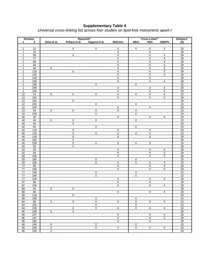
Supplementary Table 4 Universal cross-linking list across four studies on lipid-free monomeric apoA-I
Residue Reporteda Cross-Linkerb Distancec
1 2 Silva et al Pollard et al Segrest et al Melchior MDA BS3 CBDPS (Å) 1 12 X X X X X X 23 1 23 X X 28 1 59 X X X X 28 1 77 X X X 28 1 88 X X X 28 1 94 X X X 28 1 96 X X X X 28 1 118 X X X X 28 1 133 X X X 28 1 140 X X 28 1 182 X X X 28 1 195 X X 23 1 208 X X X 28 1 239 X X X 28
12 23 X X X X X X X 23 12 182 X X X 28 12 195 X 28 12 226 X X 23 12 239 X X 28 23 59 X X X X X 23 23 239 X X 23 36 40 X X X 28 40 45 X X X X 23 40 59 X 28 40 94 X X 23 40 118 X X X 28 40 133 X X X X X 23 40 140 X X X 28 40 182 X 28 40 239 X X X X X 23 45 59 X 28 52 59 X X X 28 55 59 X X X 28 58 239 X X X 28 59 195 X X 23 59 239 X X X X X 23 77 88 X X X 28 77 182 X X X 28 77 195 X X 23 77 208 X X 23 77 239 X X X 28 87 88 X X 28 87 239 X X X 28 88 94 X X 28 88 96 X X X 28 88 118 X 28 88 195 X X 23 94 96 X X X X X X X 23 94 226 X X 23 94 239 X X X X X X 23 96 106 X X 28 96 107 X X X 28 96 167 X X X 28 96 182 X X 28 96 195 X X X 23 96 208 X X X X X X 23 96 226 X 28
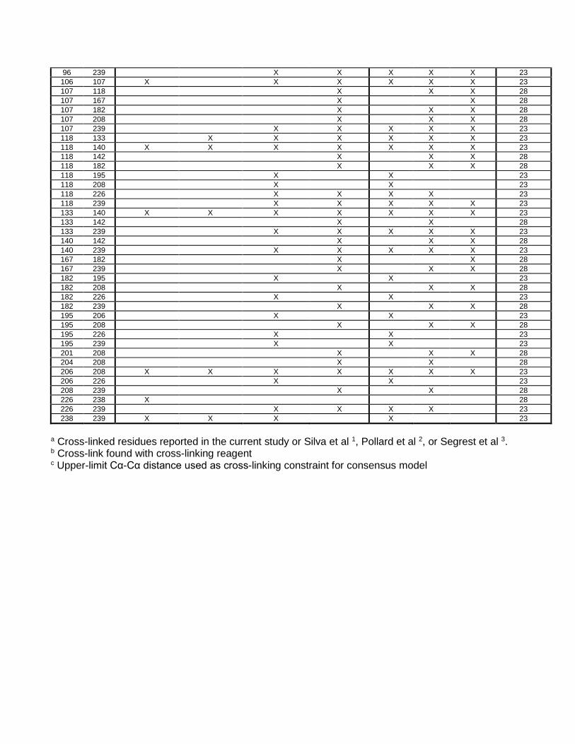
96 239 X X X X X 23 106 107 X X X X X X 23 107 118 X X X 28 107 167 X X 28 107 182 X X X 28 107 208 X X X 28 107 239 X X X X X 23 118 133 X X X X X X 23 118 140 X X X X X X X 23 118 142 X X X 28 118 182 X X X 28 118 195 X X 23 118 208 X X 23 118 226 X X X X 23 118 239 X X X X X 23 133 140 X X X X X X X 23 133 142 X X 28 133 239 X X X X X 23 140 142 X X X 28 140 239 X X X X X 23 167 182 X X 28 167 239 X X X 28 182 195 X X 23 182 208 X X X 28 182 226 X X 23 182 239 X X X 28 195 206 X X 23 195 208 X X X 28 195 226 X X 23 195 239 X X 23 201 208 X X X 28 204 208 X X 28 206 208 X X X X X X X 23 206 226 X X 23 208 239 X X 28 226 238 X 28 226 239 X X X X 23 238 239 X X X X 23
a Cross-linked residues reported in the current study or Silva et al 1, Pollard et al 2, or Segrest et al 3. b Cross-link found with cross-linking reagent c Upper-limit Cα-Cα distance used as cross-linking constraint for consensus model
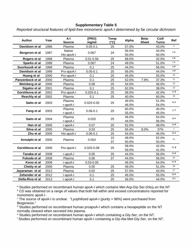
Supplementary Table 5 Reported structural features of lipid-free monomeric apoA-I determined by far circular dichroism
Author Year A-I
Source [PRO] mg/ml
Temp (°C) Alpha Beta-
Sheet Coil/
Turns Ref
Davidson et al 1996 Plasma 0.05-0.1 25 57.0% 43.0% 4
Bergeron et al 1997 Native 0.067 24 56.0% 44.0% 5 a His-apoA-I 50.0% 50.0% Rogers et al 1998 Plasma 0.01-0.56 25 68.0% 32.0% 6 b
Sparks et al 1999 Plasma 0.067 24 49.0% 51.0% 7 c Suurkuusk et al 1999 Plasma 0.1 25 44.0% 56.0% 8 d Davidson et al 1999 Pro-apoA-I 0.05-0.1 25 56.0% 44.0% 9 d Huang et al 2000 Pro-apoA-I 0.1 25 45.0% 55.0% 10 Panzenbock et al 2000 Plasma 0.1 25 52.0% 7.9% 37.3% 11
Weinberg et al 2000 Plasma 0.08 25 54.0% 46.0% 12 Sigalov et al 2001 Plasma 0.1 25 62.0% 38.0% 13
Gorshkova et al 2002 Pro-apoA-I 0.025-0.1 25 58.0% 42.0% 14 d Reschly et al 2002 Plasma 0.1 25 40.0% 60.0% 15
Saito et al 2003 Plasma 0.025-0.05 25 49.0% 51.0% 16 e r-apoA-I 46.0% 54.0%
Fang et al 2003 Plasma 0.06-0.1 25 60.0% 40.0% 17 f r-apoA-I 55.0% 45.0%
Saito et al 2004 Plasma 0.025 25 46.0% 54.0% 18 e r-apoA-I 44.0% 56.0% Han et al 2005 r-apoA-I 0.07 25 52.0% 48.0% 19 d
Silva et al 2005 Plasma 0.03 25 55.0% 8.0% 37% 1 Zhu et al 2005 His-apoA-I 0.06-0.1 25 54.0% 46.0% 20 a
Arnulphi et al 2005 Plasma 0.054 15 48.0% 52.0% 21 37 50.0% 50.0%
Gorshkova et al 2006 Pro-apoA-I 0.025-0.08 25 58.0% 42.0% 22 g 59.0% 41.0% Tanaka et al 2008 r-apoA-I 0.05 25 44.0% 56.0% 23 d Fukuda et al 2008 Plasma 0.06 37 44.0% 56.0% 24
Kono et al 2009 r-apoA-I 0.03-0.05 46.0% 54.0% 25 d Chetty et al 2009 Plasma 0.05 25 49.0% 51.0% 26
Jayaraman et al 2012 Plasma 0.02 25 57.0% 43.0% 27 Zehender et al 2012 r-apoA-I 0.1 25 45.0% 55.0% 28 h
Della-Riva et al 2013 r-apoA-I 0.1 25 56.0% 44.0% 29 i
a Studies performed on recombinant human apoA-I which contains Met-Arg-Gly-Ser-(His)6 on the NT b CD was obtained on a range of values that both fall within and exceed concentrations reported for monomeric apoA-I . c The source of apoA-I is unclear. “Lyophilized apoA-I (purity > 96%) were purchased from Biogenesis.” d Studies performed on recombinant human proapoA-I which contains a hexapeptide on the NT normally cleaved when secreted into plasma. e Studies performed on recombinant human apoA-I which containing a Gly-Ser, on the NT. f Studies performed on recombinant human apoA-I containing a Gly-Ala-Met-Gly-Ser, on the NT.

g Studies performed on recombinant human proapoA-I containing a hexapeptide on the NT normally cleaved when secreted into plasma. Protein was expressed in either a baculovirus (top) or adenovirus (bottom) system. h Studies performed on recombinant human apoA-I containing a Gly-Gly on the NT. i Studies performed on recombinant human apoA-I which contains a point mutation, E2D at the N-terminus.
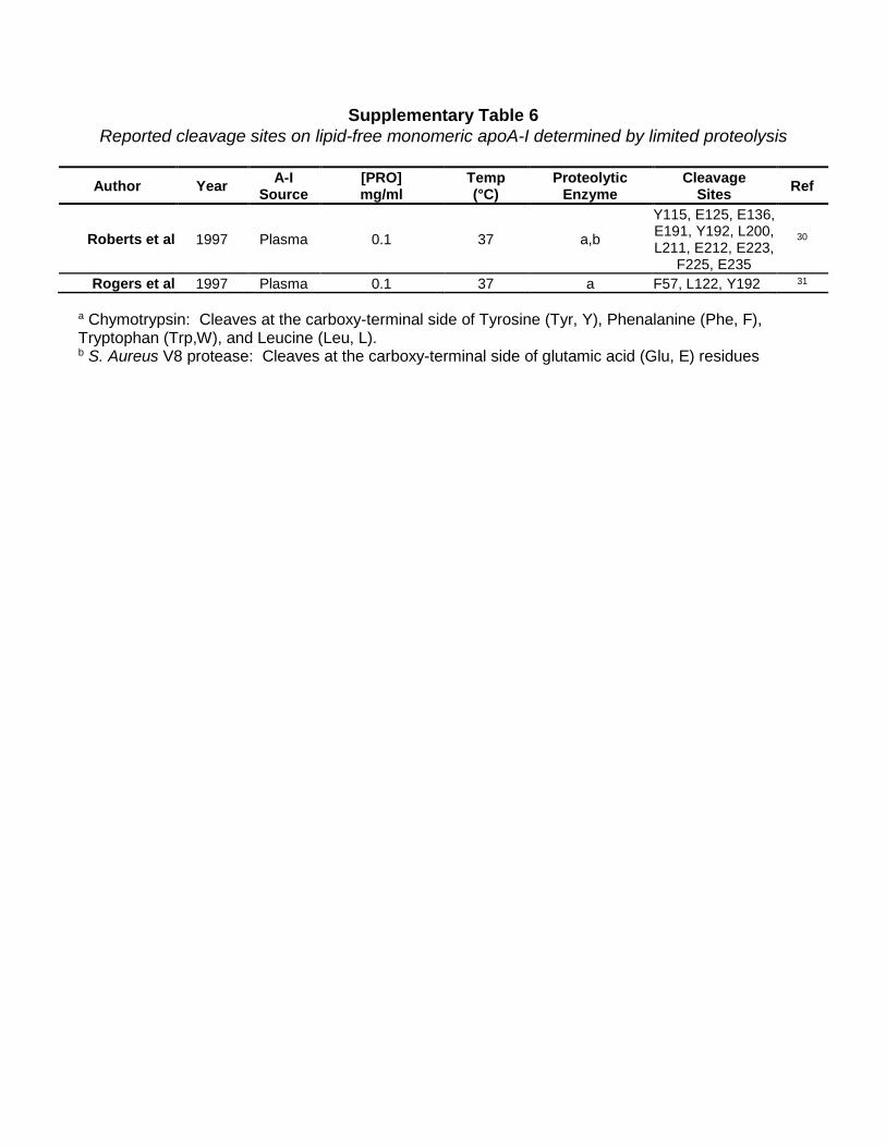
Supplementary Table 6 Reported cleavage sites on lipid-free monomeric apoA-I determined by limited proteolysis
Author Year A-I
Source [PRO] mg/ml
Temp (°C)
Proteolytic Enzyme
Cleavage Sites Ref
Roberts et al 1997 Plasma 0.1 37 a,b
Y115, E125, E136, E191, Y192, L200, L211, E212, E223,
F225, E235
30
Rogers et al 1997 Plasma 0.1 37 a F57, L122, Y192 31
a Chymotrypsin: Cleaves at the carboxy-terminal side of Tyrosine (Tyr, Y), Phenalanine (Phe, F), Tryptophan (Trp,W), and Leucine (Leu, L). b S. Aureus V8 protease: Cleaves at the carboxy-terminal side of glutamic acid (Glu, E) residues
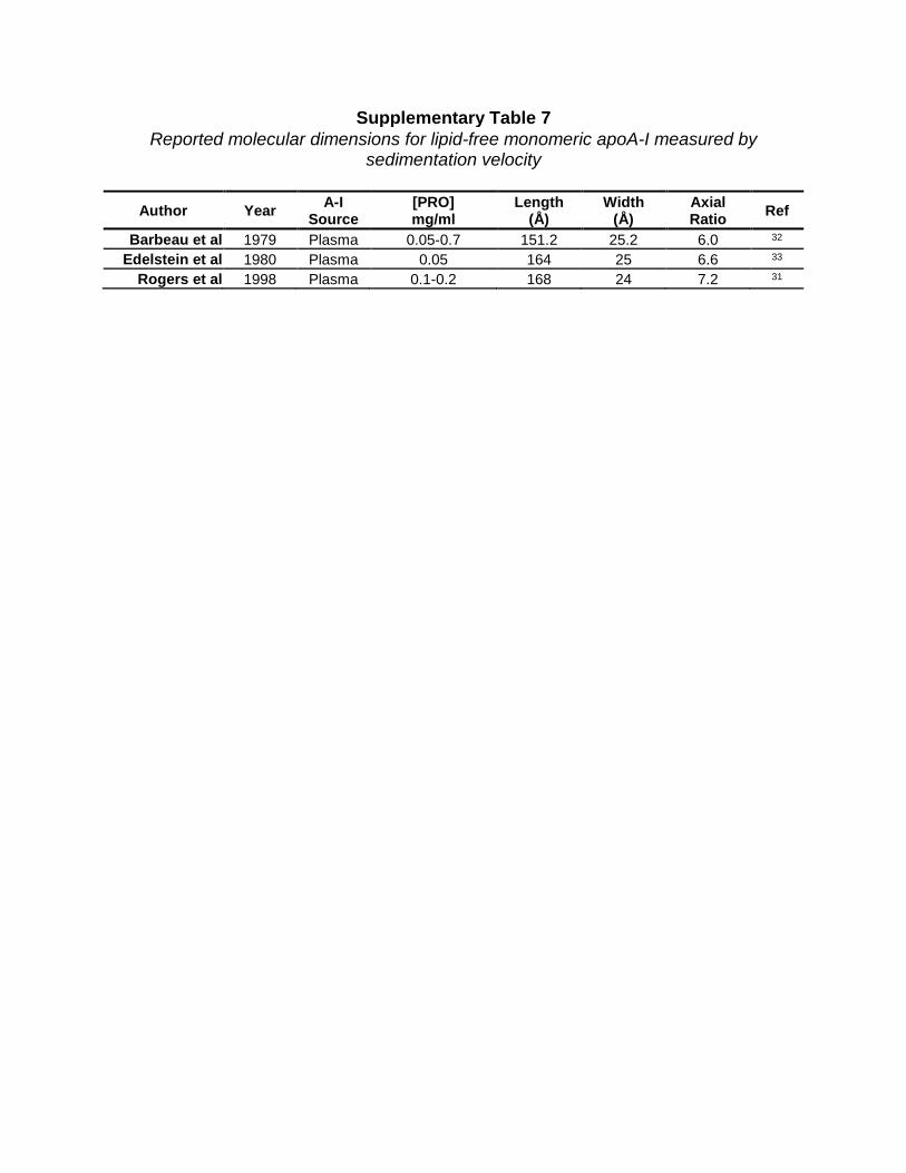
Supplementary Table 7 Reported molecular dimensions for lipid-free monomeric apoA-I measured by
sedimentation velocity
Author Year A-I Source
[PRO] mg/ml
Length (Å)
Width (Å)
Axial Ratio Ref
Barbeau et al 1979 Plasma 0.05-0.7 151.2 25.2 6.0 32 Edelstein et al 1980 Plasma 0.05 164 25 6.6 33
Rogers et al 1998 Plasma 0.1-0.2 168 24 7.2 31
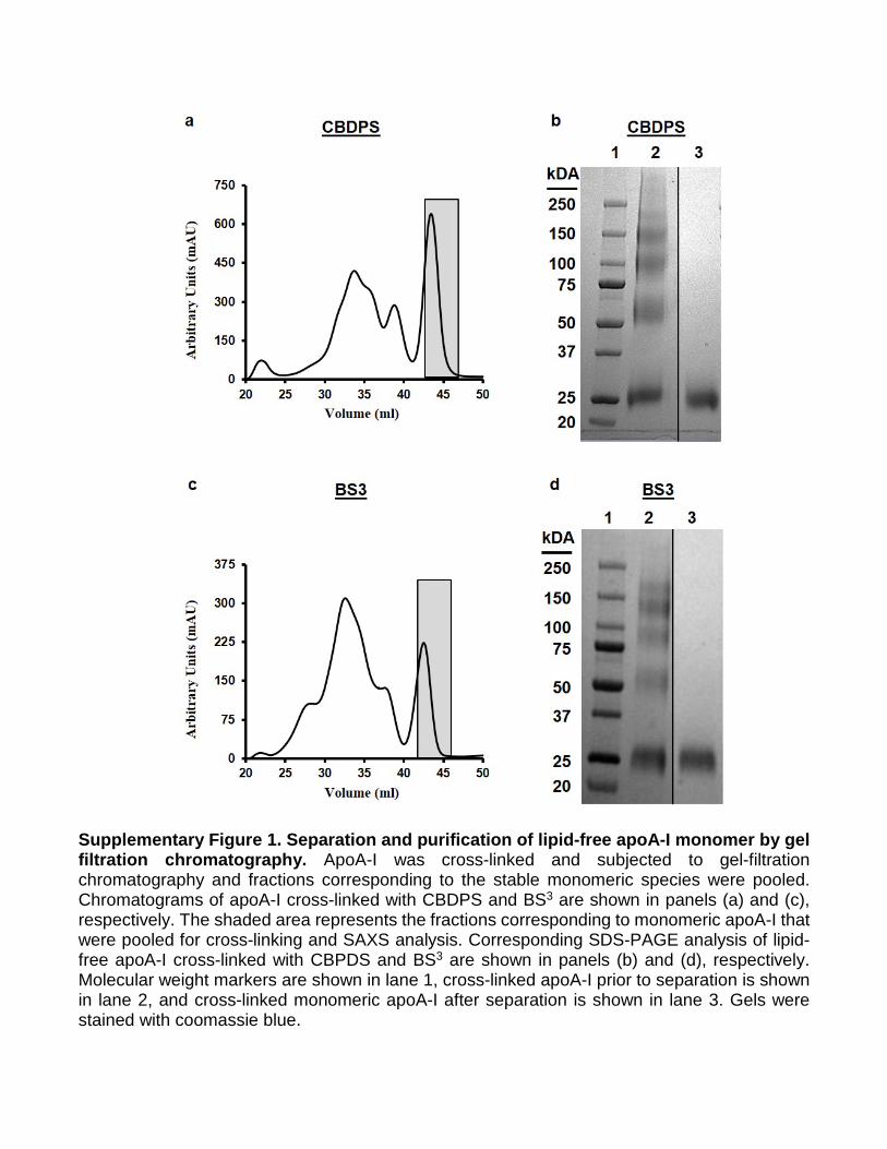
Supplementary Figure 1. Separation and purification of lipid-free apoA-I monomer by gel filtration chromatography. ApoA-I was cross-linked and subjected to gel-filtration chromatography and fractions corresponding to the stable monomeric species were pooled. Chromatograms of apoA-I cross-linked with CBDPS and BS3 are shown in panels (a) and (c), respectively. The shaded area represents the fractions corresponding to monomeric apoA-I that were pooled for cross-linking and SAXS analysis. Corresponding SDS-PAGE analysis of lipid-free apoA-I cross-linked with CBPDS and BS3 are shown in panels (b) and (d), respectively. Molecular weight markers are shown in lane 1, cross-linked apoA-I prior to separation is shown in lane 2, and cross-linked monomeric apoA-I after separation is shown in lane 3. Gels were stained with coomassie blue.
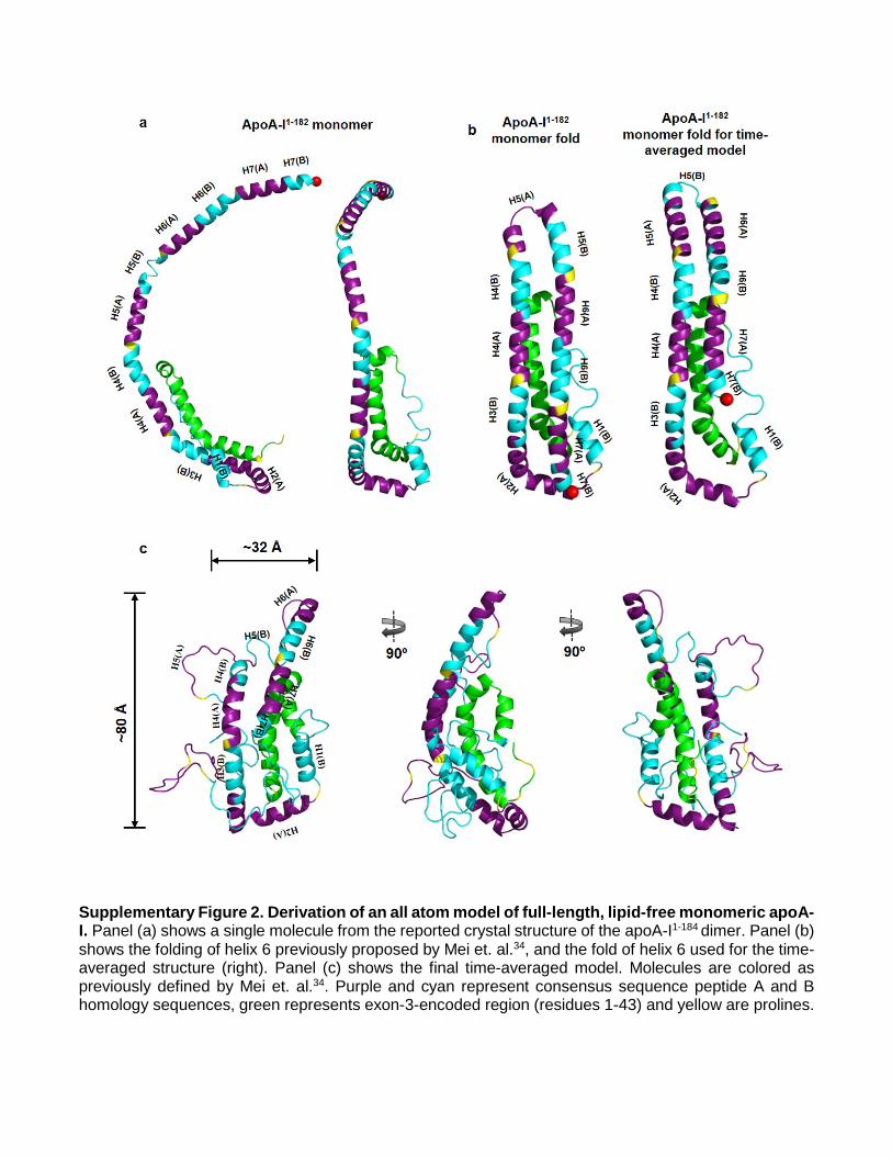
Supplementary Figure 2. Derivation of an all atom model of full-length, lipid-free monomeric apoA-I. Panel (a) shows a single molecule from the reported crystal structure of the apoA-I1-184 dimer. Panel (b) shows the folding of helix 6 previously proposed by Mei et. al.34, and the fold of helix 6 used for the time-averaged structure (right). Panel (c) shows the final time-averaged model. Molecules are colored as previously defined by Mei et. al.34. Purple and cyan represent consensus sequence peptide A and B homology sequences, green represents exon-3-encoded region (residues 1-43) and yellow are prolines.
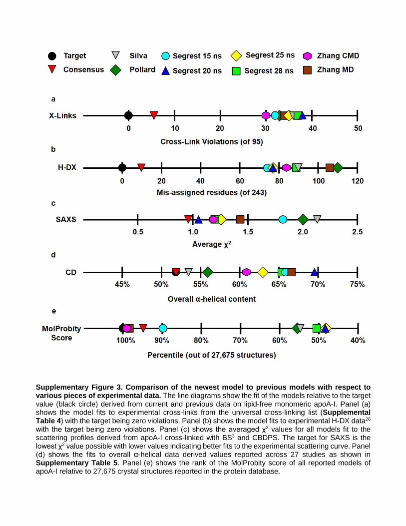
Supplementary Figure 3. Comparison of the newest model to previous models with respect to various pieces of experimental data. The line diagrams show the fit of the models relative to the target value (black circle) derived from current and previous data on lipid-free monomeric apoA-I. Panel (a) shows the model fits to experimental cross-links from the universal cross-linking list (Supplemental Table 4) with the target being zero violations. Panel (b) shows the model fits to experimental H-DX data26 with the target being zero violations. Panel (c) shows the averaged χ2 values for all models fit to the scattering profiles derived from apoA-I cross-linked with BS3 and CBDPS. The target for SAXS is the lowest χ2 value possible with lower values indicating better fits to the experimental scattering curve. Panel (d) shows the fits to overall α-helical data derived values reported across 27 studies as shown in Supplementary Table 5. Panel (e) shows the rank of the MolProbity score of all reported models of apoA-I relative to 27,675 crystal structures reported in the protein database.
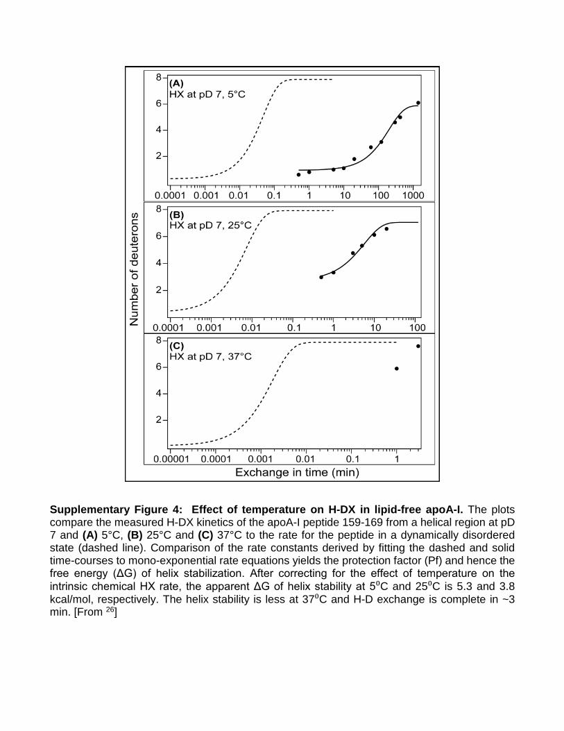
Supplementary Figure 4: Effect of temperature on H-DX in lipid-free apoA-I. The plots compare the measured H-DX kinetics of the apoA-I peptide 159-169 from a helical region at pD 7 and (A) 5°C, (B) 25°C and (C) 37°C to the rate for the peptide in a dynamically disordered state (dashed line). Comparison of the rate constants derived by fitting the dashed and solid time-courses to mono-exponential rate equations yields the protection factor (Pf) and hence the free energy (ΔG) of helix stabilization. After correcting for the effect of temperature on the intrinsic chemical HX rate, the apparent ΔG of helix stability at 5⁰C and 25⁰C is 5.3 and 3.8 kcal/mol, respectively. The helix stability is less at 37⁰C and H-D exchange is complete in ~3 min. [From 26]
Supplementary Note
Due to space restrictions of the Journal, we were asked to abbreviate our discussion of the model in the main paper. What follows is the original, more complete discussion.
Consistency with previously reported data. Analytical ultracentrifugation (AUC) can provide low-resolution molecular shape information. Supplementary Table 7 summarizes three analytical ultracentrifugation studies31-33 on lipid-free apoA-I, which conclude that monomeric apoA-I is asymmetrical in shape with an axial ratio of ~6.5 (161 x 24 Å). This contrasts with our SAXS envelopes which had an axial ratio of 2.1-2.8. The discrepancy between AUC and SAXS measurements may reflect the dynamics of apoA-I in solution vs the ‘locked’ state after cross-linking. Using Forster resonance energy transfer, Brouillette and colleagues later concluded apoA-I is more compact35 and that the AUC results31 were probably reflective of an unfolded protein due to high external centrifugal forces. The molecular length measured by AUC is about double what we measured by SAXS while the width is about half. This is consistent with the notion that monomeric apoA-I, like apoA-IV36, can open up like a pocket knife to oligomerize or bind lipids. This may be further supported by EPR studies identifying residues 26, 44, 64, 167, 217, and 226 to be in the same plane37. In the time-averaged structure all but residues 217 and 226 fall within the same plane. Given that EPR studies were executed on multimeric apoA-I, it’s plausible that when H6 opens and apoA-I self-associates, residues 217 and 226 fall in-plane with the remaining residues. Several laboratories38-40 have suggested that helix 5 may be the center of such a hinge. Additionally, the positioning of H6 on the new model appears poised to unfold for interaction with lipid or another molecule. The exact nature of this unfolding must await further studies focused specifically on apoA-I oligomerization.
Limitations of the model. A limitation of the current model stems from our attempt to represent a highly dynamic protein with a single, time-averaged model. Numerous studies have documented the “molten globule” nature of apoA-I16,41. H-DX quantifies the rate of proton transfer to amide groups in the protein and allows one to determine locations and stabilities of elements of secondary structure26. Given that the observed rate constant for hydrogen exchange is lower than the rate constant for helical closing and H-bond formation in apoA-I, the observed hydrogen exchange rate is related to the α-helix open-closed equilibrium constant (Kop), a measure of the free energy (ΔG) of helix stabilization. This concept is illustrated in Supplementary Figure 4 which compares hydrogen-exchange rates at three temperatures for a peptide segment in an unprotected random coil state (dashed line) and in a protected helical state (solid line). Given the degree of protection, expressed as the protection factor Pf (=1/Kop), one can derive Kop and the ΔG of helix stabilization. ApoA-I has ΔG of helix stabilization ranging from ~3-5 kcal/mol26. At neutral pH and room temperature, Pf for these helices is ~10e4, which corresponds with complete hydrogen exchange into the α-helical segments occurring in ~10 minutes (Supplementary Figure 4B); i.e. all the helical segments of native apoA-I have opened and closed at least once in this timeframe. To put this in context, a more stable globular protein such as cytochrome C has a ΔG of helix stabilization of 10 kcal/mol corresponding to a Pf of ~10e8, indicating that complete hydrogen exchange would require ~10 weeks. Given this degree of secondary structure dynamics, the overall high content of random coil, and the number of solvent exposed hydrophobic residues in apoA-I, it may not be possible to fully capture apoA-I structure in a single model. At physiological temperatures, apoA-I may adopt many of the structures shown in Fig. 1. However, our model is a time-averaged structure derived from experimental data obtained on a time scale that is much longer than typical secondary structure oscillations. For this reason, we think of it as a base model upon which hypothesized dynamics and conformational alterations can be further tested or modeled.
Another issue relates to the notion of solvent accessibility of the cross-linking reagents. While most cross-links fit the model in terms of Euclidian distance (‘as the crow flies’), nearly half are impeded by some obstruction. For example, the cross-link path may be sterically hindered by a side chain rotamer from a non-participating residue or it may pass through the backbone of an adjacent helical domain. The answer to how this can happen most likely lies in protein dynamics. Cross-linking experiments are
completed on the time scale of minutes to hours, substantially longer than the timescale described above for helix opening and closing. Thus, α-helical domains in apoA-I have unfolded and refolded multiple times allowing cross-linker access to amines that are otherwise inaccessible. Furthermore, it is possible that cross-links may stabilize low probability structures, which would facilitate these observations. However, previous reports have shown excellent consistency between observed cross-links on solution structures with crystal structures of apoA-I1-184 (42) and apoA-IV36 and non-apolipoproteins43-46 validating the approach. It’s important to recognize that in vitro studies presented here are performed on an ensemble of structures that vary at any given time point during the experiment. Additionally, it’s likely that most of the dynamics are localized; i.e. lipid-free apoA-I likely exists as a discrete structure that exhibits characteristics of a molten globular protein. Thus, the model represents a time-average of those ensembles and cross-links that appear sterically hindered or solvent inaccessible likely occur on an alternate conformation within the boundaries of the experimental system. Additional studies are needed to better define these boundaries and the extent of rearrangement apoA-I can achieve in vivo and in vitro. Finally, despite its consistency with much of the known structural data, we note that the model is still limited in resolution compared to NMR or X-ray crystallography. The general backbone configuration is likely correct, but more refined molecular interactions such as salt-bridging and hydrogen bonding are still unclear. While the protein clearly has a somewhat defined structure and shape as captured by SAXS, the exact lengths of helical domains, the precise positions of N- and C-termini and even the integrity of the helical bundle itself are likely to be in flux on the timescale of seconds. Highly unstable helices (Pf<10) that have ΔG stabilization of < 1.3 kcal/mol and are open > 10% of the time are not detected in the timescale of the H-DX kinetic experiments but are detected by CD measurements. However, truncation of the C-terminus from residue 243 to residues 221 and 231 reduces CD-detectable helix content by 14 and 7 amino acid residues, respectively16,40. On the basis of such observations, Mei and Atkinson40 suggested that the segment spanning residues 231-241 contains α-helical structure. This C-terminal segment is located near the N-terminus (Fig. 5c) and it may contribute to the stabilization of the NT helix bundle induced by the C-terminal domain47. The existence of such a C-terminal helical domain is further supported by preliminary molecular dynamics simulations of the new model (Segrest et al, unpublished observation).
References
1. Silva, R.A., Hilliard, G.M., Fang, J., Macha, S. & Davidson, W.S. A three-dimensional molecular
model of lipid-free apolipoprotein A-I determined by cross-linking/mass spectrometry and sequence threading. Biochemistry 44, 2759-69 (2005).
2. Pollard, R.D., Fulp, B., Samuel, M.P., Sorci-Thomas, M.G. & Thomas, M.J. The conformation of lipid-free human apolipoprotein A-I in solution. Biochemistry 52, 9470-81 (2013).
3. Segrest, J.P., Jones, M.K., Shao, B. & Heinecke, J.W. An experimentally robust model of monomeric apolipoprotein A-I created from a chimera of two X-ray structures and molecular dynamics simulations. Biochemistry 53, 7625-40 (2014).
4. Davidson, W.S., Hazlett, T., Mantulin, W.W. & Jonas, A. The role of apolipoprotein AI domains in lipid binding. Proc Natl Acad Sci U S A 93, 13605-10 (1996).
5. Bergeron, J. et al. Characterization of human apolipoprotein A-I expressed in Escherichia coli. Biochim Biophys Acta 1344, 139-52 (1997).
6. Rogers, D.P. et al. Truncation of the amino terminus of human apolipoprotein A-I substantially alters only the lipid-free conformation. Biochemistry 36, 288-300 (1997).
7. Sparks, D.L., Frank, P.G., Braschi, S., Neville, T.A. & Marcel, Y.L. Effect of apolipoprotein A-I lipidation on the formation and function of pre-beta and alpha-migrating LpA-I particles. Biochemistry 38, 1727-35 (1999).
8. Suurkuusk, M. & Hallen, D. Denaturation of apolipoprotein A-I and the monomer form of apolipoprotein A-I(Milano). Eur J Biochem 265, 346-52 (1999).
9. Davidson, W.S. et al. Structural organization of the N-terminal domain of apolipoprotein A-I: studies of tryptophan mutants. Biochemistry 38, 14387-95 (1999).
10. Huang, W. et al. A single amino acid deletion in the carboxy terminal of apolipoprotein A-I impairs lipid binding and cellular interaction. Arterioscler Thromb Vasc Biol 20, 210-6 (2000).
11. Panzenbock, U., Kritharides, L., Raftery, M., Rye, K.A. & Stocker, R. Oxidation of methionine residues to methionine sulfoxides does not decrease potential antiatherogenic properties of apolipoprotein A-I. J Biol Chem 275, 19536-44 (2000).
12. Weinberg, R.B. et al. Structure and interfacial properties of chicken apolipoprotein A-IV. J Lipid Res 41, 1410-8 (2000).
13. Sigalov, A.B. & Stern, L.J. Oxidation of methionine residues affects the structure and stability of apolipoprotein A-I in reconstituted high density lipoprotein particles. Chem Phys Lipids 113, 133-46 (2001).
14. Gorshkova, I.N., Liu, T., Zannis, V.I. & Atkinson, D. Lipid-free structure and stability of apolipoprotein A-I: probing the central region by mutation. Biochemistry 41, 10529-39 (2002).
15. Reschly, E.J. et al. Apolipoprotein A-I alpha -helices 7 and 8 modulate high density lipoprotein subclass distribution. J Biol Chem 277, 9645-54 (2002).
16. Saito, H. et al. Domain structure and lipid interaction in human apolipoproteins A-I and E, a general model. J Biol Chem 278, 23227-32 (2003).
17. Fang, Y., Gursky, O. & Atkinson, D. Structural studies of N- and C-terminally truncated human apolipoprotein A-I. Biochemistry 42, 6881-90 (2003).
18. Saito, H. et al. alpha-Helix formation is required for high affinity binding of human apolipoprotein A-I to lipids. Journal of Biological Chemistry 279, 20974-20981 (2004).
19. Han, J.M., Jeong, T.S., Lee, W.S., Choi, I. & Cho, K.H. Structural and functional properties of V156K and A158E mutants of apolipoprotein A-I in the lipid-free and lipid-bound states. J Lipid Res 46, 589-96 (2005).
20. Zhu, X., Wu, G., Zeng, W., Xue, H. & Chen, B. Cysteine mutants of human apolipoprotein A-I: a study of secondary structural and functional properties. J Lipid Res 46, 1303-11 (2005).
21. Arnulphi, C., Sanchez, S.A., Tricerri, M.A., Gratton, E. & Jonas, A. Interaction of human apolipoprotein A-I with model membranes exhibiting lipid domains. Biophys J 89, 285-95 (2005).
22. Gorshkova, I.N. et al. Structure and stability of apolipoprotein a-I in solution and in discoidal high-density lipoprotein probed by double charge ablation and deletion mutation. Biochemistry 45, 1242-54 (2006).
23. Tanaka, M. et al. Influence of tertiary structure domain properties on the functionality of apolipoprotein A-I. Biochemistry 47, 2172-80 (2008).
24. Fukuda, M. et al. Conformational change of apolipoprotein A-I and HDL formation from model membranes under intracellular acidic conditions. J Lipid Res 49, 2419-26 (2008).
25. Kono, M. et al. Disruption of the C-terminal helix by single amino acid deletion is directly responsible for impaired cholesterol efflux ability of apolipoprotein A-I Nichinan. J Lipid Res 51, 809-18 (2010).
26. Chetty, P.S. et al. Helical structure and stability in human apolipoprotein A-I by hydrogen exchange and mass spectrometry. Proc.Natl.Acad.Sci.U.S.A 106, 19005-19010 (2009).
27. Jayaraman, S., Cavigiolio, G. & Gursky, O. Folded functional lipid-poor apolipoprotein A-I obtained by heating of high-density lipoproteins: relevance to high-density lipoprotein biogenesis. Biochemical Journal 442, 703-712 (2012).
28. Zehender, F., Ziegler, A., Schonfeld, H.J. & Seelig, J. Thermodynamics of protein self-association and unfolding. The case of apolipoprotein A-I. Biochemistry 51, 1269-80 (2012).
29. Ryan, R.O., Forte, T.M. & Oda, M.N. Optimized bacterial expression of human apolipoprotein A-I. Protein Expr Purif 27, 98-103 (2003).
30. Roberts, L.M. et al. Structural analysis of apolipoprotein A-I: limited proteolysis of methionine-reduced and -oxidized lipid-free and lipid-bound human apo A-I. Biochemistry 36, 7615-24 (1997).
31. Rogers, D.P., Roberts, L.M., Lebowitz, J., Engler, J.A. & Brouillette, C.G. Structural analysis of apolipoprotein A-I: effects of amino- and carboxy-terminal deletions on the lipid-free structure. Biochemistry 37, 945-55 (1998).
32. Barbeau, D.L., Jonas, A., Teng, T. & Scanu, A.M. Asymmetry of apolipoprotein A-I in solution as assessed from ultracentrifugal, viscometric, and fluorescence polarization studies. Biochemistry 18, 362-9 (1979).
33. Edelstein, C. & Scanu, A.M. Effect of guanidine hydrochloride on the hydrodynamic and thermodynamic properties of human apolipoprotein A-I in solution. J Biol Chem 255, 5747-54 (1980).
34. Mei, X. & Atkinson, D. Crystal structure of C-terminal truncated apolipoprotein A-I reveals the assembly of high density lipoprotein (HDL) by dimerization. J Biol Chem 286, 38570-82 (2011).
35. Brouillette, C.G. et al. Forster resonance energy transfer measurements are consistent with a helical bundle model for lipid-free apolipoprotein A-I. Biochemistry 44, 16413-25 (2005).
36. Walker, R.G. et al. The Structure of Human Apolipoprotein A-IV as Revealed by Stable Isotope-assisted Cross-linking, Molecular Dynamics, and Small Angle X-ray Scattering. Journal of Biological Chemistry 289, 5596-5608 (2014).
37. Lagerstedt, J.O. et al. The "beta-clasp" model of apolipoprotein A-I--a lipid-free solution structure determined by electron paramagnetic resonance spectroscopy. Biochim Biophys Acta 1821, 448-55 (2012).
38. Rogers, D.P. et al. The lipid-free structure of apolipoprotein A-I: effects of amino-terminal deletions. Biochemistry 37, 11714-25 (1998).
39. Pollard, R.D., Fulp, B., Sorci-Thomas, M.G. & Thomas, M.J. High-Density Lipoprotein Biogenesis: Defining the Domains Involved in Human Apolipoprotein A-I Lipidation. Biochemistry 55, 4971-81 (2016).
40. Mei, X., Liu, M., Herscovitz, H. & Atkinson, D. Probing the C-terminal domain of lipid-free apoA-I demonstrates the vital role of the H10B sequence repeat in HDL formation. J Lipid Res 57, 1507-17 (2016).
41. Gursky, O. & Atkinson, D. Thermal unfolding of human high-density apolipoprotein A-1: implications for a lipid-free molten globular state. Proc Natl Acad Sci U S A 93, 2991-5 (1996).
42. Melchior, J.T. et al. An Evaluation of the Crystal Structure of C-terminal Truncated Apolipoprotein A-I in Solution Reveals Structural Dynamics Related to Lipid Binding. J Biol Chem 291, 5439-51 (2016).
43. Huang, B.X., Kim, H.Y. & Dass, C. Probing three-dimensional structure of bovine serum albumin by chemical cross-linking and mass spectrometry. J Am Soc Mass Spectrom 15, 1237-47 (2004).
44. Jacobsen, R.B. et al. Structure and dynamics of dark-state bovine rhodopsin revealed by chemical cross-linking and high-resolution mass spectrometry. Protein Sci 15, 1303-17 (2006).
45. Young, M.M. et al. High throughput protein fold identification by using experimental constraints derived from intramolecular cross-links and mass spectrometry. Proc Natl Acad Sci U S A 97, 5802-6 (2000).
46. Peng, L., Rasmussen, M.I., Chailyan, A., Houen, G. & Hojrup, P. Probing the structure of human protein disulfide isomerase by chemical cross-linking combined with mass spectrometry. J Proteomics 108, 1-16 (2014).
47. Koyama, M. et al. Interaction between the N- and C-terminal domains modulates the stability and lipid binding of apolipoprotein A-I. Biochemistry 48, 2529-2537 (2009).