Embed Size (px)
Citation preview
nutrients
Article
A Combination of Flaxseed Oil and AstaxanthinImproves Hepatic Lipid Accumulation and ReducesOxidative Stress in High Fat-Diet Fed Rats
Jiqu Xu 1,2,3,†, Shuang Rong 4,†, Hui Gao 5, Chang Chen 6,7, Wei Yang 5, Qianchun Deng 1,2,3,Qingde Huang 1,2,3, Lingyun Xiao 8 and Fenghong Huang 1,2,3,*
1 Department of Nutriology, Oil Crops Research Institute, Chinese Academy of Agricultural Sciences,2 Xudong Second Road, Wuhan 430062, China; [email protected] (J.X.); [email protected] (Q.D.);[email protected] (Q.H.)
2 Hubei Key Laboratory of Lipid Chemistry and Nutrition, Oil Crops Research Institute, Chinese Academy ofAgricultural Sciences, 2 Xudong Second Road, Wuhan 430062, China
3 Key Laboratory of Biology and Genetic Improvement of Oil Crops, Ministry of Agriculture, 2 XudongSecond Road, Wuhan 430062, China
4 Department of Nutrition and Food Hygiene, School of Public Health, Medical College, Wuhan University ofScience and Technology, No. 2, Huangjiahu Road, Wuhan 430065, China; [email protected]
5 Department of Nutrition and Food Hygiene, School of Public Health, Tongji Medical College,Huazhong University of Science and Technology, 13 Hangkong Road, Wuhan 430030, China;[email protected] (H.G.); [email protected] (W.Y.)
6 Department of Gastroenterology, The First People’s Hospital of Yichang, The People’s Hospital of ChinaThree Gorges University, 2 Jiefang Road, Yichang 443000, China; [email protected]
7 Department of Gastroenterology, The People’s Hospital of China Three Gorges University, 2 Jiefang Road,Yichang 443000, China
8 Functional Oil Laboratory Associated by Oil Crops Research Institute, Chinese Academy of AgriculturalSciences and Infinite (China) Co., LTD., 66 Jianzhong Road, Guangzhou 510665, China;[email protected]
* Correspondence: [email protected]; Tel.: +86-27-86827874; Fax: +86-27-86815916† These authors contributed equally to this work.
Received: 19 January 2017; Accepted: 6 March 2017; Published: 13 March 2017
Abstract: Hepatic lipid accumulation and oxidative stress are crucial pathophysiological mechanismsfor non-alcoholic fatty liver disease (NAFLD). Thus, we examined the effect of a combination offlaxseed oil (FO) and astaxanthin (ASX) on hepatic lipid accumulation and oxidative stress in rats fed ahigh-fat diet. ASX was dissolved in flaxseed oil (1 g/kg; FO + ASX). Animals were fed diets containing20% fat, where the source was lard, or 75% lard and 25% FO + ASX, or 50% lard and 50% FO + ASX,or FO + ASX, for 10 weeks. Substitution of lard with FO + ASX reduced steatosis and reduced hepatictriacylglycerol and cholesterol. The combination of FO and ASX significantly decreased hepatic sterolregulatory element-binding transcription factor 1 and 3-hydroxy-3-methylglutaryl-CoA reductase butincreased peroxisome proliferator activated receptor expression. FO + ASX significantly suppressedfatty acid synthase and acetyl CoA carboxylase but induced carnitine palmitoyl transferase-1 and acylCoA oxidase expression. FO + ASX also significantly elevated hepatic SOD, CAT and GPx activityand GSH, and markedly reduced hepatic lipid peroxidation. Thus, FO and ASX may reduce NAFLDby reversing hepatic steatosis and reducing lipid accumulation and oxidative stress.
Keywords: flaxseed oil; astaxanthin; high fat diet; lipid accumulation; oxidant stress
Nutrients 2017, 9, 271; doi:10.3390/nu9030271 www.mdpi.com/journal/nutrients
Nutrients 2017, 9, 271 2 of 13
1. Introduction
Nonalcoholic fatty liver disease (NAFLD), which includes benign hepatic steatosis and cirrhosis,has been shown to be associated with obesity, hyperlipidemia and type II diabetes [1,2]. As theprevalence of these diseases is increasing worldwide, NAFLD has become a serious health concern [3].NAFLD and associated increased prevalence of coronary artery disease (CAD) [4] contribute to moremorbidity and mortality and create greater health-care costs. The pathogenesis of NAFLD is thoughtto occur via a “double-hit”: hepatocellular lipid accumulation and subsequent oxidative stress [5].
N-3 polyunsaturated fatty acids (n-3 PUFAs) have been of interest because of their purportedbenefits for many chronic diseases including cardiovascular disease, cancer, and type II diabetes [6].The plant-derived n-3 PUFA, α-linolenic acid (ALA), is nutritionally essential and cannot be synthesizedde novo in vertebrate tissue [7]. ALA can compete with linoleic acid to reduce arachidonic acid or act asa precursor for longer chain n-3 PUFAs (such as EPA and DHA), or it can directly interact with nuclearreceptors and ion channels to produce positive effects [8]. Because it is high in ALA, flaxseed oil (FO)may be a promising functional food ingredient. Recently, ALA has been shown to lower hepatic lipidsand reduce NAFLD [9]. However, ALA is particularly susceptible to oxidation, so addition of FO mayincrease hepatic lipid peroxidation [10], which may adversely affect hepatoprotection.
As a lipophilic xanthophyll carotenoid, astaxanthin (ASX) is produced de novo by microorganismsand plants, but it is also found in diverse microalgae, fungi and crustaceans. ASX has a hydroxyl(OH) and keto (CdO) moiety on each ionone ring and its molecular structure lends it powerfulantioxidant activity [11]. Recently, ASX has been studied for its anti-inflammatory, anti-tumor,anti-diabetic, cardiovascular and neuroprotective activities as well for its ability to modulate immunefunction [11–15]. ASX has been shown to protect against hepatic endoplasmic reticular stress,inflammation and lipid deposition in high fat, high-fructose fed mice [16,17], as well as protect againstdioxin-induced hepatotoxicity [18]. However, the effectiveness of administration of antioxidants aloneis suboptimal to inhibit the progression of NAFLD [19]. We previously reported that FO and ASXreduced plasma oxidative stress, lipids, and inflammation, and may contribute to cardiovascularprotection [20]. Thus, we studied whether this combination can reduce liver damage in rats fed ahigh-fat diet.
2. Methods
2.1. Chemical Sources
Flaxseed oil was obtained from Caoyuankangshen Food Co., Ltd. (Inner Mongolia, China).Commercial deodorized lard was purchased from a local supermarket. Astaxanthin was obtainedfrom Jingzhou Natural Astaxanthin Inc. (Jingzhou, China) and was dissolved in flaxseed oil to a finalconcentration of 1 g/kg (FO + ASX) when used.
2.2. Animals and Diets
Forty male Sprague–Dawley rats (initially 150–170 g) were purchased from Sino-British Sippr/BK(Shanghai, China). Animals were housed individually and maintained at a controlled ambienttemperature (22 ± 1 ◦C) under diurnal conditions (light–dark: 08:00–20:00) with access to laboratorychow and tap water ad libitum. After 1 week of acclimatization, rats were randomized into a high-fatdiet (CON) group and three experimental groups (n = 10 animals/group). All animals were fedpurified experimental diets as shown in Table 1. Dietary fat was provided by lard (CON group), or 75%lard and 25% FO + ASX (L-FO + ASX group), or 50% lard and 50% FO + ASX (M-FO + ASX group),or 100% FO + ASX (H-FO + ASX group). Weekly purified diets were mixed, formed into a doughwith purified water, rolled into pellets, sealed in air-tight plastic bags under nitrogen gas and storedat −80 ◦C until use. Food in the cages was shaded from light and changed daily. All animals wereweighed twice a week and food intake was measured weekly. Animals were cared for in accordancewith The Guiding Principles in the Care and Use of Animals. The experiment was approved by the

Nutrients 2017, 9, 271 3 of 13
Oil Crops Research Institute Council on Animal Care Committee, Chinese Academy of AgriculturalSciences ((2013) IACUC Number: 0021).
Table 1. Nutrient content and ingredient composition of the experimental diets.
Diet g kcal
Nutrient,%
Protein 20 17Carbohydrate 50 43
Fat 20 39Total 100
Ingredient
Casein 200 800DL-methionine 3 12Maize starch 350 1400
Sucrose 150 600Cellulose 50 0
Mineral mixture (AIN-93M) 35 0Vitamin mixture (AIN-93M) 10 40
Choline bitartrate 2 0Fat 200 1800
Total 1000 4652
2.3. Tissue Preparation
After 10 weeks of treatment, rats were fasted for 16 h and then killed under anesthesia. Then,cardiac blood was collected with sodium heparin and centrifuged at 1500× g for 10 min at 4 ◦C toremove plasma which was stored at −80 ◦C. Livers were rapidly dissected, weighed, and a small pieceof the right liver lobe was fixed in 4% paraformaldehyde for light microscopy. Remaining liver tissuewas stored at −80 ◦C until analysis.
2.4. Serum Assays
Plasma aspartate aminotransferase (AST) and alanine aminotransferase (ALT) were quantifiedusing a Hitachi 7020 auto-analyzer with commercial kits (Wako Pure Chemicals, Osaka, Japan).
2.5. Liver Histology
Liver was sectioned at 5 µm thickness and stained with hematoxylin and eosin (H&E). Fifteendigital images per animal were selected for estimating volume density (Vv) of liver steatosis bypoint-counting hepatic fat droplets as previously described [21].
2.6. Liver Lipid Content
Lipids were extracted from 1 g liver with a mixture of chloroform/methanol (2:1, v/v) by themethod of Folch [22]. Hepatic triglyceride (TG) and total cholesterol (TC) were measured withcommercial kits (Zhongsheng Beikong Biotech Company, Beijing, China).
2.7. Western Blot
Liver samples were lysed in ice-cold RIPA buffer supplemented with 1 mM PMSF (Sigma, St. Louis,MO, USA) and then kept at 4 ◦C for 2 h. Supernatant was collected by centrifuge at 10,000× g for15 min at 4 ◦C. For SDS–PAGE, samples were mixed with SDS sample buffer and incubated at98 ◦C for 5 min. Western blot was performed with the following antibodies: β-actin mAb (#3700,Cell Signaling, Danvers, MA, USA), peroxisome proliferator activated receptor α (PPARα) pAb(ab8934, Abcam, Cambridge, UK), sterol regulatory element-binding transcription factor 1 (SREBP1)

Nutrients 2017, 9, 271 4 of 13
mAb (sc-365513, Santa Cruz Technology, Santa Cruz, CA, USA), and 3-hydroxy-3-methylglutaryl-CoAreductase (HMGCR) mAb (ab174830, Abcam).
2.8. Quantitative Real-Time PCR
Total RNA of the liver was isolated using Trizol (Invitrogen, Grand Island, NY, USA) as indicatedby kit instructions. Using the RT system (Takara Bio, Dalian, China), cDNA was synthesizedfrom total RNA. Real-time PCR was performed with SYBR Premix Ex Taq II (Takara Bio, Dalian,China), using the ABI 7900HT real-time thermocycler (Applied Biosystems, Forster, CA, USA). Thedissociation curve of each gene was performed and analyzed using ABI 7900HT software, and theresult confirmed product specificity. Each sample was analyzed three times and normalized toβ-actin. Results of real-time PCR were analyzed with the 2−∆∆Ct method as previously described [23].Primer sequences for genes in this study were as follows. fatty acid synthase (FAS): forward5′-GGACATGGTCACAGACGATGAC-3′, reverse 5′-GTCGAACTTGGACAGATCCTTCA-3′; acetylCoA carboxylase (ACC): forward 5′-GCCTCTTCCTGACAAACGAG-3′, reverse 5′-TCCATACGCCTGAAACATGA-3′; carnitine palmitoyl transferase-1 (CPT-1): forward 5′-AACTTTGTGCAGGCCATGATG-3′, reverse 5′-AGCTTGTGAGAAGCACCAGCA-3′; acyl CoA oxidase (ACO): forward5′-ATCTCTGTGGTTGCTGTGGAGTCA-3′, reverse 5′-TCTGGATG CTTCCTTCTCCAAGGT-3′.
2.9. Assay of Hepatic Antioxidant Activity and Lipid Peroxidation
Livers were weighed and a 10% homogenate was prepared in a 50 mmol/L phosphate buffer(pH 7.0) containing 0.1 mmol/L EDTA. Homogenate was centrifuged at 1000× g for 10 min at 4 ◦Cfor subsequent tests. Superoxide dismutase (SOD) activity was measured according to the method ofKono [24]. Glutathione peroxidase (GPx) activity was assayed by the method of Sazuka [25]. Catalase(CAT) activity was estimated using the method of Goth [26]. Glutathione (GSH) was quantified usingmethods published by Moron [27]. Thiobarbituric acid reactive substances (TBARS) were quantifiedusing the method of Buege [28], and all methods used have been previously described [29].
2.10. Protein Measurement
Protein was quantified using the Lowry method [30] and bovine serum albumin (BSA) asa standard.
2.11. Statistical Analyses
Results were expressed as means± SEM (standard error of the mean), and analyses were based onone-way ANOVA, followed by the Fisher PLSD post hoc test if differences were significant. All statisticalanalyses were performed using SPSS 13.0 statistical software (SPSS Inc., Chicago, IL, USA), and thelimit of statistical significance was set at p < 0.05.
3. Results
3.1. Food Intake and Body Weight Gain
There were no differences in food intake and weight gain among all groups during the studyperiod (data not shown).
3.2. Effects of FO and ASX Combination on Hepatic Enzymes in Plasma
Plasma ALT and AST decreased in all FO + ASX groups (Figure 1), but these changes neverreached statistical significance (p > 0.05).

Nutrients 2017, 9, 271 5 of 13Nutrients 2017, 9, 271 5 of 13
Groups
CON L-FO+ASX M-FO+ASX H-FO+ASXA
LT
and
AS
T (
U/L
)0
20
40
60
80
100
120
140
160
ALTAST
Figure 1. Effects of flaxseed oil (FO) and astaxanthin (ASX) on plasma alanine aminotransferase
(ALT) and aspartate aminotransferase (AST) in rats fed a high‐fat diet. CON: high‐fat diet group; L‐.
M‐ and H‐FO + ASX: low, moderate, and high FO and ASX groups. Bars represent means ± SEM (n =
10/group).
3.3. Effects of FO and ASX on Liver Morphology
Photomicrographs of H&E‐stained liver sections appear in Figure 2. Rats fed a high‐fat diet for
10 weeks developed extensive microvesicular steatosis and scattered macrovesicular steatosis.
However, when rats received FO + ASX, circular lipid droplets were reduced and even fewer were
present in H‐FO + ASX animals. Steatosis was less in all treated animals fed a high‐fat diet as
compared to controls.
Figure 1. Effects of flaxseed oil (FO) and astaxanthin (ASX) on plasma alanine aminotransferase(ALT) and aspartate aminotransferase (AST) in rats fed a high-fat diet. CON: high-fat diet group;L-, M- and H-FO + ASX: low, moderate, and high FO and ASX groups. Bars represent means ± SEM(n = 10/group).
3.3. Effects of FO and ASX on Liver Morphology
Photomicrographs of H&E-stained liver sections appear in Figure 2. Rats fed a high-fat dietfor 10 weeks developed extensive microvesicular steatosis and scattered macrovesicular steatosis.However, when rats received FO + ASX, circular lipid droplets were reduced and even fewer werepresent in H-FO + ASX animals. Steatosis was less in all treated animals fed a high-fat diet as comparedto controls.
Nutrients 2017, 9, 271 5 of 13
Groups
CON L-FO+ASX M-FO+ASX H-FO+ASX
AL
T a
nd A
ST
(U
/L)
0
20
40
60
80
100
120
140
160
ALTAST
Figure 1. Effects of flaxseed oil (FO) and astaxanthin (ASX) on plasma alanine aminotransferase
(ALT) and aspartate aminotransferase (AST) in rats fed a high‐fat diet. CON: high‐fat diet group; L‐.
M‐ and H‐FO + ASX: low, moderate, and high FO and ASX groups. Bars represent means ± SEM (n =
10/group).
3.3. Effects of FO and ASX on Liver Morphology
Photomicrographs of H&E‐stained liver sections appear in Figure 2. Rats fed a high‐fat diet for
10 weeks developed extensive microvesicular steatosis and scattered macrovesicular steatosis.
However, when rats received FO + ASX, circular lipid droplets were reduced and even fewer were
present in H‐FO + ASX animals. Steatosis was less in all treated animals fed a high‐fat diet as
compared to controls.
Figure 2. Cont.

Nutrients 2017, 9, 271 6 of 13Nutrients 2017, 9, 271 6 of 13
Groups
CON L-FO+ASX M-FO+ASX H-FO+ASX
Ste
ato
sis(
%)
0
10
20
30
40
*
**
**
E
Figure 2. Liver histology after hematoxylin and eosin (H&E) staining of sections from representative
rats from each group: (A) high‐fat diet group; (B) low FO and ASX group; (C) moderate FO and ASX
group and (D) high FO and ASX group. Arrow/arrowheads indicate micro‐ and macrovesicular
steatosis, respectively. Asterisks mark scattered foci of macrovesicular steatosis; Volume density
quantitation of hepatic steatosis (E). Bars represent means ± SEM (n = 5 animals/group). * p < 0.05 and
** p < 0.01 compared to CON group.
3.4. Effects of FO and ASX on Liver Lipids
As shown in Figure 3, hepatic TGs were lower in the L‐FO + ASX, M‐FO + ASX, and H‐FO +
ASX group than in controls. Similarly, livers from M‐FO + ASX and H‐FO + ASX animals had less
TC compared to control rats.
A B
Figure 3. Effects of FO and ASX on hepatic triglyceride (TG) (A) and total cholesterol (TC) (B) in rats
fed a high‐fat diet. CON: high‐fat diet group; L‐. M‐ and H‐FO + ASX: low, moderate, and high FO
and ASX groups. Bars represent means ± SEM (n = 10 animals/group). * p < 0.05 and ** p < 0.01
compared to CON group.
3.5. Effects of FO and ASX on Liver Protein Expression
Compared to controls (Figure 4), all FO + ASX had less SREBP1 and HMGCR protein expression
but more PPARα protein expression was observed in M‐ FO + ASX and H‐FO + ASX groups.
Groups
CON L-FO+ASX M-FO+ASX H-FO+ASX
TG
(um
ol/g
live
r)
0
20
40
60
80
100
**
*
**
Groups
CON L-FO+ASX M-FO+ASX H-FO+ASX
TC
(um
ol/g
live
r)
0
5
10
15
20
25
30
35
**
**
Figure 2. Liver histology after hematoxylin and eosin (H&E) staining of sections from representativerats from each group: (A) high-fat diet group; (B) low FO and ASX group; (C) moderate FO andASX group and (D) high FO and ASX group. Arrow/arrowheads indicate micro- and macrovesicularsteatosis, respectively. Asterisks mark scattered foci of macrovesicular steatosis; Volume densityquantitation of hepatic steatosis (E). Bars represent means ± SEM (n = 5 animals/group). * p < 0.05 and** p < 0.01 compared to CON group.
3.4. Effects of FO and ASX on Liver Lipids
As shown in Figure 3, hepatic TGs were lower in the L-FO + ASX, M-FO + ASX, and H-FO + ASXgroup than in controls. Similarly, livers from M-FO + ASX and H-FO + ASX animals had less TCcompared to control rats.
Nutrients 2017, 9, 271 6 of 13
Groups
CON L-FO+ASX M-FO+ASX H-FO+ASX
Ste
ato
sis(
%)
0
10
20
30
40
*
**
**
E
Figure 2. Liver histology after hematoxylin and eosin (H&E) staining of sections from representative
rats from each group: (A) high‐fat diet group; (B) low FO and ASX group; (C) moderate FO and ASX
group and (D) high FO and ASX group. Arrow/arrowheads indicate micro‐ and macrovesicular
steatosis, respectively. Asterisks mark scattered foci of macrovesicular steatosis; Volume density
quantitation of hepatic steatosis (E). Bars represent means ± SEM (n = 5 animals/group). * p < 0.05 and
** p < 0.01 compared to CON group.
3.4. Effects of FO and ASX on Liver Lipids
As shown in Figure 3, hepatic TGs were lower in the L‐FO + ASX, M‐FO + ASX, and H‐FO +
ASX group than in controls. Similarly, livers from M‐FO + ASX and H‐FO + ASX animals had less
TC compared to control rats.
A B
Figure 3. Effects of FO and ASX on hepatic triglyceride (TG) (A) and total cholesterol (TC) (B) in rats
fed a high‐fat diet. CON: high‐fat diet group; L‐. M‐ and H‐FO + ASX: low, moderate, and high FO
and ASX groups. Bars represent means ± SEM (n = 10 animals/group). * p < 0.05 and ** p < 0.01
compared to CON group.
3.5. Effects of FO and ASX on Liver Protein Expression
Compared to controls (Figure 4), all FO + ASX had less SREBP1 and HMGCR protein expression
but more PPARα protein expression was observed in M‐ FO + ASX and H‐FO + ASX groups.
Groups
CON L-FO+ASX M-FO+ASX H-FO+ASX
TG
(um
ol/g
live
r)
0
20
40
60
80
100
**
*
**
Groups
CON L-FO+ASX M-FO+ASX H-FO+ASX
TC
(um
ol/g
live
r)
0
5
10
15
20
25
30
35
**
**
Figure 3. Effects of FO and ASX on hepatic triglyceride (TG) (A) and total cholesterol (TC) (B) in ratsfed a high-fat diet. CON: high-fat diet group; L-, M- and H-FO + ASX: low, moderate, and high FO andASX groups. Bars represent means ± SEM (n = 10 animals/group). * p < 0.05 and ** p < 0.01 comparedto CON group.
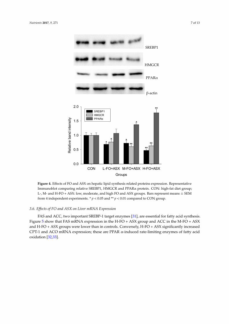
3.5. Effects of FO and ASX on Liver Protein Expression
Compared to controls (Figure 4), all FO + ASX had less SREBP1 and HMGCR protein expressionbut more PPARα protein expression was observed in M-FO + ASX and H-FO + ASX groups.
Nutrients 2017, 9, 271 7 of 13Nutrients 2017, 9, 271 7 of 13
SREBP1
HMGCR
PPARα
β‐actin
Groups
CON L-FO+ASX M-FO+ASX H-FO+ASX
Rela
tive
band
inte
nsity
0.0
0.5
1.0
1.5
2.0SREBP1HMGCRPPARα
**
**
***
****
*
Figure 4. Effects of FO and ASX on hepatic lipid synthesis related proteins expression.
Representative Immunoblot comparing relative SREBP1, HMGCR and PPARα protein. CON: high‐fat
diet group; L‐. M‐ and H‐FO + ASX: low, moderate, and high FO and ASX groups. Bars represent
means ± SEM from 4 independent experiments. * p < 0.05 and ** p < 0.01 compared to CON group.
3.6. Effects of FO and ASX on Liver mRNA Expression
FAS and ACC, two important SREBP‐1 target enzymes [31], are essential for fatty acid
synthesis. Figure 5 show that FAS mRNA expression in the H‐FO + ASX group and ACC in the
M‐FO + ASX and H‐FO + ASX groups were lower than in controls. Conversely, H‐FO + ASX
significantly increased CPT‐1 and ACO mRNA expression; these are PPAR α‐induced rate‐limiting
enzymes of fatty acid oxidation [32,33].
Figure 4. Effects of FO and ASX on hepatic lipid synthesis related proteins expression. RepresentativeImmunoblot comparing relative SREBP1, HMGCR and PPARα protein. CON: high-fat diet group;L-, M- and H-FO + ASX: low, moderate, and high FO and ASX groups. Bars represent means ± SEMfrom 4 independent experiments. * p < 0.05 and ** p < 0.01 compared to CON group.
3.6. Effects of FO and ASX on Liver mRNA Expression
FAS and ACC, two important SREBP-1 target enzymes [31], are essential for fatty acid synthesis.Figure 5 show that FAS mRNA expression in the H-FO + ASX group and ACC in the M-FO + ASXand H-FO + ASX groups were lower than in controls. Conversely, H-FO + ASX significantly increasedCPT-1 and ACO mRNA expression; these are PPAR α-induced rate-limiting enzymes of fatty acidoxidation [32,33].

Nutrients 2017, 9, 271 8 of 13Nutrients 2017, 9, 271 8 of 13
Groups
CON L-FO+ASX M-FO+ASX H-FO+ASX
Re
lativ
e m
RN
A le
vels
0
1
2
3
4
5
FASACCCPT-1ACO
**
**
***
**
Figure 5. Effects of FO and ASX on hepatic FAS, ACC, CPT‐1 and ACO mRNA. CON: high‐fat diet
group; L‐. M‐ and H‐FO + ASX: low, moderate, and high FO and ASX groups. Bars represent means ±
SEM from 4 independent experiments. * p < 0.05 and ** p < 0.01 compared to CON group.
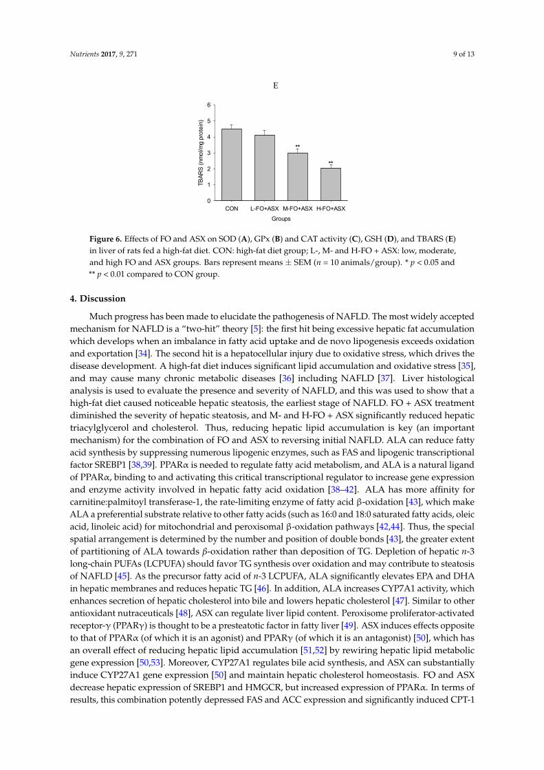
3.7. Effects of FO and ASX on Liver Antioxidant Capacity and Lipid Peroxidation
Figure 6 shows that SOD activity in the H‐FO + ASX group was increased compared to controls,
as was CAT activity in all FO + ASX groups. GPx activity was greater in the M‐ FO + ASX and H‐FO
+ ASX groups. After M‐ FO + ASX and H‐FO + ASX treatment, GSH was elevated compared with
controls. TBARS were lower in the M‐ FO + ASX and H‐FO + ASX groups compared to controls.
A B
C D
Groups
CON L-FO+ASX M-FO+ASX H-FO+ASX
GP
x (U
nits
/mg
pro
tein
)
0
100
200
300
400
500
600
**
**
Groups
CON L-FO+ASX M-FO+ASX H-FO+ASX
GS
H (
mg/
g pr
ote
in)
0
1
2
3
4
5
6
7**
**
Groups
CON L-FO+ASX M-FO+ASX H-FO+ASX
SO
D (
Uni
ts/m
g pr
ote
in)
0
20
40
60
80
100
**
Groups
CON L-FO+ASX M-FO+ASX H-FO+ASX
CA
T (
Uni
ts/m
g pr
ote
in)
0
20
40
60
80
**
**
*
Figure 5. Effects of FO and ASX on hepatic FAS, ACC, CPT-1 and ACO mRNA. CON: high-fatdiet group; L-, M- and H-FO + ASX: low, moderate, and high FO and ASX groups. Bars representmeans ± SEM from 4 independent experiments. * p < 0.05 and ** p < 0.01 compared to CON group.
3.7. Effects of FO and ASX on Liver Antioxidant Capacity and Lipid Peroxidation
Figure 6 shows that SOD activity in the H-FO + ASX group was increased compared to controls,as was CAT activity in all FO + ASX groups. GPx activity was greater in the M-FO + ASX andH-FO + ASX groups. After M-FO + ASX and H-FO + ASX treatment, GSH was elevated comparedwith controls. TBARS were lower in the M-FO + ASX and H-FO + ASX groups compared to controls.
Nutrients 2017, 9, 271 8 of 13
Groups
CON L-FO+ASX M-FO+ASX H-FO+ASX
Re
lativ
e m
RN
A le
vels
0
1
2
3
4
5
FASACCCPT-1ACO
**
**
***
**
Figure 5. Effects of FO and ASX on hepatic FAS, ACC, CPT‐1 and ACO mRNA. CON: high‐fat diet
group; L‐. M‐ and H‐FO + ASX: low, moderate, and high FO and ASX groups. Bars represent means ±
SEM from 4 independent experiments. * p < 0.05 and ** p < 0.01 compared to CON group.
3.7. Effects of FO and ASX on Liver Antioxidant Capacity and Lipid Peroxidation
Figure 6 shows that SOD activity in the H‐FO + ASX group was increased compared to controls,
as was CAT activity in all FO + ASX groups. GPx activity was greater in the M‐ FO + ASX and H‐FO
+ ASX groups. After M‐ FO + ASX and H‐FO + ASX treatment, GSH was elevated compared with
controls. TBARS were lower in the M‐ FO + ASX and H‐FO + ASX groups compared to controls.
A B
C D
Groups
CON L-FO+ASX M-FO+ASX H-FO+ASX
GP
x (U
nits
/mg
pro
tein
)
0
100
200
300
400
500
600
**
**
Groups
CON L-FO+ASX M-FO+ASX H-FO+ASX
GS
H (
mg/
g pr
ote
in)
0
1
2
3
4
5
6
7**
**
Groups
CON L-FO+ASX M-FO+ASX H-FO+ASX
SO
D (
Uni
ts/m
g pr
ote
in)
0
20
40
60
80
100
**
Groups
CON L-FO+ASX M-FO+ASX H-FO+ASX
CA
T (
Uni
ts/m
g pr
ote
in)
0
20
40
60
80
**
**
*
Figure 6. Cont.
Nutrients 2017, 9, 271 9 of 13Nutrients 2017, 9, 271 9 of 13
E
Groups
CON L-FO+ASX M-FO+ASX H-FO+ASX
TB
AR
S (
nmo
l/mg
pro
tein
)0
1
2
3
4
5
6
**
**
Figure 6. Effects of FO and ASX on SOD (A), GPx (B) and CAT activity (C), GSH (D), and TBARS (E)
in liver of rats fed a high‐fat diet. CON: high‐fat diet group; L‐. M‐ and H‐FO + ASX: low, moderate,
and high FO and ASX groups. Bars represent means ± SEM (n = 10 animals/group). * p < 0.05 and ** p
< 0.01 compared to CON group.
4. Discussion
Much progress has been made to elucidate the pathogenesis of NAFLD. The most widely
accepted mechanism for NAFLD is a “two‐hit” theory [5]: the first hit being excessive hepatic fat
accumulation which develops when an imbalance in fatty acid uptake and de novo lipogenesis
exceeds oxidation and exportation [34]. The second hit is a hepatocellular injury due to oxidative
stress, which drives the disease development. A high‐fat diet induces significant lipid accumulation
and oxidative stress [35], and may cause many chronic metabolic diseases [36] including NAFLD
[37]. Liver histological analysis is used to evaluate the presence and severity of NAFLD, and this was
used to show that a high‐fat diet caused noticeable hepatic steatosis, the earliest stage of NAFLD. FO
+ ASX treatment diminished the severity of hepatic steatosis, and M‐ and H‐FO + ASX significantly
reduced hepatic triacylglycerol and cholesterol. Thus, reducing hepatic lipid accumulation is key (an
important mechanism) for the combination of FO and ASX to reversing initial NAFLD. ALA can
reduce fatty acid synthesis by suppressing numerous lipogenic enzymes, such as FAS and lipogenic
transcriptional factor SREBP1 [38,39]. PPARα is needed to regulate fatty acid metabolism, and ALA
is a natural ligand of PPARα, binding to and activating this critical transcriptional regulator to
increase gene expression and enzyme activity involved in hepatic fatty acid oxidation [38–42]. ALA
has more affinity for carnitine:palmitoyl transferase‐1, the rate‐limiting enzyme of fatty acid
β‐oxidation [43], which make ALA a preferential substrate relative to other fatty acids (such as 16:0
and 18:0 saturated fatty acids, oleic acid, linoleic acid) for mitochondrial and peroxisomal
β‐oxidation pathways [42,44]. Thus, the special spatial arrangement is determined by the number
and position of double bonds [43], the greater extent of partitioning of ALA towards β‐oxidation
rather than deposition of TG. Depletion of hepatic n‐3 long‐chain PUFAs (LCPUFA) should favor TG
synthesis over oxidation and may contribute to steatosis of NAFLD [45]. As the precursor fatty acid
of n‐3 LCPUFA, ALA significantly elevates EPA and DHA in hepatic membranes and reduces
hepatic TG [46]. In addition, ALA increases CYP7A1 activity, which enhances secretion of hepatic
cholesterol into bile and lowers hepatic cholesterol [47]. Similar to other antioxidant nutraceuticals
[48], ASX can regulate liver lipid content. Peroxisome proliferator‐activated receptor‐γ (PPARγ) is
thought to be a presteatotic factor in fatty liver [49]. ASX induces effects opposite to that of PPARα
(of which it is an agonist) and PPARγ (of which it is an antagonist) [50], which has an overall effect of
reducing hepatic lipid accumulation [51,52] by rewiring hepatic lipid metabolic gene expression
[50,53]. Moreover, CYP27A1 regulates bile acid synthesis, and ASX can substantially induce
CYP27A1 gene expression [50] and maintain hepatic cholesterol homeostasis. FO and ASX decrease
hepatic expression of SREBP1 and HMGCR, but increased expression of PPARα. In terms of results,
this combination potently depressed FAS and ACC expression and significantly induced CPT‐1 and
Figure 6. Effects of FO and ASX on SOD (A), GPx (B) and CAT activity (C), GSH (D), and TBARS (E)in liver of rats fed a high-fat diet. CON: high-fat diet group; L-, M- and H-FO + ASX: low, moderate,and high FO and ASX groups. Bars represent means ± SEM (n = 10 animals/group). * p < 0.05 and** p < 0.01 compared to CON group.
4. Discussion
Much progress has been made to elucidate the pathogenesis of NAFLD. The most widely acceptedmechanism for NAFLD is a “two-hit” theory [5]: the first hit being excessive hepatic fat accumulationwhich develops when an imbalance in fatty acid uptake and de novo lipogenesis exceeds oxidationand exportation [34]. The second hit is a hepatocellular injury due to oxidative stress, which drives thedisease development. A high-fat diet induces significant lipid accumulation and oxidative stress [35],and may cause many chronic metabolic diseases [36] including NAFLD [37]. Liver histologicalanalysis is used to evaluate the presence and severity of NAFLD, and this was used to show that ahigh-fat diet caused noticeable hepatic steatosis, the earliest stage of NAFLD. FO + ASX treatmentdiminished the severity of hepatic steatosis, and M- and H-FO + ASX significantly reduced hepatictriacylglycerol and cholesterol. Thus, reducing hepatic lipid accumulation is key (an importantmechanism) for the combination of FO and ASX to reversing initial NAFLD. ALA can reduce fattyacid synthesis by suppressing numerous lipogenic enzymes, such as FAS and lipogenic transcriptionalfactor SREBP1 [38,39]. PPARα is needed to regulate fatty acid metabolism, and ALA is a natural ligandof PPARα, binding to and activating this critical transcriptional regulator to increase gene expressionand enzyme activity involved in hepatic fatty acid oxidation [38–42]. ALA has more affinity forcarnitine:palmitoyl transferase-1, the rate-limiting enzyme of fatty acid β-oxidation [43], which makeALA a preferential substrate relative to other fatty acids (such as 16:0 and 18:0 saturated fatty acids, oleicacid, linoleic acid) for mitochondrial and peroxisomal β-oxidation pathways [42,44]. Thus, the specialspatial arrangement is determined by the number and position of double bonds [43], the greater extentof partitioning of ALA towards β-oxidation rather than deposition of TG. Depletion of hepatic n-3long-chain PUFAs (LCPUFA) should favor TG synthesis over oxidation and may contribute to steatosisof NAFLD [45]. As the precursor fatty acid of n-3 LCPUFA, ALA significantly elevates EPA and DHAin hepatic membranes and reduces hepatic TG [46]. In addition, ALA increases CYP7A1 activity, whichenhances secretion of hepatic cholesterol into bile and lowers hepatic cholesterol [47]. Similar to otherantioxidant nutraceuticals [48], ASX can regulate liver lipid content. Peroxisome proliferator-activatedreceptor-γ (PPARγ) is thought to be a presteatotic factor in fatty liver [49]. ASX induces effects oppositeto that of PPARα (of which it is an agonist) and PPARγ (of which it is an antagonist) [50], which hasan overall effect of reducing hepatic lipid accumulation [51,52] by rewiring hepatic lipid metabolicgene expression [50,53]. Moreover, CYP27A1 regulates bile acid synthesis, and ASX can substantiallyinduce CYP27A1 gene expression [50] and maintain hepatic cholesterol homeostasis. FO and ASXdecrease hepatic expression of SREBP1 and HMGCR, but increased expression of PPARα. In terms ofresults, this combination potently depressed FAS and ACC expression and significantly induced CPT-1
Nutrients 2017, 9, 271 10 of 13
and ACO expression. Thus, the combination of FO and ASX may decrease hepatic triacylglyceroland cholesterol by suppressing hepatic lipogenesis and cholesterol synthesis as well as by promotinglipid oxidation.
Data show that high-fat diet diminishes hepatic antioxidant status and elevates oxidative stressand lipid peroxidation [54,55]. After a FO + LA diet, H-FO + ASX treated animals had more SOD,CAT and GPx activity, and more GSH. More antioxidant activity and reduced lipid peroxidationsuggested alleviation of hepatic oxidative stress. Although FO may reduce hepatic GSH depletion bylowering cholesterol and triacylglycerol [9], the potent antioxidant properties of ASX should be mainlyresponsible for the dramatic hepatic oxidative stress improvement effect after FO + LA consumption.Due to polar moieties on both ends of the polyene chain, ASX had more free radical scavengingcapability than α-tocopherol or β-carotene [56–58]. Also, structurally damaged mitochondria is amajor source of oxidative stress in NAFLD [59], and ASX protects the mitochondrial redox state and itsfunctional integrity [60] to reduce ROS production. In addition, ASX can elevate hepatic antioxidantSOD, CAT and GPx expression and activity by inducing, at least partly, the Nrf2 pathway [52,53]and many non-enzymatic antioxidants such as GSH, vitamins E and C after a high-fat diet or otherpathological conditions [16,52,61].
5. Conclusions
FO plus ASX can reduce hepatic steatosis, TG, cholesterol and oxidative stress, suggesting that thismay reduce NAFLD induced by a high-fat diet. Thus, FO + ASX may be promising for hepatoprotectionbut more work is required in additional NAFLD models to confirm this hypothesis.
Acknowledgments: This work was supported by National Natural Science Foundation of China (NSFC-31171681)and the earmarked fund for Modern Agro-industry Technology Research System (CARS-17), China.
Author Contributions: Jiqu Xu and Shuang Rong designed the overall research experiments. Hui Gao,Chang Chen, Wei Yang and Qianchun Deng performed the experiments and data analysis. Qingde Huangand Lingyun Xiao collected and analyzed the references. Jiqu Xu and Fenghong Huang wrote and editedthe manuscript.
Conflicts of Interest: The authors declare no conflict of interest.
References
1. Vuppalanchi, R.; Chalasani, N. Nonalcoholic fatty liver disease and nonalcoholic steatohepatitis: Selectedpractical issues in their evaluation and management. Hepatology 2009, 49, 306–317. [CrossRef] [PubMed]
2. Anderson, N.; Borlak, J. Molecular mechanisms and therapeutic targets in steatosis and steatohepatitis.Pharmacol. Rev. 2008, 60, 311–357. [CrossRef] [PubMed]
3. Amarapurkar, D.; Kamani, P.; Patel, N.; Gupte, P.; Kumar, P.; Agal, S.; Baijal, R.; Lala, S.; Chaudhary, D.;Deshpande, A. Prevalence of non-alcoholic fatty liver disease: Population based study. Ann. Hepatol. 2007, 6,161–163. [PubMed]
4. Ciccone, M.M.; Principi, M.; Ierardi, E.; Di Leo, A.; Ricci, G.; Carbonara, S.; Gesualdo, M.; Devito, F.; Zito, A.;Cortese, F.; et al. Inflammatory bowel disease, liver diseases and endothelial function: Is there a linkage?J. Cardiovasc. Med. (Hagerstown) 2015, 16, 11–21. [CrossRef] [PubMed]
5. Day, C.P.; James, O.F. Steatohepatitis: A tale of two “hits”? Gastroenterology 1998, 114, 842–845. [CrossRef]6. Anderson, B.M.; Ma, D.W. Are all n-3 polyunsaturated fatty acids created equal? Lipids Health Dis. 2009, 8,
33. [CrossRef] [PubMed]7. Holman, R.T. Control of polyunsaturated acids in tissue lipids. J. Am. Coll. Nutr. 1986, 5, 183–211. [CrossRef]
[PubMed]8. Barcelo-Coblijn, G.; Murphy, E.J. Alpha-linolenic acid and its conversion to longer chain n-3 fatty acids:
Benefits for human health and a role in maintaining tissue n-3 fatty acid levels. Prog. Lipid Res. 2009, 48,355–374. [CrossRef] [PubMed]
9. Yang, S.F.; Tseng, J.K.; Chang, Y.Y.; Chen, Y.C. Flaxseed oil attenuates nonalcoholic fatty liver ofhyperlipidemic hamsters. J. Agric. Food Chem. 2009, 57, 5078–5083. [CrossRef] [PubMed]
Nutrients 2017, 9, 271 11 of 13
10. Trebušak, T.; Levart, A.; Voljc, M.; Tomažin, U.; Pirman, T. The effect of linseed oil supplementation onperformance, fatty acid composition and oxidative status of rabbits. Acta Agric. Slov. 2011, 98, 119–125.[CrossRef]
11. Hussein, G.; Sankawa, U.; Goto, H.; Matsumoto, K.; Watanabe, H. Astaxanthin, a carotenoid with potentialin human health and nutrition. J. Nat. Prod. 2006, 69, 443–449. [CrossRef] [PubMed]
12. Hussein, G.; Nakamura, M.; Zhao, Q.; Iguchi, T.; Goto, H.; Sankawa, U.; Watanabe, H. Antihypertensive andneuroprotective effects of astaxanthin in experimental animals. Biol. Pharm. Bull. 2005, 28, 47–52. [CrossRef][PubMed]
13. Fassett, R.G.; Coombes, J.S. Astaxanthin: A potential therapeutic agent in cardiovascular disease. Mar. Drugs2011, 9, 447–465. [CrossRef] [PubMed]
14. Ambati, R.R.; Phang, S.M.; Ravi, S.; Aswathanarayana, R.G. Astaxanthin: Sources, extraction, stability,biological activities and its commercial applications—A review. Mar. Drugs 2014, 12, 128–152. [CrossRef][PubMed]
15. Ciccone, M.M.; Cortese, F.; Gesualdo, M.; Carbonara, S.; Zito, A.; Ricci, G.; De Pascalis, F.; Scicchitano, P.;Riccioni, G. Dietary intake of carotenoids and their antioxidant and anti-inflammatory effects incardiovascular care. Mediat. Inflamm. 2013, 2013, 782137. [CrossRef] [PubMed]
16. Bhuvaneswari, S.; Arunkumar, E.; Viswanathan, P.; Anuradha, C.V. Astaxanthin restricts weight gain,promotes insulin sensitivity and curtails fatty liver disease in mice fed a obesity-promoting diet.Process Biochem. 2010, 45, 1406–1414. [CrossRef]
17. Bhuvaneswari, S.; Yogalakshmi, B.; Sreeja, S.; Anuradha, C.V. Astaxanthin reduces hepatic endoplasmicreticulum stress and nuclear factor-kappab-mediated inflammation in high fructose and high fat diet-fedmice. Cell Stress Chaperones 2014, 19, 183–191. [CrossRef] [PubMed]
18. Turkez, H.; Geyikoglu, F.; Yousef, M.I.; Togar, B.; Gurbuz, H.; Celik, K.; Akbaba, G.B.; Polat, Z.Hepatoprotective potential of astaxanthin against 2,3,7,8-tetrachlorodibenzo-p-dioxin in cultured rathepatocytes. Toxicol. Ind. Health 2014, 30, 101–112. [CrossRef] [PubMed]
19. Takitani, K.; Miyazaki, H.; Yoden, A.; Tamai, H. Children's toxicology from bench to bed—Liver injury (2):Mechanism of antioxidant therapy for nonalcoholic fatty liver disease. J. Toxicol. Sci. 2009, 34, SP223–SP228.[CrossRef] [PubMed]
20. Xu, J.; Gao, H.; Zhang, L.; Chen, C.; Yang, W.; Deng, Q.; Huang, Q.; Huang, F. A combination of flaxseed oiland astaxanthin alleviates atherosclerosis risk factors in high fat diet fed rats. Lipids Health Dis. 2014, 13, 63.[CrossRef] [PubMed]
21. Catta-Preta, M.; Mendonca, L.S.; Fraulob-Aquino, J.; Aguila, M.B.; Mandarim-de-Lacerda, C.A. A criticalanalysis of three quantitative methods of assessment of hepatic steatosis in liver biopsies. Virchows Arch.2011, 459, 477–485. [CrossRef] [PubMed]
22. Folch, J.; Lees, M.; Sloane Stanley, G.H. A simple method for the isolation and purification of total lipidesfrom animal tissues. J. Biol. Chem. 1957, 226, 497–509. [PubMed]
23. Livak, K.J.; Schmittgen, T.D. Analysis of relative gene expression data using real-time quantitative pcr andthe 2(-delta delta c(t)) method. Methods 2001, 25, 402–408. [CrossRef] [PubMed]
24. Kono, Y. Generation of superoxide radical during autoxidation of hydroxylamine and an assay for superoxidedismutase. Arch. Biochem. Biophys. 1978, 186, 189–195. [CrossRef]
25. Sazuka, Y.; Tanizawa, H.; Takino, Y. Effect of adriamycin on the activities of superoxide dismutase,glutathione peroxidase and catalase in tissues of mice. Jpn. J. Cancer Res. 1989, 80, 89–94. [CrossRef][PubMed]
26. Goth, L. A simple method for determination of serum catalase activity and revision of reference range.Clin. Chim. Acta 1991, 196, 143–151. [CrossRef]
27. Moron, M.S.; Depierre, J.W.; Mannervik, B. Levels of glutathione, glutathione reductase and glutathiones-transferase activities in rat lung and liver. Biochim. Biophys. Acta 1979, 582, 67–78. [CrossRef]
28. Buege, J.A.; Aust, S.D. Microsomal lipid peroxidation. Methods Enzymol. 1978, 52, 302–310. [PubMed]29. Xu, J.; Zhou, X.; Deng, Q.; Huang, Q.; Yang, J.; Huang, F. Rapeseed oil fortified with micronutrients reduces
atherosclerosis risk factors in rats fed a high-fat diet. Lipids Health Dis. 2011, 10, 96. [CrossRef] [PubMed]30. Lowry, O.H.; Rosebrough, N.J.; Farr, A.L.; Randall, R.J. Protein measurement with the folin phenol reagent.
J. Biol. Chem. 1951, 193, 265–275. [PubMed]
Nutrients 2017, 9, 271 12 of 13
31. Shimano, H. Sterol regulatory element-binding protein-1 as a dominant transcription factor for generegulation of lipogenic enzymes in the liver. Trends Cardiovasc. Med. 2000, 10, 275–278. [CrossRef]
32. Clemenz, M.; Frost, N.; Schupp, M.; Caron, S.; Foryst-Ludwig, A.; Bohm, C.; Hartge, M.; Gust, R.; Staels, B.;Unger, T.; et al. Liver-specific peroxisome proliferator-activated receptor alpha target gene regulation by theangiotensin type 1 receptor blocker telmisartan. Diabetes 2008, 57, 1405–1413. [CrossRef] [PubMed]
33. Hashimoto, T.; Fujita, T.; Usuda, N.; Cook, W.; Qi, C.; Peters, J.M.; Gonzalez, F.J.; Yeldandi, A.V.; Rao, M.S.;Reddy, J.K. Peroxisomal and mitochondrial fatty acid beta-oxidation in mice nullizygous for both peroxisomeproliferator-activated receptor alpha and peroxisomal fatty acyl-coa oxidase. Genotype correlation with fattyliver phenotype. J. Biol. Chem. 1999, 274, 19228–19236. [CrossRef] [PubMed]
34. Musso, G.; Gambino, R.; Cassader, M. Recent insights into hepatic lipid metabolism in non-alcoholic fattyliver disease (nafld). Prog. Lipid Res. 2009, 48, 1–26. [CrossRef] [PubMed]
35. Lieber, C.S.; Leo, M.A.; Mak, K.M.; Xu, Y.; Cao, Q.; Ren, C.; Ponomarenko, A.; DeCarli, L.M. Model ofnonalcoholic steatohepatitis. Am. J. Clin. Nutr. 2004, 79, 502–509. [PubMed]
36. Heinonen, I.; Rinne, P.; Ruohonen, S.T.; Ruohonen, S.; Ahotupa, M.; Savontaus, E. The effects of equal calorichigh fat and western diet on metabolic syndrome, oxidative stress and vascular endothelial function in mice.Acta Physiol. 2014, 211, 515–527. [CrossRef] [PubMed]
37. Omagari, K.; Kato, S.; Tsuneyama, K.; Inohara, C.; Kuroda, Y.; Tsukuda, H.; Fukazawa, E.; Shiraishi, K.;Mune, M. Effects of a long-term high-fat diet and switching from a high-fat to low-fat, standard diet onhepatic fat accumulation in sprague-dawley rats. Dig. Dis. Sci. 2008, 53, 3206–3212. [CrossRef] [PubMed]
38. Devarshi, P.P.; Jangale, N.M.; Ghule, A.E.; Bodhankar, S.L.; Harsulkar, A.M. Beneficial effects of flaxseedoil and fish oil diet are through modulation of different hepatic genes involved in lipid metabolism instreptozotocin-nicotinamide induced diabetic rats. Genes Nutr. 2013, 8, 329–342. [CrossRef] [PubMed]
39. Ide, T.; Kobayashi, H.; Ashakumary, L.; Rouyer, I.A.; Takahashi, Y.; Aoyama, T.; Hashimoto, T.; Mizugaki, M.Comparative effects of perilla and fish oils on the activity and gene expression of fatty acid oxidationenzymes in rat liver. Biochim. Biophys. Acta 2000, 1485, 23–35. [CrossRef]
40. Ide, T. Effect of dietary alpha-linolenic acid on the activity and gene expression of hepatic fatty acid oxidationenzymes. BioFactors 2000, 13, 9–14. [CrossRef] [PubMed]
41. Murase, T.; Aoki, M.; Tokimitsu, I. Supplementation with alpha-linolenic acid-rich diacylglycerolsuppresses fatty liver formation accompanied by an up-regulation of beta-oxidation in zucker fatty rats.Biochim. Biophys. Acta 2005, 1733, 224–231. [CrossRef] [PubMed]
42. Ide, T.; Murata, M.; Sugano, M. Stimulation of the activities of hepatic fatty acid oxidation enzymes bydietary fat rich in alpha-linolenic acid in rats. J. Lipid Res. 1996, 37, 448–463. [PubMed]
43. Clouet, P.; Niot, I.; Bezard, J. Pathway of alpha-linolenic acid through the mitochondrial outer membranein the rat liver and influence on the rate of oxidation. Comparison with linoleic and oleic acids. Biochem. J.1989, 263, 867–873. [CrossRef] [PubMed]
44. DeLany, J.P.; Windhauser, M.M.; Champagne, C.M.; Bray, G.A. Differential oxidation of individual dietaryfatty acids in humans. Am. J. Clin. Nutr. 2000, 72, 905–911. [PubMed]
45. Videla, L.A.; Rodrigo, R.; Araya, J.; Poniachik, J. Oxidative stress and depletion of hepatic long-chainpolyunsaturated fatty acids may contribute to nonalcoholic fatty liver disease. Free Radic. Biol. Med. 2004, 37,1499–1507. [CrossRef] [PubMed]
46. Kim, H.K.; Choi, H. Dietary alpha-linolenic acid lowers postprandial lipid levels with increase ofeicosapentaenoic and docosahexaenoic acid contents in rat hepatic membrane. Lipids 2001, 36, 1331–1336.[CrossRef] [PubMed]
47. Morise, A.; Serougne, C.; Gripois, D.; Blouquit, M.F.; Lutton, C.; Hermier, D. Effects of dietary alpha linolenicacid on cholesterol metabolism in male and female hamsters of the lpn strain. J. Nutr. Biochem. 2004, 15,51–61. [CrossRef] [PubMed]
48. Scicchitano, P.; Cameli, M.; Maiello, M.; Modesti, P.A.; Muiesan, M.L.; Novo, S.; Palmiero, P.; Saba, P.S.;Pedrinelli, R.; Ciccone, M.M.; et al. Nutraceuticals and dyslipidaemia: Beyond the common therapeutics.J. Funct. Foods 2014, 6, 11–32. [CrossRef]
49. Moran-Salvador, E.; Lopez-Parra, M.; Garcia-Alonso, V.; Titos, E.; Martinez-Clemente, M.; Gonzalez-Periz, A.;Lopez-Vicario, C.; Barak, Y.; Arroyo, V.; Claria, J. Role for ppargamma in obesity-induced hepatic steatosis asdetermined by hepatocyte- and macrophage-specific conditional knockouts. FASEB J. 2011, 25, 2538–2550.[CrossRef] [PubMed]

Nutrients 2017, 9, 271 13 of 13
50. Jia, Y.; Kim, J.Y.; Jun, H.J.; Kim, S.J.; Lee, J.H.; Hoang, M.H.; Hwang, K.Y.; Um, S.J.; Chang, H.I.; Lee, S.J. Thenatural carotenoid astaxanthin, a ppar-alpha agonist and ppar-gamma antagonist, reduces hepatic lipidaccumulation by rewiring the transcriptome in lipid-loaded hepatocytes. Mol. Nutr. Food Res. 2012, 56,878–888. [CrossRef] [PubMed]
51. Ikeuchi, M.; Koyama, T.; Takahashi, J.; Yazawa, K. Effects of astaxanthin in obese mice fed a high-fat diet.Biosci. Biotechnol. Biochem. 2007, 71, 893–899. [CrossRef] [PubMed]
52. Yang, Y.; Seo, J.M.; Nguyen, A.; Pham, T.X.; Park, H.J.; Park, Y.; Kim, B.; Bruno, R.S.; Lee, J. Astaxanthin-richextract from the green alga haematococcus pluvialis lowers plasma lipid concentrations and enhancesantioxidant defense in apolipoprotein e knockout mice. J. Nutr. 2011, 141, 1611–1617. [CrossRef] [PubMed]
53. Yang, Y.; Pham, T.X.; Wegner, C.J.; Kim, B.; Ku, C.S.; Park, Y.K.; Lee, J.Y. Astaxanthin lowers plasma tagconcentrations and increases hepatic antioxidant gene expression in diet-induced obesity mice. Br. J. Nutr.2014, 112, 1797–1804. [CrossRef] [PubMed]
54. Jadeja, R.N.; Thounaojam, M.C.; Dandekar, D.S.; Devkar, R.V.; Ramachandran, A.V. Clerodendronglandulosum.Coleb extract ameliorates high fat diet/fatty acid induced lipotoxicity in experimental modelsof non-alcoholic steatohepatitis. Food Chem. Toxicol. 2010, 48, 3424–3431. [CrossRef] [PubMed]
55. Raso, G.M.; Esposito, E.; Iacono, A.; Pacilio, M.; Cuzzocrea, S.; Canani, R.B.; Calignano, A.; Meli, R.Comparative therapeutic effects of metformin and vitamin E in a model of non-alcoholic steatohepatitis inthe young rat. Eur. J. Pharmacol. 2009, 604, 125–131. [CrossRef] [PubMed]
56. Shimidzu, N.; Goto, M.; Miki, W. Carotenoids as singlet oxygen quenchers in marine organisms. Fish. Sci.1996, 62, 134–137.
57. Krinsky, N.I. Antioxidant functions of carotenoids. Free Radic. Biol. Med. 1989, 7, 617–635. [CrossRef]58. Miki, W. Biological functions and activities of animal carotenoids. Pure Appl. Chem. 1991, 63, 141–146.
[CrossRef]59. McCarty, M.F. Full-spectrum antioxidant therapy featuring astaxanthin coupled with lipoprivic strategies
and salsalate for management of non-alcoholic fatty liver disease. Med. Hypotheses 2011, 77, 550–556.[CrossRef] [PubMed]
60. Wolf, A.M.; Asoh, S.; Hiranuma, H.; Ohsawa, I.; Iio, K.; Satou, A.; Ishikura, M.; Ohta, S. Astaxanthin protectsmitochondrial redox state and functional integrity against oxidative stress. J. Nutr. Biochem. 2010, 21, 381–389.[CrossRef] [PubMed]
61. Sangeetha, R.K.; Baskaran, V. Retinol-deficient rats can convert a pharmacological dose of astaxanthin toretinol: Antioxidant potential of astaxanthin, lutein, and beta-carotene. Can. J. Physiol. Pharmacol. 2010, 88,977–985. [CrossRef] [PubMed]
© 2017 by the authors. Licensee MDPI, Basel, Switzerland. This article is an open accessarticle distributed under the terms and conditions of the Creative Commons Attribution(CC BY) license (http://creativecommons.org/licenses/by/4.0/).