Embed Size (px)
Citation preview

A CLINICAL EVALUATION OF PATHOLOGICAL MYOPIA
Dissertation Submitted to
THE TAMIL NADU DR. M.G.R. MEDICAL UNIVERSITY
In partial fulfilment of the regulations
for the award of the degree of
M.S. BRANCH – III
OPHTHALMOLOGY
GOVT. THANJAVUR MEDICAL COLLEGE & HOSPITAL
THE TAMIL NADU DR. M.G.R. MEDICAL UNIVERSITY
CHENNAI, INDIA.
April 2013

2
Acknowledgement
I owe my thanks to the Dean, Govt. Thanjavur Medical
College & Hospital, Dr. C.GUNASEKARAN, M.D.,D.C.H., for
allowing me to avail the facilities needed for my dissertation work.
I take great pleasure in expressing my deep sense of
gratitude and respect for Prof. P.S.GIRIDHAR, M.S., D.O., Professor and
Head of the Department of Ophthalmology and my unit Chief at Govt.
Thanjavur Medical College and Hospital for his constant encouragement
and permitting me to do the study.
I am extremely grateful to Prof. P. NALLAMUTHU,
M.S., D.O., Professor of Ophthalmology, unit II, for his guidance and
encouragement and also for his constructive criticism which helped my
work to take its present shape.
I am indebted to Dr. U.VIJAYSHANMUGAM, M.S.,
Assistant Professor, Department of Ophthalmology for giving me
guidance to conduct my study.
I am extremely grateful to all the Assistant Professors of
the Department of Ophthalmology Dr.AMUDHAVADIVU,M.S.,

3
Dr.RAJA M.S., Dr. ANBUSELVI, M.S., Dr. THAIYALNAYAGI, M.S.,
Thanjavur Medical College for their support and guiding me to complete
the study.
I thank all my colleagues and all staff of the Department for
their cooperation. Last but not the least, my sincere thanks to all the
patients who co-operated for this study, without whom this study could
not have been possible.

4
Certificate
CERTIFICATE This is to certify that the dissertation entitled “A CLINICAL
STUDY OF PATHOLOGICAL MYOPIA” is the bonafide original work
of Dr. KUMARAVEL.T. in partial fulfilment of the requirements for
M.S. Branch – III (Ophthalmology) Examination of the Tamilnadu Dr.
M.G.R. Medical University to be held in April 2013
DEAN PROF.P.S. GIRIDHAR M.S
Govt. Thanjavur Medical College & Head of the Department
Hospital, Govt.Thanjavur medical college
Thanjavur Thanjavur

5
Declaration I, Dr. KUMARVEL.T., solemnly declare that
dissertation titled, “A CLINICAL STUDY OF PATHOLOGICAL
MYOPIA” is a bonafide work done by me at Govt. Thanjavur Medical
College & Hospital during 2011-2013 under the expert guidance and
supervision of Prof. P.S.GIRIDHAR, M.S.,D.O. Head of the Department,
Department of Ophthalmology.
The dissertation is submitted to The Tamilnadu, Dr. M.G.R.Medical
University, towards partial fulfilment of requirement for the award of
M.S. Degree (Branch – III) in Ophthalmology.
Place : Thanjavur.
Date :
(Dr. KUMARAVEL)
6
CONTENTS S.NO TITLE PAGE NO
PART I REVIEW OF LITERATURE 1 HISTORY 7
2 DEFINITION & CLASSIFICTION OF MYOPIA
8
3 PREVALENCE AND ETIOPATHOGENESIS OF DEGENERATIVE MYOPIA
14
5 PATHOLOGICAL CHANGES IN DEGENERTIVE MYOPIA
19
6 ASSOCIATIONS OF DEGENERATIVE MYOPIA
33
7 MANAGEMENT OF PATHOLOGICAL MYOPIA
37
PART II
8 AIM OF STUDY 52
9 PURPOSE OF STUDY 53
10 INCLUSION AND EXCLUSION CRITERIA 54
11 MATERIALS AND METHODS 55
12 MASTER CHART 59
13 INTERPRETATION OF RESULTS 64
14 SUMMARY AND CONCLUSION 86
PART III
15 ABBREVIATIONS 89
16 PROFOMA 90
17 BIBLIOGRAPHY 92
18 PLAGIARISM SCREENSHOT 94
7
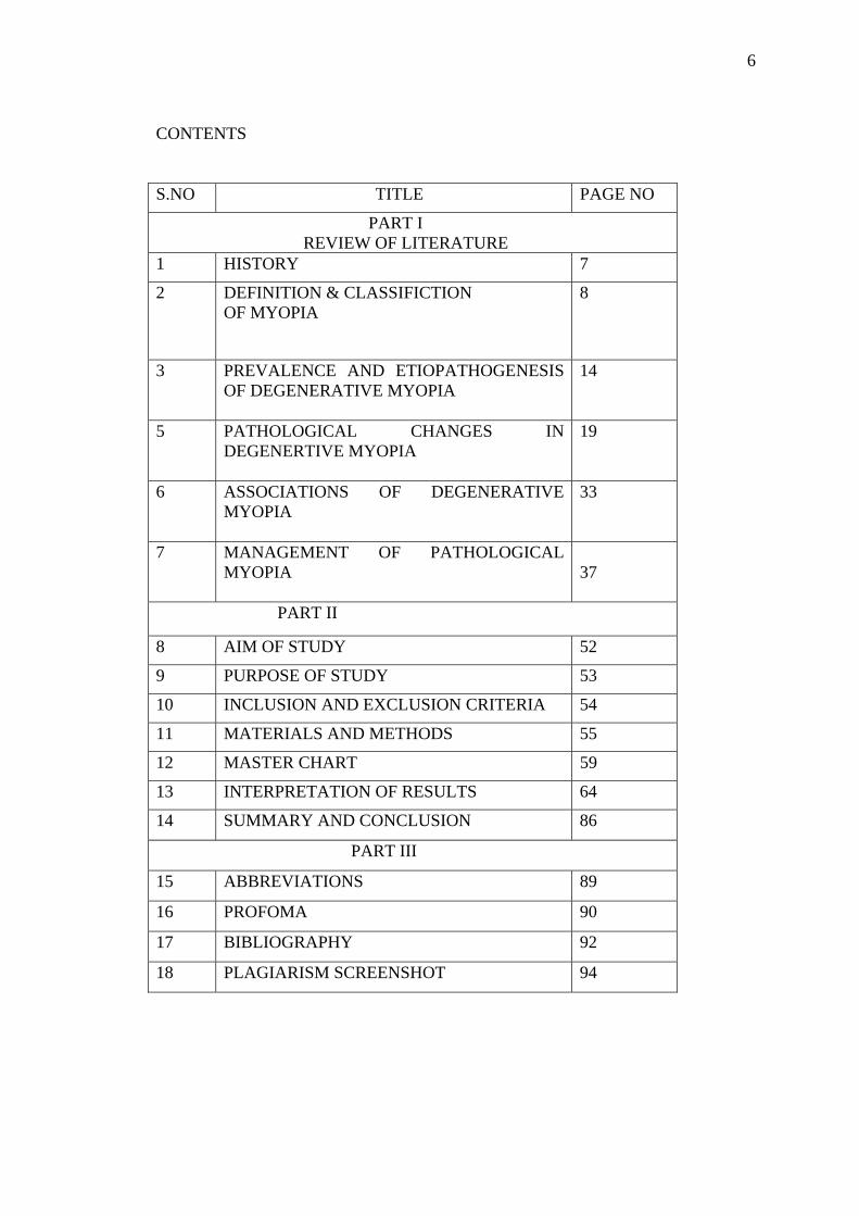
HISTORY
Myopia is well known even since ancient period. The word Myopia is a
Greek word, which means “to shut” or “to close the eye”. The original
meaning of the word Myopia was meant to describe the condition where
the patient attempts to see clearly by partially shutting or closing their
eyes.
Aristotle was the first person who was thought to have distinguished
between myopia and hypermetropia. Gallen(138-201 AD) who belongs to
Rome , used the word “myopia”. It is known by other terminologies like
“near – sightedness” or “short sightedness”
Initially the term hypometropia was used to differentiate from
hypermetropia, an opposite condition(2). But the term “myopia” still
holds good. Environmental factors were first blamed to cause myopia.
Cohn was the first to describe the “environmental theory of Myopia.
Later the “genetic theory” was also proposed. The two theories, genetic
and environmental were together referred to as “Nature vs Nurture
theory”

8
DEFINITION
Myopia or short sight is a type of refractive error in which parallel rays of
light coming from infinity are focussed in front of the retina when the
accommodation is at rest.(2)
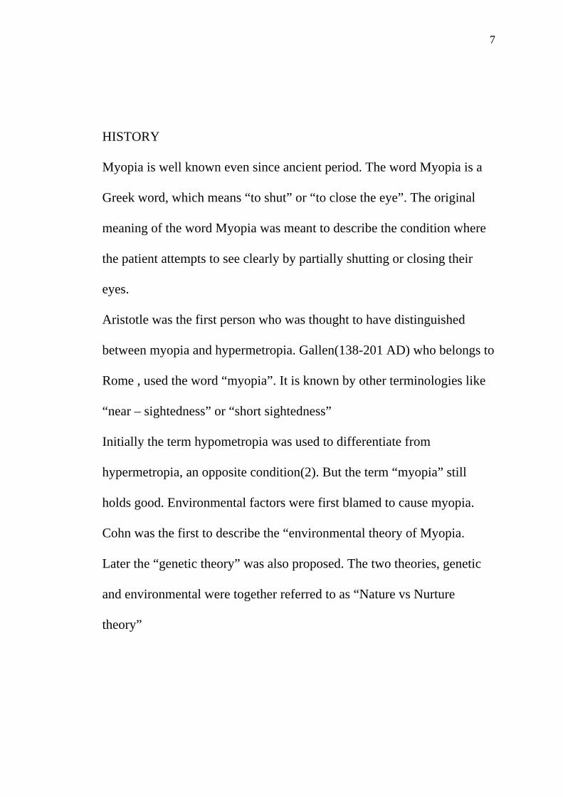
CLASSIFICATION (17)
Various classification systems have been described for myopia
1. Clinical classification:
Simple myopia, nocturnal myopia, pseudo myopia, degenerative
myopia, and induced (acquired) myopia.
2 Other systems classify myopia by degree
(i.e., low, medium, or high) or
3. by age of onset
(i.e., congenital, youth onset, early adult-onset, late adult-onset)
Classification Systems for Myopia
9
Type of Classification Classes of Myopia
Clinical entity Simple myopia
Nocturnal myopia
Pseudo myopia
Degenerative myopia
Induced myopia
Degree Low myopia (<3.00 D)
Medium myopia (3.00 D-6.00D)
High myopia(<6.00D)
Age of onset Congenital myopia (present at birth
and persisting
through infancy)
Youth-onset myopia
(<20 years of age)
Early adult-onset myopia
(2-40 years of age)
Late adult-onset myopia
(>40years of age)
10
CLINICAL VARIETIES OF MYOPIA
1. Congenital myopia
2. simple or developmental myopia
3. pathological or degenerative myopia
4. acquired myopia
CONGENITAL MYOPIA
Congenital myopia is present since birth, however usually
diagnosed by the age of 2-3 years. It is seen more frequently in
children born prematurely or with various birth defects such as
marfans syndrome or homocystinuria. Usually the error is about 8 –
10 D dioptres.
In most of the them the error is unilateral and may be associated
with convergent squint. The condition is not progressive and full
cycloplegic refractive error including astigmatic correction should
be prescribed.
11
SIMPLE MYOPIA
Simple or developmental myopia is the commonest variety. It
is considered as a physiological error not associated with any disease
of the eye. It results from normal biological variations in the
development of eye. Inheritance is considered to be autosomal
dominant. However there are number of reports which claim
recessive mode of inheritance is common.(5)
Simple myopia begins between 7 and 10 years of age and may
increase during the years of growth until stabilising round mid
teens. Refractive error is usually less than 6 D.
DEGENERATIVE MYOPIA
Pathological/degenerative/progressive myopia as the
name indicates, is a rapidly progressive condition resulting in
high myopia during early adult life which is usually associated
with degenerative changes in the eye, particularly in the
posterior segment of globe.

12
It is usually but not invariably results from a rapid axial
growth of the eyeball which is outside the normal biological
variations of development.
Duke elder(1) says ‘myopes should be classified not by
retinoscopy but by ophthalmoscopy’.
Low myopes and indeed eyes with normal axial length may
show degenerative changes characteristic of myopia while
over -17 dioptre may show no abnormal changes.
From medical point of view degenerative myopia is the most
important of all refractive errors for it is relatively common,
leading frequently to much visual disability and not infrequently
to eventual blindness. Its economic and social implications are
therefore considerable.
TYPES OF ACQUIRED MYOPIA:
INDEX MYOPIA:
This occurs in condition such as nuclear sclerosis, incipient stage
of cortical cataract and in diabetes.
13
CURVATURE MYOPIA:
Conditions where there is increase in corneal curvature as in keraroconus
may produce curvature myopia.
POSITIONAL MYOPIA:
This may occur in conditions producing anterior subluxation of lens.
CONSECUTIVE MYOPIA:
The condition follows overcorrection of hypermetropia or wrong
implantation of intraocular lens.
PSEUDOMYOPIA:
So called artificial myopia may be produced in excessive accommodation
or spasm of accommodation.
SPACE MYOPIA:
The condition occurs when the individual has no stimulation for distant
fixation.(5)
DRUG INDUCED MYOPIA:
Seen in patients using cholinergic drugs such as pilocarpine.
14
PREVALENCE
The prevalence of myopia is difficult to access from
literature since the available data deals with all types of short
sight. Myopes of over-6 dioptre represents 27-32% of myopic
population and of over -8 dioptre 6-18%. Although there are no
studies regarding prevalence of pathological myopia in India,
according to national program for control of blindness and
world health organisation (NPCB-WHO) study(8) refractive
errors are the second leading cause of low vision and blindness
counting for 18.87% of low vision and 7.35% of both low
vision and blindness. This is second most common cause of
blindness only secondary to cataract.
SEX
Sex appears to have an influence on the incidence.
Although males and females are equally affected in lower
degrees, in higher degrees females are more prone to
degenerative changes

15
RACE
High degree with degenerative changes is more common in
Chinese Japanese Arabs and Jews. Uncommon among Negroes.
ETIOLOGY
As already said, degenerative myopia is usually but not invariably
associated with increased axial length of the eyeball and degenerative
changes in the posterior segment of eye, various theories has been put
forward to explain the facts.
ROLE OF HEREDITY
Genetic factors play a major role in progressive myopia. in one study
hereditary element was present in 32.89%.father alone affected in
12.43% mother alone in 17.36% and both parent in 3.1% neither present
in 67.09%.(1)

16
It is presumed the heredity like growth of retina is the determinant in the
development of myopia. The sclera due to its distensibility follows retinal
growth but the choroid undergoes degeneration due to stretching, which
in turn causes degeneration of retina. The classical view is that due to
mechanical stretching of the posterior part of the globe causing to the
weakness of sclera. Inherited weakness of the sclera must also be
postulated.
Yet not known whether mesoderm or ectoderm is the primary fault(5)
.Mesoderm theory-
This theory says that there is disparity between sclera and extraocular
muscles. When the muscles cease to grow they exercise a strain on the
sclera. Arrest of the development of sclera at the fifth month might result
in thinness and weakening of this structure.
Neuroectodermal theory-
Each coat of the retina, choroids and the sclera has its own growth
potential. In myopia overgrowth of retina is usually genetically
determined. As the retina enlarges it pushes towards the posterior pole,
the sclera, adapting to this growth becomes thinned.

17
The choroid rendered susceptible to stretching becomes atrophied and the
retina which depends on the choroid for its nutrition degenerates
secondarily.
Another theory considers ciliary muscle as a pivotal mechanism in
controlling ocular growth by acting as a counter force to excessive ocular
expansion. According to this theory, underdevelopment of ciliary muscle
leads to excessive axial growth due to a decrease in the inhibitory activity
of the ciliary body on ocular growth.
Inheritance in lower degrees of myopia is autosomal dominant. In
higher degrees various forms of inheritance exists of which recessive
form appears to be common. Autosomal dominant type associated with
nystagmus and sex inheritance has also been reported.
In a study by institute of genetics, Fudan University, china it was found
that HLA-DQB1 gene was altered in pathological myopia
ROLE OF GENERAL GROWTH PROCESS
The role of general growth process though minor cannot be denied in
the progress of myopia. Lengthening of the posterior segment of globe
commences during the period of active growth and probably ends with
the termination of active growth.(4) Therefore, factors such as nutritional
deficiency, debilitating illness and endocrine disturbances which affect
18
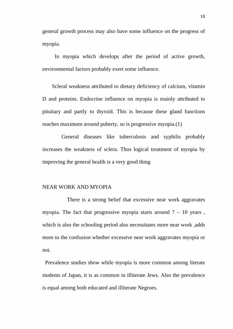
general growth process may also have some influence on the progress of
myopia.
In myopia which develops after the period of active growth,
environmental factors probably exert some influence.
Scleral weakness attributed to dietary deficiency of calcium, vitamin
D and proteins. Endocrine influence on myopia is mainly attributed to
pituitary and partly to thyroid. This is because these gland functions
reaches maximum around puberty, so is progressive myopia.(1)
General diseases like tuberculosis and syphilis probably
increases the weakness of sclera. Thus logical treatment of myopia by
improving the general health is a very good thing
NEAR WORK AND MYOPIA
There is a strong belief that excessive near work aggravates
myopia. The fact that progressive myopia starts around 7 – 10 years ,
which is also the schooling period also necessitates more near work ,adds
more to the confusion whether excessive near work aggravates myopia or
not.
Prevalence studies show while myopia is more common among literate
students of Japan, it is as common in illiterate Jews. Also the prevalence
is equal among both educated and illiterate Negroes.

19
The influence of close work is secondary and incidental in the aetiology
of the condition which is essentially predetermined.(4) As regards
the development of myopia to processes of growth, it is said that the
lengthening of the posterior segment of the eye commences only during
the period of active growth. The eye and the brain show precocious
growth at the age of 4 years; the brain is 84% and the eye 78% and the
rest of the body is 21%. After this, both the eye and the brain increase
slowly while the body grows more rapidly. However, when axial myopia
continues to progress, it is interpreted as a precocious growth which has
failed to get arrested.
PATHOLOGICAL CHANGES IN DEGENERATIVE MYOPIA:
The gross appearance of highly myopic eye is characteristic both in size
and shape. Instead of being globular it is egg shaped.
It is enlarged but the elongation of eye is almost entirely confined to the
posterior pole.
Initially thought to be inflammatory condition, it is now established all
changes characteristic of myopia are those of degenerative process.
SCLERA:
Electron microscopic studies of the sclera in high myopes show thinning
of meridional bundles with separation and splaying of cross bundles. In

20
classical view, the cause of thinning sclera was due to mechanical
stretching but atrophic element does co exist.
CHOROID:
The choroidal changes are essentially atrophic in nature (myopic
choroidal degeneration). The chief change is a generalized thinning of the
choroidal coat which becomes very attenuated and may even disappear
completely in larger area.
First change is usually a disappearance of lumina of small vessels, which
eventually appear as solid sclerotic white threads.
Vessels contain leucocytes which are not normally seen. As the
degeneration continues the choriocapillaries fails completely and the
larger vessels are alone left. Finally they too become obliterated(1)
Along with vascular changes the chromotophores lose their pigment,
disintegrate, and last of all the elastic elements disappear.
Slits occur in the elastic lamina, usually have clefts which may form
branching or reticular figures resembling cracks in lacqunae.
RETINA:
Atrophy involving retinal pigment epithelium occurs before atrophy oy
rods and cones and chorioretinal fusion occurs. The regular hexagonal
pattern of normal cells is replaced by irregular pattern of misshapen cells
with much of the pigment lying extracellularly.
21
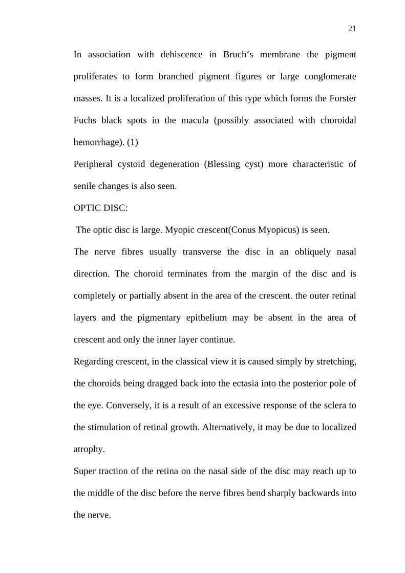
In association with dehiscence in Bruch‘s membrane the pigment
proliferates to form branched pigment figures or large conglomerate
masses. It is a localized proliferation of this type which forms the Forster
Fuchs black spots in the macula (possibly associated with choroidal
hemorrhage). (1)
Peripheral cystoid degeneration (Blessing cyst) more characteristic of
senile changes is also seen.
OPTIC DISC:
The optic disc is large. Myopic crescent(Conus Myopicus) is seen.
The nerve fibres usually transverse the disc in an obliquely nasal
direction. The choroid terminates from the margin of the disc and is
completely or partially absent in the area of the crescent. the outer retinal
layers and the pigmentary epithelium may be absent in the area of
crescent and only the inner layer continue.
Regarding crescent, in the classical view it is caused simply by stretching,
the choroids being dragged back into the ectasia into the posterior pole of
the eye. Conversely, it is a result of an excessive response of the sclera to
the stimulation of retinal growth. Alternatively, it may be due to localized
atrophy.
Super traction of the retina on the nasal side of the disc may reach up to
the middle of the disc before the nerve fibres bend sharply backwards into
the nerve.

22
Sometimes inverse crescent (inverse myopia) does occur where the
myopic crescent is situated on the nasal side and super traction on the
temporal side.
Although many ocular and systemic conditions are associated with
pathological; myopia, certain entities such as retinal venous thrombosis,
hypertensive and diabetic retinopathy are rare. Arterial occlusions,
papilloedema are also rare, while closed angle glaucoma is an exception.
In case of unilateral myopia the pathological changes may be confined to
the normal eye alone.
SYMPTOMS OF DEGERATIVE MYOPIA(3)
• Decreased visual acuity. this may greatly vary from
6/9 to loss of perception of light
• muscae volitantes .this may be really annoying to
the patients.
• Defective dark adaptation
• Colour vision defects –particularly for blue colour
• Night blindness
• Visual field defects

23
Visual field defects
Visual defects in degenerative myopia is divided in to typical
and atypical
Typical visual field defects
Enlargement of blind spot.
Loss in the superior temporal quadrant of peripheral field.
Atypical visual field
Centrocaecal scotoma
Hemianopia
Nasal scotoma
Annular scotoma
Tubular vision
24
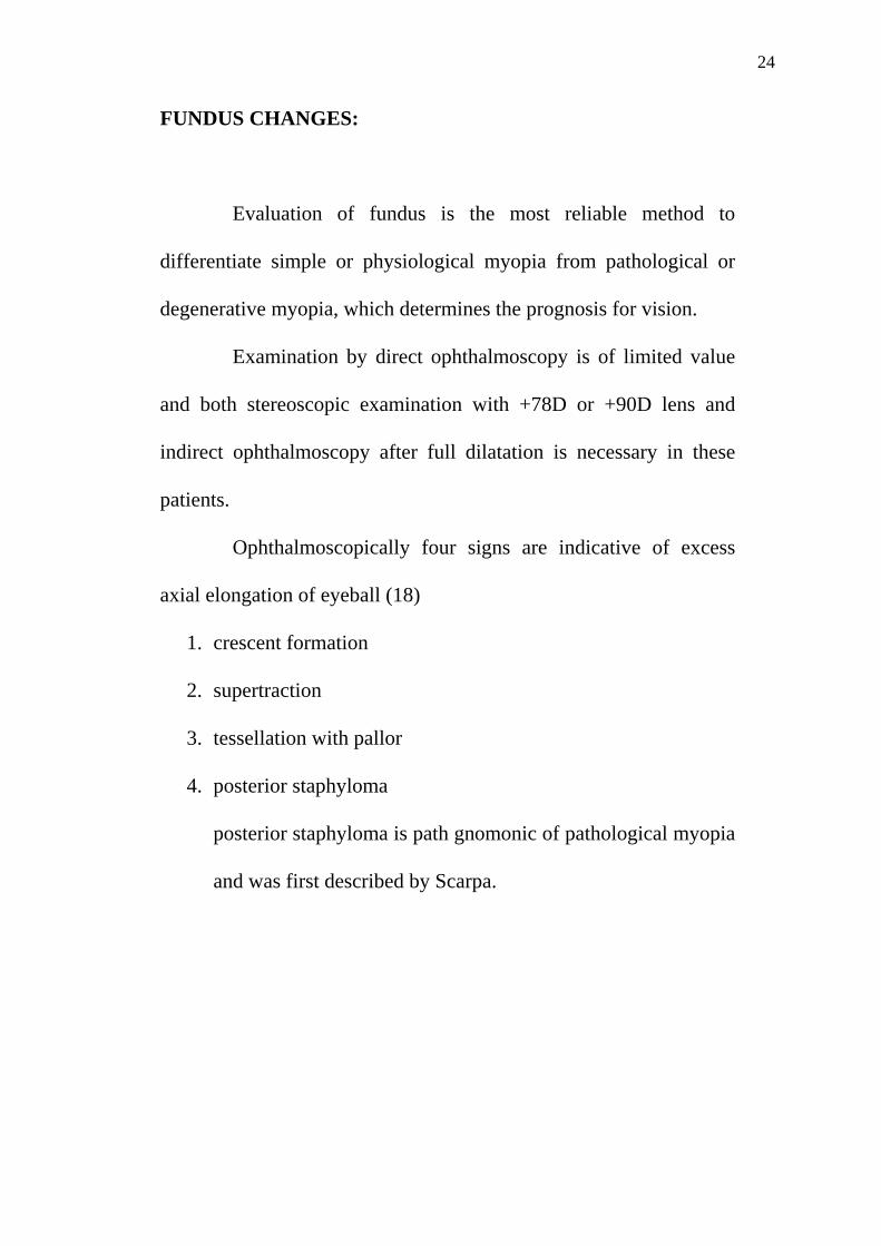
FUNDUS CHANGES:
Evaluation of fundus is the most reliable method to
differentiate simple or physiological myopia from pathological or
degenerative myopia, which determines the prognosis for vision.
Examination by direct ophthalmoscopy is of limited value
and both stereoscopic examination with +78D or +90D lens and
indirect ophthalmoscopy after full dilatation is necessary in these
patients.
Ophthalmoscopically four signs are indicative of excess
axial elongation of eyeball (18)
1. crescent formation
2. supertraction
3. tessellation with pallor
4. posterior staphyloma
posterior staphyloma is path gnomonic of pathological myopia
and was first described by Scarpa.
25
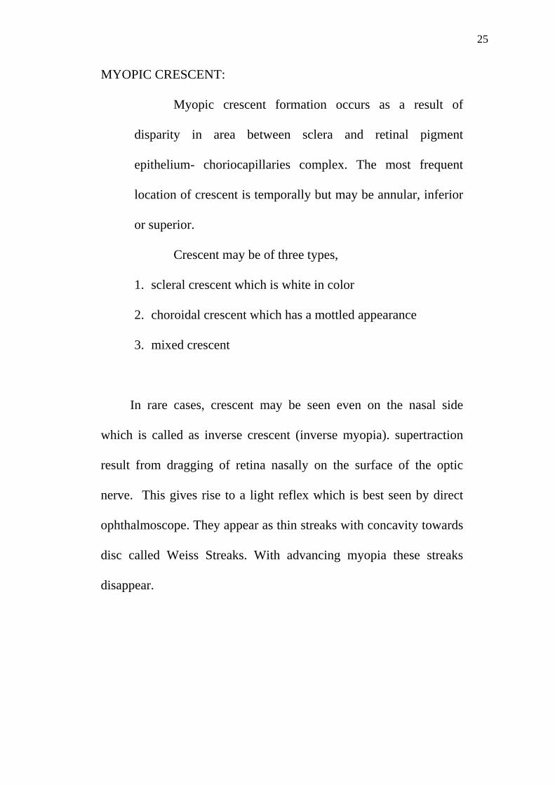
MYOPIC CRESCENT:
Myopic crescent formation occurs as a result of
disparity in area between sclera and retinal pigment
epithelium- choriocapillaries complex. The most frequent
location of crescent is temporally but may be annular, inferior
or superior.
Crescent may be of three types,
1. scleral crescent which is white in color
2. choroidal crescent which has a mottled appearance
3. mixed crescent
In rare cases, crescent may be seen even on the nasal side
which is called as inverse crescent (inverse myopia). supertraction
result from dragging of retina nasally on the surface of the optic
nerve. This gives rise to a light reflex which is best seen by direct
ophthalmoscope. They appear as thin streaks with concavity towards
disc called Weiss Streaks. With advancing myopia these streaks
disappear.
26
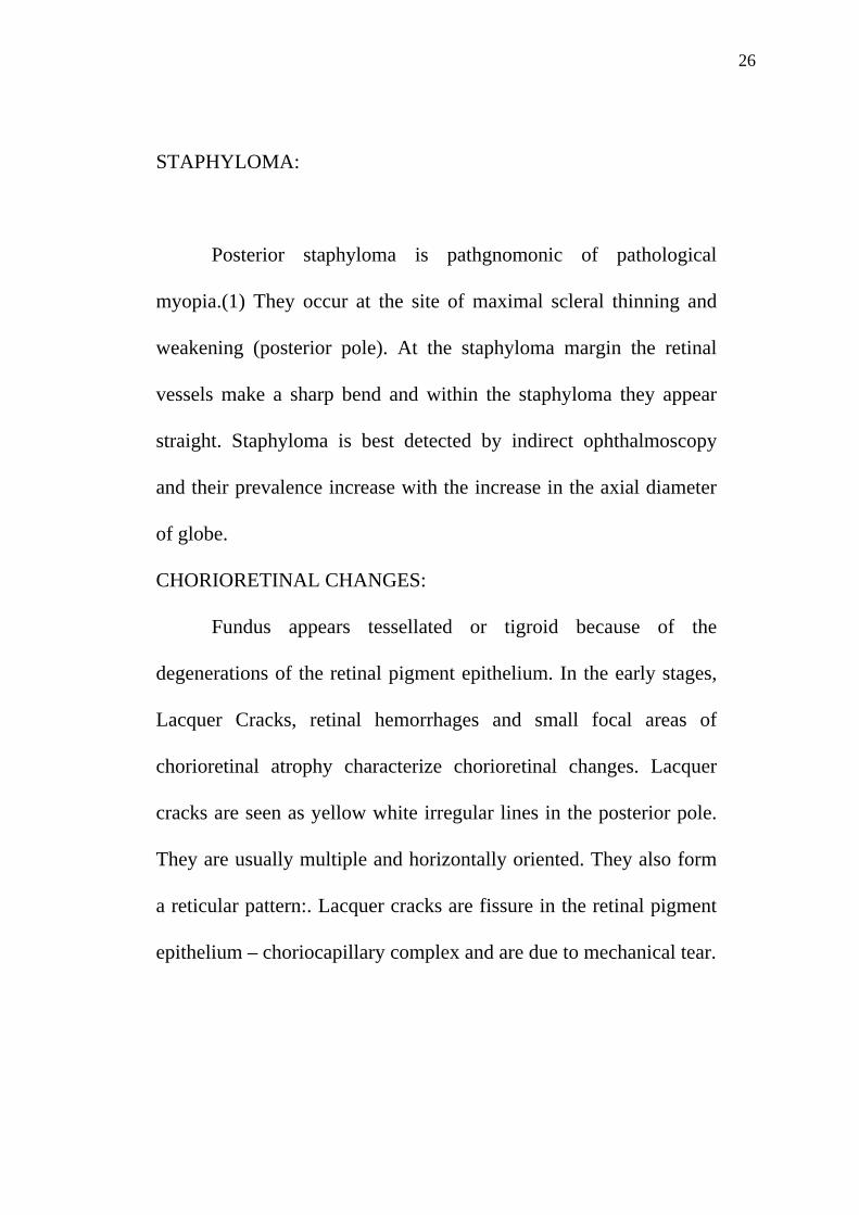
STAPHYLOMA:
Posterior staphyloma is pathgnomonic of pathological
myopia.(1) They occur at the site of maximal scleral thinning and
weakening (posterior pole). At the staphyloma margin the retinal
vessels make a sharp bend and within the staphyloma they appear
straight. Staphyloma is best detected by indirect ophthalmoscopy
and their prevalence increase with the increase in the axial diameter
of globe.
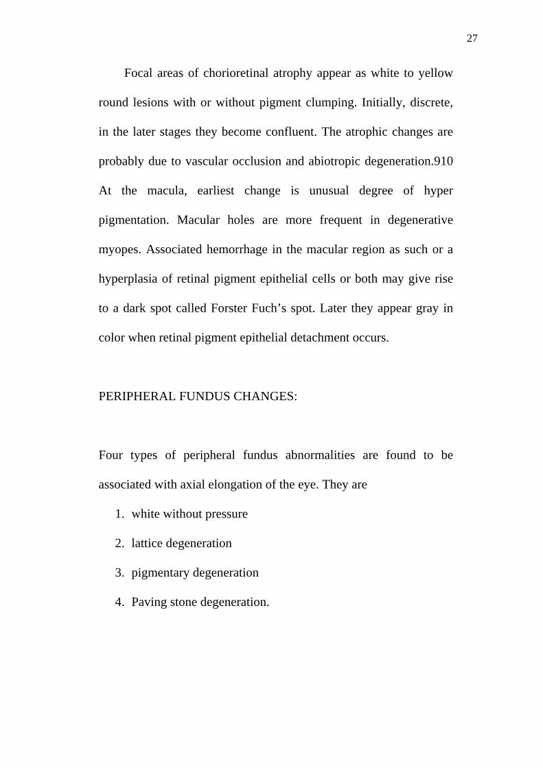
CHORIORETINAL CHANGES:
Fundus appears tessellated or tigroid because of the
degenerations of the retinal pigment epithelium. In the early stages,
Lacquer Cracks, retinal hemorrhages and small focal areas of
chorioretinal atrophy characterize chorioretinal changes. Lacquer
cracks are seen as yellow white irregular lines in the posterior pole.
They are usually multiple and horizontally oriented. They also form
a reticular pattern:. Lacquer cracks are fissure in the retinal pigment
epithelium – choriocapillary complex and are due to mechanical tear.
27
Focal areas of chorioretinal atrophy appear as white to yellow
round lesions with or without pigment clumping. Initially, discrete,
in the later stages they become confluent. The atrophic changes are
probably due to vascular occlusion and abiotropic degeneration.910
At the macula, earliest change is unusual degree of hyper
pigmentation. Macular holes are more frequent in degenerative
myopes. Associated hemorrhage in the macular region as such or a
hyperplasia of retinal pigment epithelial cells or both may give rise
to a dark spot called Forster Fuch’s spot. Later they appear gray in
color when retinal pigment epithelial detachment occurs.
PERIPHERAL FUNDUS CHANGES:
Four types of peripheral fundus abnormalities are found to be
associated with axial elongation of the eye. They are
1. white without pressure
2. lattice degeneration
3. pigmentary degeneration
4. Paving stone degeneration.
28
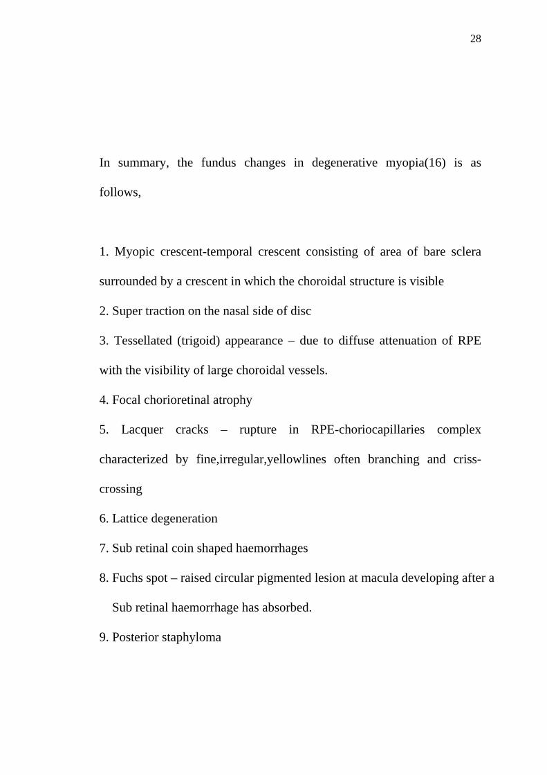
In summary, the fundus changes in degenerative myopia(16) is as
follows,
1. Myopic crescent-temporal crescent consisting of area of bare sclera
surrounded by a crescent in which the choroidal structure is visible
2. Super traction on the nasal side of disc
3. Tessellated (trigoid) appearance – due to diffuse attenuation of RPE
with the visibility of large choroidal vessels.
4. Focal chorioretinal atrophy
5. Lacquer cracks – rupture in RPE-choriocapillaries complex
characterized by fine,irregular,yellowlines often branching and criss-
crossing
6. Lattice degeneration
7. Sub retinal coin shaped haemorrhages
8. Fuchs spot – raised circular pigmented lesion at macula developing after a
Sub retinal haemorrhage has absorbed.
9. Posterior staphyloma
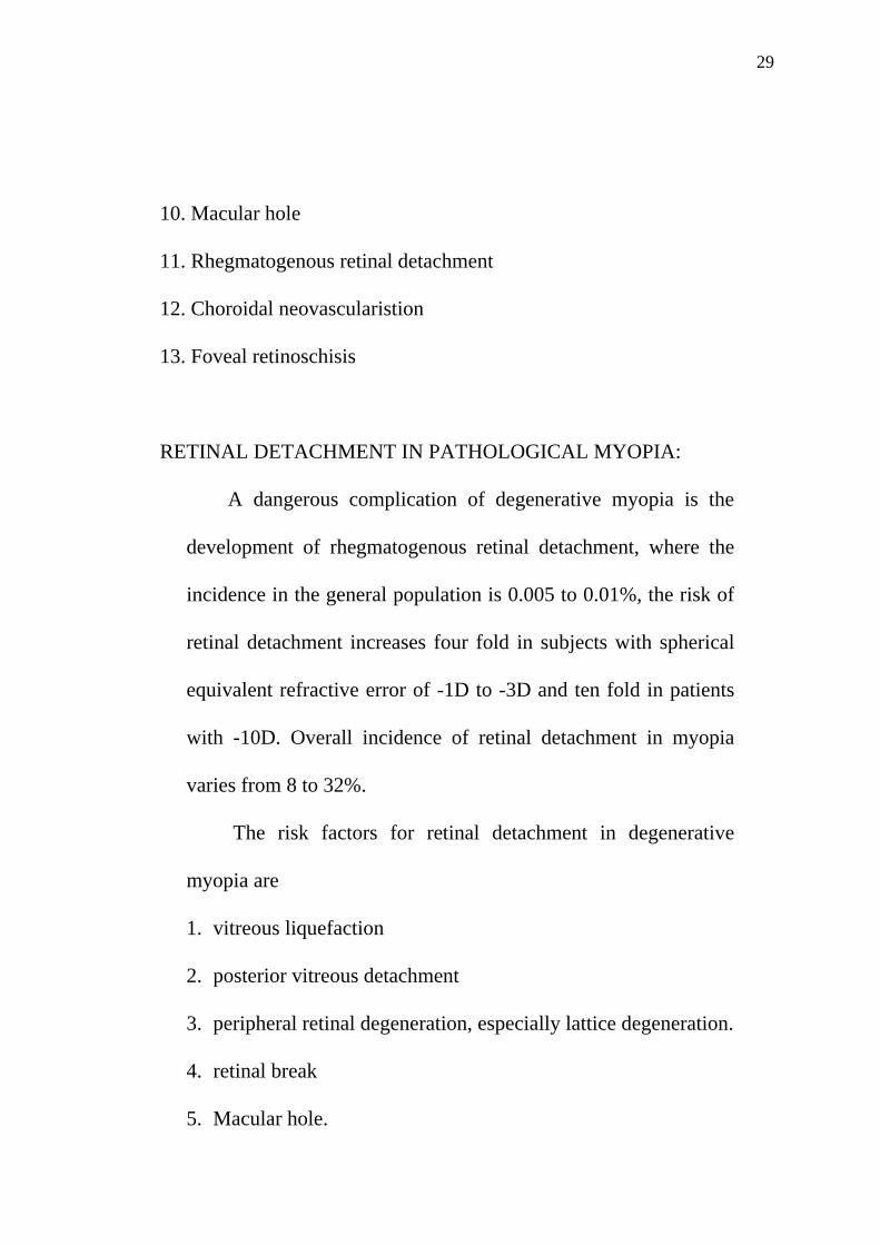
29
10. Macular hole
11. Rhegmatogenous retinal detachment
12. Choroidal neovascularistion
13. Foveal retinoschisis
RETINAL DETACHMENT IN PATHOLOGICAL MYOPIA:
A dangerous complication of degenerative myopia is the
development of rhegmatogenous retinal detachment, where the
incidence in the general population is 0.005 to 0.01%, the risk of
retinal detachment increases four fold in subjects with spherical
equivalent refractive error of -1D to -3D and ten fold in patients
with -10D. Overall incidence of retinal detachment in myopia
varies from 8 to 32%.
The risk factors for retinal detachment in degenerative
myopia are
1. vitreous liquefaction
2. posterior vitreous detachment
3. peripheral retinal degeneration, especially lattice degeneration.
4. retinal break
5. Macular hole.

30
Macular holes are more frequent in myopic eyes with retinal
detachment and before considering them as a cause of retinal
detachment, peripheral retinal detachment should be ruled out.
CHOROIDAL NEOVASCULARISATION IN DEGENERATIVE
MYOPIA:
Choroidal neovacularisation is a major cause of vision loss
in pathological myopia. CNV may take origin from lacquer
cracks. The prevalence of choroidal neovascularisation is about
5-10% in degenerative myopia. The subfoveal location is quite
frequent, accounting for 58 -74% of cases.
Myopic CNV is generally small, <1 disc area, flat, greyish,
with hyper pigmented margins. Most myopic CNVs are type II,
located in the space between sensory retina and the retinal
pigment epithelium
In CNVs due to age related macular degeneration, it is
mainly of type I, which is situated in the subretinal pigment
epithelial space.
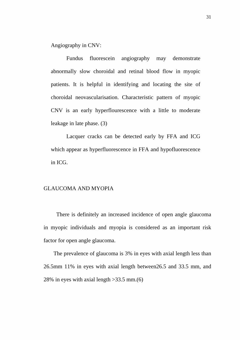
31
Angiography in CNV:
Fundus fluorescein angiography may demonstrate
abnormally slow choroidal and retinal blood flow in myopic
patients. It is helpful in identifying and locating the site of
choroidal neovascularisation. Characteristic pattern of myopic
CNV is an early hyperflourescence with a little to moderate
leakage in late phase. (3)
Lacquer cracks can be detected early by FFA and ICG
which appear as hyperfluorescence in FFA and hypofluorescence
in ICG.
GLAUCOMA AND MYOPIA
There is definitely an increased incidence of open angle glaucoma
in myopic individuals and myopia is considered as an important risk
factor for open angle glaucoma.
The prevalence of glaucoma is 3% in eyes with axial length less than
26.5mm 11% in eyes with axial length between26.5 and 33.5 mm, and
28% in eyes with axial length >33.5 mm.(6)

32
The clinical importance of the association lies in the fact that the
glaucoma is usually of an insidious type without high tension and
therefore readily missed by indentation tonometry.
The importance of using applanation tonometry is important
owing to the low ocular rigidity.
Another fact in the concept of normal or low tension glaucoma
is that individuals with normal or low tension actually have a higher value
of intraocular pressure and is falsely measured due to low ocular rigidity
as in myopia.
Previously it was thought that increased axial length in
degenerative myopia is due to increased intraocular pressure, which
determines or accentuates myopia. Also pathological myopia and
glaucoma are of the same degenerative process. It was thought that
glaucoma is sequelae to a generalised atrophy of choroids.
Not only the measurement of intraocular pressure makes difficult
in the diagnosis of glaucoma, the appearance of disc and fundus adds to
the problem. Myopic eyes have abnormal disc and their diagnosis
represents special problem in the management of glaucoma.
The distance between the level of lamina cribirosa and the level of
retina is much less when compared to myopic eyes. The average value is
0.7mm whereas in myopics it is 0.2-0.5mm.therefore a completely
cupped disc in myopia will have only half the depth of the usual

33
glaucomatous cup. Such shallow excavation is difficult to appreciate
clinically.
Also incidence of pigmentary glaucoma is more common among
young myopic males.
OCULAR AND SYSTEMIC ASSOCIATIONS OF
PATHOLOGICAL MYOPIA(7)
OCULAR ASSOCIATIONS
Retinopathy of prematurity
Congenital glaucoma
Albinism
Congenital stationary night blindness
Ectopia lentis
Retinitis pigmentosa
Wagner’s syndrome
SYSTEMIC ASSOCIATIONS
Marfans syndrome
Ehlers danlos syndrome
Downs syndrome
Alport’syndrome
34
Albinism
Congenital rubella
De Lange’s syndrome
Foetal alcohol syndrome
Gyrate atrophy-hyper ornithinemia
Laurence Moon beidel Bardet syndrome
Stickler syndrome
Pierre robin syndrome.
DIFFERENTIAL DIAGNOSIS:
RETINITIS PIGMENTOSA
A very careful history and a meticulous clinical examination will have no
difficulty in diagnosing myopia.,
Although patients with retinitis pigmentosa are frequently myopic, show
secondary cataract and vitreous liquefaction, can develop macular
degeneration and have peripheral visual field defects. These are easily
differentiated by other fundus changes.
OCULAR HISTOPLASMOSIS
Peripapillary atrophic changes, retinal pigment epithelial punched out
defects and macular neovascularisations are also seen in Ocular
Histoplasmosis Syndrome.
35
ARMD:
May develop CNV and a similar macular appearance to high myopia, but
typically drusen are present and typically myopic disc featutes are absent.
TILTED DISC:
Anolomous disc with scleral crescent inferonasally and an irregular
vascular pattern as the vessels emerge from the disc(Situs Inverses) Is
associated with areas fundus ectasia in the direction of tilt. Most patients
have myopia and astigmatism but no chorioretinal degeneration or
lacquer cracks.
TOXOPLASMOSIS:
A well circumscribed chorioretinal scar that does not typically develop
CNV. Active disease show retinitis and vitritis which are absent in high
myopia.
GYRATE ATROPHY:
The condition is Rare. Well demarcated multiple areas of chorioretinal
atrophy beginning in the mid periphery in childhood. Later they coalesce
to involve a larger portion. Blood levels of ornithine are increased.
Patients are often high myopic.
36
INVESTIGATIVE WORK –UP(15)
1. Visual acuity measurement
2. IOP measurement by applanation tonometry.(indentation
tonometry like Schiotz may underestimate IOP in high myopes.
3. Refractive error estimation
4. Slit lamp examination and fundus stereoscopic examination with
78D or 90D lens of the macula to identify CNV which may appear
as grey or green lesion beneath the retina.
5. Detailed evaluation of fundus periphery with indirect
ophthalmoscopy to search for retinal breaks or detachment. Care
should be taken in doing a scleral depression over a staphyloma.
6. FFA and ICG in suspected CNV patients and in those where
lacquer cracks transverse the macula
7. Ocular coherence tomography when a macular detachment is
suspected

37
MANAGEMENT:
Degenerative Myopia as such cannot be prevented by any
known treatment. It is advisable to forewarn a couple with high
myopia about a strong possibility to be affected At present, treatment
is focused on controlling the possible complications related to the
progression of degenerative myopia, with special attention to
choroidal neovascularisation and retinal detachment.
A periodic examination is always indicated in all these patients
.
Management of degenerative myopia should run along the
following general guidelines.(3)
1. correction of the refractive error
2. early identification and treatment of the complications so as to
prevent the patient from becoming virtually blind
3. building up of adequate visual hygiene and general health
and visual rehabilitation in case of low vision patients.
38
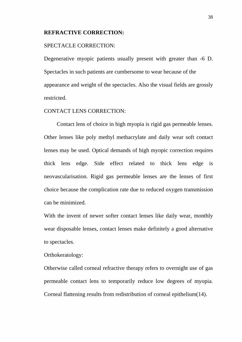
REFRACTIVE CORRECTION:
SPECTACLE CORRECTION:
Degenerative myopic patients usually present with greater than -6 D.
Spectacles in such patients are cumbersome to wear because of the
appearance and weight of the spectacles. Also the visual fields are grossly
restricted.
CONTACT LENS CORRECTION:
Contact lens of choice in high myopia is rigid gas permeable lenses.
Other lenses like poly methyl methacrylate and daily wear soft contact
lenses may be used. Optical demands of high myopic correction requires
thick lens edge. Side effect related to thick lens edge is
neovascularisation. Rigid gas permeable lenses are the lenses of first
choice because the complication rate due to reduced oxygen transmission
can be minimized.
With the invent of newer softer contact lenses like daily wear, monthly
wear disposable lenses, contact lenses make definitely a good alternative
to spectacles.
Orthokeratology:
Otherwise called corneal refractive therapy refers to overnight use of gas
permeable contact lens to temporarily reduce low degrees of myopia.
Corneal flattening results from redistribution of corneal epithelium(14).

39
REFRACTIVE SURGERY
Keratorefractive surgeries may be helpful only to correct
the refractive component of the degenerative myopia but does not arrest
the degenerative changes.
Refractive surgery includes any procedure done to alter the
refractive condition of the eye to improve uncorrected refractive error.
The history dates back to nineteenth century when both contact lenses
and laser were not available. With the discovery of contact lenses in
1950’s refractive surgery became secondary interest due to simplicity and
relative safety of contact lenses. Interest in refractive surgeries renewed
in 1970 because occupations which demanded good vision without
glasses and contact lenses like pilots and athletes sought refractive
surgeries.
PRINCIPLES OF REFRACTIVE SURGERIES:
The anterior surface of cornea is responsible for 60 – 70%of optical
power of eye. This significant contribution makes it the most operated
part upon the eye in most refractive surgeries. Myopia can be corrected
by making the central cornea more flat. Phakic intraocular lenses change
the refractive state by changing the refractive media of the eye. Surgical
techniques can be classified into incisional, thermal, lamellar and
intraocular.
40
Incisional methods include procedures such as radial keratotomy.
Thermal based refractive surgeries use heat to shrink the collagen in the
corneal stroma. The latest technique uses Holmium: Yag to produce the
shrinkage.
Lamellar procedures alter directly the shape of cornea by ablation or
placement of pre designed corneal lenticules in the cornea. These include
keratomileusis, keratophakia, epikeratoplasty, photorefractive
keratectomy and intrastromal photoablation. Among these procedures
laser assisted in situ keratomileusis (LASIK) is the most popular.
INCISIONAL CORNEAL SURGERIES:
Since 1980s incisional corneal surgeries have had periods of adoption,
refinement, and abandonment. Incisional surgery for myopia has been
replaced by laser procedures. The role of incisional surgeries reserved for
correcting astigmatism after cataract surgery (limbal relaxing incision)
and after penetrating keratoplasty(arcuarte keratotomy). Contribution to
incisional corneal surgeries were made by Sato of japan and Fyodorov of
Russia. Sato observed central corneal flattening and improvement of
vision after healing of spontaneous rupture of descemets membrane in
keratoconus patients. Fyodorov established that the central corneal clear
zone was inversely related to the amount of refractive correction.

41
In radial keratotomy, radial peripheral corneal incision are made
either posterior (Japanese technique) or anteriorly (Russian technique).
Upon healing of the incision, flattening of the incision occurs.(14)
Radial keratotomy was used to treat patients with refractive error of -1
to -4 D of myopia and its role in pathological myopia is insignificant.
ONLAY AND INLAY TECHNIQUES:
In these techniques, the refractive error is corrected by preformed
tissue or synthetic material onto or into the cornea. Corneal ring segments
are under investigation to treat ectactic condition such as keratoconus or
ectasia after refractive surgeries. As for as now laser procedures replaced
all of the older procedures.
PHOTOABLATIVE TECHNIQUES:
The Argon fluoride Excimer Laser reduces the refractive error by
ablating the anterior corneal stroma to a new radius of curvature.
There are three major refractive surgical techniques that employ Excimer
Laser ablation(14)
1. photorefractive keratectomy(PRK), where the epithelium is derided
2. LASEK , where the epithelium is preserved as a flap
42
3. LASIK.here the Excimer laser ablation is performed under a
lamellar flap made with a microkeratome or using Nd;Yag
femtosecond laser(Intralase)
PHOTOREFRACTIVE KERATECTOMY:
Here the epithelium is removed by a sharp blade or blunt spatula after
application of diluted absolute alcohol (20%) to the corneal surface to
loosen the epithelium. Epithelium can also be removed by transepithelial
ablation by the Excimer Laser itself. Photo ablation is then applied and
postoperatively the patient is fitted with bandage soft contact lens for 48
to 72 hrs.
The popularity of PRK decreased when LASIK came because of faster
visual recovery and decreased post operative discomfort with LASIK.
PRK is an alternative to LASIK in conditions where very low refractive
correction is needed, epithelial basement disease, thin cornea and for
treatment of LASIK flap related complications like button holed flap.
With the advent of wave front guided laser ablation the popularity of
PRK may increase.
LASIK in high myopia:
Although LASIK is done for myopia up to -12 to -16D, in this high
range the predictability of the procedure is markedly reduced. In addition,
while treating such a high myopia, there was a high incidence of loss of

43
best corrected visual acuity than in the correction of low levels of
myopia. However, patients with high myopia often gain best corrected
visual acuity after LASIK, probably due to decreased image
magnification preoperatively due to spectacles. The required ablation
depths for high corrections may leave an inadequate stromal bed for long
term instability of cornea. The stromal bed thickness after refractive
surgery should be at least 300 microns in the minimum. In addition,
higher order corrections have an unacceptable of high side effects,
including halos, glare and loss of contrast sensitivity. These side effects
are due to increased high order aberrations like spherical aberrations.
CONTRAINDICATIONS OF LASIK(5)
1. thin cornea
2. ectactic corneal disorder
3. dry eyes
4. blepharophimosis
5. glaucoma
6. large pupil size
7. monocular patients
8. retinal vascular disease
9. autoimmune disease
10. Pregnancy.
44
COMPLICATIONS OF LASIK:
Intraoperative complications
Flap related complication:
Flap of variable and suboptimal thickness and diameter
Incomplete flap:
Tear or hole in the flap
Free flap
Damage, destruction or dislocation of flap
Complications during laser application:
Decentration of ablation
Incorrect ablation
Ablation of hinge
Complication during flap reposition
Incorrect placement of flap
Wrinkling on reposition of flap.
Damage of flap due to increased handling
Postoperative complication:
poor adhesion and anchoring of flap with the stromal bed
Infective keratitis
Non-specific intrastromal / intralamellar keratitis (Sands of Sahara)
Epithelial ingrowth under the flap
Undercorrection or overcorrection

45
Regression
Haze at the interface
Corneal ectasia
INTRAOCULAR SURGERIES:
1. PHAKIC IOL
2. CLEAR LENS EXTRACTION(REFRACTIVE LENS
EXCHANGE)
PHAKIC IOL represents a new category of IOL that expand the range of
keratorefractive surgery. While LASIK surgery can only be done for
refractive error up to -14D , PIOL can be done for up to -20D. PIOL can
be an alternative if PRK and LASIK are contraindicated.(14)
Types of phakic IOL
1. angle supported PIOL
2. iris supported PIOL
3. sulcus supported PIOL
The combination of corneal and intraocular refractive surgeries is
called BIOPTICS. This may ultimately allow patients at the extremes
of refractive error to achieve predictable outcome.
PIOL are contraindicated in pre-existing intraocular diseases such as
corneal endothelial decompensation, iritis, rubeosis iridis, and cataract
with glaucoma.
46
CLEAR LENS EXCHANGE may be preferable to PIOL in the presence
of lens opacity.
EARLY IDENTIFICATION AND TREATMENT OF
COMPLICATIONS:
The most important causes of vision loss in degenerative myopia are
choroidal neovascularisation, macular degeneration and retinal
detachment. Every effort should be made to examine thoroughly all
highly myopic patients by indirect ophthalmoscopy to identify the
peripheral retinal degenerations. Prophylactic laser treatment of the
identified patients may avoid retinal detachment.
Choroidal noeovascularisation in early stages may not be detected by
clinical examination alone. Special investigations like fundus flourescein
angiography, indocyanine green angiography and optical coherence
tomography are necessary to identify early CNV.
Laser photocoagulation has been applied successfully in
juxtrafoveal and extrafoveal choroidal neovascularisation. Laser is not
indicated in subfoveal choroidal neovascularisation. Treatment options
other than laser include surgery, photodynamic therapy and anti-
Vascular Endothelial growth Factors.

47
Successful surgery for choroidal neovascularisation is surgical
removal of CNV and macular translocation with 360 degree
retinotomy. Vertiporfin in photodynamic therapy trials have shown
that it is effective in retaining vision by stabilizing or improving visual
acuity and contrast sensitivity particularly when used in combination
with anti Vascular Endothelial Growth factors. This is also useful in
recurrent cases.
OTHER MODES OF APPROACH
The management of stretching of the sclera and the arrest of the
degenerative process to date is not rewarding. Attempts are made to
strengthen the thinned out sclera by patch graft but was not very
successful. Bearing in mind, the scleral thinning is due to intra ocular
pressure, use of anti glaucoma medications have been tried with
variable results. Treatment in animal models based on biochemical
and genetic approach are promising. One such is the modification of
the scleral proteoglycan synthesis which might be able to inhibit the
development of globe enlargement

48
VISUAL REHABILITATION:
One of the major causes of visual disability is pathological myopia which
is largely unaddressed.
Rehabilitation is as important as the prevention and control of blindness.
The visually challenged persons may need the following types of
rehabilitation
1. Medical rehabilitation
By low vision aids many visually challenged persons may have
some useful vision. A low vision patient is a person who because of an
irreversible disorder of the visual system cannot perform customary
visual activities without special vision enhancing devices.
A low vision aid refers to an optical device that improves the residual
vision by magnifying the image of the object at the retinal level. Also
there are some non optical aids which may be helpful in improving the
residual vision.
Principle of low vision aids is based on the fact that with sufficient
magnification the normal retina surrounding the damaged retina can be
used for central vision. Though the surrounding retina is less sensitive
than the fovea or parafoveal region some useful vision may be obtained.
49
Types of optical low vision aids(5)
The currently available optical low vision aids are as follows
1. Magnifying spectacles
2. Hand held magnifiers
3. Stand magnifyiers
4. Telescops
5. close-circuit television
Magnifying spectacles are the most commonly prescribed low
vision aids and many patients achieve a high degree of success with their
use. They may be uniocular or binocular spectacles. the vantages of
magnifying spectacles re more comfortable to use ,both hands are free
field of vision is large, simultaneous near and distant vision possible and
are less expensive.
Closed circuit television (CCTV)
In CCTV the camera picks up the reading material, magnifies and
displays it on the television screen. They provide excellent contrast
and magnification. These can be modified for a variety of purposes like
reading, writing, computer works, crafts etc.
A CCTV provides a number of advantages over the optical systems. it
provides a distortion free , brighter, magnified image with enhanced
50
contrast on a magnified screen. The letters on the back screen also helps
in image improvement in some cases. The major limiting factor is that it
is expensive, heavy, and difficult to mobilise.
Non optical devices
The various types of non optical methods are
Approach magnification
Proper lightning
Contrast enhancement
Auditory aids such as talking clocks, computers with speech synthesizers
are available for use by visually challenged persons.
Fibre tipped pens- with black ink provides best contrast while
writing. The writing ids are quit useful for signature and cheque
writing.
2. TRAINING AND PSYCHOLOGICL REHABILITATION
It is the most important aspect. First of all they should be assured
and made to feel that they are equally useful and not inferior to
sighted persons. Their training should include mobility training
with help of a stick, training in daily activities like washing, putting
clothes, shaving, cooking and other household works.
3. EDUCATIONAL REHABILITTION
51
It includes education in blind schools with the facility of Braille
system of education.
4. VOCATIONAL REHBILITATION
It will help them to earn their livelihood. Blind persons can be
trained in making handicrafts, book binding, candle and chalk
making, etc.
To conclude, the strategies must ensure that
No citizens go blind needlessly due to preventable causes
All avenues are exhausted to restore the best of possible vision
to curable blind persons.
Blinds not amenable to curable measure receive comprehensive
rehabilitation.
52
PART – II
AIM OF THE STUDY
1. To analyse the ocular parameters associated with degenerative
myopia
2. To study the corneal thickness, axial length and Refractive
parameters with reference pathological myopia
3. To assess the ocular associations of pathological myopia
4. To assess the visual status associated with pathological myopia
5. To determine the visual disability and the major causes of vision
loss in pathological myopia
6. To categorise and assess the percentage of visual disability
53
PURPOSE OF STUDY
Degenerative myopia is common in India and is a major cause for visual
disability According to NPCB-WHO SURVEY 1980 uncorrected
refractive errors, particularly pathological myopia, accounts for 7.355 of
all causes of bilateral blindness which is only second to cataract among
all leading causes of blindness. In Tamilnadu the percentage goes to 21.5
% .also in the survey for causes of low vision in India, refractive errors
accounts for 18.875 and is the second leading cause for low vision.
While prevention is not possible at least blindness from pathological
myopia may be prevented, at least to some extent. this is possible by early
detection of the most common cause of vision loss the choroidal
neovascularisation by appropriate investigation and treating them. also
early detection of peripheral retinal degeneration And prophylactic laser
treatment would be helpful.
Vision loss in myopes is not only because of their high refractive error
but also due to the associated conditions with it.
One among them which gains importance is open angle glaucoma which
is easily missed because of the falsely recorded visual acuity and the
misleaded optic disc apperaence.prompt recognition of these associated
conditions and addressing them will be greatly helpful to them in
retaining their vision.
54
INCLUSION AND EXCLUSION CRITERIA
INCLUSION CRITERIA
1. All patients with refractive error greater than – 6 dioptre
spherical equivalents in one or both eyes were included in the
study.
2. Patients with age 10 – 40 who filled the above criteria were
included in the study.
3. Both sexes, males and females ,were included in the study
4. Patients were included irrespective of the visual acuity
EXCLUSION CRITERIA
1. Patients with previous intraocular surgery were excluded
from the study.
2. Patients aged less than 10 years and greater than 40 years
were excluded from the study
3. Patients wearing contact lenses were excluded from the
Study.

55
MATERIALS AND METHODS
This is a retrospective study done in eye department, Thanjavur medical
college, Thanjavur from June 2011 to October 2012.
A total of fifty patients attending outpatient department were selected.
Informed consent was obtained from all the patients.
All patients underwent a complete ocular examination including
1. Visual acuity measurement- by snellens visual acuity chart
2. Anterior segment examination by torch light followed by slit
lamp examination.
3. Corneal curvature measurement and retinoscopy by automated
refractometer.
4. Intraocular measurement by goldmann applanation tonometer
after instillation of 4 % lignocaine and staining with sterile
fluorescin strips.
5. Central corneal thickness measurement by pachymeter
56
6. Axial length measurement by ultrasound A scan
7. Gonioscopy by Goldman single mirror
8. Dilated fundus examination using direct, bio microscopic
examination with + 78 dioptre lens, indirect method.
9. In selective cases, ultrasound –B scan and was also done.
10 .In selective cases visual field examination was done on another
day with Octopus automated perimeter.
All the measurements and examination were done by single trained
person to avoid inter observer variations.
57
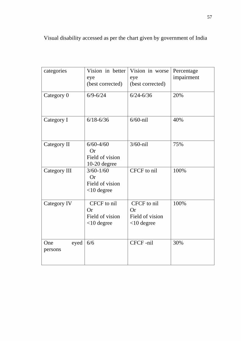
Visual disability accessed as per the chart given by government of India categories Vision in better
eye (best corrected)
Vision in worse eye (best corrected)
Percentage impairment
Category 0 6/9-6/24 6/24-6/36 20%
Category I 6/18-6/36 6/60-nil 40%
Category II 6/60-4/60 Or Field of vision 10-20 degree
3/60-nil 75%
Category III 3/60-1/60 Or Field of vision <10 degree
CFCF to nil 100%
Category IV CFCF to nil Or Field of vision <10 degree
CFCF to nil Or Field of vision <10 degree
100%
One eyed persons
6/6 CFCF -nil 30%

58
KEY TO MASTER CHART:
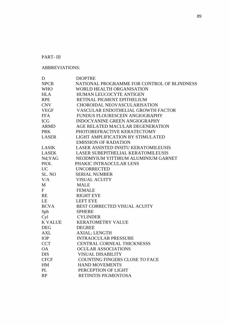
SL. NO SERIAL NUMBER
V/A VISUAL ACUITY
M MALE
F FEMALE
RE RIGHT EYE
LE LEFT EYE
BCVA BEST CORRECTED VISUAL ACUITY
Sph SPHERE
Cyl CYLINDER
K VALUE KERATOMETRY VALUE
DEG DEGREE
AXL AXIAL; LENGTH
IOP INTRAOCULAR PRESSURE
CCT CENTRAL CORNEAL THICKNESSS
OA OCULAR ASSOCIATIONS
DIS VISUAL DISABILITY
CFCF COUNTING FINGERS CLOSE TO FACE
HM HAND MOVEMENTS
PL PERCEPTION OF LIGHT
RP RETINITIS PIGMENTOSA

59
MASTER CHART
TABLE 1.1
SL.NO
NAME AGE/ SEX
V/A
REFRACTION K VALUE
IOP
CCT AXL OA DIS %
UC
BCVA
Sph Cyl AXIS
1 MANIKANDAN
17/M
RE 2/60 6/60 -6.00 -1.00
70 45.75 45.75
14
0.527 24.07 -
30
LE 6/6 6/6 - - - 45.25 45.25
18
0.528 23.45
2 JOSHI 20/M
RE CFCF 4/60 -19.75 -1.0 25 43.25 43.00
16
0.512 33.25 - 75
LE HM HM -22.00 - - 43.00 43.00
16
0.487 35.06
3 PERIYAVOTT AIYI
46/F RE 6/60 6/12 -6.50 - - 42.25 44.25
16
0.512 22.18 - 30
LE CFCF 3/60 -9.00 - - 45.25 45.50
14
0.496 27.17
4 VIGNESH 17/M
RE 5/60 6/18 -11.00 -2.00
180 42.00 44.50
14
0.476 26.47
- -
LE 5/60 6/18 -11.00 -1.50
120 42.75 44.75
12
0.475 28.99
5 MANIKANDAN
15/M
RE 2/60 6/60 -10.00 - - 41.25 43.00
16
0.528 24.12 - 75
LE 1/60 1/60 -12.00 - - 41.30 42.25
14
0.562 24.56
6 SIVARANJINI
9/F RE CFCF 3/60 -18.00 -2.0 172 47.00 48.75
12
0.419 26.55 RP, KERATOCONUS
100
LE CFCF 2/60 -24.00 -3.50
29 47.25 48.75
12
0.465 26.42
7 VAISHNAVI 22/F RE 5/60 6/18 -3.75 -4.75
32 40.50 42.25
18
0.465 23.75 - -
LE 4/60 6/18 -3.75 -3.25
179 40.00 42.75
18
0.479 24.01
8 MURALI 32/M
RE 4/60 6/36 -5.00 -2.00
120 43.25 43.25
16
0.512 23.82 - -
LE 6/60 6/18 -4.50 -2.00
30 44.12 44.25
18
0.514 23.89
9 BOSE 18/M
RE 3/60 6/60 -8.00 - - 42.25 43.00
14
0.504 24.18 - -
LE 3/60 6/60 -9.00 - - 41.25 43.75
14
0.502 24.25
10 SUJATHA 30/F RE 6/60 6/12 -8.50 -2.50
70 42.00 43.75
14
0.482 27.79 - 30
LE CFCF CFCF
-6.00 -3.50
180 42.00 44.75
12
0.481 28.13

60
TABLE 1.2
S.NO
NAME AGE/ SEX
V/A REFRACTION K VALUE
IOP CCT 0.521
AXL
OA DIS %
UC
BCVA
Sph Cyl AXIS
11 KASTHURI 24/M RE 1/60 1/60 -7.00 -2.00
70 41.30 42.25
26 0.521 24.81 GLAUCOMA 75
LE 2/60 6/60 -6.00 -1.50
150 41.75 43.00
20 0.493 25.01
12 SHANKAR 22/M RE 6/60 6/12 -6.00 - - 44.15 44.50
18 0.518 23.18 - -
LE 5/60 6/12 -7.00 - - 44.25 44.25
18 0.522 23.45
13 KRISHNAN 42/M RE CFCF 2/60 -8.00 - - 43.25 42.75
22 0.488 24.26 GLAUCOMA 100
LE CFCF 2/60 -8.00 - - 42.50 43.00
22 0.482 24.18
14 MANI 18/M RE 6/60 6/18 -7.00 - - 43.25 43.50
18 0.522 24.03 - 30
LE 1/60 1/60 -18.00
- - 44.75 45.25
16 0.514 25.72
15 JAMEELA 28/F RE 6/60 6/24 -6.00 -1.00
180 42.25 42.75
16 0.522 23.72 - -
LE 4/60 6/36 -6.00 -1.50
180 43.25 43.25
16 0.514 23.68
16 SANDIYAGU 26/M RE HM HM -5.25 -2.50
16 44.25 43.75
14 0.469 22.21 RP, KERATO CONUS
100
LE HM HM -6.75 -2.00
150 43.25 43.25
14 0.467 24.38
17 JOSEPH 4O/M RE 6/60 6/24 -7.00 - - 44.25 43.75
18 0.524 24.01 - -
LE 6/60 6/18 -6.00 - - 44.75 44.50
18 0.518 23.82
18 PREMA 29/F RE 1/60 1/60 -9.00 -1.00
90 45.25 44.75
14 0.498 25.12 - 30
LE 6/60 6/12 -6.00 - - 43.25 43.75
16 0.504 24.01
19 SIVAKUMAR 22/M RE 5/60 6/12 -7.00 - - 43.50 43.75
18 0.510 24.13 - -
LE 5/60 6/12 -7.00 - - 44.25 44.25
16 0.515 24.18
20 RANI 28/F RE NO PL
NO PL
-18.00
-2.00
73 46.75 47.25
12 0.482 25.88 OPTIC ATROPHY
30
LE 6/60 6/12 -6.50 - - 44.25 44.50
18 0.522 24.14
61
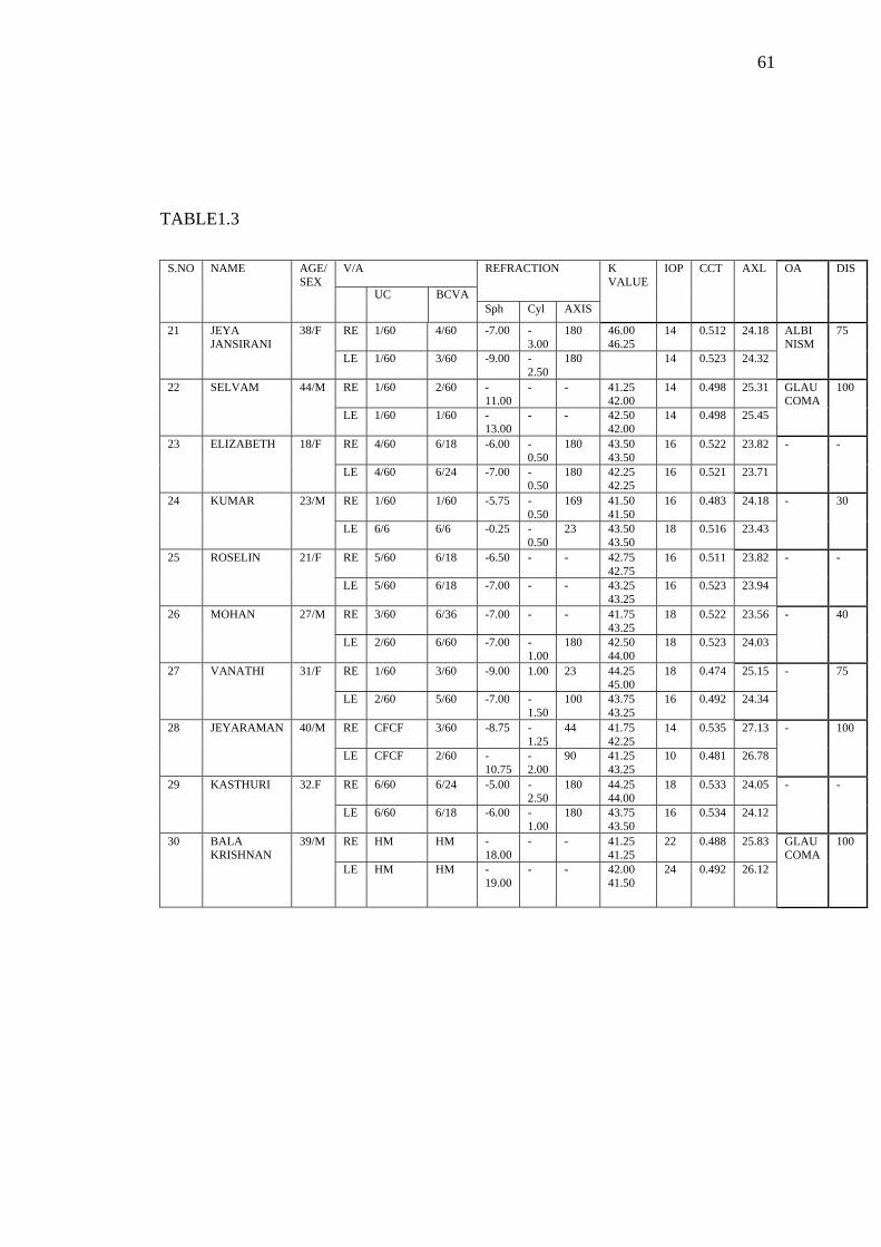
TABLE1.3
S.NO NAME AGE/ SEX
V/A REFRACTION K VALUE
IOP CCT AXL OA DIS
UC BCVA Sph Cyl AXIS
21 JEYA JANSIRANI
38/F RE 1/60 4/60 -7.00 -3.00
180 46.00 46.25
14 0.512 24.18 ALBI NISM
75
LE 1/60 3/60 -9.00 -2.50
180 14 0.523 24.32
22 SELVAM 44/M RE 1/60 2/60 -11.00
- - 41.25 42.00
14 0.498 25.31 GLAU COMA
100
LE 1/60 1/60 -13.00
- - 42.50 42.00
14 0.498 25.45
23 ELIZABETH 18/F RE 4/60 6/18 -6.00 -0.50
180 43.50 43.50
16 0.522 23.82 - -
LE 4/60 6/24 -7.00 -0.50
180 42.25 42.25
16 0.521 23.71
24 KUMAR 23/M RE 1/60 1/60 -5.75 -0.50
169 41.50 41.50
16 0.483 24.18 - 30
LE 6/6 6/6 -0.25 -0.50
23 43.50 43.50
18 0.516 23.43
25 ROSELIN 21/F RE 5/60 6/18 -6.50 - - 42.75 42.75
16 0.511 23.82 - -
LE 5/60 6/18 -7.00 - - 43.25 43.25
16 0.523 23.94
26 MOHAN 27/M RE 3/60 6/36 -7.00 - - 41.75 43.25
18 0.522 23.56 - 40
LE 2/60 6/60 -7.00 -1.00
180 42.50 44.00
18 0.523 24.03
27 VANATHI 31/F RE 1/60 3/60 -9.00 1.00 23 44.25 45.00
18 0.474 25.15 - 75
LE 2/60 5/60 -7.00 -1.50
100 43.75 43.25
16 0.492 24.34
28 JEYARAMAN 40/M RE CFCF 3/60 -8.75 -1.25
44 41.75 42.25
14 0.535 27.13 - 100
LE CFCF 2/60 -10.75
-2.00
90 41.25 43.25
10 0.481 26.78
29 KASTHURI 32.F RE 6/60 6/24 -5.00 -2.50
180 44.25 44.00
18 0.533 24.05 - -
LE 6/60 6/18 -6.00 -1.00
180 43.75 43.50
16 0.534 24.12
30 BALA KRISHNAN
39/M RE HM HM -18.00
- - 41.25 41.25
22 0.488 25.83 GLAU COMA
100
LE HM HM -19.00
- - 42.00 41.50
24 0.492 26.12

62
TABLE1.4
S.NO NAME AGE/ SEX
V/A REFRACTION K VALUE
IOP CCT AXL OA DIS
UC BCVA Sph Cyl AXIS
31 ARUL ANAN DHAM
35/M RE PL PL -14.00 - - 41.00 43.25
14 0.489 26.12 RP 100
LE PL PL -16.00 - - 41.00 41.75
14 0.492 25.63
32 MALLIKA 40/F RE 6/60 6/12 -5.00 -1.00
68 43.25 43.25
18 0.531 23.82 - -
LE 6/60 6/18 -6.00 -0.50
121 43.25 43.50
18 0.533 23.75
33 ANTONY 41/M RE 6/60 6/24 -6.00 -0.50
21 43.50 42.75
18 0.522 23.51 - -
LE 5/60 6/24 -6.00 -1.00
104 42.50 42.75
16 0.523 23.13
34 JEEVA 18/M RE 3/60 6/36 -4.00 -3.00
90 43.50 43.75
18 0.521 23.82 - -
LE 6/6 6/6 - - - 44.25 44.25
18 0.521 23.82
35 DEVAKI 38/F RE 2/60 6/60 -5.50 -2.50
42 43.50 42.75
18 0.528 23.18 - 40
LE 4/60 6/36 -.5.00 -2.00
117 43.50 43.50
16 0.518 23.22
36 ANANDRAJ 17/M RE 4/60 6/36 -6.00 -1.00
37 41.50 42.00
16 0.513 24.08 - 40
LE 6/60 6/18 -6.00 -1.50
134 41.00 42.25
18 0.522 24.83
37 JOSEPH 28/M RE 6/60 6/18 -5.50 -1.00
90 42.25 41.75
18 0.512 23.72 - -
LE 5/60 6/18 -6.00 - - 42.00 42.25
18 0.513 23.66
38 ARUL 18/M RE 6/60 6/18 -6.00 - - 42.25 43.00
16 0.522 23.12 - 20
LE 4/60 6/36 -7.00 - - 41.75 42.25
16 0.522 23.18
39 BALASUBRAMA NIYAM
22/M RE 3/60 6/60 -7.00 -2.00
90 41.25 43.75
16 0.511 23.89 - 40
LE 4/60 6/36 -7.00 -1.00
90 41.50 42.50
18 0.507 24.03
40 SELVAM 40/M RE 6/60 6/12 -5.50 -0.50
180 43.50 44.50
18 0.522 23.54 - -
LE 5/60 6/18 -6.00 - - 43.50 43.25
18 0.523 23.81

63
TABLE1.5
S.NO NAME AGE/ SEX
V/A REFRACTION K VALUE
IOP CCT AXL OA DIS
UC BCVA Sph Cyl AXIS
41 KOWSALYA 31/F RE CFCF 2/60 -8.00 -1.50
4 41.75 43.25
16 0.489 25.72 - 100
LE CFCF 2/60 -9.00 - - 42.00 41.75
16 0.491 25.18
42 MARY 38/F RE 5/60 6/24 -6.00 -.0.50
90 43.50 43.25
18 0.503 24.08 - -
LE 4/60 6/18 -6.00 - - 44.00 44.25
18 0.506 24.13
43 MOHAN 35/M RE CFCF 1/60 -13.00
- - 41.00 41.25
12 0.472 27.11 - 30
LE 5/60 6/12 -7.00 - - 43.25 45.25
18 0.514 24.28
44 VIMALA 23/F RE HM HM -16.00
-.3.00
82 41.25 41.75
12 0.466 26.83 100
LE HM HM -19.00
-3.00
90 41.00 40.75
12 0.473 26.56
45 ISMAIL 41/M RE 4/60 6/18 -6.00 -1.00
180 42.75 42.50
16 0.512 24.12 - -
LE 5/60 6/24 -6.00 -1.00
180 43.25 42.75
16 0.518 24.66
46 DEEPA 16/F RE 5/60 6/18 -5.50 -1.50
180 41.50 43.25
16 0.502 23.92 - -
LE 4/60 6/18 -6.50 - - 42.75 44.25
16 0.496 24.16
47 RANI 32/F RE 1/60 5/60 -8.00 - - 43.25 43.00
18 0.492 24.82 - 40
LE 1/60 6/60 -7.50 - - 43.25 43.25
18 0.488 24.67
48 BALAJI 14/M RE 3/60 6/36 -6.00 -1.50
180 44.25 44.00
18 0.511 24.12 - 20
LE 4/60 6/18 -6.00 - - 43.75 43.75
18 0.513 24.08
49 PAREETHA 18/F RE 5/60 6/18 -5.50 -1.00
180 41.75 43.25
18 0.512 23.98 - -
LE 5/60 6/18 -5.50 -1.50
180 41.50 43.25
18 0.522 23.82
50 RAJKUMAR 21/M RE 6/60 6/12 -6.00 - - 43.50 43.50
18 0.523 24.07 - -
LE 6/60 6/18 -6.00 - - 43.25 43.50
18 0.534 24.13
64
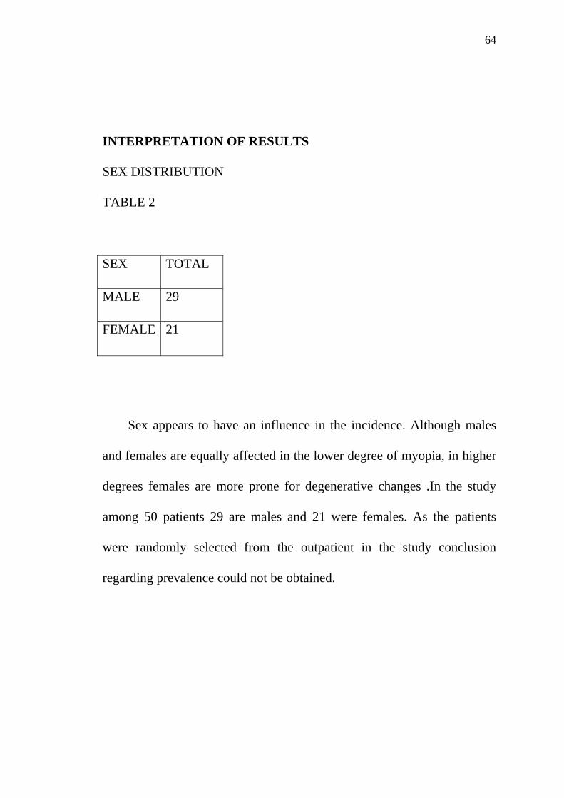
INTERPRETATION OF RESULTS
SEX DISTRIBUTION
TABLE 2
SEX TOTAL
MALE 29
FEMALE 21
Sex appears to have an influence in the incidence. Although males
and females are equally affected in the lower degree of myopia, in higher
degrees females are more prone for degenerative changes .In the study
among 50 patients 29 are males and 21 were females. As the patients
were randomly selected from the outpatient in the study conclusion
regarding prevalence could not be obtained.
65
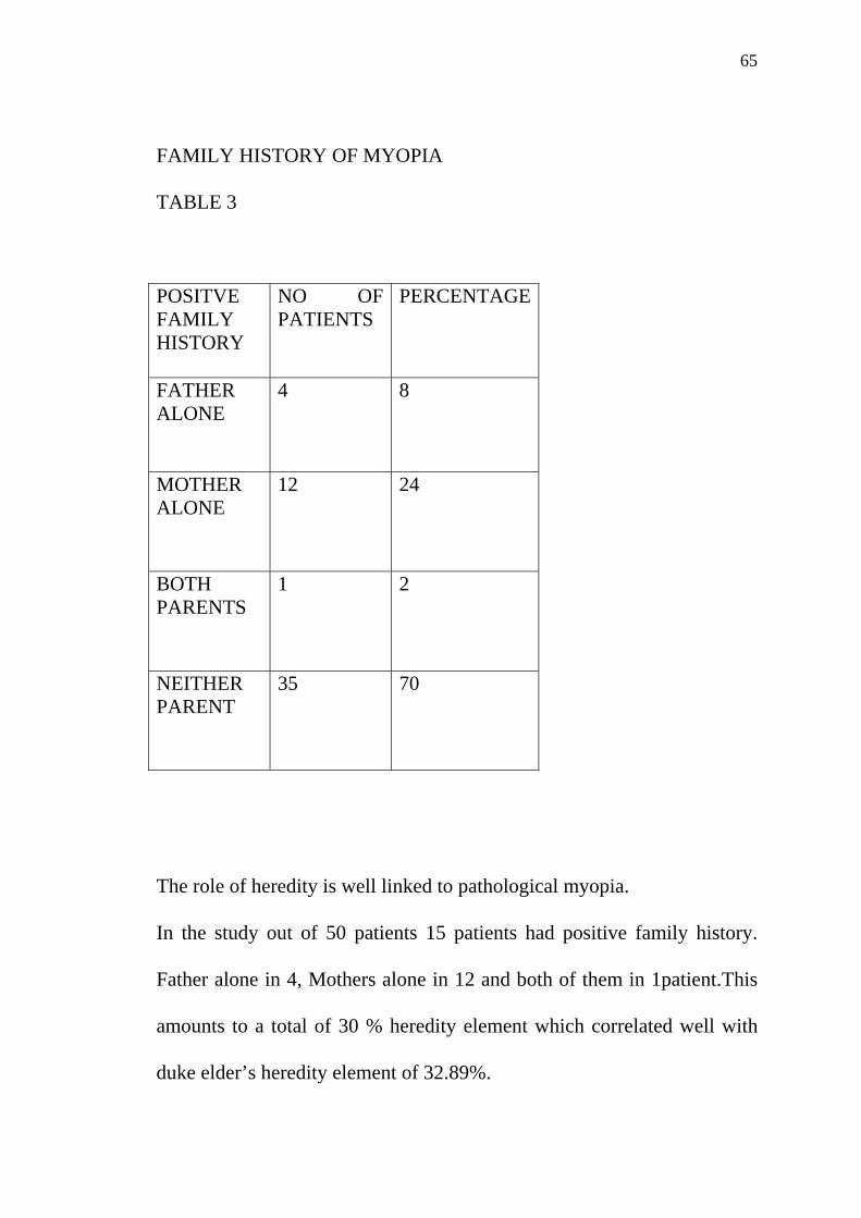
FAMILY HISTORY OF MYOPIA
TABLE 3
POSITVE FAMILY HISTORY
NO OF PATIENTS
PERCENTAGE
FATHER ALONE
4 8
MOTHER ALONE
12 24
BOTH PARENTS
1 2
NEITHER PARENT
35 70
The role of heredity is well linked to pathological myopia.
In the study out of 50 patients 15 patients had positive family history.
Father alone in 4, Mothers alone in 12 and both of them in 1patient.This
amounts to a total of 30 % heredity element which correlated well with
duke elder’s heredity element of 32.89%.
66
Although heredity element is found in 30% there was no family history in
70% which again adds more to the conclusion that factors other than
heredity like environmental factors ,visual hygine,general health,
increased near work, increased intraocular pressure etc., may play role in
pathogenesis of Pathological myopia.
LATERALITY According to duke elder unilateral high myopia is relatively rare.
In the study two cases of unilateral myopia was found among 50 patients.
Analysis from literature tends to show predominance for higher degrees
of myopia and unilateral myopia in right eye.
In the study no laterality for higher degrees of myopia in right eye was
found and out of 48 cases of bilateral myopia 24 cases had higher degrees
in right eye and 24 patients had higher degrees in left eye.
However in the study the affected eye was the right side in both two cases
of unilateral myopia.
67
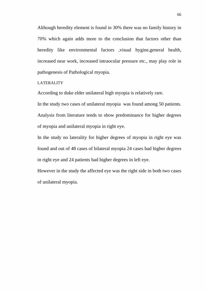
PICTURE 1 (AR value showing unilateral high myopic
refractive error in left eye. Case no. 24)
68
AXIAL LENGTH
TABLE 4
AXIALLENGTH NO OF
EYES
<22.50 mm 7
22.51-24.00 25
24.01-26.00 41
>26.00 25
The axial length well correlated with both the retinoscopy valuesand the
amount of visual impairment. The more the axial length the more was the
retinoscopy value and poorer is the visual acuity.
Among the 98 eyes( with the exception and two eyes in patients with
unilateral myopia)25 eyes and axial length >26 mm, 41 eyes between 24
and 26 mm,25 eyes Between 22.51 to 24.00 mm and only 7 eyes < 22.50
mm. Also patients with higher axial length had decreased central corneal
thickness and the intraocular pressure relatively low.

69
PICTURE 2 (Ultrasound A Scan value in LE
Showing 35.06mm Case no.2)
70
In my study the maximum recorded axial length was 35.06mm.
Even patients whose axial length were normal showed fundus changes of
Pathological myopia confirming the fact that the degeneration of retina is
not only due to increase in axial length and stretching of sclera but in fact
may be genetically determined.
71
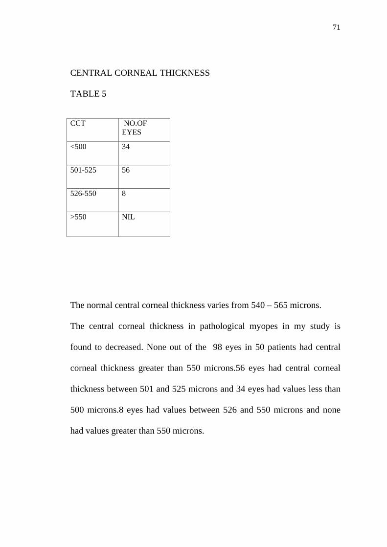
CENTRAL CORNEAL THICKNESS
TABLE 5
CCT NO.OF
EYES
<500 34
501-525 56
526-550 8
>550 NIL
The normal central corneal thickness varies from 540 – 565 microns.
The central corneal thickness in pathological myopes in my study is
found to decreased. None out of the 98 eyes in 50 patients had central
corneal thickness greater than 550 microns.56 eyes had central corneal
thickness between 501 and 525 microns and 34 eyes had values less than
500 microns.8 eyes had values between 526 and 550 microns and none
had values greater than 550 microns.
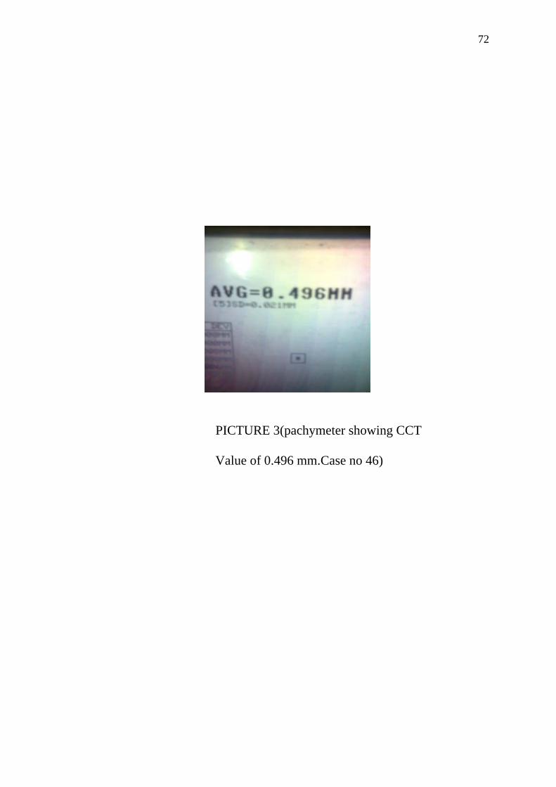
72
PICTURE 3(pachymeter showing CCT
Value of 0.496 mm.Case no 46)

73
This is an important fact because the decreased CCT in pathological
myopes results in falsely low recording of a really high intraocular
pressure and thus a coexisting glaucoma may be easily missed. Recording
of intraocular pressure by applanation tonometry becomes essential in all
pathological myopes. This may give amore accurate value than the falsely
recorded low value of indentation tonometry because of decreased CCT
and low ocular rigidity.
Previously it was said that corneal thickness maybe thicker than normal
in pathological myopes. But studies have shown varying thickness may
occur in degenerative myopia and in fact thinner corneas are more
common in myopes .Too thin corneas in these patients makes
contraindication for the laser corrections in these patients
The decrease in CCT makes to think that the stretching may not be
limited to the posterior segment alone and may be a generalised process
involving whole eyeball. Other anterior segment changes such as rupture
in descemets membrane, zonular dehiscence adds more to the fact that
degeneration may be a generalised process but much attention is given to
the posterior segment changes because of the effect on vision caused by
it.

74
REFRACTIVE POWER OF CORNEA IN DEGENERATIVE MYOPIA: TABLE6
AVERAGE REFRACTIVE POWER IN DIOPTRE
NO. OF EYES
<42.00D 17
42.01 – 43.00D 16
43.01- 44.00D
34
44.01 – 45.00D 20
>45.00D 11
75
In the study 17 out of the 98 eyes had refractive power less than 42 D,
16 eyes had had refractive power between 42 and 43 D , 34 out of 98 eyes
had values between 43 and 44D and 20 eyes between 44 and 45 D . Only
11 eyes had values greater than 45 D.
The refractive power of cornea in degenerative myopia in this study is
more clumped in the lower normal values with about 75%o having
refractive power <44D . This is correlating with the previous facts that
the cornea in the degenerative myopia is mire flatter than normal to
compensate the axial elongation. However. two patients with keratoconus
had refractive power of about 48D
However values greater than 45 D should always arise suspicion of
keratoconus and along with gross change in refractive error within short
period without degenerative changes in patients aged more than 18 years
corneal topographic studies should always be done.
76
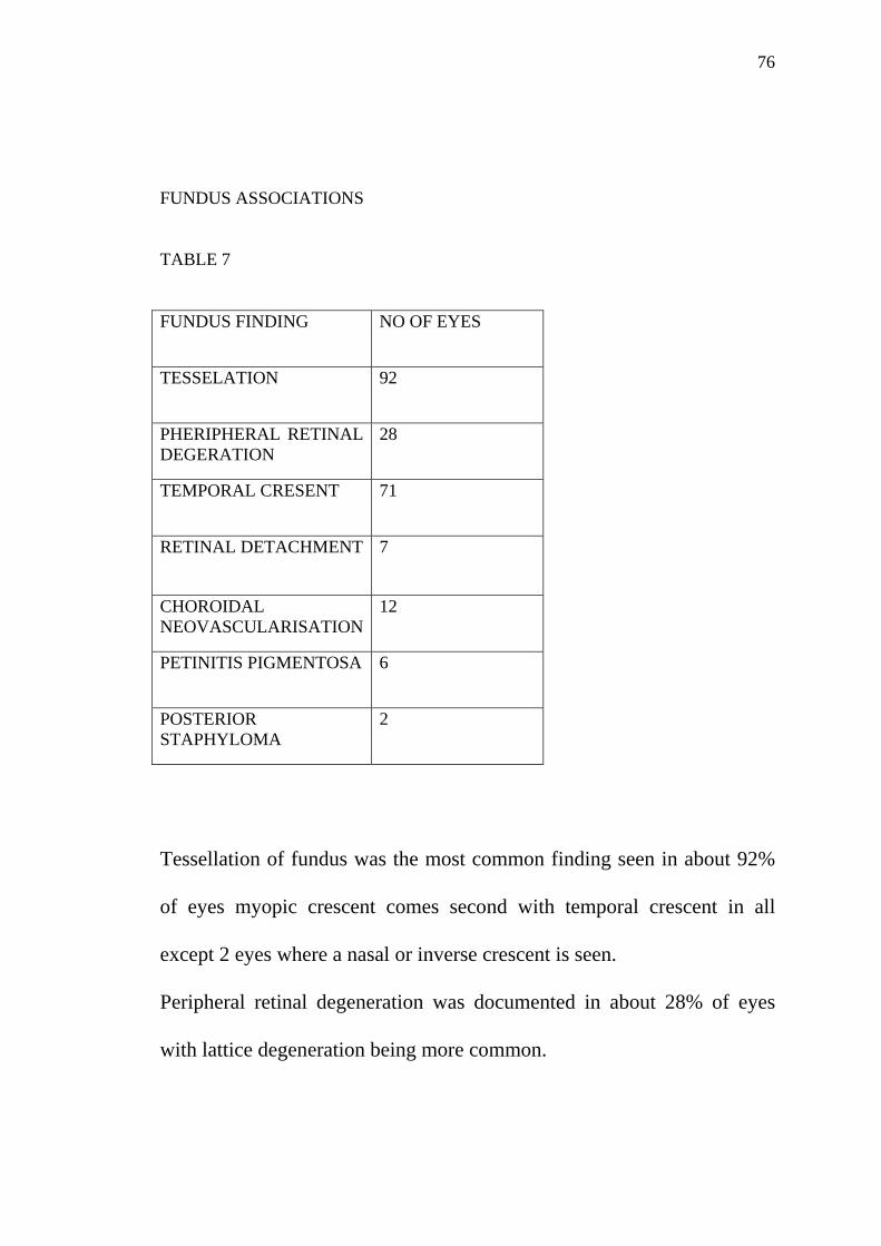
FUNDUS ASSOCIATIONS TABLE 7 FUNDUS FINDING NO OF EYES
TESSELATION 92
PHERIPHERAL RETINAL DEGERATION
28
TEMPORAL CRESENT 71
RETINAL DETACHMENT 7
CHOROIDAL NEOVASCULARISATION
12
PETINITIS PIGMENTOSA 6
POSTERIOR STAPHYLOMA
2
Tessellation of fundus was the most common finding seen in about 92%
of eyes myopic crescent comes second with temporal crescent in all
except 2 eyes where a nasal or inverse crescent is seen.
Peripheral retinal degeneration was documented in about 28% of eyes
with lattice degeneration being more common.
77
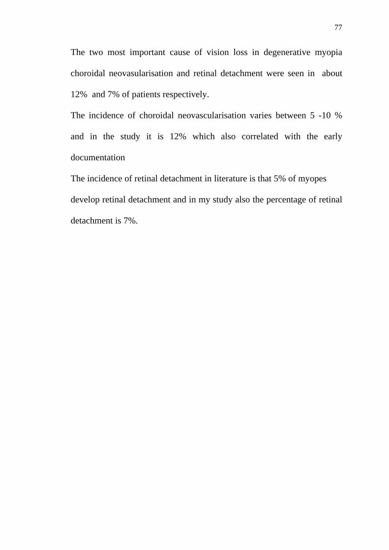
The two most important cause of vision loss in degenerative myopia
choroidal neovasularisation and retinal detachment were seen in about
12% and 7% of patients respectively.
The incidence of choroidal neovascularisation varies between 5 -10 %
and in the study it is 12% which also correlated with the early
documentation
The incidence of retinal detachment in literature is that 5% of myopes
develop retinal detachment and in my study also the percentage of retinal
detachment is 7%.
78
PICTURE 4 (Ultrasound B scan showing
retinal detachment In RE . Case no 43)
79
The incidence of choridal neovascularisation is 8% which is diagnosed
by slit lamp bio microscopic examination of fundus with 78 dioptre
lens under high magnification. The actual prevalence of CNV
May be much higher as in the early stages may be missed by
ophthalmoscopy,
This necessitates the need for special investigations like fundus
Fluroscein angiography, indocyanin green angiography and ocular
coherence tomography to diagnose early CNV and their prompt treatment
with Photodynamic therapy and anti VGEF may prevent the patient from
Vision loss and becoming virtually blind. Posterior staphyloma is seen is
2 eyes which is confirmed by Ultrasound – B Scan.
80
OCULAR ASSOCIATIONS TABLE 8 OCULAR ASSOCIATIONS
NO OF EYES
OPEN ANGLE GLAUCOMA
8
PETINITIS PIGMENTOSA
6
KERATOCONUS 4
ALBINISM 2
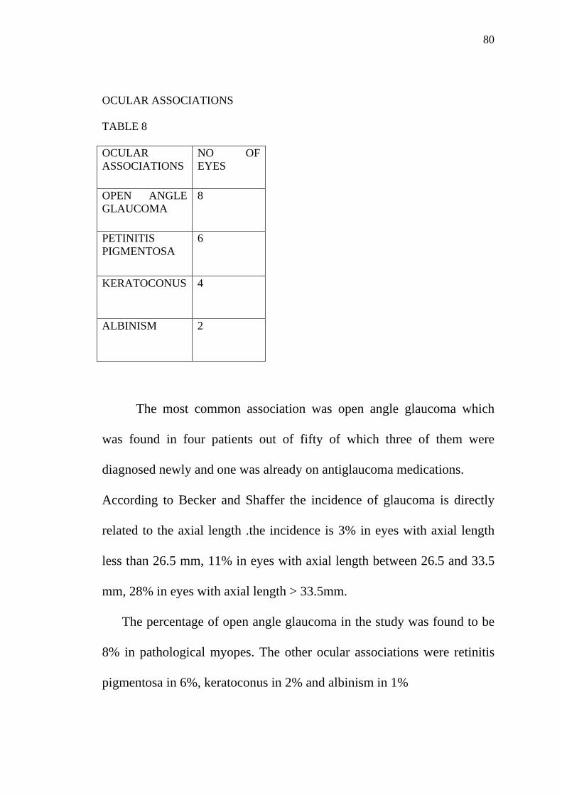
The most common association was open angle glaucoma which
was found in four patients out of fifty of which three of them were
diagnosed newly and one was already on antiglaucoma medications.
According to Becker and Shaffer the incidence of glaucoma is directly
related to the axial length .the incidence is 3% in eyes with axial length
less than 26.5 mm, 11% in eyes with axial length between 26.5 and 33.5
mm, 28% in eyes with axial length > 33.5mm.
The percentage of open angle glaucoma in the study was found to be
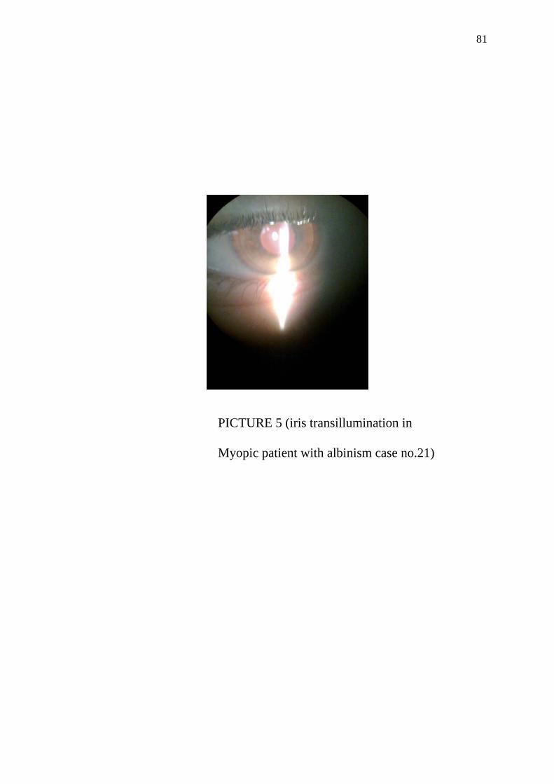
8% in pathological myopes. The other ocular associations were retinitis
pigmentosa in 6%, keratoconus in 2% and albinism in 1%
81
PICTURE 5 (iris transillumination in
Myopic patient with albinism case no.21)
82
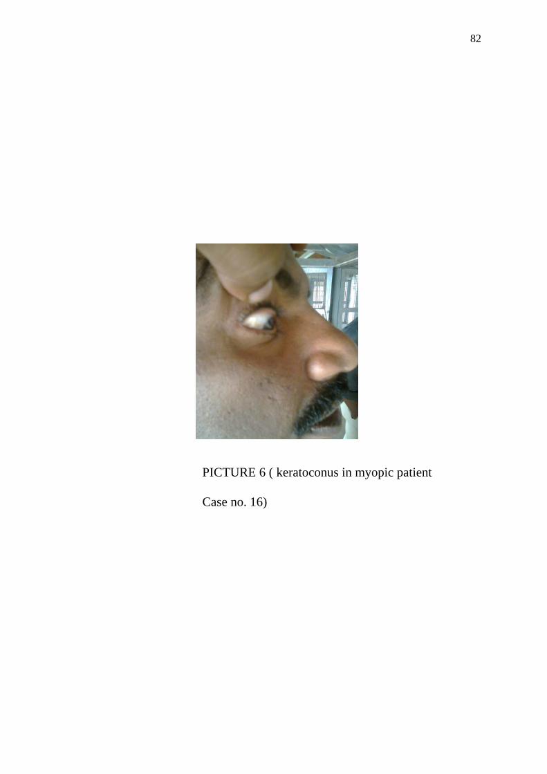
PICTURE 6 ( keratoconus in myopic patient
Case no. 16)

83
Although the corneal curvature in pathological myopia is said to be
flatter than normal, its thickness is very much less when compared to
normal which may results in bulging of the cornea. The phenomenon of
keratoconus in myopia may be well explained. Similar to the posterior
staphyloma where the sclera gets thinned and protrudes out. The
prevalence of both keratoconus and posterior staphyloma is 4% although
both does not occurred in the same individuals.

84
VISUAL DISABILITY
TABLE 9
DISABILITY PERCENTAGE
NO OF PERSONS PERCENTAGE
NO DISABILITY 25 50%
20% DISABILITY 2 4%
30% DISABILITY 7 14%
40%DISABILITY 3 6%
75% DISABILITY 5 10%
100%DISABILITY 8 16%
85
Out of 50 patients 25 patients had some percentage of visual
disability which means 50% of all pathological myopes are visually
disabled. 8 patients had 100% disability, 5 had 75% disbility, and 3 had
40% disability. 7 had 30% disability and 2 had 20 % disability. In East
Asian cities, myopia is very common and appears to be rising in some
parts of the world. The most common causes for blindness in pathological
myopia in my study were choroidal neovascularisation , retinal
detachment and optic Atrophy due to open angle glaucoma. While the
occurrence of pathological myopia cannot be prevented early recognition
of the above said cause and treatment of the same may much reduce the
visual disability.

86
SUMMARY AND CONCLUSION The literature on degenerative myopia was reviewed. Materials and methods employed are stated. In the study there was 30% association of family history in
pathological myopia. The ocular parameters were greatly altered in
pathological myopia. The axial length is increased and their increase is
proportional to the refractive error and vision loss in majority of cases.
The central corneal thickness is reduced to less than 525 microns in most
of the patients which is very important finding because it may results in
the false recording of intraocular pressure. The common ocular
associations found in pathological myopia are 1) open angle glaucoma, 2)
retinitis pigmentosa , 3) keratoconus and 4)albinism. The common cause
of vision loss associated with pathological myopia were choroidal
neovascularistion, and retinal detachment.
The associated conditions which were responsible for vision loss were
Optic atrophy due to glaucoma and retinitis pigmentosa.
Regarding visual disability in pathological myopia 50% of all
myopic patients suffered from some form of visual disability.
87
8 patients had 100% disability (16%), 5 patients had 75% disability
(10%), 3 patients had 40% disability (6%), 7 patients had 30% disability
(14%) and 2 patients had 20 % disability (4%)
Degenerative myopia deserves recognition as one of the truly
neglected areas of ophthalmology. Degenerative myopia is an important
world-health issue with an unfortunate history of ineffectual treatments
that have led most eye specialists to believe that it is something of a "lost
cause." As a result, this condition, which is responsible for the loss of
vision in so many people during the middle years of life and in old age,
seems destined to run its natural course, save for attention to correction of
the refractive error . Much of the sight loss in high axial myopia is due to
the complications such as choroidal neovascularisation and retinal
detachment. If early diagnosis of the CNV is done in pathological myopes
by doing fundus Fluorescein angiography and indocyanine green
angiography, prompt treatment with laser photocoagulation,
photodynamic therapy and anti- VGEF factors, vision loss due to these
major complications may be prevented. Early diagnosis of peripheral
retinal degeneration by indirect ophthalmoscopy and prophylactic
treatment with lasers may prevent a retinal detachment.
88
Diagnosis of coexisting factors such as open angle glaucoma in myopes
should not be missed as it is one of the leading causes of blindness
associated with degenerative myopia.In doubtful cases prophylactic
antiglaucoma medications can be prescribed.
There is an increased vulnerability for trauma in these patients
due to prominent globe .so necessary preventive measures should be
taken. Myopia related visual impairment may affect the productivity,
mobility, quality of life and activities of daily living of the individuals.
Potentially blinding-myopia related pathologies are often irreversible in
nature, especially if diagnosed late. So early recognition of the coexisting
conditions and complications, and their management may prevent the
degenerative myopic patients from becoming essentially blind.
89
PART- III ABBREVIATIONS: D DIOPTRE NPCB NATIONAL PROGRAMME FOR CONTROL OF BLINDNESS WHO WORLD HEALTH ORGANISATION HLA HUMAN LEUCOCYTE ANTIGEN RPE RETINAL PIGMENT EPITHELIUM CNV CHOROIDAL NEOVASCULARISATION VEGF VASCULAR ENDOTHELIAL GROWTH FACTOR FFA FUNDUS FLOURESCEIN ANGIOGRAPHY ICG INDOCYANINE GREEN ANGIOGRAPHY ARMD AGE RELATED MACULAR DEGENERATION PRK PHOTOREFRACTIVE KERATECTOMY LASER LIGHT AMPLIFICATION BY STIMULATED EMISSION OF RADATION LASIK LASER ASSISTED INSITU KERATOMILEUSIS LASEK LASER SUBEPITHELIAL KERATOMILEUSIS Nd;YAG NEODMYIUM YITTIRUM ALUMINIUM GARNET PIOL PHAKIC INTRAOCULAR LENS UC UNCORRECTED SL. NO SERIAL NUMBER V/A VISUAL ACUITY M MALE F FEMALE RE RIGHT EYE LE LEFT EYE BCVA BEST CORRECTED VISUAL ACUITY Sph SPHERE Cyl CYLINDER K VALUE KERATOMETRY VALUE DEG DEGREE AXL AXIAL; LENGTH IOP INTRAOCULAR PRESSURE CCT CENTRAL CORNEAL THICKNESSS OA OCULAR ASSOCIATIONS DIS VISUAL DISABILITY CFCF COUNTING FINGERS CLOSE TO FACE HM HAND MOVEMENTS PL PERCEPTION OF LIGHT RP RETINITIS PIGMENTOSA
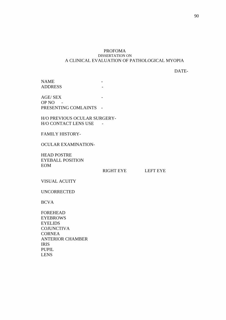
90
PROFOMA DISSERTATION ON A CLINICAL EVALUATION OF PATHOLOGICAL MYOPIA DATE- NAME - ADDRESS - AGE/ SEX - OP NO - PRESENTING COMLAINTS - H/O PREVIOUS OCULAR SURGERY- H/O CONTACT LENS USE - FAMILY HISTORY- OCULAR EXAMINATION- HEAD POSTRE EYEBALL POSITION EOM RIGHT EYE LEFT EYE VISUAL ACUITY UNCORRECTED BCVA FOREHEAD EYEBROWS EYELIDS COJUNCTIVA CORNEA ANTERIOR CHAMBER IRIS PUPIL LENS

91
RIGHT EYE LEFT EYE KERATOMETRY CCT IOP USG-A SCAN RETINOSCOPY FUNDUS USG-B SCAN VISUAL FIELDS OCULAR ASSOCIATIONS VISUAL DISABILITY%
92
Bibliography 1. Duke‐Elder S. Pathological refractive errors. System
of ophthalmology.Volume V. Ophthalmic optics and refraction.
St Louis: Mosby, 1970. 297 -373.
2. Duke Elders practice of refraction-David Abrams, Stewart Duke Elder
3. Mordern Ophthalmology- volume 3 lc Dutta 3rd edition
4. Parsons disease of the eye-19th edition
5. Theory and practice of refraction-A K KHURANA
6. Becker Shaffer‘s diagnosis and therapy of glaucoma -8th edition
7. Ophthalmology 2nd edition Myron Yanoff/ Jay s. Duker
8. The principles nd practice of community ophthalmology.
NPCB-Gvs Moorthy- Sanjev K. Guptha
9. Degenerative Myopia: a Review of its Nature and Current Treatment
by BrianWard Ph.D., M.D.December 2011
10. Environmental factors in the epidemiology of malignant myopia.
Amj Optom Physiol Opt 1982. Daubs J.
.11. Tokoro T. On the definition of pathological myopia in group studies.
Ophthalmology Supplement 1988.
93
12. Oxford textbook of ophthalmology.volume 2 edited by David L.Easty
And John .M.Sparrow
13. Prevalence and progression of myopic retinopathy in an older
population. Ophthalmology 2002. Vongphanit J, Mitchell P, Wang J J.
14. American academy of ophthalmology. basic and clinical science
Courses. Section 13 refractive surgery pg 87-134
15. The Wills eye manual – office and emergency room diagnosis and
treatment of eye diseases – 5th edition chapter11.22.high myopia page31
16. Jack j Kanski clinical ophthalmology –a systematic approach
7th edition
17. Optometric clinical practice guideline, care of the patient with
myopia., American optometric association.

94