Embed Size (px)
Citation preview
A Dissertation on
A COMPARATIVE STUDYBETWEEN MODIFIED
UNDERLAY MYRINGOPLASTY WITH GRAFT
OVER THE HANDLE OF MALLEUS AND
CLASSICAL UNDERLAY TECHNIQUE WITH
GRAFT UNDER THE HANDLE OF MALLEUS
Submitted to the
THE TAMILNADU DR. M.G.R. MEDICAL UNIVERSITY
In partial fulfilment of the requirements
For the award of the degree of
M.S.BRANCH IV (OTORHINOLARYNGOLOGY)
GOVERNMENT STANLEY MEDICAL
COLLEGE & HOSPITAL
THE TAMILNADU DR. M.G.R. MEDICAL UNIVERSITY,
CHENNAI, TAMILNADU
APRIL 2013
DECLARATION
I, Dr.V.A.KARTHIKEYAN, Solemnly declare that the
dissertation, titled “A COMPARATIVE STUDY BETWEEN
MODIFIED UNDERLAY MYRINGOPLASTYWITH GRAFT
OVER THE HANDLE OF MALLEUS AND CLASSICAL
UNDERLAY TECHNIQUE WITH GRAFT UNDER THE HANDLE
OF MALLEUS”is a bonafide work done by me during the period from
FEB 2011 to SEP 2012 at Government Stanley Medical College and
Hospital, Chennai under the expert supervision of PROF.DR.T.
BALASUBRAMANIAN, M.S., D.L.O., Professor and Head,
Department Of Otorhinolaryngology, Government Stanley Medical
College and Hospital, Chennai.
This dissertation is submitted to The Tamil Nadu Dr. M.G.R.
Medical University in partial fulfilment of the rules and regulations for
the M.S. degree examinations in Otorhinolaryngology to be held in April
2013.
Chennai-1 DR.V.A.KARTHIKEYAN.
Date:
CERTIFICATE
This is to certify that the dissertation presented “A
COMPARATIVE STUDY BETWEEN MODIFIED UNDERLAY
MYRINGOPLASTY WITH GRAFT OVER THE HANDLE OF
MALLEUS AND CLASSICAL UNDERLAY TECHNIQUE WITH
GRAFT UNDER THE HANDLE OF MALLEUS” herein by
DR.V.A.KARTHIKEYAN, is an original work done in the Department
of Otorhinolaryngology, Government Stanley Medical College and
Hospital, Chennai in partial fulfillment of regulations of the Tamilnadu
Dr. M.G.R. Medical University for the award of degree of M.S.
(Otorhinolaryngology) Branch IV, under my supervision during the
academic period 2010-2013.
Prof. DR.T.BALASUBRAMANIAN, Professor and Head Of Dept, Dept. of Otorhinolaryngology, Govt. Stanley Medical College, Chennai-1.
THE DEAN, Govt. Stanley Medical college, Chennai-1
ACKNOWLEDGEMENTS
I wish to express my sincere thanks to Prof. Dr.
S.GEETHALAKSHMI, M.D., PhD, DEAN, Government Stanley
Medical College and Hospital for having permitted me to utilize the
facilities of the hospital for the conduct of the study.
My heartfelt gratitude to Prof. Dr. T.BALASUBRAMANIAN,
M.S., D.L.O., Professor and Head, Department of Otorhinolaryngology,
Government Stanley Medical College and Hospital for his motivation,
valuable suggestions, expert supervision and for making all necessary
arrangements for conducting this study.
I owe my sincere thanks to Prof. Dr. M. RAMANIRAJ.M.S,
D.L.O., Additional professor, Otorhinolaryngology, Prof. Dr. N.
SEETHALAKSHMI M.S., D.L.O Associate professor,
Otorhinolaryngology for supporting, guiding and encouraging me in this
study.
I thank Prof. Dr. R. MUTHUKUMAR.M.S, D.L.O for
suggesting this topic and for giving valuable inputs.
I wish to thank my Assistant professors DR.ATHIYAMAN M.S,
DR.KARUPPASAMY M.S., D.L.O., DR.CHANDRAMOULI M.S.,
DR.NANMULLAI M.S., and DR.BHARANIDHARAN D.L.O. for their
valuable suggestions and help.
I also thank Mrs.Radha Kalaiselvan, Audiologist and speech
pathologist of ENT Department, Government Stanley hospital for her
expert assistance.
My sincere thanks to all those post graduates who helped me
during this study period.
I thank the staff nurses and theatre personnel, Government Stanley
Hospital for their cooperation and assistance.
Last but not least, my indebtedness and gratitude to all the patients
who are the cornerstone of my study, and who most willingly and
selflessly subjected themselves to this study for the sake of the benefit of
the community.
CONTENTS
S.NO TOPIC P.NO
01. INTRODUCTION 1
02. AIMS AND OBJECTIVES 3
03. MATERIALS AND METHODS 4
04. REVIEW OF LITERATURE 20
05. RESULTS AND OBSERVATION 68
06. DISCUSSION 80
07. CONCLUSION 85
08. ANNEXURE
BIBLIOGRAPHY
MASTERCHART
PROFORMA
PATIENT INFORMATION SHEET
INFORMED CONSENT FORM
PLAGIARISM- SCREEN SHOT
86
1 �
INTRODUCTION
Chronic otitis media (COM) is defined as chronic inflammation
of mucoperiosteal lining of part or whole of the middle ear cleft. It has
been recognized since prehistoric times. It is classified into two types,
mucosal and squamous, of which mucosal type is characterized by
intermittent mucoid or mucopurulent discharge through a perforated
tympanic membrane.
Although the introduction of antibiotics has reduced the
mortality in COM, still surgery remains the definitive treatment
modality for closure of tympanic membrane perforation.
From seventeenth to nineteenth century several attempts at
closing tympanic membrane perforations were made using prosthetic
materials like paper patch and cauterising agents.
Surgical repair of tympanic membrane was first attempted by
Banzer (1640) with pig’sbladder1. In1878 Berthold devised the term
myringoplasty 1, 3. In 1952,Wullstein formally announced2, 3a technique
of closing perforation. That time he used split thickness skin graft2, 3.
2 �
In 1958 Heerman began to use temporalis fascia.3First Shea
performed underlay tympanoplasty. He performed stapedectomy and
he accidentally tore the ear drum. That time he successfully repaired
the ear drum perforation with medial vein graft3. He placed the graft
medial to the ear drum perforation. The use of fascia as an underlay
graft was first reported by Storrs3, 4 in 1961.In their classic underlay
myringoplasty they placed the graft medial to the handle of malleus.3, 4.
In our study we compare the results of two techniques of
underlay myringoplasty graftunder the handle of malleus and the graft
over the handle of malleus in terms of graft uptake and hearing
outcome.
3 �
AIMS AND OBJECTIVES
• To compare the two techniques of underlay myringoplasty
modified underlay technique with graft over the handle of
malleus and classical underlay technique with graft under the
handle of malleus.
• To assess the graft uptake rate of these two techniques.
• To assess the hearing outcome of these two techniques.
4 �
MATERIALS AND METHODS
STUDY DESIGN - Prospective study.
STUDY PLACE - Department of E.N.T. Stanley Medical
College.
STUDY PERIOD - Feb 2011 to Sep 2012
SAMPLE SIZE - 60 patients.
FOLLOW UP PERIOD - 6 Months
INCLUSION CRITERIA:
Both male and female patients between 18 to 60 years attending
E.N.T. OPD at Stanley Medical College with following clinical
findings were included
• Central perforation.
• Dry perforation.
• Ossicular chain intact and mobile.
• Mucosa of the middle ear normal and healthy.
• Good cochlear reserve.
EXCLUSION CRITERIA:
• Patients with adenoid enlargement.
5 �
• Presence of septic foci in nose or throat.
• Revision cases.
• Traumatic perforation
• Cases with mixed and sensorineural hearing loss.
• Anomaly of external or middle ear.
• Cases with extracranial or intracranial complications of
CSOM
MATERIALS:
Surgical technique adopted in our study is underlay
myringoplasty. All cases were done with assistance of endoscope. The
equipment’s used for this surgery listed below:
1) 0 degree Hopkins endoscope, 4 millimetres wide angled5, 6.
(Same one that is used for nasal surgery).
2) CCD camera (storz, single chip).
3) Rosen’s, sickle knife and other middle ear micro instruments
were used.
4) Colour monitor which is facing the surgeon and light source
cable.6
5) Maico ma 52 clinical diagnostic two channel audiometer
provided with sound proof room for audiological assessment.
6 �
METHODOLOGY:
This study was conducted in a group of 60 patients in the
department of E.N.T. Stanley Medical College in the time periods from
February 2011 to September 2012 with a follow up period of 6 months.
The patients who were selected for surgery were admitted in the
ward. Detail history was recorded in the case sheet. Clinical
examinations were performed including tuning fork tests. Complete
examination of both the ears was done. Presence of septic foci was
ruled out by thorough examination of nose, paranasal sinuses, pharynx
and larynx. Examination under microscope or endoscope was done in
all cases to assess the ossicular status, condition of middle ear
etc...Audiometric tests were done to document pre-operative hearing
status. Diagnostic nasal endoscopy was done for all cases to rule out
focal sepsis. Following investigations are taken for all patients for the
purpose of anaesthetic fitness.
• complete haemogram
• Renal function tests
• Blood sugar
• HIV-ELISA (after consent)
• Blood grouping and typing
7 �
• Urine analysis
• chest X ray PA View
• ECG
were done for all cases included in the study.
Pure Tone Audiometry was done in a sound proof room using
maico ma 52 clinical diagnostic two channel audiometer.
Patient and their relatives were clearly informed about nature of
the disease, surgery and all possible outcomes and complications of
surgery. Informed consent was obtained from each patient and one of
his/her relative.
All cases were done under local anaesthesia. Pre medication and
local infiltration used were same for all cases. All cases were done with
the aid of endoscope.
About 30 patients with dry ear for more than 6 weeks were
subjected to surgery, underlay myringoplasty graft, over the handle of
malleus were considered group A. Of these 24 patients with unilateral
disease and 6 patients with bilateral disease were taken up for the
study. For patients with bilateral disease, worse ear in terms of hearing
was taken up for surgery.
Another 30 patients with dry ear more than 6 weeks were
subjected to underlay myringoplasty graft, under the handle of malleus
8 �
were considered as group B. Of these 22 patients with unilateral
disease and 8 patients with bilateral disease were taken up for the
study. For patients with bilateral disease, worse ear in terms of hearing
was taken for surgery.
Graft material of choice in all cases was temporalis fascia.4, 7
following surgery mastoid dressing was done, that was changed on the
post-operative day two. Sutures were removed on 7th post-operative
day from the graft harvested site. Patients were treated with i.v
antibiotics for 2- 3 days and oral antibiotics were continued for another
one week.
In the first month patients were followed up every week. Next
two months they followed up every 15 days. Then once in 2 or 3
months till the end of the study.
Post operatively following parameters were noted:
1) Graft taken
2) Graft not taken
3) Graft lateralisation
4) Atelectasis.
Pure tone audiometry was done after 3 months and documented.
Pre and post-operative air bone (A-B) gap calculated by taking the
9 �
averages of bone conduction and air conduction at the frequencies of
500,1000 and 2000 Hz. 7
Even though socio-economic status influences the disease and its
outcome, all the patients attending Stanley Medical College & Hospital
belong to lower socio-economic group. Hence this parameter was
excluded in this study.
Myringoplasty:
Myringoplasty can be done under local or general
anaesthesia.5But in this study all cases were done under local
anaesthesia.
GENERAL ANESTHESIA -ADVANTAGES:
• Airway is better secured
• Better choice in paediatric population.
• Apprehensive patients are benefited.
• Patient is more comfortable under GA
LOCAL ANAESTHESIA -ADVANTAGES:
• Minimal bleeding and better field of surgery
• Early ambulation
• low cost
OPERATIVE TECHNIQUE:
10 �
Patient positioning8:
For the surgeon to accomplish the goal of grafting, the head
must be positioned. Bringing the patient close to the edge of the bed
prevents the surgeon from overextending his/her hands, thus increasing
stability. It also assists in correct upright posture. The patient’s arm on
the operating side should be padded and tucked close to the body. The
patient’s head should be placed on a doughnut-shaped pillow
approximately 3 to 4 inches thick. Care should be taken to ensure that
the contralateral ear is not folded. This arrangement provides stability
for the patient’s head and allows the head to be rotated along the neck
axis. A hydraulic chair without armrests greatly assists the surgeon by
allowing better angles of approach with minimal repositioning. An
operating table that can rotate about its long axis assists in obviating
the need of the surgeon to change position and facilitates achieving the
desired angle of exposure with less effort. Finally, the patient’s head
needs to be placed at the foot of the bed so there is adequate room for
the surgeon’s legs beneath the table. The anaesthesiologist should be at
the foot of the bed. The blood pressure cuff should be placed on the
arm contralateral to the surgeon.
Skin preparation and sterility:
11 �
A small area of skin of the scalp over the temporal region must
be shaved to harvest the fascia so that 2 to 3 cm of hairless skin is
visible. This operating area painted with povidone iodine solution and
the same flushed into the external auditory canal and then sterile saline.
TECHNIQUE OF LOCAL ANAESTHESIA9:
All cases were sedated using injection fortwin 30 mg and
injection promethazine 25 mg given intramuscularly half an hour
before surgery. Pulse and oxygen saturation were monitored while
giving local anaesthetic infiltration and throughout the surgery. Local
anaesthesia consists of 2 per cent xylocaine mixed with 1 in 2, 00,000
adrenaline was used for local infiltration.
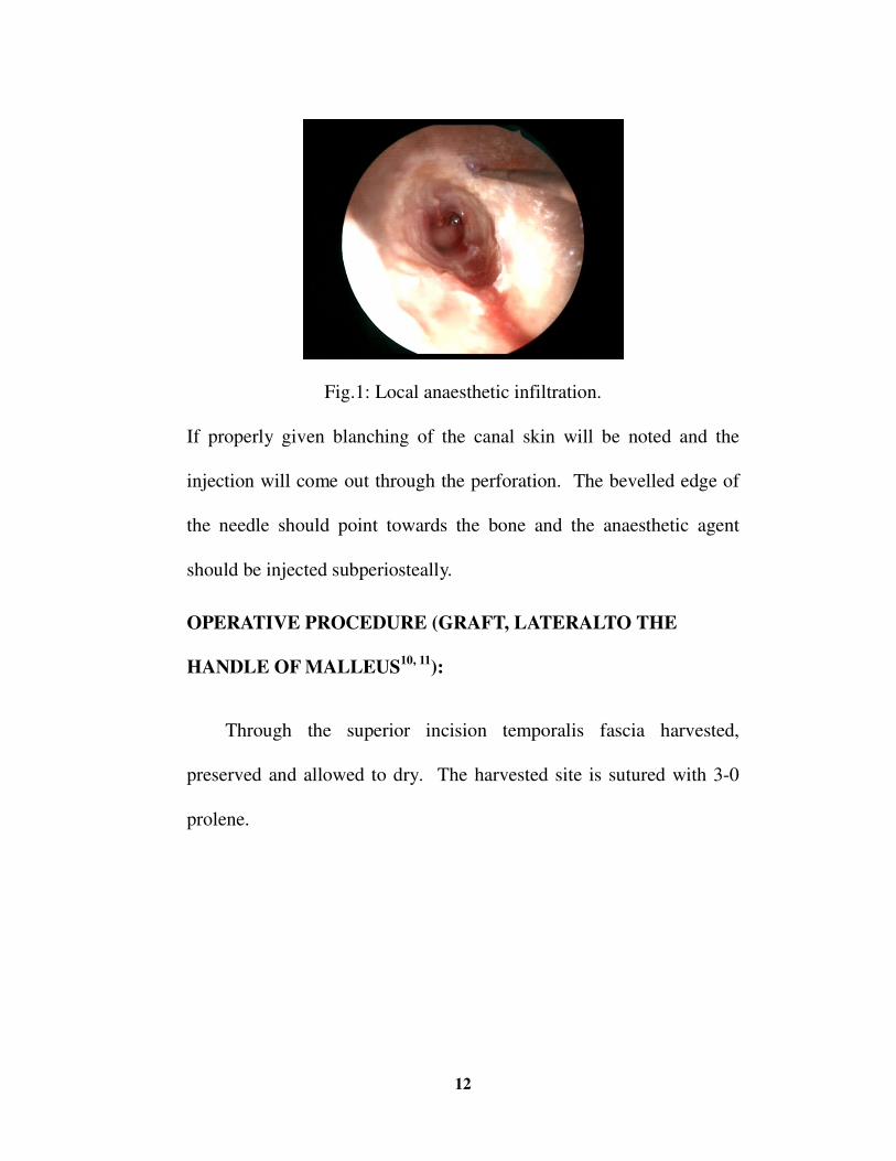
Infiltration of local anaesthetic agent was done under endoscopic
visualisation. The four quadrants of the cartilaginous external auditory
canal and the vascular strip region of the bony canal were injected9 at
3, 6, 9, 12 o’clock positions.
12 �
Fig.1: Local anaesthetic infiltration.
If properly given blanching of the canal skin will be noted and the
injection will come out through the perforation. The bevelled edge of
the needle should point towards the bone and the anaesthetic agent
should be injected subperiosteally.
OPERATIVE PROCEDURE (GRAFT, LATERALTO THE
HANDLE OF MALLEUS10, 11):
Through the superior incision temporalis fascia harvested,
preserved and allowed to dry. The harvested site is sutured with 3-0
prolene.
13 �
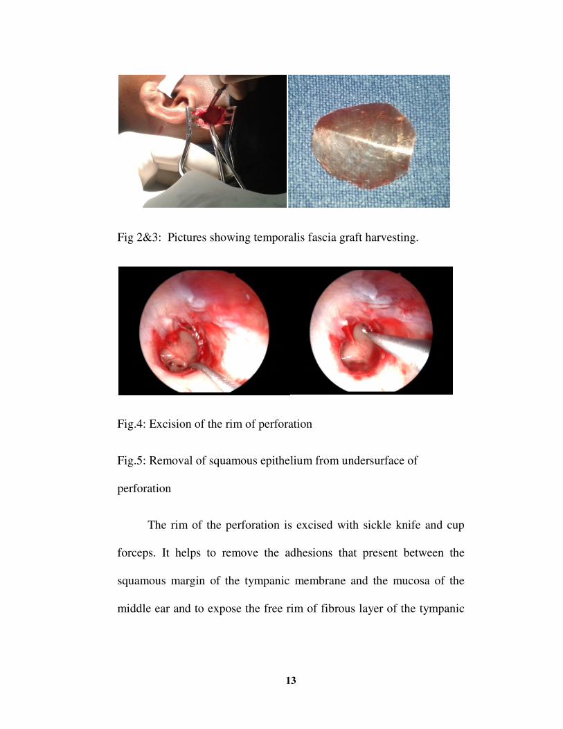
Fig 2&3: Pictures showing temporalis fascia graft harvesting.
Fig.4: Excision of the rim of perforation
Fig.5: Removal of squamous epithelium from undersurface of
perforation
The rim of the perforation is excised with sickle knife and cup
forceps. It helps to remove the adhesions that present between the
squamous margin of the tympanic membrane and the mucosa of the
middle ear and to expose the free rim of fibrous layer of the tympanic
14 �
membrane. Then, the under surface of the drum remnant was denuded
of squamous epithelium with Rosen’s round knife.
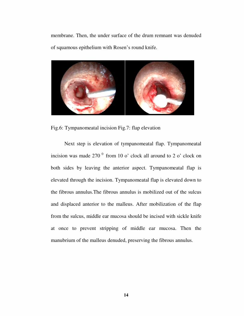
Fig.6: Tympanomeatal incision Fig.7: flap elevation
Next step is elevation of tympanomeatal flap. Tympanomeatal
incision was made 270 0 from 10 o’ clock all around to 2 o’ clock on
both sides by leaving the anterior aspect. Tympanomeatal flap is
elevated through the incision. Tympanomeatal flap is elevated down to
the fibrous annulus.The fibrous annulus is mobilized out of the sulcus
and displaced anterior to the malleus. After mobilization of the flap
from the sulcus, middle ear mucosa should be incised with sickle knife
at once to prevent stripping of middle ear mucosa. Then the
manubrium of the malleus denuded, preserving the fibrous annulus.
15 �
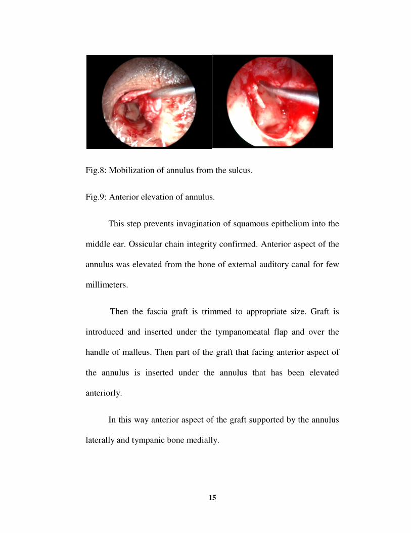
Fig.8: Mobilization of annulus from the sulcus.
Fig.9: Anterior elevation of annulus.
This step prevents invagination of squamous epithelium into the
middle ear. Ossicular chain integrity confirmed. Anterior aspect of the
annulus was elevated from the bone of external auditory canal for few
millimeters.
Then the fascia graft is trimmed to appropriate size. Graft is
introduced and inserted under the tympanomeatal flap and over the
handle of malleus. Then part of the graft that facing anterior aspect of
the annulus is inserted under the annulus that has been elevated
anteriorly.
In this way anterior aspect of the graft supported by the annulus
laterally and tympanic bone medially.
16 �
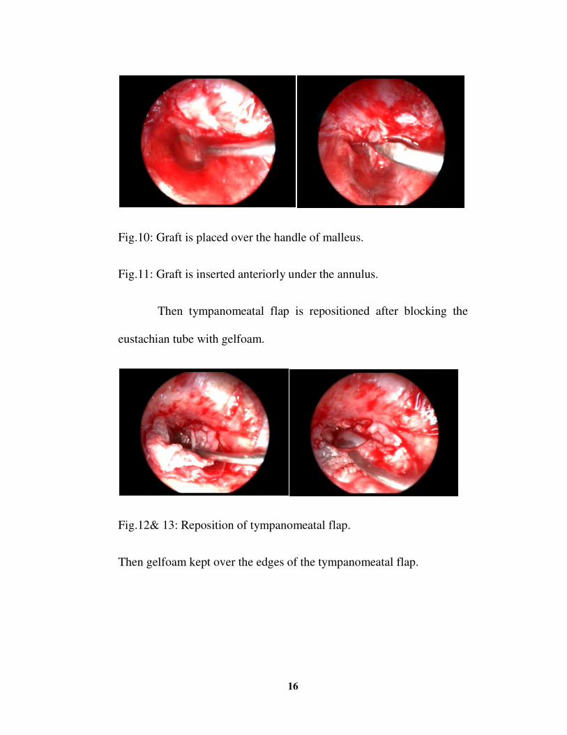
Fig.10: Graft is placed over the handle of malleus.
Fig.11: Graft is inserted anteriorly under the annulus.
Then tympanomeatal flap is repositioned after blocking the
eustachian tube with gelfoam.
Fig.12& 13: Reposition of tympanomeatal flap.
Then gelfoam kept over the edges of the tympanomeatal flap.
17 �
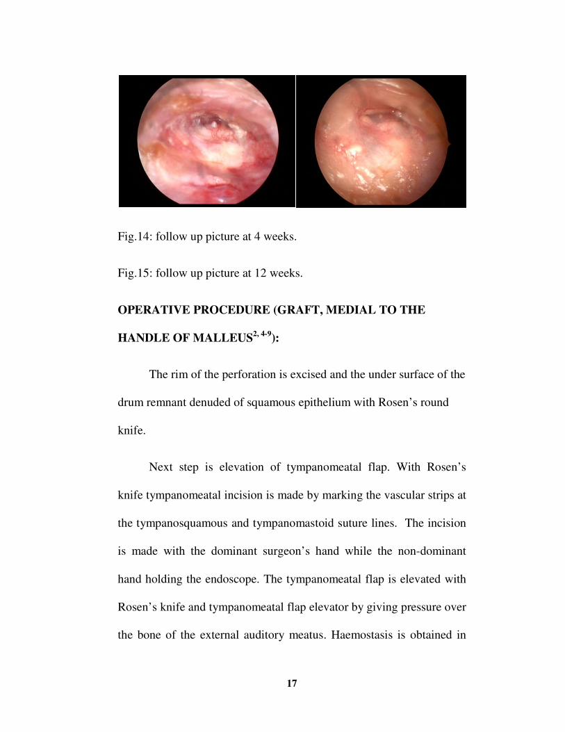
Fig.14: follow up picture at 4 weeks.
Fig.15: follow up picture at 12 weeks.
OPERATIVE PROCEDURE (GRAFT, MEDIAL TO THE
HANDLE OF MALLEUS2, 4-9):
The rim of the perforation is excised and the under surface of the
drum remnant denuded of squamous epithelium with Rosen’s round
knife.
Next step is elevation of tympanomeatal flap. With Rosen’s
knife tympanomeatal incision is made by marking the vascular strips at
the tympanosquamous and tympanomastoid suture lines. The incision
is made with the dominant surgeon’s hand while the non-dominant
hand holding the endoscope. The tympanomeatal flap is elevated with
Rosen’s knife and tympanomeatal flap elevator by giving pressure over
the bone of the external auditory meatus. Haemostasis is obtained in
18 �
between with the aid of adrenaline soaked cotton balls and 20 gauge
needle suction tip.
The tympanomeatal flap is elevated down to the fibrous annulus.
The fibrous annulus is mobilized out of the sulcus and displaced
anterior to the malleus. After mobilization of annulus from the sulcus,
mucosa of the middle ear should be incised in the same manner. Then
the manubrium of the malleus denuded, preserving the fibrous annulus.
Working through the endoscope, the surgeon can obtain an excellent
view of the anterior annulus simply by rotating the endoscope
anteriorly. Even with a prominent anterior canal wall bulge, it is
seldom necessary to remove bone in this area to see the annulus.
Haemostasis should be achieved prior to graft placement with
Gelfoam saturated in 1:1,000 adrenaline packed into the middle ear
space while the graft is being trimmed. The dried graft is trimmed to
appropriate size. A slit is made in the superior aspect of the graft to
accommodate placement medial to the manubrium. The adrenaline
soaked gelfoam is removed and the middle ear space is filled with
plain Gelfoam12. Particular care is exercised in packing the middle ear
with Gelfoam. Packing is started at the eustachian tube region and
proceeds posteriorly. Adequate Gelfoam should be kept in the
19
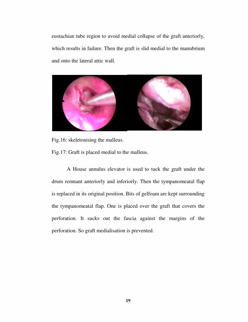
eustachian tube region to avoid medial collapse of the graft anteriorly,
which results in failure. Then the graft is slid medial to the manubrium
and onto the lateral attic wall.
Fig.16: skeletonising the malleus.
Fig.17: Graft is placed medial to the malleus.
A House annulus elevator is used to tuck the graft under the
drum remnant anteriorly and inferiorly. Then the tympanomeatal flap
is replaced in its original position. Bits of gelfoam are kept surrounding
the tympanomeatal flap. One is placed over the graft that covers the
perforation. It sucks out the fascia against the margins of the
perforation. So graft medialisation is prevented.
20 �
REVIEW OF LITERATURE
EMBRYOLOGY:
Development of Temporal Bone, External Auditory Canal,
Tympanic Ring, and Tympanic Membrane:
The adult temporal bone is an amalgam of the squamous,
petrous, mastoid, tympanic, and styloid bones. The close association of
the external auditory canal, tympanic ring, and tympanic membrane
justifies the inclusion of their developmental process in conjunction
with that of the temporal bone as a whole. The development of the
bony labyrinth and petrosa, however, because of its intricacy, warrants
separate discussion. The following account of the development of the
external auditory canal, tympanic ring, tympanic membrane, and
temporal bone is derived from the works of Anson and associates as
well as Pearson. The dorsal part of the first branchial groove, which
gives rise to the external auditory canal, progressively deepens during
the second month. The ectoderm of the groove briefly abuts on the
endoderm of the tubotympanic recess (first pharyngeal pouch), but
during the sixth week, a mesodermal in growth breaks this contact.
Beginning at 8 weeks, the inferior portion of the first branchial groove
21 �
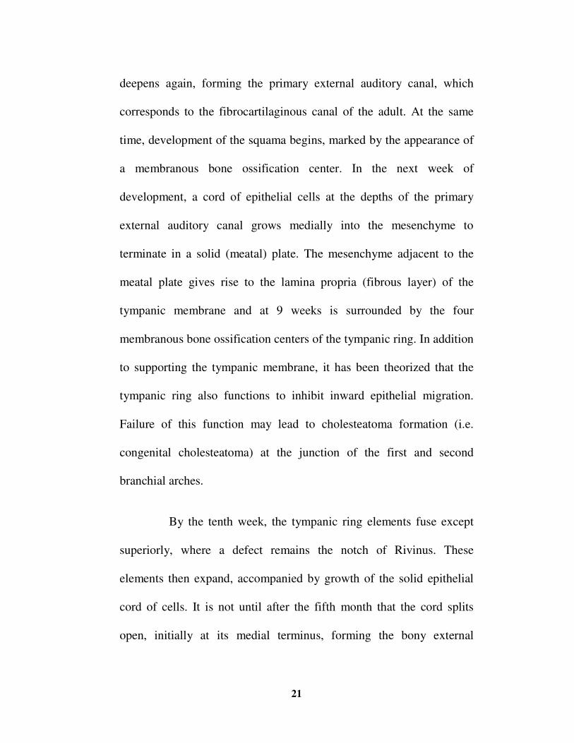
deepens again, forming the primary external auditory canal, which
corresponds to the fibrocartilaginous canal of the adult. At the same
time, development of the squama begins, marked by the appearance of
a membranous bone ossification center. In the next week of
development, a cord of epithelial cells at the depths of the primary
external auditory canal grows medially into the mesenchyme to
terminate in a solid (meatal) plate. The mesenchyme adjacent to the
meatal plate gives rise to the lamina propria (fibrous layer) of the
tympanic membrane and at 9 weeks is surrounded by the four
membranous bone ossification centers of the tympanic ring. In addition
to supporting the tympanic membrane, it has been theorized that the
tympanic ring also functions to inhibit inward epithelial migration.
Failure of this function may lead to cholesteatoma formation (i.e.
congenital cholesteatoma) at the junction of the first and second
branchial arches.
By the tenth week, the tympanic ring elements fuse except
superiorly, where a defect remains the notch of Rivinus. These
elements then expand, accompanied by growth of the solid epithelial
cord of cells. It is not until after the fifth month that the cord splits
open, initially at its medial terminus, forming the bony external
22 �
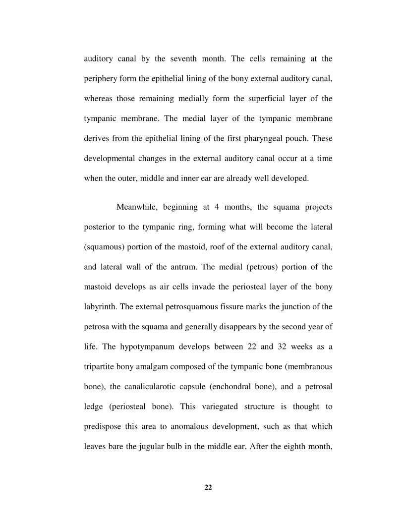
auditory canal by the seventh month. The cells remaining at the
periphery form the epithelial lining of the bony external auditory canal,
whereas those remaining medially form the superficial layer of the
tympanic membrane. The medial layer of the tympanic membrane
derives from the epithelial lining of the first pharyngeal pouch. These
developmental changes in the external auditory canal occur at a time
when the outer, middle and inner ear are already well developed.
Meanwhile, beginning at 4 months, the squama projects
posterior to the tympanic ring, forming what will become the lateral
(squamous) portion of the mastoid, roof of the external auditory canal,
and lateral wall of the antrum. The medial (petrous) portion of the
mastoid develops as air cells invade the periosteal layer of the bony
labyrinth. The external petrosquamous fissure marks the junction of the
petrosa with the squama and generally disappears by the second year of
life. The hypotympanum develops between 22 and 32 weeks as a
tripartite bony amalgam composed of the tympanic bone (membranous
bone), the canalicularotic capsule (enchondral bone), and a petrosal
ledge (periosteal bone). This variegated structure is thought to
predispose this area to anomalous development, such as that which
leaves bare the jugular bulb in the middle ear. After the eighth month,
23 �
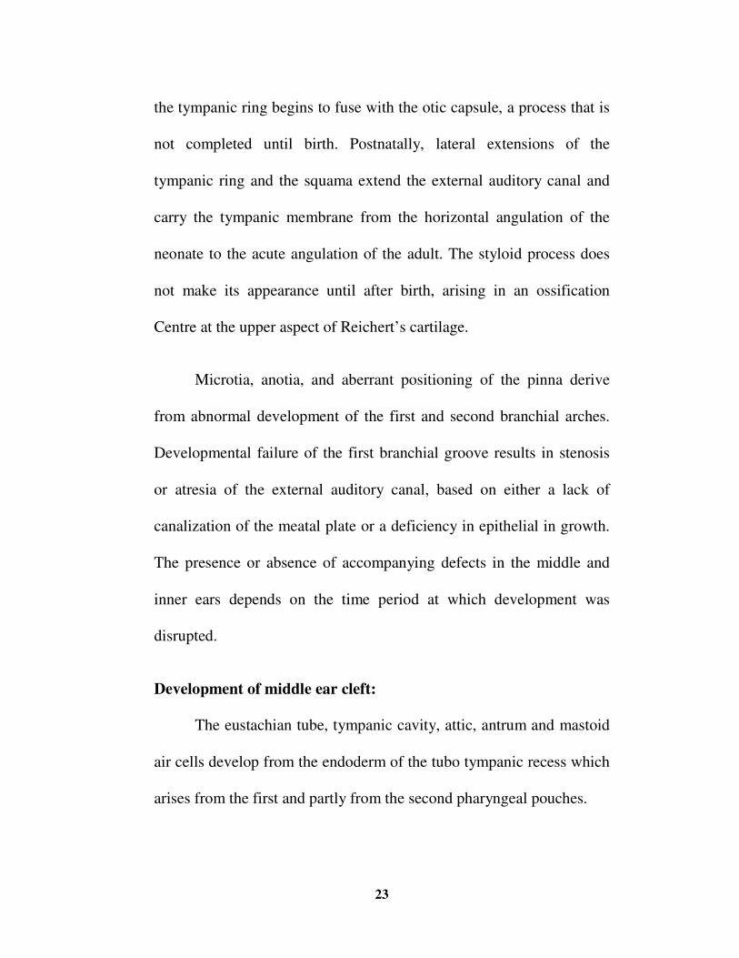
the tympanic ring begins to fuse with the otic capsule, a process that is
not completed until birth. Postnatally, lateral extensions of the
tympanic ring and the squama extend the external auditory canal and
carry the tympanic membrane from the horizontal angulation of the
neonate to the acute angulation of the adult. The styloid process does
not make its appearance until after birth, arising in an ossification
Centre at the upper aspect of Reichert’s cartilage.
Microtia, anotia, and aberrant positioning of the pinna derive
from abnormal development of the first and second branchial arches.
Developmental failure of the first branchial groove results in stenosis
or atresia of the external auditory canal, based on either a lack of
canalization of the meatal plate or a deficiency in epithelial in growth.
The presence or absence of accompanying defects in the middle and
inner ears depends on the time period at which development was
disrupted.
Development of middle ear cleft:
The eustachian tube, tympanic cavity, attic, antrum and mastoid
air cells develop from the endoderm of the tubo tympanic recess which
arises from the first and partly from the second pharyngeal pouches.
24 �
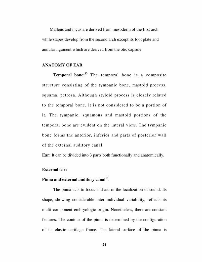
Malleus and incus are derived from mesoderm of the first arch
while stapes develop from the second arch except its foot plate and
annular ligament which are derived from the otic capsule.
ANATOMY OF EAR
Temporal bone:13 The temporal bone is a composite
structure consisting of the tympanic bone, mastoid process,
squama, petrosa. Although styloid process is closely related
to the temporal bone, it is not considered to be a portion of
it. The tympanic, squamous and mastoid portions of the
temporal bone are evident on the lateral view. The tympanic
bone forms the anterior, inferior and parts of posterior wall
of the external auditory canal.
Ear: It can be divided into 3 parts both functionally and anatomically.
External ear:
Pinna and external auditory canal13:
The pinna acts to focus and aid in the localization of sound. Its
shape, showing considerable inter individual variability, reflects its
multi component embryologic origin. Nonetheless, there are constant
features. The contour of the pinna is determined by the configuration
of its elastic cartilage frame. The lateral surface of the pinna is
25 �
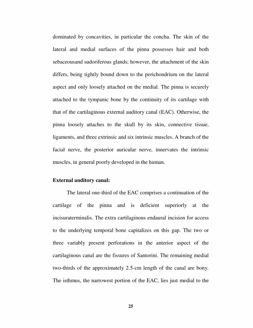
dominated by concavities, in particular the concha. The skin of the
lateral and medial surfaces of the pinna possesses hair and both
sebaceousand sudoriferous glands; however, the attachment of the skin
differs, being tightly bound down to the perichondrium on the lateral
aspect and only loosely attached on the medial. The pinna is securely
attached to the tympanic bone by the continuity of its cartilage with
that of the cartilaginous external auditory canal (EAC). Otherwise, the
pinna loosely attaches to the skull by its skin, connective tissue,
ligaments, and three extrinsic and six intrinsic muscles. A branch of the
facial nerve, the posterior auricular nerve, innervates the intrinsic
muscles, in general poorly developed in the human.
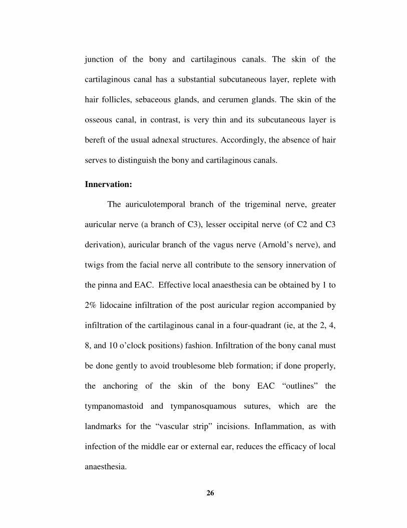
External auditory canal:
The lateral one-third of the EAC comprises a continuation of the
cartilage of the pinna and is deficient superiorly at the
incisuraterminalis. The extra cartilaginous endaural incision for access
to the underlying temporal bone capitalizes on this gap. The two or
three variably present perforations in the anterior aspect of the
cartilaginous canal are the fissures of Santorini. The remaining medial
two-thirds of the approximately 2.5-cm length of the canal are bony.
The isthmus, the narrowest portion of the EAC, lies just medial to the
26 �
junction of the bony and cartilaginous canals. The skin of the
cartilaginous canal has a substantial subcutaneous layer, replete with
hair follicles, sebaceous glands, and cerumen glands. The skin of the
osseous canal, in contrast, is very thin and its subcutaneous layer is
bereft of the usual adnexal structures. Accordingly, the absence of hair
serves to distinguish the bony and cartilaginous canals.
Innervation:
The auriculotemporal branch of the trigeminal nerve, greater
auricular nerve (a branch of C3), lesser occipital nerve (of C2 and C3
derivation), auricular branch of the vagus nerve (Arnold’s nerve), and
twigs from the facial nerve all contribute to the sensory innervation of
the pinna and EAC. Effective local anaesthesia can be obtained by 1 to
2% lidocaine infiltration of the post auricular region accompanied by
infiltration of the cartilaginous canal in a four-quadrant (ie, at the 2, 4,
8, and 10 o’clock positions) fashion. Infiltration of the bony canal must
be done gently to avoid troublesome bleb formation; if done properly,
the anchoring of the skin of the bony EAC “outlines” the
tympanomastoid and tympanosquamous sutures, which are the
landmarks for the “vascular strip” incisions. Inflammation, as with
infection of the middle ear or external ear, reduces the efficacy of local
anaesthesia.
27 �
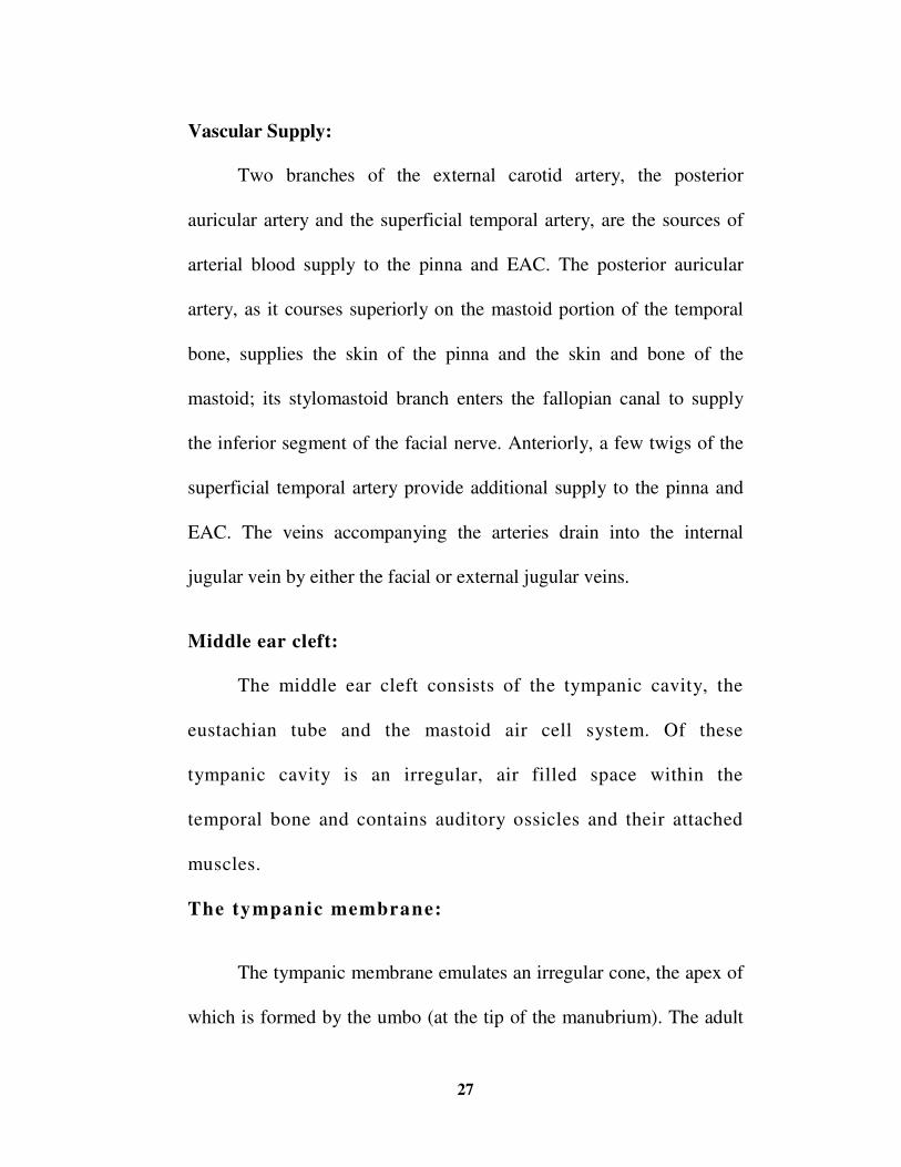
Vascular Supply:
Two branches of the external carotid artery, the posterior
auricular artery and the superficial temporal artery, are the sources of
arterial blood supply to the pinna and EAC. The posterior auricular
artery, as it courses superiorly on the mastoid portion of the temporal
bone, supplies the skin of the pinna and the skin and bone of the
mastoid; its stylomastoid branch enters the fallopian canal to supply
the inferior segment of the facial nerve. Anteriorly, a few twigs of the
superficial temporal artery provide additional supply to the pinna and
EAC. The veins accompanying the arteries drain into the internal
jugular vein by either the facial or external jugular veins.
Middle ear cleft:
The middle ear cleft consists of the tympanic cavity, the
eustachian tube and the mastoid air cell system. Of these
tympanic cavity is an irregular, air filled space within the
temporal bone and contains auditory ossicles and their attached
muscles.
The tympanic membrane:
The tympanic membrane emulates an irregular cone, the apex of
which is formed by the umbo (at the tip of the manubrium). The adult
28 �
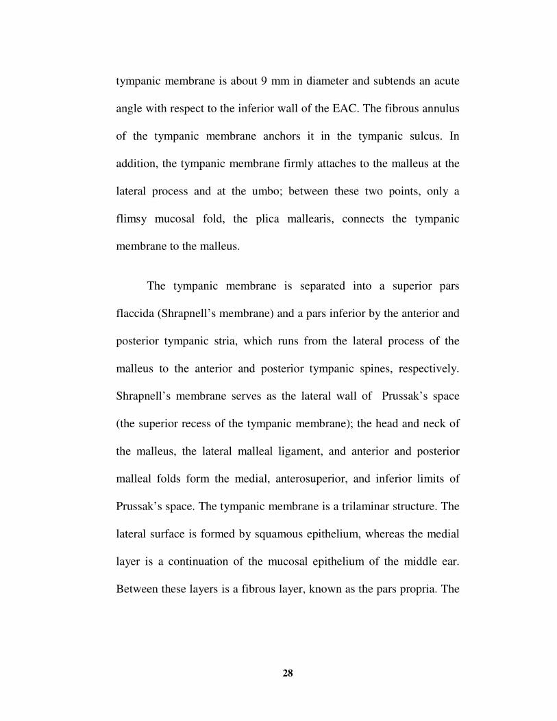
tympanic membrane is about 9 mm in diameter and subtends an acute
angle with respect to the inferior wall of the EAC. The fibrous annulus
of the tympanic membrane anchors it in the tympanic sulcus. In
addition, the tympanic membrane firmly attaches to the malleus at the
lateral process and at the umbo; between these two points, only a
flimsy mucosal fold, the plica mallearis, connects the tympanic
membrane to the malleus.
The tympanic membrane is separated into a superior pars
flaccida (Shrapnell’s membrane) and a pars inferior by the anterior and
posterior tympanic stria, which runs from the lateral process of the
malleus to the anterior and posterior tympanic spines, respectively.
Shrapnell’s membrane serves as the lateral wall of Prussak’s space
(the superior recess of the tympanic membrane); the head and neck of
the malleus, the lateral malleal ligament, and anterior and posterior
malleal folds form the medial, anterosuperior, and inferior limits of
Prussak’s space. The tympanic membrane is a trilaminar structure. The
lateral surface is formed by squamous epithelium, whereas the medial
layer is a continuation of the mucosal epithelium of the middle ear.
Between these layers is a fibrous layer, known as the pars propria. The
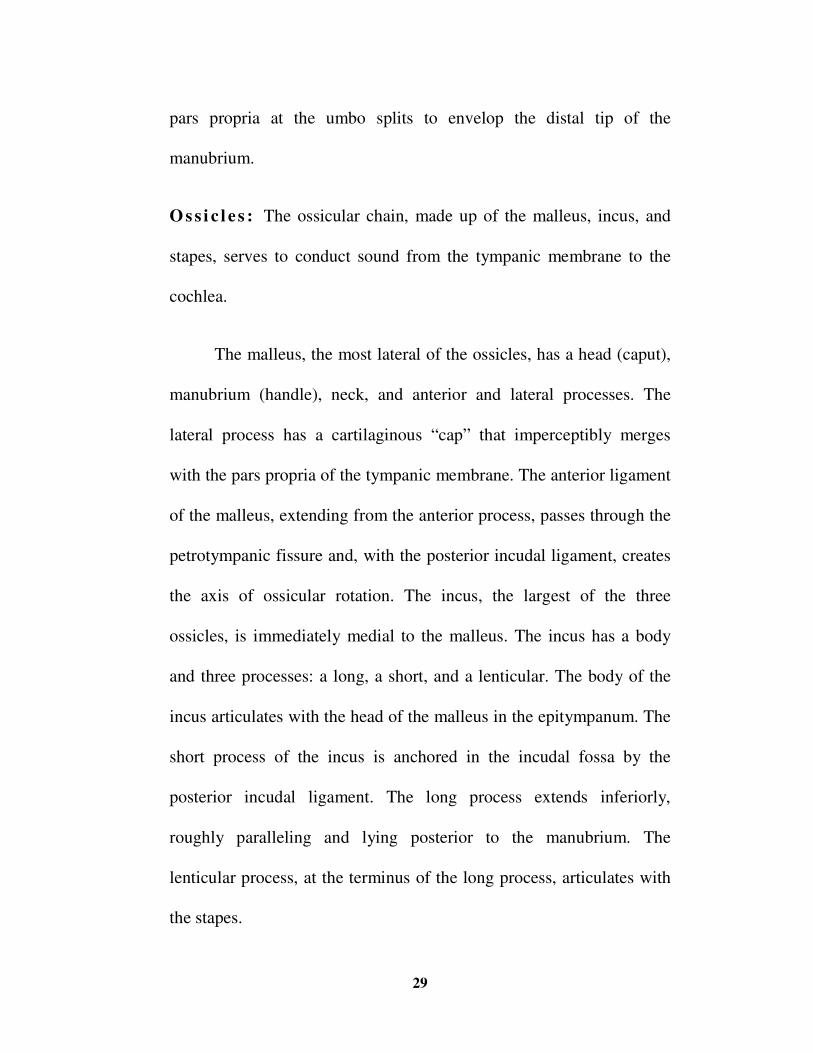
29 �
pars propria at the umbo splits to envelop the distal tip of the
manubrium.
O s s i c l e s : The ossicular chain, made up of the malleus, incus, and
stapes, serves to conduct sound from the tympanic membrane to the
cochlea.
The malleus, the most lateral of the ossicles, has a head (caput),
manubrium (handle), neck, and anterior and lateral processes. The
lateral process has a cartilaginous “cap” that imperceptibly merges
with the pars propria of the tympanic membrane. The anterior ligament
of the malleus, extending from the anterior process, passes through the
petrotympanic fissure and, with the posterior incudal ligament, creates
the axis of ossicular rotation. The incus, the largest of the three
ossicles, is immediately medial to the malleus. The incus has a body
and three processes: a long, a short, and a lenticular. The body of the
incus articulates with the head of the malleus in the epitympanum. The
short process of the incus is anchored in the incudal fossa by the
posterior incudal ligament. The long process extends inferiorly,
roughly paralleling and lying posterior to the manubrium. The
lenticular process, at the terminus of the long process, articulates with
the stapes.
30 �
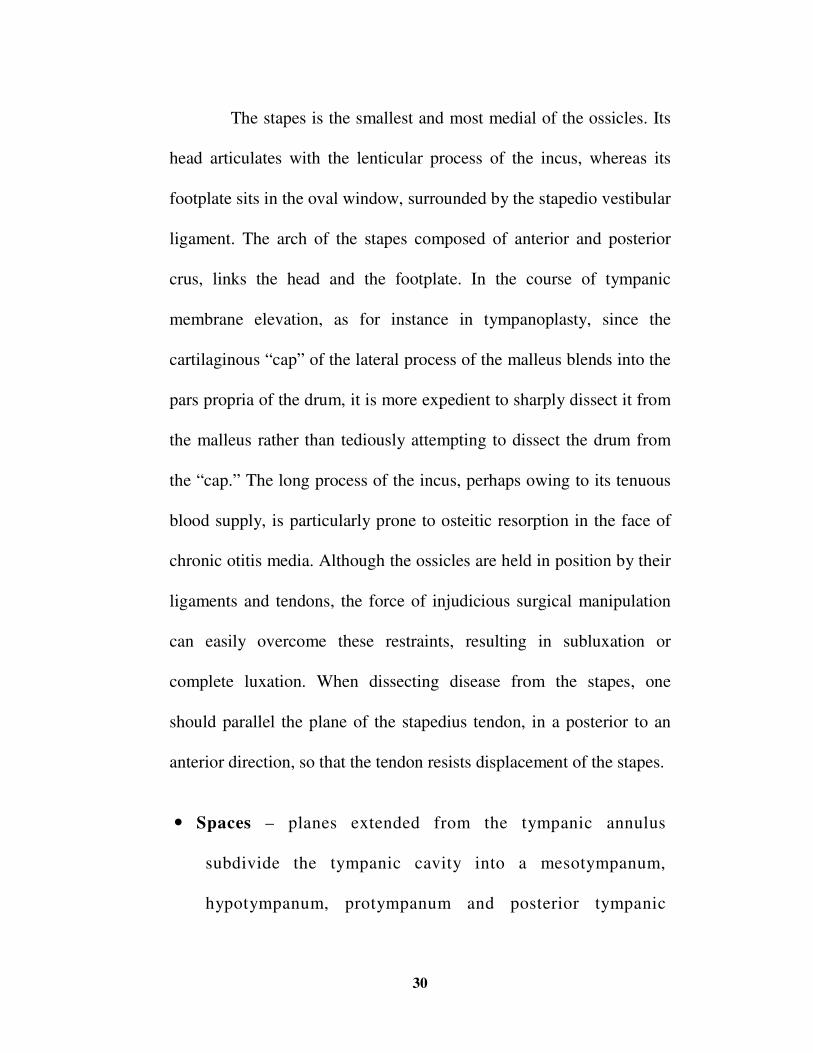
The stapes is the smallest and most medial of the ossicles. Its
head articulates with the lenticular process of the incus, whereas its
footplate sits in the oval window, surrounded by the stapedio vestibular
ligament. The arch of the stapes composed of anterior and posterior
crus, links the head and the footplate. In the course of tympanic
membrane elevation, as for instance in tympanoplasty, since the
cartilaginous “cap” of the lateral process of the malleus blends into the
pars propria of the drum, it is more expedient to sharply dissect it from
the malleus rather than tediously attempting to dissect the drum from
the “cap.” The long process of the incus, perhaps owing to its tenuous
blood supply, is particularly prone to osteitic resorption in the face of
chronic otitis media. Although the ossicles are held in position by their
ligaments and tendons, the force of injudicious surgical manipulation
can easily overcome these restraints, resulting in subluxation or
complete luxation. When dissecting disease from the stapes, one
should parallel the plane of the stapedius tendon, in a posterior to an
anterior direction, so that the tendon resists displacement of the stapes.
• Spaces – planes extended from the tympanic annulus
subdivide the tympanic cavity into a mesotympanum,
hypotympanum, protympanum and posterior tympanic
31 �
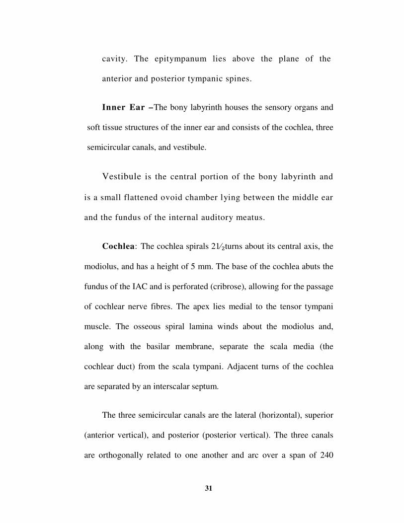
cavity. The epitympanum lies above the plane of the
anterior and posterior tympanic spines.
Inner Ear –The bony labyrinth houses the sensory organs and
soft tissue structures of the inner ear and consists of the cochlea, three
semicircular canals, and vestibule.
Vestibule is the central portion of the bony labyrinth and
is a small flattened ovoid chamber lying between the middle ear
and the fundus of the internal auditory meatus.
Cochlea: The cochlea spirals 21⁄�turns about its central axis, the
modiolus, and has a height of 5 mm. The base of the cochlea abuts the
fundus of the IAC and is perforated (cribrose), allowing for the passage
of cochlear nerve fibres. The apex lies medial to the tensor tympani
muscle. The osseous spiral lamina winds about the modiolus and,
along with the basilar membrane, separate the scala media (the
cochlear duct) from the scala tympani. Adjacent turns of the cochlea
are separated by an interscalar septum.
The three semicircular canals are the lateral (horizontal), superior
(anterior vertical), and posterior (posterior vertical). The three canals
are orthogonally related to one another and arc over a span of 240
32 �
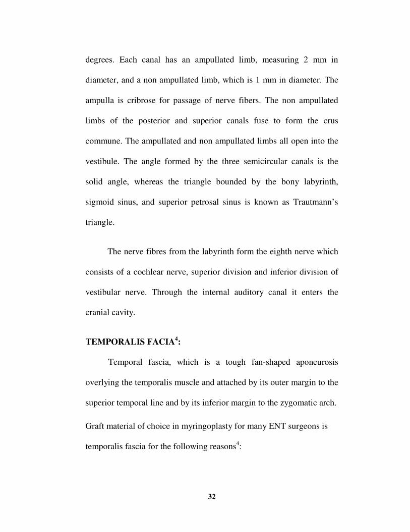
degrees. Each canal has an ampullated limb, measuring 2 mm in
diameter, and a non ampullated limb, which is 1 mm in diameter. The
ampulla is cribrose for passage of nerve fibers. The non ampullated
limbs of the posterior and superior canals fuse to form the crus
commune. The ampullated and non ampullated limbs all open into the
vestibule. The angle formed by the three semicircular canals is the
solid angle, whereas the triangle bounded by the bony labyrinth,
sigmoid sinus, and superior petrosal sinus is known as Trautmann’ s
triangle.
The nerve fibres from the labyrinth form the eighth nerve which
consists of a cochlear nerve, superior division and inferior division of
vestibular nerve. Through the internal auditory canal it enters the
cranial cavity. �
TEMPORALIS FACIA4:
Temporal fascia, which is a tough fan-shaped aponeurosis
overlying the temporalis muscle and attached by its outer margin to the
superior temporal line and by its inferior margin to the zygomatic arch.�
Graft material of choice in myringoplasty for many ENT surgeons is
temporalis fascia for the following reasons4:
33 �
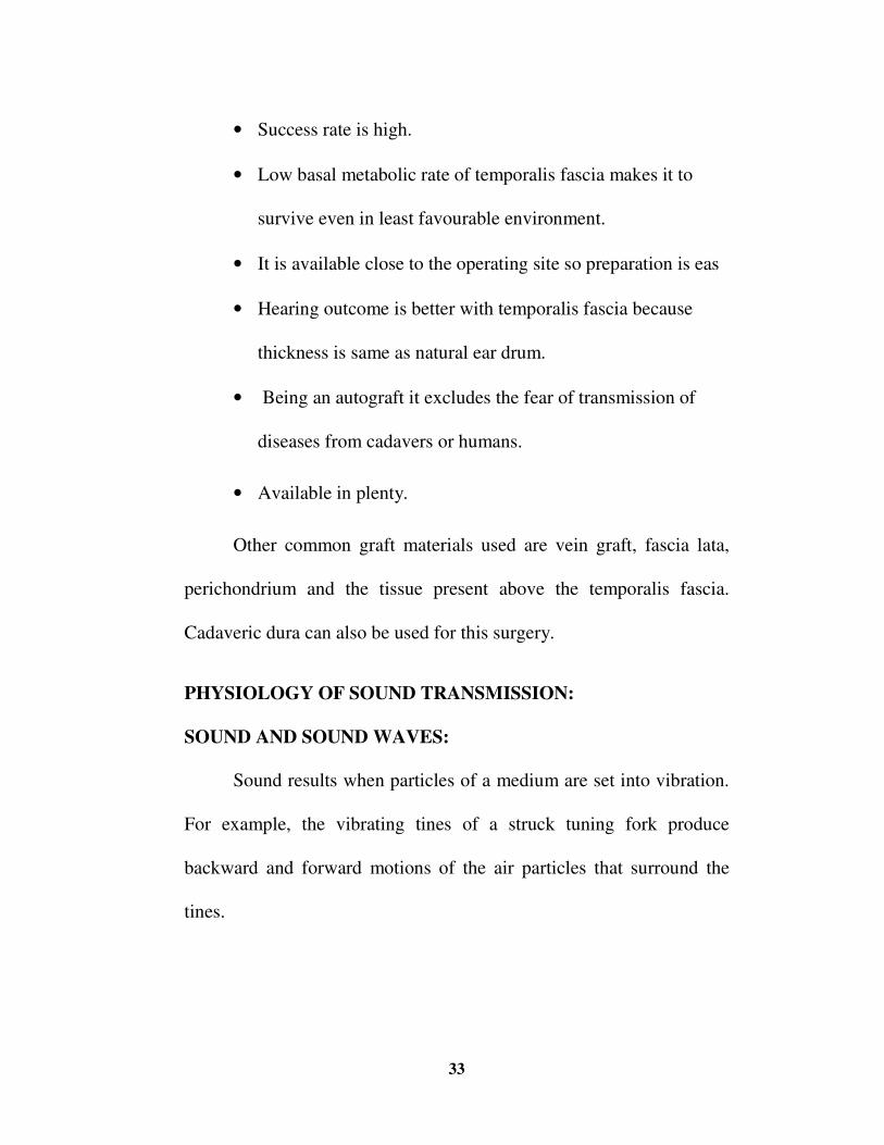
• Success rate is high.
• Low basal metabolic rate of temporalis fascia makes it to
survive even in least favourable environment.
• It is available close to the operating site so preparation is eas
• Hearing outcome is better with temporalis fascia because
thickness is same as natural ear drum.
• Being an autograft it excludes the fear of transmission of
diseases from cadavers or humans.
• Available in plenty.
Other common graft materials used are vein graft, fascia lata,
perichondrium and the tissue present above the temporalis fascia.
Cadaveric dura can also be used for this surgery.
PHYSIOLOGY OF SOUND TRANSMISSION:
SOUND AND SOUND WAVES:
Sound results when particles of a medium are set into vibration.
For example, the vibrating tines of a struck tuning fork produce
backward and forward motions of the air particles that surround the
tines.
34 �
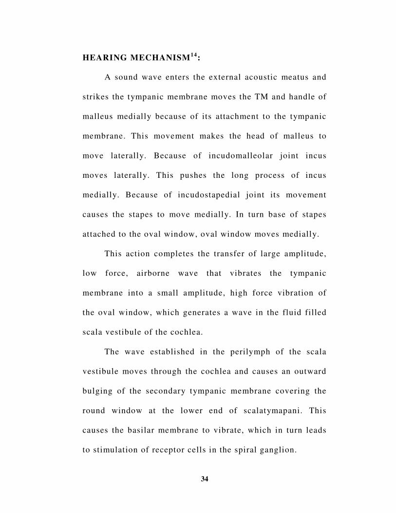
HEARING MECHANISM14:
A sound wave enters the external acoustic meatus and
strikes the tympanic membrane moves the TM and handle of
malleus medially because of its attachment to the tympanic
membrane. This movement makes the head of malleus to
move laterally. Because of incudomalleolar joint incus
moves laterally. This pushes the long process of incus
medially. Because of incudostapedial joint its movement
causes the stapes to move medially. In turn base of stapes
attached to the oval window, oval window moves medially.
This action completes the transfer of large amplitude,
low force, airborne wave that vibrates the tympanic
membrane into a small amplitude, high force vibration of
the oval window, which generates a wave in the fluid filled
scala vestibule of the cochlea.
The wave established in the perilymph of the scala
vestibule moves through the cochlea and causes an outward
bulging of the secondary tympanic membrane covering the
round window at the lower end of scalatymapani. This
causes the basilar membrane to vibrate, which in turn leads
to stimulation of receptor cells in the spiral ganglion.
35 �
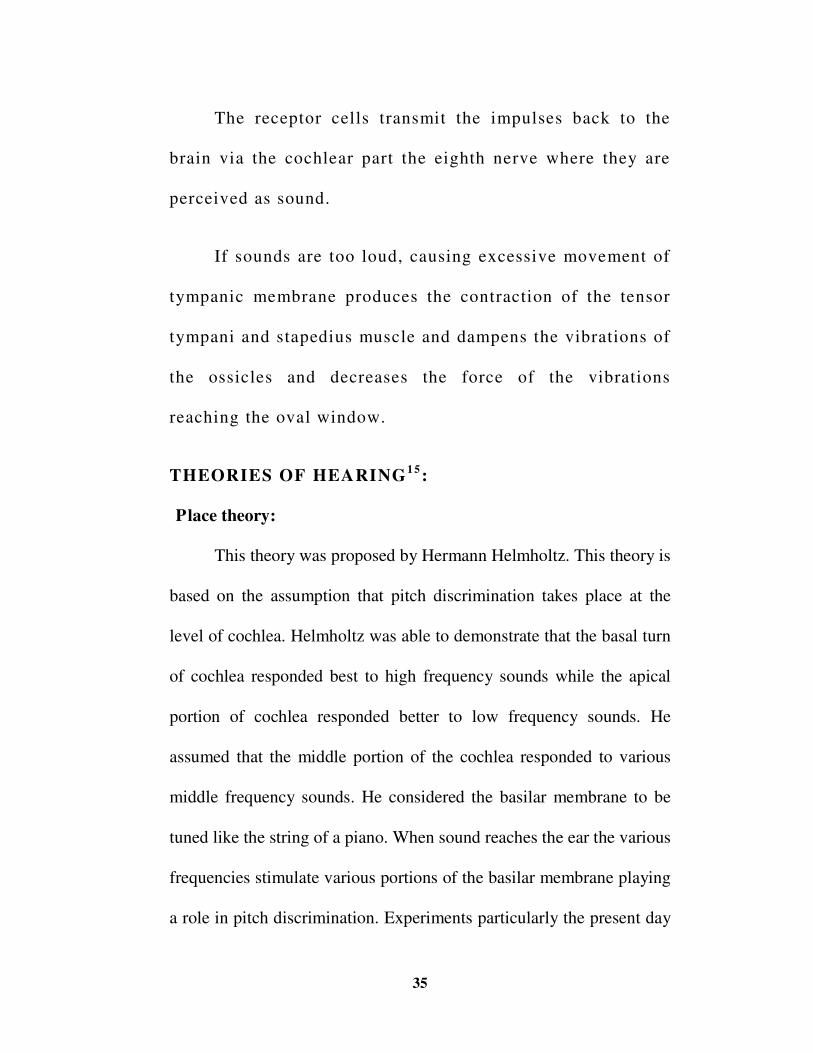
The receptor cells transmit the impulses back to the
brain via the cochlear part the eighth nerve where they are
perceived as sound.
If sounds are too loud, causing excessive movement of
tympanic membrane produces the contraction of the tensor
tympani and stapedius muscle and dampens the vibrations of
the ossicles and decreases the force of the vibrations
reaching the oval window.
THEORIES OF HEARING1 5:
Place theory:
This theory was proposed by Hermann Helmholtz. This theory is
based on the assumption that pitch discrimination takes place at the
level of cochlea. Helmholtz was able to demonstrate that the basal turn
of cochlea responded best to high frequency sounds while the apical
portion of cochlea responded better to low frequency sounds. He
assumed that the middle portion of the cochlea responded to various
middle frequency sounds. He considered the basilar membrane to be
tuned like the string of a piano. When sound reaches the ear the various
frequencies stimulate various portions of the basilar membrane playing
a role in pitch discrimination. Experiments particularly the present day
36 �
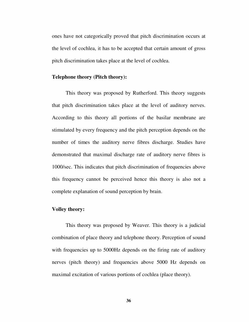
ones have not categorically proved that pitch discrimination occurs at
the level of cochlea, it has to be accepted that certain amount of gross
pitch discrimination takes place at the level of cochlea.
Telephone theory (Pitch theory):
This theory was proposed by Rutherford. This theory suggests
that pitch discrimination takes place at the level of auditory nerves.
According to this theory all portions of the basilar membrane are
stimulated by every frequency and the pitch perception depends on the
number of times the auditory nerve fibres discharge. Studies have
demonstrated that maximal discharge rate of auditory nerve fibres is
1000/sec. This indicates that pitch discrimination of frequencies above
this frequency cannot be perceived hence this theory is also not a
complete explanation of sound perception by brain.
Volley theory:
This theory was proposed by Weaver. This theory is a judicial
combination of place theory and telephone theory. Perception of sound
with frequencies up to 5000Hz depends on the firing rate of auditory
nerves (pitch theory) and frequencies above 5000 Hz depends on
maximal excitation of various portions of cochlea (place theory).
37 �
Travelling wave theory of Bekesy:
This is one type of place theory. This theory holds that pitch
discrimination is determined when a certain place along the basilar
membrane is set into maximum vibration. The auditory nerve fibres
supplying the maximally vibrating portion of basilar membrane start to
fire in response to it.
MIDDLE EAR TRANSFORMERS:16
Catenary lever:
Helmholtz was first to propose a concept of a catenary lever to
the action of tympanic membrane. A familiar example of this type of
lever is tennis net. The tighter the tennis net stretched, the greater force
is exerted on the posts holding it. Because the bony annulus is
immobile, sound energy applies to the tympanic membrane is
amplified at the central attachment, the malleus. It is estimated that
even though the curvature of the tympanic membrane is variable, the
catenary lever provides at least two times gain in sound pressure at the
malleus. Force exerted on the stretched curved fibers of the tympanic
membrane are amplified at its point of attachment, the annular bone is
immobile, so that the malleus is the recipient of this magnified energy,
directing it into the ossicular chain for transmission to the
perilymphatic fluid.
38 �
Ossicular lever:
The lever action that results from the different lengths of the
rotating malleus and incus arms around the axis of rotation of the
ossicles. The axis of rotation is an imaginary line joining the anterior
malleal ligament to the incudal ligament that anchors the short process
of the incus. The malleus and incus lever arms in humans are nearly
the same length. Hence, the ratio of these lengths, which is 1.3,
predicts a small, 2-dB increase in sound pressure applied by the stapes
to the inner ear.
Hydraulic lever:
Helmholtz’ s third concept of impedance matching is referred as
area ratio. The effective vibratory area of tympanic membrane is 55
mm sq. whereas foot plate area is 3.2 mm sq. hence effective area ratio
is 14:1. This is a mechanical advantage provided by tympanic
membrane. The product of areal ratio into lever ratio is known as
transformer ratio i.e. 14x1.3=18:1
Phase difference:
In the normal ear sound pressure waves never reach the oval
window and round window in the same phase, due to the presence of
tympanic membrane, middle ear and air cushions. If air waves reach
39 �
round window and the oval window at the same time it cancel the
effect of sound waves leading to stasis of perilymph. This reciprocal
action at oval window and round window is cancelled as phase
difference. Therefore, loss of this phase difference (large or posteriorly
based perforations) may lead to more conductive hearing loss.
However in normal case sound wave reaches oval window earlier than
round window which is also an added advantage of hearing.
Ossicular and acoustic coupling:
The contribution of the middle ear to the window pressure
difference that stimulates the inner ear can be split into several
stimulus pathways. A previous section described how the tympano-
ossicular system transforms sound pressure in the ear canal to sound
pressure at the oval window. This pathway has been termed ossicular
coupling. There is another mechanism, called acoustic coupling,
through which the middle ear can stimulate the inner ear. Motion of the
tympanic membrane in response to ear canal sound creates sound
pressure in the middle ear cavity. Because the cochlear windows are
separated by a few millimetres, the acoustic sound pressures at the oval
and round windows, respectively, are similar but not identical. Small
differences between the magnitudes and phases of the sound pressures
40 �
outside the two windows result in a small but measurable difference in
sound pressure between the two windows. In the normal ear, the
magnitude of this acoustically coupled window pressure difference is
small, on the order of 60 dB less than ossicular coupling. Hence,
ossicular coupling dominates normal middle ear function, and one can
ignore acoustic coupling. However, as will be seen later, acoustic
coupling can play an important role when ossicular coupling is
compromised, as in some diseased and reconstructed ears.
MIDDLE EAR GAS EXCHANGE MECHANISM:
Like the lung, the middle ear is an organ in the human body that
must maintain an aerated cavity within it for the fulfillment of its
function. When hypoventilation occurs in the lungs it threatens the
maintenance of life, and when it occurs in the middle ear it causes loss
of hearing, which is an important function for maintaining the quality
of life. Because of similarity in their functions, the lung and middle ear
also have many similarities in structure, bluestone et al. (1981)
compared the eustachian tube with the larynx and the middle ear and
mastoid to the lung, and suggested that the eustachian tube, middle ear,
and mastoid are involved in the ventilation of the middle ear just the
larynx and lungs are involved with that of the body. He also pointed
41 �
out the similarities of otitis media atelectatic ear in the middle ear and
pneumonia or atelectasis in the lungs as pathophysiological conditions
of these organs caused by their organic or functional failures
Thus, in order to maintain physiologicalcondition of the middle
ear, ventilation and pressure regulation are regarded as the most
important functions.
In short middle ear is also ventilated by transmucosal gas
exchange, which is passive exchange of gases through the middle ear
mucosa. The gas exchange is slow that it cannot cope with a sudden
change in atmospheric pressure, but it has the advantage that it works
constantly even during sleep. The gas exchange function is impaired by
inflammatory thickening of mucosa, and stops completely when the air
space in the middle ear is lost.
CHRONIC OTITIS MEDIA:
DEFINITION:
Chronic otitis media (COM) is, defined as a chronic
inflammation of the middle ear and mastoid cavity, which presents
with recurrent ear discharges or otorrhea through a tympanic
perforation.
42 �
AETIOLOGY OF TYMPANIC MEMBRANE
PERFORATIONS:
BACTERIAL17:
Bacterial infection of the middle ear causes acute otitis media,
which often results in a small perforation through which purulent
material discharges. These perforations heal spontaneously in a short
time unless complicating factors coexists. Eustachian tube dysfunction
is the major factor that results in a permanent perforation. In such a
middle ear, the mucus membrane is exposed to repeated infection, both
through the external auditory canal and through the Eustachian tube,
with chronic continuous discharge or with recurring episodes of
suppurative otitis media. Allergic sensitization of the exposed middle
ear mucosa frequently occurs. Such disease is termed active chronic
otitis media without cholesteatoma. The incidence of the condition
varies widely by geography, race, and genetic predisposition, as well as
socioeconomic factors.
Beta-haemolytic streptococci are capable of inciting a
Necrotizing form of acute suppurative otitis media, especially in
children suffering from measles or scarlet fever. The pathogenic
43 �
characteristics of the infecting species of bacterium cause capillary
infarction and subsequent ischemic necrosis of tissue. Thus, large
portions of the tympanic membrane (where the blood supply is
poorest) become necrotic and slough, usually leaving intact the better
nourished annular rim, the area near the handle of the malleus, and the
pars flaccida. The resulting kidney shaped perforation is called a
central perforation because of the marginal rim that remains. After the
acute episode of necrotizing otitis media has subsided, the perforation
may heal spontaneously, leaving a thin atrophic scar, or may remain
permanently open. The microcirculation of the middle ear also is
disrupted, leading to tympanosclerosis, in which healing involves an
ossification process with variable involvement of the middle ear
structures.
Mycobacterial17:
Mycobacterial infection of the tympanic membrane is primarily
caused by Mycobacterium tuberculosis and, to a lesser degree, atypical
mycobacteria. This condition manifests as a relentless, low-grade
inflammation of the tympanic membrane that is refractory to
conventional (oral or topical) antibiotic treatment. It eventually results
in multiple small perforations that subsequently coalesce.
44 �
PATHOLOGY OF TYMPANIC MEMBRANE
PERFORATIONS:
As documented in the experimental animal, proliferation of
stratified squamous epithelium at the rim of a perforation begins within
12 hours, and granulation tissue growth begins at 36 hours.
Regeneration of the epithelium of the inner (mucosal) surface is more
sluggish and begins only after several days. As long as there is a
suitably flat surface, stratified squamous epithelium grows at the rate
of 1 mm a day. Histopathologic examination of permanent perforations
showed that stratified squamous epithelium grows medially over the
edge of the perforation, which appears to arrest the subsequent closure
of the perforation. Removal of this medialized epithelium forms the
basis of some of the treatments for tympanic membrane perforation.
The cytokines implicated in this arrest of healing may be multiple, but
transforming growth factor- 1 (TGF-1) is found at the border of the
chronic perforation and may mediate the arrest of healing. This is the
reason why we excising the margin of the perforation while doing
myringoplasty.
45 �
EFFECTS OF TYMPANIC MEMBRANE PERFORATIONS
ON HEARING:18
Perforations of the tympanic membrane cause a conductive
hearing loss that can range from negligible to 50 dB. The primary
mechanism of conductive loss caused by a perforation is a reduction in
ossicular coupling caused by a loss in the sound pressure difference
across the tympanic membrane. The sound pressure difference across
the tympanic membrane provides the primary drive to the motion of
the drum and ossicles. Perforation-induced physical changes such as
reduction in tympanic membrane area or changes in coupling of
tympanic membrane motion to the malleus do not appear to contribute
significantly to the hearing loss caused by a perforation. Perforations
cause a loss that depends on frequency, perforation size, and middle
ear air space volume. Perforation-induced losses are greatest at the
lowest frequencies and generally decrease as frequency increases.
Perforation size is an important determinant of the loss; larger
perforations result in larger hearing losses. The volume of the middle
ear air space (combined tympanic cavity and mastoid air volume) is
also an important parameter that determines the amount of hearing loss
caused by a perforation; small middle ear air space volumes result in
46 �
larger air–bone gaps. Other things being equal, for a given sound
pressure in the ear canal and a given perforation, the resulting sound
pressure within the middle ear cavity will vary inversely with middle
ear volume. Hence, the transtympanic membrane sound pressure
difference will be smaller (and the conductive loss correspondingly
greater) with smaller middle ear volumes. Identical perforations in two
different ears can have conductive losses that differ by up to 20to 30
dB if the middle ear air space volumes differ substantially (within
normal ears, middle ear air space volume can range from 2 cm3 to 20
cm3).
PATHOGENESIS AND CLASSIFICATION OF
CHRONIC OTITIS MEDIA1 9 :
Central perforation of the tympanic membrane can remain dry,
with only rare intermittent drainage, that is, inactive chronic otitis
media19. More typically, chronic or recurrent mucoid otorrhea, that is,
active chronic otitis media19, is provoked by exposure of the tympanic
mucosa to bacteria of the external auditory canal as well as of the
eustachian tube.
Pseudomonas aeruginosa is the most common offending
organism causing chronic otitis media.17Other isolates include enteric
47 �
aerobic gram-negative bacilli such as Proteus, E.coli and gram positive
Staphylococcus aureus, Streptococcus, gram negative Haemophilus
influenza. Anaerobic organisms are associated with foul smelling
discharge include Bacteroids and Peptostreptococcus species.17
Otoscopy, or preferably examination with an operating
microscope, reveals a tympanic membrane perforation and, in active
disease, mucoid or mucopurulent discharge. The presence of an aural
polyp or malodorous otorrhea should raise the clinician’ s suspicion
regarding the presence of cholesteatoma. After careful aspiration of
any debris, the status of the middle ear mucosa can be assessed through
the perforation and can appear only slightly thickened, with ciliary
activity visualized, or can be markedly thickened, with polypoid
degeneration. The ossicular chain can be intact, or disrupted, with the
long process of the incus most prone to resorption. The mobility of the
stapes can be assessed by microscopic examination combined with
Barany noise box stimulation of the contralateral ear, taking advantage
of the crossed stapes reflex.
On histopathologic examination, the middle ear mucosa is
oedematous, with sub mucosal fibrosis and Infiltration by chronic
inflammatory cells. The mucosal oedema can lead to the formation of
48 �
aural polyps, which can even project into the external auditory canal.
Bone erosion, including ossicular resorption, mucosal ulceration, and
the formation of granulation tissue, is the correlate of persistent
otorrhea. Mucosal thickening can impair ossicular and tympanic
membrane mobility, aggravated by the deposition of hyalinised
cartilage (i.e., tympanosclerosis).
The treatment of chronic otitis media focuses on the mucosal
infection in the tympanomastoid compartment. After aspiration of
retained debris, aural toilet comprising 1.5% acetic acid (one part white
vinegar to two parts water) irrigation (at body temperature, three times
daily with an ear/ulcer syringe) followed by the instillation of topical
antibiotic drops (e.g., ciprofloxacin, sulfacetamide) Suffices to bring
most infections under control. The acetic acid irrigation removes
accumulated debris and acidifies the external auditory to canal,
discouraging growth of Pseudomonas and other bacteria. Any
underlying allergies and/or nasopharyngeal disorder should be
managed and closure of the tympanic membrane perforation
undertaken as soon as the ear becomes dry.
49 �
AUDIOLOGICAL ASSESSMENT:
Clinical tests of hearing:
• Finger friction test
• Watch test
• Speech test
• Tuning fork test20
These tests are performed with tuning forks of different
frequencies such as 256, 512, 1024 Hz but for the routine clinical
practice, tuning fork of 512 Hz is ideal.20
A Prediction of air- bone gap can be made if tuning forks of
256, 512, and 1024 Hz are used.
• A Rinne test equal or negative for 256 Hz but positive for 512
Hz indicates air bone gap of 20-30 decibel.
• A Rinne test equal or negative for 256 and 512 Hz but positive
for 1024 Hz indicates air bone gap of 30- 45 decibel.
• A Rinne test negative for all the three tuning forks of 256, 512
and 1024 Hz, indicates air bone of 45- 60 decibel.
50 �
Weber’ s lateralized to the worse ear in conductive hearing
loss. Weber’ s can be lateralized even when the Rinne is positive
with 256 Hz tuning fork. Because weber’ s will be lateralized
when there is 5 decibel difference between two ears. But at least
20 decibel hearing loss needed for the Rinne to get lateralized. For
these reasons weber’ s test considered a more sensitive than Rinne.
Absolute bone conduction test is a measure of cochlear
function. It is reduced in sensorineural hearing loss.
PURE TONE AUDIOMETRY:21, 22
Pure-tone audiometry is used to establish hearing threshold
sensitivity at discrete frequencies across a range important for human
communication. Threshold levels are plotted on an audiogram to show
how threshold sensitivity varies across the frequency range. The
complete pure-tone audiogram consists of air- and bone-conduction
threshold curves for each ear.
The pure-tone audiogram is based on audiometric zero, or
average normal hearing, across a defined frequency range. By
definition, 0 dB HL is the average intensity level at which threshold of
sensitivity is measured in normal-hearing individuals. For clinical
51 �
purposes, the standard deviation is considered to be 5 dB, so that 95%
of the normal population will have thresholds varying from –10 to +10
dB HL. Based on the pure-tone audiogram, then, hearing loss is often
classified as minimal (15 to 25 dB), mild (25 to 40 dB), moderate (40
to 55 dB), moderately severe (55 to 70 dB), severe (70 to 90 dB), or
profound (more than 90 dB). Although hearing loss in the minimal
range may or may not result in impairment or handicap, it is incorrect
to consider thresholds in this range to be within normal limits. The
pure tone audiogram is also used to describe the shape of loss or the
audiometric contour or configuration. The audiogram also provides a
measure of interaural symmetry or the extent to which hearing
sensitivity is the same in both ears or better in one than the other. In
addition, the combination of air and bone-conduction audiometry is
used to determine type of hearing loss.
USES OF PURE TONE AUDIOGRAM:21, 22
• It is a measure of threshold of hearing by air and bone
conduction and thus the degree and type of hearing loss.
• A record can be kept for future reference.
• Audiogram is essential for the prescription of hearing aid.
• Helps to find degree of handicap for medico legal purposes.
52 �
• Helps to predict speech reception threshold.
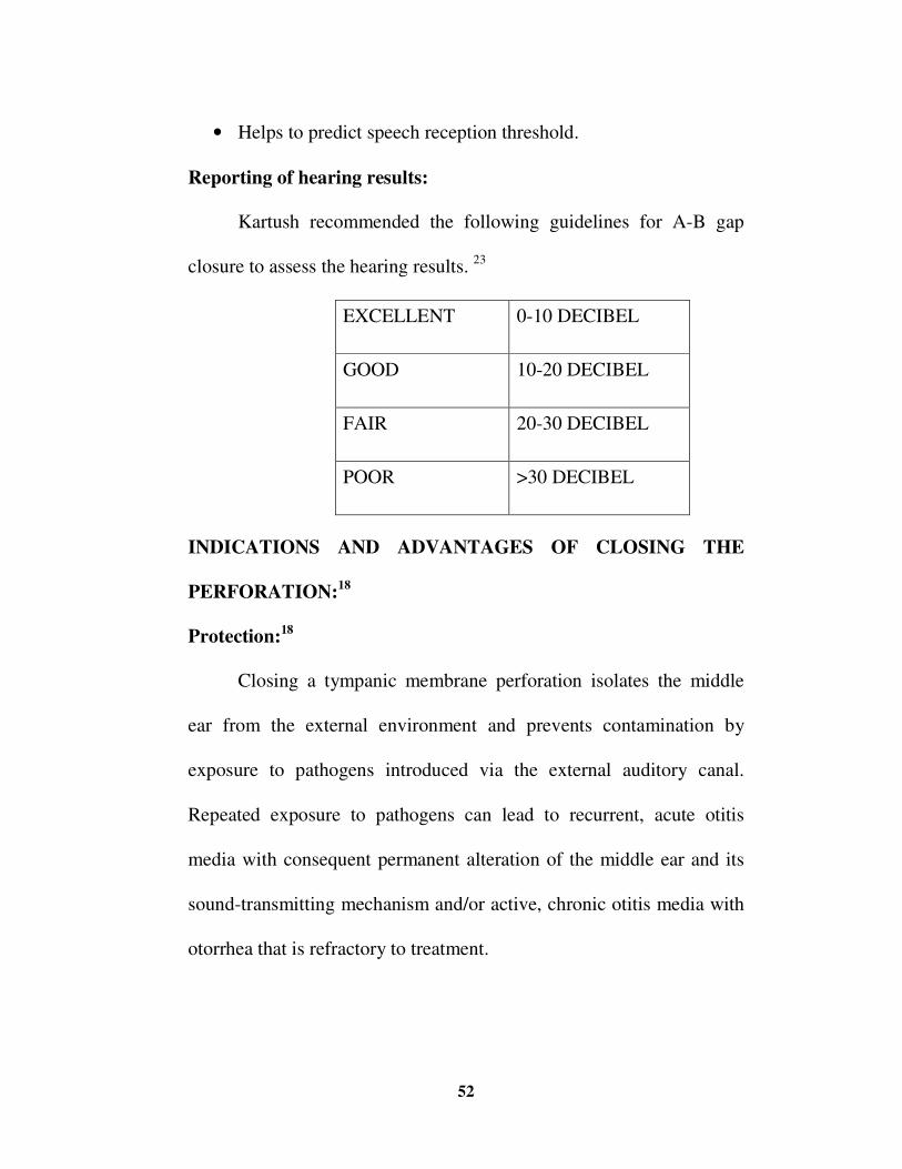
Reporting of hearing results:
Kartush recommended the following guidelines for A-B gap
closure to assess the hearing results. 23
EXCELLENT 0-10 DECIBEL
GOOD 10-20 DECIBEL
FAIR 20-30 DECIBEL
POOR >30 DECIBEL
INDICATIONS AND ADVANTAGES OF CLOSING THE
PERFORATION:18
Protection:18
Closing a tympanic membrane perforation isolates the middle
ear from the external environment and prevents contamination by
exposure to pathogens introduced via the external auditory canal.
Repeated exposure to pathogens can lead to recurrent, acute otitis
media with consequent permanent alteration of the middle ear and its
sound-transmitting mechanism and/or active, chronic otitis media with
otorrhea that is refractory to treatment.
53 �
Auditory:
Closure of a tympanic membrane perforation restores the
vibratory area of the membrane and affords round window protection,
thus improving hearing and decreasing tinnitus; however, high-
frequency audiometry demonstrates a persistent air–bone gap, despite
successful closure of a perforation.48 An approximate estimate of the
improvement in hearing and tinnitus that may be expected from closure
of a perforation is obtained by temporary patching with cigarette paper
or cellophane. If a carefully applied airtight patch does not eliminate
the conductive loss, one may assume that an additional ossicular lesion
is present, either fixation or discontinuity. In such cases, simple
myringoplasty will not suffice. Closure must be accompanied or
followed by correction of the ossicular problem by tympanoplasty.
CONTRAINDICATIONS TO CLOSURE OF A
PERFORATION:24
Not every perforation needs to, or should, be closed. Each
patient must be managed in accordance with what is best for that
patient. An elderly or debilitated patient with an asymptomatic
perforation, or a patient with a perforation in an only hearing ear, is not
a good surgical candidate. In a young child with a perforation from a
54 �
ventilation tube inserted because of poor ear ventilation, it is unwise to
repair the tympanic membrane until it is apparent that eustachian tube
function has significantly improved by the pathologic process repeat
itself.
Absolute
The presence of cholesteatoma is an absolute contraindication
for a myringoplasty. Active chronic otitis media with otorrhea
refractory to medical management indicates the presence of an
infectious focus, which needs to be surgically addressed prior to repair
of the tympanic membrane. Medical contraindications to surgical
intervention include advanced patient age. Last but not least,
unrealistic patient expectations and/or the inability of the patient to
understand the reasoning for the proposed intervention are
contraindications for repairing a tympanic membrane through
myringoplasty.
Relative
Eustachian tube dysfunction increases the chances of failure of
myringoplasty. Myringoplasty in the only hearing ear should be
evaluated carefully, and the potentially small risk of sensorineural
hearing loss should be outweighed by the benefits of the operation.
55 �
Such an example would be the presence of severe atelectasis, which
has led to ossicular erosion, or atelectasis with changes indicating
progression to cholesteatoma. Every attempt should be made to control
allergies manifested by upper respiratory symptomatology that will
increase the incidence of failure of myringoplasty.
Fate of tympanic membrane grafts:4
Histologically tympanic membrane grafts become lined by
squamous epithelium on the ear canal side and the middle ear mucosa
on the tympanic cavity side. The graft itself becomes the middle or
connective tissue portion of the reconstructed drum, but the orderly
arrangement of concentric and radial collagen fibres as seen in the
normal drum is not reconstituted in the graft.
MYRINGOPLASTY:
The term myringoplasty is reserved for the simple repair of a
tympanic membrane perforation in which no ossicular reconstruction is
involved. Temporalis fascia is the graft material of choice, although
perichondrium obtained from the tragus produces the same success
rates; however, perichondrium is not transparent and hinders future
examination of the middle ear. Two basic grafting techniques have
56 �
emerged, referred to as the overlay and under surface (or medial onlay)
techniques.
PATIENT SELECTION:
Regardless of the grafting technique chosen, the preoperative
evaluation and management of the patient with a tympanic membrane
perforation remain the same. A proper history with sufficient detail as
it relates to the tympanic membrane perforation is necessary as is a
thorough examination of the ear in question under the examining
microscope. A complete head and neck examination is essential in
identifying risk factors for failure of the proposed myringoplasty. The
record should document the findings, preferably diagramming the
perforation in question; current technology enables the physician to
obtain a high-quality photograph of the otoscopic findings. All patients
should undergo pure-tone air and bone conduction audiometric testing
along with speech discrimination evaluation. Tuning fork tests should
be done on all patients to confirm the audiologic findings. If there is
disagreement between the tuning fork examination and the audiogram,
then the latter should be repeated. High-resolution computed
tomographic (CT) scanning of the temporal bone is not indicated
unless there is a suspicion of underlying pathology or separate
57 �
indicators from the problem at hand such as revision surgery in
association with chronic otorrhea. The patient is counselled
preoperatively about the nature of the problem, the proposed treatment,
alternative therapies, expected outcome, and potential complications. It
is helpful to provide written explanations and instructions discussing
pre- and postoperative care of the ear. Videotaped discussions of the
proposed procedure are also useful.
The following factors must be considered and dealt with
preoperatively.
Eustachian Tube Function:25
Assessing the function of the eustachian tube is difficult.
Conflicting evidence exists in the literature as to the importance of
eustachian tube function for the success of tympanic membrane repair,
reflecting the inaccuracy of the methods available to assess the
function of the eustachian tube. From a practical point of view, one of
the most useful indications of proper Eustachian tube function is a
normal contralateral ear. Bilateral pathology is associated with
decreased success of surgical intervention, especially in children.
Although a functioning eustachian tube is important to the success of
the operation, a lack of eustachian tube function should not preclude
58 �
surgical intervention. In fact, eustachian tube function may improve
once the infection is removed and the middle ear space reconstructed
through normalization of the mucosal inflammation seen in the
presence of a perforation.
Control of Infection
Eradication or control of active infection in the ear under
question is crucial. Repeat visits for proper aural toilet and monitoring
of improvement in otorrhea should be instituted prior to any
consideration of surgery. The patient is instructed to irrigate the ear
three times a day with a sterile 1.5% acetic acid solution using a small
rubber bulb ear syringe. This procedure allows purulent material to be
removed from the middle ear and external canal and restores a more
physiologic pH. It is important that the solution be used at body
temperature to avoid caloric stimulation of the labyrinth. Following
acetic acid irrigation, three drops of an antibiotic otic solution are
instilled into the ear. Depending on the severity of the infection, intra
venous and oral antibiotics may also be prescribed. The fluid from
draining ears is not routinely cultured because the majority will
respond to local care. If the ear continues to drain despite aggressive
medical treatment, culture and sensitivity testing is performed and the
59 �
antibiotic regimen adjusted appropriately. The majority of patients will
respond to this treatment. If treatment fails, consideration should be
given to other factors such as patient compliance, mastoid
involvement, severe allergies, and/or incomplete treatment of the
offending organism(s).
Perioperative Antibiotics26
In the absence of signs of active infection (purulent otorrhea),
the perioperative administration of systemic antibiotics does not
influence the results of myringoplasty.26 Furthermore, perioperative
antibiotics do not prevent the emergence of bacterial pathogens in the
postoperative period. Similarly, the presence of no purulent otorrhea
preoperatively does not indicate the presence of pathogenic bacterial
flora in the middle ear and external auditory canal.
Control of Allergies and Rhinosinusitis:27
Every attempt is made to identify and treat coexistent inhalant
allergies and sinus disease prior to surgery. The status of the upper
respiratory tract directly influences eustachian tube function and
therefore the eventual outcome of surgery. Therefore, a trial of
antihistamines and/or nasal corticosteroid sprays may be indicated
empirically based on the history and examination. If there is a strong
60 �
history of allergies, skin testing with targeted desensitization should be
considered.
Age of Patient:
The success of tympanic membrane repair in the paediatric age
group has been reported from as low as 35% to as high as 93%. The
consensus holds that myringoplasty in the paediatric age group has a
lower success rate than in the adult age group. The reasons for this
discrepancy are not clear but may be related to the higher incidence of
otitis media and its predisposing factors in children. In children, it is
best to avoid elective surgery for myringoplasty between the ages of 4
and 7 years because of the risk of otitis media and from a
psychological point of view. At age 8 years and thereafter, the child
should participate in the decision process for such elective surgery.
Status of Contralateral Ear:
If the ear under question is the only hearing ear, no attempt
should be made to repair a tympanic membrane perforation. A small
but real risk of sensorineural hearing loss does exist with
myringoplasty. The rate of such hearing loss has been reported to range
from 0.1 to 2%.68 often the contralateral ear may be involved by
similar pathology. The choice of which ear to repair is then dependent
61 �
on hearing status—the worse hearing ear should be operated first. An
exception to this rule is a pathologic condition in a better hearing
contralateral ear that threatens hearing or health such as cholesteatoma,
active infection such as mastoiditis, or significant atelectasis with
likely progression to cholesteatoma or ossicular chain erosion.
Hearing Status/Need for Hearing Aids:
If there are bilateral perforations, with significant conductive
hearing loss in the absence of otorrhea, hearing aid fitting may be
preferable for children under the age of 8 years. If otorrhea is provoked
by hearing aid use in this situation, the worse hearing ear should be
repaired first. A wet ear in and of itself is not a contraindication to
myringoplasty, although purulent otorrhea is a contraindication. A wet
ear should not be an absolute contraindication for myringoplasty,
provided that factors such as allergies, eustachian tube function, and
superinfection have been controlled. In selected patients, a
mastoidectomy may be considered as an adjunct to the myringoplasty
that can improve the success rate.
HISTORY OF MYRINGOPLASTY:
From the seventeenth to the nineteenth centuries, several
attempts at closing tympanic membrane perforations using prosthetic
62 �
materials were made. In 1878 Berthhold devised the term
myringoplasty. 1, 3, 28 But the surgical repair of the tympanic membrane
was first attempted by Banzer in 1640.1,28
The use of cauterizing agents to promote healing of tympanic
membrane perforations was introduced by Roosa in 1876,28 who used
the application of silver nitrate to the rim of a perforation; the use of
trichloroacetic acid was first advocated in 1895. It was not until Joynt
combined the cautery and paper patch techniques that closure results
improved, forming the basis of the modern-day use of the paper patch
technique as popularized by Derlacki.
In 1952 Wullstein formally announced the technique of closing
perforations with a split-thickness skin graft. 2, 3 Only a year later,
Zollner described his experiences with a similar graft. At the same
time, Wullstein and Zollner introduced the use of the operating
microscope, significantly enhancing surgical results by improving the
accuracy of the technique. 2
Problems with skin grafts as the closure material for tympanic
membrane perforations soon became apparent, and in 1956, Zollner
first used fascia lata to close perforations. In 1958, Heermann began to
use temporalis fascia3. In 1960, Shea described the closure of tympanic
63 �
membrane perforations using a vein graft.3 The advantages of
connective tissue over skin as a grafting material were confirmed by
the higher percentage of successful closures; however, skin grafting of
perforations was not completely abandoned. Meatal skin, which lacks
glands and is elevated with the underlying periosteum, has distinct
advantages over skin grafts, both full and split thickness,2, 3from other
areas of the body. It has continued to be used in selective instances. In
the 1960s and 1970s, homograft (cadaveric) materials, including
tympanic membrane, dura, and pericardium, among others, were used
with varying success. None of these materials gained universal
acceptance and today pose a problem because of the potential for
transmitting disease (eg, Jakob-Creutzfeldt disease and HIV infection).
Temporalis fascia continues to be the material of choice for
reconstruction of the tympanic membrane.
Role of endoscope in ear surgeries:5, 6
Rigid Hopkins rod endoscopes and fiberoptic endoscopes are
both available for use in temporal bone surgery. Generally, the rigid
endoscopes are preferred because of their superior resolution. Newer
generation rigid endoscopes, using gradient index of refraction lenses,
are becoming ever smaller, and are closing the gap with fiber
technology. Charge-coupled device (CCD) camera microchips are now
64 �
so small that they can be placed on the end of larger endoscopic
instruments, eliminating the need for fiberoptic or long-lens systems
entirely. 5, 6 As CCD chips become more miniaturized, they have the
potential to further revolutionize the capabilities of endoscopes.
Endoscopic images have the disadvantage of spherical distortion
(“ fisheye views” ) and cannot provide the three-dimensional view
afforded with the binocular operating microscope. Endoscopic
magnification increases steeply as an object comes into close proximity
to the lens and can approach the powers achieved with the operating
microscope. Endoscopic surgeons learn to compensate for the variable
magnification and two dimensional views by watching how a structure
and its surroundings change as the endoscope is moved in and out of
proximity to it. The three-dimensionality of an image is re-created by
these changes with endoscope motion. Endoscopes intended for
introduction through the tympanic membrane must be 1.9 mm in
diameter or less. Operating room exposures permit the use of larger
endoscopes, such as 2.7 to 4.0 mm, which are preferred since they
yield larger images with improved brightness and clarity. Endoscopes
typically have 0-, 30-, and 70-degree view angles. Endoscopic
illumination is provided by a halogen or xenon fiberoptic light source,
generally using 150 to 300 watts. Images may be viewed directly
65 �
through the endoscope lens, or, more commonly, a CCD camera is
attached to the endoscope lens to deliver the images to a monitor.
Computer interfaces, digital or video recording devices, and printing
systems are optional.
ENDOSCOPIC TYMPANOPLASTY5, 6
Anterior, marginal tympanic membrane perforations are
frequently repaired using a postauricular approach to maximize
exposure. The visualization of far anterior perforations may be
especially difficult, and the anterior margin may be completely hidden
from direct view behind a prominent anterior canal bony overhang.
Anterior perforations may be managed through a transcanal approach,
using the endoscope to visualize the anterior margin. When the anterior
margin is very narrow, lying quite close to the anterior canal and
raising some question as to whether an underlay graft has sufficient
contact to hold to the anterior drum remnant, an argon laser may be
used to “ spot weld” the graft in place. The fiber delivered laser is
convenient as the tip may be bent to accommodate the anterior bony
overhang. A line-of- sight laser, such as a CO2 laser, would not be
useful in these circumstances.
66 �
CLASSIFICATION:
1. Onlay technique.
2. Underlay technique.
1. ONLAY TECHNIQUE:29, 30
In this technique graft is placed beneath the outer squamous
(skin layer) of the tympanic membrane. The procedure is difficult to
master because peeling only outer squamous layer of the ear drum is
not easy. After that graft is placed over the perforation.
2. UNDERLAY TECHNIQUE:31
In this technique tympanomeatal flap is elevated along with
fibrous annulus and remnant tympanic membrane and the graft is
placed medial to the drum remnant or fibrous annulus rather than top
of it. It is relatively easy procedure.
M E R I T S & D E M E R I T S O F T H E TECHNIQUES:
MERITS OF UNDERLAY TECHNIQUE:31
1. Middle ear can be inspected for the disease.
2. Success rate is high.
3. Comparatively easy procedure.
67 �
DEMERITS:
1. Middle ear space may become obliterated.
2. Graft medialisation
3. Anteriorly, the graft has loose contact with the remnant
ear drum leading to anterior perforation.
DEMERITS OF OVERLAY TECHNIQUE:
1. Anterior blunting.
2. Formation of Epithelial pearl
3. Graft lateralization
4. Middle ear is not inspected.
5. Difficult technique to master.
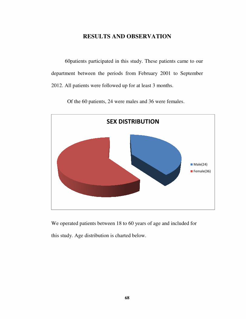
RESULTS AND OBSERVATION
60patients participated in this study
department between
2012. All patients were followed up for at least 3 months.
Of the 60 patients
We operated patients between 18 to 60 years of age and included for
this study. Age distribution is charted below.
68
RESULTS AND OBSERVATION
participated in this study. These patients came to our
the periods from February 2001 to September
2012. All patients were followed up for at least 3 months.
60 patients, 24 were males and 36 were females.
We operated patients between 18 to 60 years of age and included for
Age distribution is charted below.
������������� �
. These patients came to our
February 2001 to September
24 were males and 36 were females.
We operated patients between 18 to 60 years of age and included for
� ������
�� �����
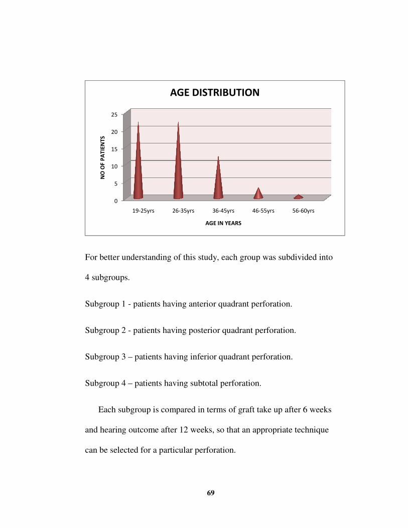
For better understandi
4 subgroups.
Subgroup 1 - patients having anterior quadrant perforation.
Subgroup 2 - patients having posterior quadrant perforation.
Subgroup 3 – patients having inferior quadrant perforation.
Subgroup 4 – patients having subtotal perforation.
Each subgroup is compared
and hearing outcome after 12 weeks
can be selected for a particular perforation
�
�
��
��
��
��
��������
���� ����������
69
For better understanding of this study, each group was subdivided
patients having anterior quadrant perforation.
patients having posterior quadrant perforation.
patients having inferior quadrant perforation.
patients having subtotal perforation.
Each subgroup is compared in terms of graft take up after 6 weeks
and hearing outcome after 12 weeks, so that an appropriate technique
can be selected for a particular perforation.
� ������ � ������ � ������ � � ����
������ ������
������������� �
bdivided into
after 6 weeks
, so that an appropriate technique
����
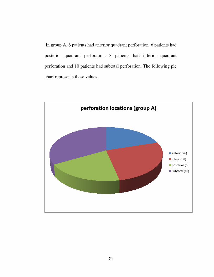
In group A, 6 patients had anterior quadrant perforation. 6 patients
posterior quadrant perforation. 8 patients had inferior quadrant
perforation and 10 patients had subtotal perforation.
chart represents these values.
�������������������������� ���!
70
In group A, 6 patients had anterior quadrant perforation. 6 patients
posterior quadrant perforation. 8 patients had inferior quadrant
and 10 patients had subtotal perforation. The following pie
chart represents these values.
�������������������������� ���!
In group A, 6 patients had anterior quadrant perforation. 6 patients had
posterior quadrant perforation. 8 patients had inferior quadrant
The following pie
����������
�����������
�����������
������������
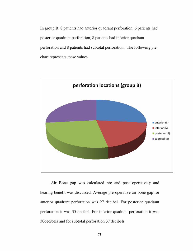
In group B, 8 patients had anterior quadrant perforation. 6 patients had
posterior quadrant perforation
perforation and 8 patients had subtotal perforation.
chart represents these values.
Air Bone gap was calculated pre and post operatively and
hearing benefit was discuss
anterior quadrant perforation was 27 decibel. For posterior quadrant
perforation it was 35 decibel. For inferior quadrant perforation it was
30decibels and for subtotal perforation 37 decibels.
�������������������������� ��!
71
In group B, 8 patients had anterior quadrant perforation. 6 patients had
perforation, 8 patients had inferior quadrant
and 8 patients had subtotal perforation. The following pie
chart represents these values.
Air Bone gap was calculated pre and post operatively and
hearing benefit was discussed. Average pre-operative air bone gap for
anterior quadrant perforation was 27 decibel. For posterior quadrant
perforation it was 35 decibel. For inferior quadrant perforation it was
for subtotal perforation 37 decibels.
�������������������������� ��!
In group B, 8 patients had anterior quadrant perforation. 6 patients had
, 8 patients had inferior quadrant
The following pie
Air Bone gap was calculated pre and post operatively and
operative air bone gap for
anterior quadrant perforation was 27 decibel. For posterior quadrant
perforation it was 35 decibel. For inferior quadrant perforation it was
�����������
����������
������������
�����������
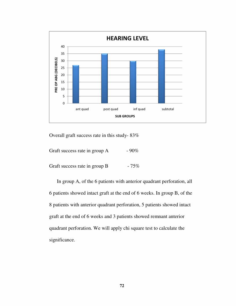
Overall graft success rate in this study
Graft success rate in group A
Graft success rate in group B
In group A, of the
6 patients showed intact graft at the end of 6 we
8 patients with anterior quadrant perforation
graft at the end of 6 weeks and 3 patients showed remnant anterior
quadrant perforation. We will apply chi square test to calculate the
significance.
�
�
��
��
��
��
��
��
��
���� ��!
�������������"��#�!
72
success rate in this study- 83%
Graft success rate in group A - 90%
Graft success rate in group B - 75%
the 6 patients with anterior quadrant perforation,
6 patients showed intact graft at the end of 6 weeks. In group B, of
8 patients with anterior quadrant perforation, 5 patients showed intact
graft at the end of 6 weeks and 3 patients showed remnant anterior
quadrant perforation. We will apply chi square test to calculate the
����� ��! ���� ��! ��������
����� ��
$����� � �#�%�#
rant perforation, all
eks. In group B, of the
5 patients showed intact
graft at the end of 6 weeks and 3 patients showed remnant anterior
quadrant perforation. We will apply chi square test to calculate the
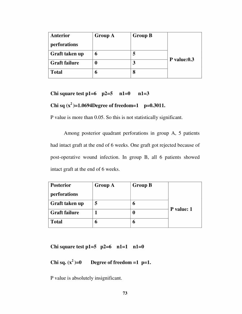
73 �
Anterior
perforations
Group A Group B
P value:0.3 Graft taken up 6 5
Graft failure 0 3
Total 6 8
Chi square test p1=6 p2=5 n1=0 n1=3
Chi sq (x2 )=1.0694Degree of freedom=1 p=0.3011.
P value is more than 0.05. So this is not statistically significant.
Among posterior quadrant perforations in group A, 5 patients
had intact graft at the end of 6 weeks. One graft got rejected because of
post-operative wound infection. In group B, all 6 patients showed
intact graft at the end of 6 weeks.
Posterior
perforations
Group A Group B
P value: 1 Graft taken up 5 6
Graft failure 1 0
Total 6 6
Chi square test p1=5 p2=6 n1=1 n1=0
Chi sq. (x2 )=0 Degree of freedom =1 p=1.
P value is absolutely insignificant.
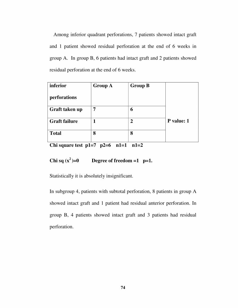
74 �
Among inferior quadrant perforations, 7 patients showed intact graft
and 1 patient showed residual perforation at the end of 6 weeks in
group A. In group B, 6 patients had intact graft and 2 patients showed
residual perforation at the end of 6 weeks.
inferior
perforations
Group A Group B
P value: 1
Graft taken up 7 6
Graft failure 1 2
Total 8 8
Chi square test p1=7 p2=6 n1=1 n1=2
Chi sq (x2 )=0 Degree of freedom =1 p=1.
Statistically it is absolutely insignificant.
In subgroup 4, patients with subtotal perforation, 8 patients in group A
showed intact graft and 1 patient had residual anterior perforation. In
group B, 4 patients showed intact graft and 3 patients had residual
perforation.
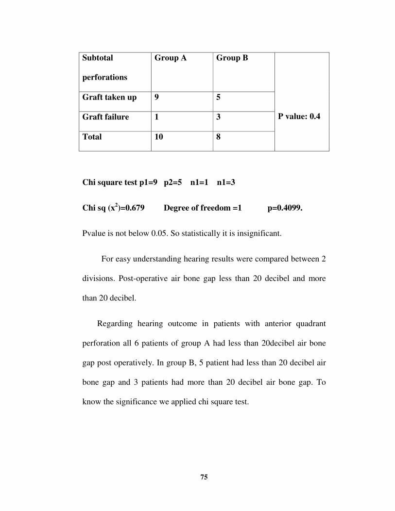
75 �
Subtotal
perforations
Group A Group B
P value: 0.4
Graft taken up 9 5
Graft failure 1 3
Total 10 8
Chi square test p1=9 p2=5 n1=1 n1=3
Chi sq (x2)=0.679 Degree of freedom =1 p=0.4099.
Pvalue is not below 0.05. So statistically it is insignificant.
For easy understanding hearing results were compared between 2
divisions. Post-operative air bone gap less than 20 decibel and more
than 20 decibel.
Regarding hearing outcome in patients with anterior quadrant
perforation all 6 patients of group A had less than 20decibel air bone
gap post operatively. In group B, 5 patient had less than 20 decibel air
bone gap and 3 patients had more than 20 decibel air bone gap. To
know the significance we applied chi square test.
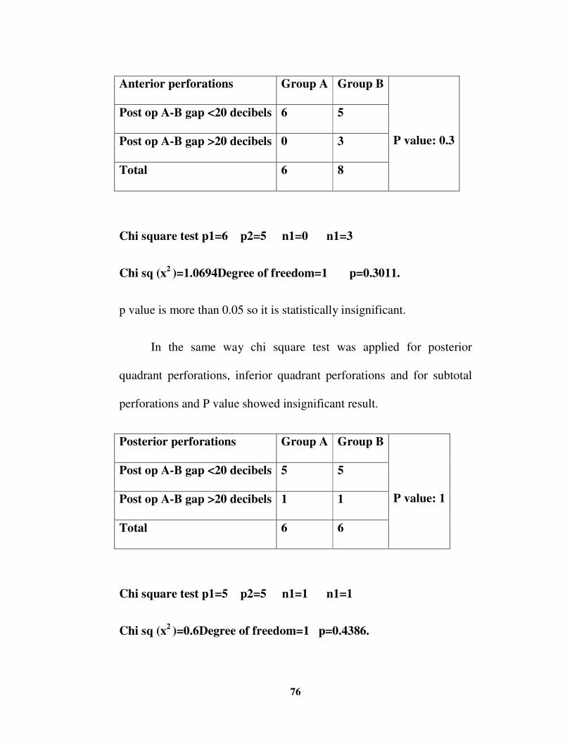
76 �
Anterior perforations Group A Group B
P value: 0.3
Post op A-B gap <20 decibels 6 5
Post op A-B gap >20 decibels 0 3
Total 6 8
Chi square test p1=6 p2=5 n1=0 n1=3
Chi sq (x2 )=1.0694Degree of freedom=1 p=0.3011.
p value is more than 0.05 so it is statistically insignificant.
In the same way chi square test was applied for posterior
quadrant perforations, inferior quadrant perforations and for subtotal
perforations and P value showed insignificant result.
Posterior perforations Group A Group B
P value: 1
Post op A-B gap <20 decibels 5 5
Post op A-B gap >20 decibels 1 1
Total 6 6
Chi square test p1=5 p2=5 n1=1 n1=1
Chi sq (x2 )=0.6Degree of freedom=1 p=0.4386.
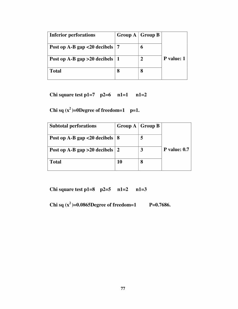
77 �
Inferior perforations Group A Group B
P value: 1
Post op A-B gap <20 decibels 7 6
Post op A-B gap >20 decibels 1 2
Total 8 8
Chi square test p1=7 p2=6 n1=1 n1=2
Chi sq (x2 )=0Degree of freedom=1 p=1.
Subtotal perforations Group A Group B
P value: 0.7
Post op A-B gap <20 decibels 8 5
Post op A-B gap >20 decibels 2 3
Total 10 8
Chi square test p1=8 p2=5 n1=2 n1=3
Chi sq (x2 )=0.0865Degree of freedom=1 P=0.7686.
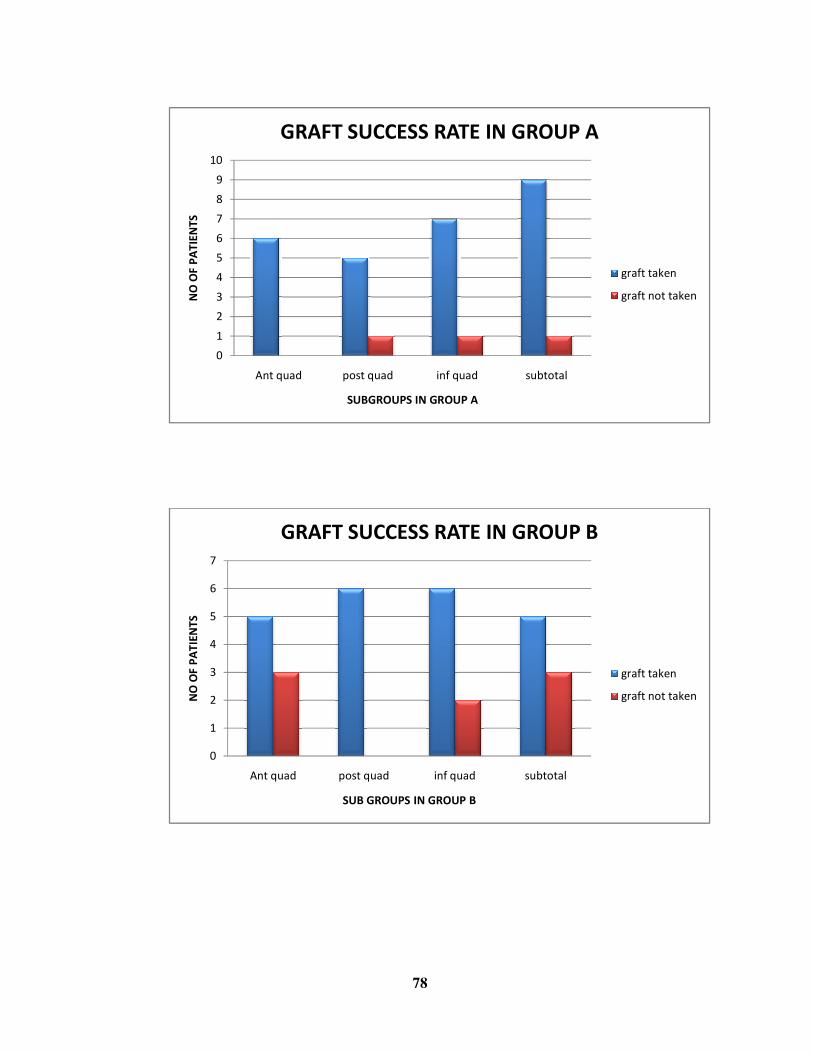
�
�
�
�
�
�
�
�
"
�
�
��
#��� ��!
���� ����������
��� ��� ""����������� ���� ���
�
�
�
�
�
�
"
#��� ��!
���� ���������
��� ���""����������� ���� ��
78
����� ��! ���� ��! ��������
���� ����� ���� ���
��� ��� ""����������� ���� ���
����� ��! ���� ��! ��������
����� ����� ���� ��
��� ��� ""����������� ���� ��
�
$�������%��
$�����������%��
$�������%��
$�����������%��
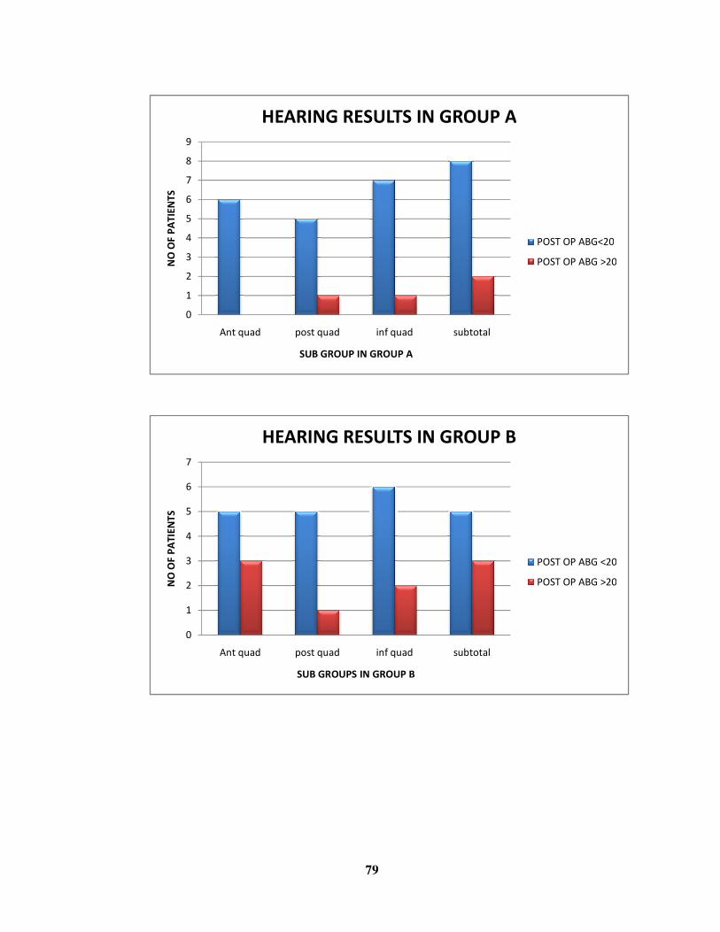
�
�
�
�
�
�
�
"
�
�
#��� ��!
���� ���������
$����� � ���� #����� ���� ���
�
�
�
�
�
�
"
#��� ��!
���� ���������
$����� � ����#����� ���� ��
79
����� ��! ���� ��! ��������
����� ���� ���� ���
$����� � ����#����� ���� ���
&' �(�' &�#)*+��
&' �(�' &�#)* �,��
����� ��! ���� ��! ��������
����� ����� ���� ��
$����� � ���� #����� ���� ��
&' �(�' &�#)* �+��
&' �(�' &�#)* �,��
�
�
&' �(�' &�#)*+��
&' �(�' &�#)* �,��
&' �(�' &�#)* �+��
&' �(�' &�#)* �,��
80 �
DISCUSSION
Surgical repair of the ear drum perforations located anterior to
the handle of malleus can present a problem. 10 The lack of anterior
support for the graft frequently leads to graft failure if classical
underlay technique is performed. 10
To overcome this problem Sheehy and Anderson reported Onlay
myringoplasty in their study series in 1980.29, 30 Following them many
studies were conducted on Onlay technique. But lateraliation of the
graft is a common problem in this technique and the procedure itself is
difficult to master. To overcome these pitfalls many modifications of
classical underlay myringoplasty have been emerging.
Sharp, Terzis, Robinson reported their experience with Kerr flap
to repair anterior and subtotal perforations.32 Abolhassan et al reported
mucosal pocket myringoplasty, a modification of classical underlay
technique. 33
The technique of myringoplasty “ underlay technique graft
placed lateral to the handle of malleus” was reported by Stage J, Bak –
piedersen K in 1992. They repaired 39 tympanic membrane
perforations with predominantly large or anterior pars
81 �
tensaperforations.10, 9. They placed the graft lateral to the handle of
malleus. After a median observation time of 20 months one ear was
found to have a small reperforation. All ears they operated have normal
tympanomeatal angles. Analysis of hearing showed good hearing
improvement and no adverse effects were attributable to conventional
underlay technique in ears with perforation involving the area anterior
to the handle of malleus.
Shrestha, Sinha reported their study in 2006. They used temporalis
fascia as grafting material and studied hearing outcomes of underlay
technique. They reported 49 cases of underlay myringoplasty and in
one case modified underlay technique was performed where graft
placed lateral to the handle of malleus for the patient with central
malleolar perforation with tip of malleus touching the promontory.
Post-operative hearing improvement was 18 decibels in that patient at
10th week.9
In the present study 2 groups are compared. Group A
consists of 30 patients operated with modified underlay technique. In
them, graft was placed over the handle of malleus. Group B consists of
30 patients operated with classical underlay technique.
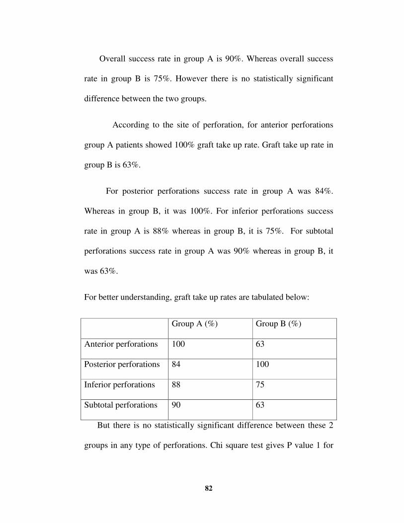
82 �
Overall success rate in group A is 90%. Whereas overall success
rate in group B is 75%. However there is no statistically significant
difference between the two groups.
According to the site of perforation, for anterior perforations
group A patients showed 100% graft take up rate. Graft take up rate in
group B is 63%.
For posterior perforations success rate in group A was 84%.
Whereas in group B, it was 100%. For inferior perforations success
rate in group A is 88% whereas in group B, it is 75%. For subtotal
perforations success rate in group A was 90% whereas in group B, it
was 63%.
For better understanding, graft take up rates are tabulated below:
Group A (%) Group B (%)
Anterior perforations 100 63
Posterior perforations 84 100
Inferior perforations 88 75
Subtotal perforations 90 63
But there is no statistically significant difference between these 2
groups in any type of perforations. Chi square test gives P value 1 for
83 �
anterior and inferior perforations. It states that absolutely there is no
significant difference between these 2 groups. But P value for anterior
quadrant perforation was 0.3 and for subtotal perforation was 0.4.
These two values are less than 1. So even though these values are not
statistically significant values of anterior and inferior quadrant
perforations shows some significance than posterior and inferior
quadrant perforations.
So here we can come to one conclusion. That is placing graft
lateral to the handle of malleus is a good alternative technique to
classical underlay technique for anterior and subtotal perforations.
Regarding hearing, 87% of patients in group A have less than 20
decibel Air bone gap post operatively. 70% of patients in group B
showed less than 20 decibel
Hearing success rate of each subgroup is tabulated below for
better understanding.
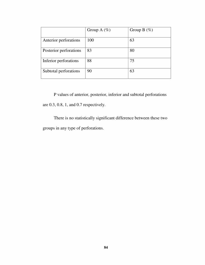
84 �
Group A (%) Group B (%)
Anterior perforations 100 63
Posterior perforations 83 80
Inferior perforations 88 75
Subtotal perforations 90 63
P values of anterior, posterior, inferior and subtotal perforations
are 0.3, 0.8, 1, and 0.7 respectively.
There is no statistically significant difference between these two
groups in any type of perforations.
85 �
CONCLUSION
• Modified underlay technique, placing the graft lateral to the
handle of malleus is a good alternative technique to
classical underlay myringoplasty for anterior and subtotal
perforations.
• There is no statistically significant difference in hearing
outcome between these two groups.
86 �
BIBLIOGRAPHY
1. Joshua S. Sckolnick, MD; Belinda Mantle, MD; Jia Li, MS;
David H. Chi,MD. Paediatric myringoplasty: Factors that
affect success- A Retrospective study. Laryngoscope, 118:
723-729, 2008.
2. Wullstien H. Theory and practice of tympanoplasty. The
Laryngoscope 1956; 66: 1076-1093.
3. Farr MR, De R, Irving RM. Cautery of the tympanic
membrane:the lesser known history of myringoplasty.
OtolNeurotol Feb; 33(2):270-6 2012.
4. Stross LA. Myringoplasty with use of fascia grafts. Arch
Otolaryngol 1961; 49: 44-45.
5. Dennis SP. Endoscopic assisted middle ear surgery. In:
Glasscock ME III, Gulya AJ, eds. Surgery of the Ear. 5th ed.
Hamilton: Elsevier, 2003: 325-34.
6. Balasubramnian Thiagarajan, Venkatesan Ulaganathan.
Endoscope assisted myringoplasty. Otolaryngology online
journal (2012) Volume 2 supplement 1. No 1.5
87 �
7. Black J H, Wormald P J. Myringoplasty effects on hearing and
contributing factors. S Afr Med J 1995; 85: 41-3.
8. Ross David Triolo, Stephen J O Leary. Hearing outcomes
following myringoplasty for patients from developing countries
with chronic otitis media. The internet journal of
otorhinolaryngology. 2010 vol 11 number2.
9. Shrestha S, Sinha BK. Hearing results after myringoplasty.
Kathmandu University Medical Journal (2006), Vol.4, No.4,
Issue 16, 455-459.
10. Stage J, Pedersen K. Underlay tympanoplasty with the graft
lateral to the malleus handle. Clin Otolaryngol Allied sci 1992
Feb: 17(1):6-9.
11. Ashfaque Ahmed Shaikh, Maisam Abbas Shiraz Onali, Salman
MatiullahShaikh and Tariq Rafi. Outcome of Tympanoplasty
Type-1 by Underlay Technique. JLUMHS JANUARY - APRIL
2009; Vol: 08 No. 01.
12. Yi Shen, MBBS; Bing Mei Teh, MBBS; Peter L. Friedland,
FRACS; Robert Henry Eikelboom, PhD; Marcus David Atlas,
FRACS. To Pack or Not to Pack? A Contemporary Review of
88 �
Middle Ear packing agents. Laryngoscope, 121:1040–1048,
2011.
13. Debora Bunzen 1, Alexandre Campos 1, Fabianasperandio 2,
Silvio Caldas Neto 3 intraoperative findings influence in
myringoplasty Anatomical result. International archives of
otorhinolaryngology.2006 vol10 (4), 391.
14. M.Karela. Sandeep Berry. A.Watkins.j.j.phillips.
Myringoplasty: surgical outcomes and hearing improvement.is
it worth performing to hearing? Eur Arch Otorhinolaryngol
(2008) 265:1039-1042.
15. J.Acoust.Soc.Am. Early theories of Hearing Journal of the
Acoustical society of America Volume 20, issue 4,pp.591-
591.(1948); 1 (page).
16. M.McDonogh The middle ear transformer mechanism: man
versus mouse. The journal of Laryngology and Otology. Vol
100, issue 01, jan1986, pp 15-20.
17. Shrestha B L, Amatya R C M, Shrestha I, Ghosh I
Microbiological profile of chronic suppurative otitis media.
89 �
Napalese Journal of ENT Head & Neck surgery. Vol 2 No2
Issue2(july- Dec 2011) page 6-7
18. HasanYasan, OguzErdgan. The effects of tympanic membrane
perforation on bone conduction hearing level.05/2011;
39(1):25-7.
19. Jack L.Pulec, Christian Dequine. Classification of chronic
suppurative otitis media operative techniques in
otolaryngology.vol 6,issue 1 pages 2-4, march 1995.
20. Tschiassny K. Tuning fork tests. A historical review. Ann
OtolRhinolLaryngol. 1946; 55:433-430.
21. Carlos Antonio Rodrigues de Faria, Fabio Akira Suzuki. Pure
tone audiometry with and without specific ear protectors.Rev
Bras Otorhinolaryngol 2008; 74(3): 417-22.
22. I Holtby, DP Forster, U Kumar. Pure tone audiometry and
impedance screening of school entrant children by nurses:
evaluation in a practical setting. J Epidemiology Community
Health 1997; 51:6 711-75
90 �
23. S Iurato, G Marioni, M Onofri Hearing results of ossiculoplasty
in Austin- Kartushgroup A patients. Journal article 22(2): 140-4
04/2001.
24. M.W. Yung. Myringoplasty for subtotal perforation. Clinical
otolaryngology and Allied sciences. June 1995 .Volume 20,
issue 3, pages 241-245.
25. V.K.Mittal Assessment of eustachian tube functions in patients
with ear drum perforation. Indian journal of otolaryngology
march 1985, vol 37, issue 1, pp 23-25.
26. C.H.Raine, A.C.Swift. Antibiotic prophylaxis – a survey. The
journal of Laryngology and Otology Feb 1985.vol 99.pp 183-
185.
27. Kamal G Effat. Results of inlay cartilage myringoplasty in
terms of closure of central tympanic membrane perforations.
Journal of Laryngology and Otology vol 119, issue 08, Aug
2005, pp 611-613.
28. Lausanne. History of myringoplasty and tympanoplasty type I.
Otolaryngol Head Neck Surg. 2008 Nov: 139(5): 613-4.
91 �
29. Sheehy JL, Anderson RG. Myringoplasty: a review of 472
Cases. Ann OtolRhinol Laryngol1980; 89:331–334.
30. BeneckeJE.Myringoplasty : The lateral graft technique with
fascia. Actaotorhinolaryngol.Belq 1995; 49(2):201-6.
31. M.Sakagami, R.Yuasa and Y Yuasa. Simple underlay
myringoplasty. The journal of laryngology and otology volume
121, issue 09, September 2007. Page 840-844.
32. Sharp JF, Terzis TF, Rbinson J. Myringoplasty for the anterior
perforation: experience with the Kerr flap. J Laryngol Otol.
1992 jan; 106 (1): 14-6.
33. AbolhassanFaramarzi, MD, SeyedBasirHashemi, MD,
AhmadrezqRajee, MD. Mucosal pocket myringoplasty: a
modification of underlay technique anterior or subtotal
perforations. American journal of otolaryngology- Head and
Neck medicine and surgery 33 (2012) 708-713.
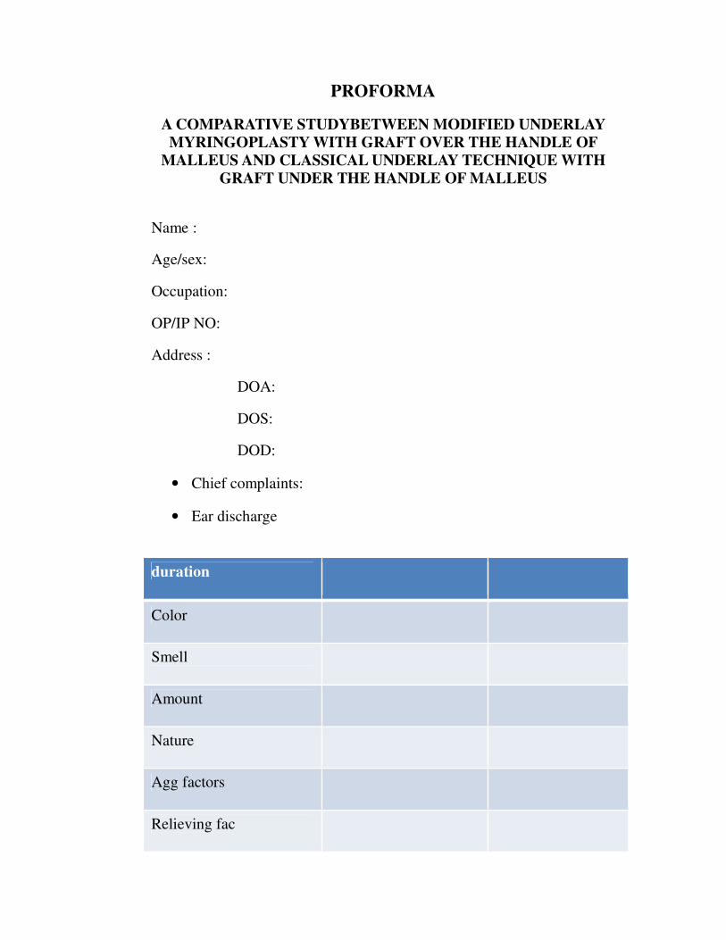
PROFORMA
A COMPARATIVE STUDYBETWEEN MODIFIED UNDERLAY MYRINGOPLASTY WITH GRAFT OVER THE HANDLE OF
MALLEUS AND CLASSICAL UNDERLAY TECHNIQUE WITH GRAFT UNDER THE HANDLE OF MALLEUS
Name :
Age/sex:
Occupation:
OP/IP NO:
Address :
DOA:
DOS:
DOD:
• Chief complaints:
• Ear discharge
duration
Color
Smell
Amount
Nature
Agg factors
Relieving fac
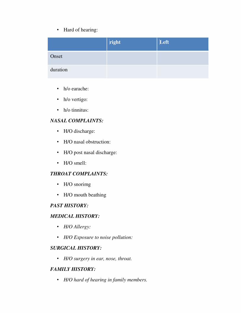
• Hard of hearing:
right Left
Onset
duration
• h/o earache:
• h/o vertigo:
• h/o tinnitus:
NASAL COMPLAINTS:
• H/O discharge:
• H/O nasal obstruction:
• H/O post nasal discharge:
• H/O smell:
THROAT COMPLAINTS:
• H/O snorimg
• H/O mouth beathing
PAST HISTORY:
MEDICAL HISTORY:
• H/O Allergy:
• H/O Exposure to noise pollution:
SURGICAL HISTORY:
• H/O surgery in ear, nose, throat.
FAMILY HISTORY:
• H/O hard of hearing in family members.
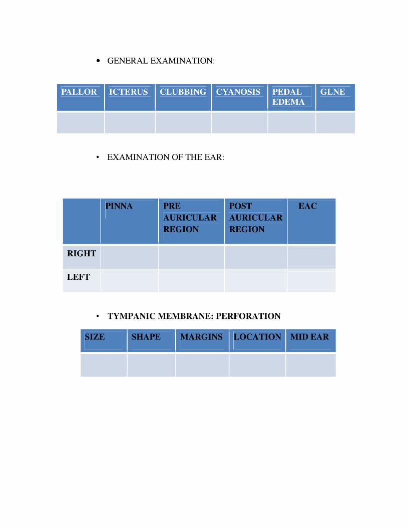
• GENERAL EXAMINATION:
PALLOR ICTERUS CLUBBING CYANOSIS PEDAL EDEMA
GLNE
• EXAMINATION OF THE EAR:
PINNA PRE
AURICULAR REGION
POST AURICULAR REGION
EAC
RIGHT
LEFT
• TYMPANIC MEMBRANE: PERFORATION
SIZE SHAPE MARGINS LOCATION MID EAR
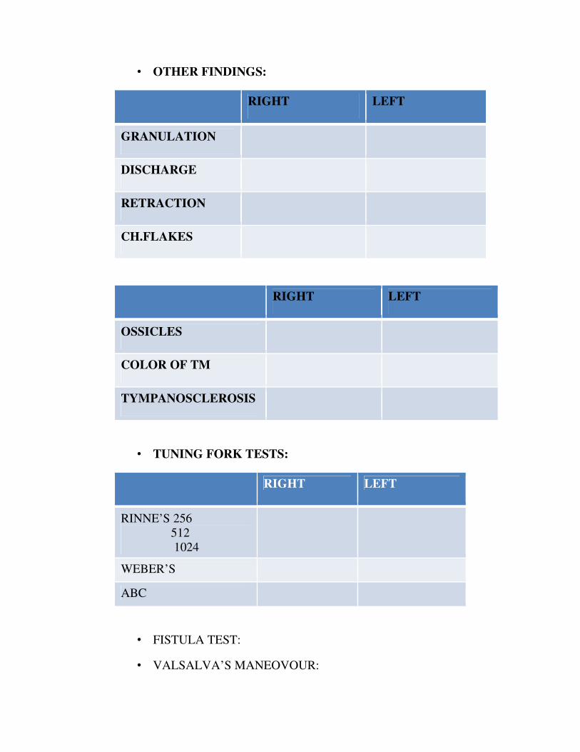
• OTHER FINDINGS:
RIGHT LEFT
GRANULATION
DISCHARGE
RETRACTION
CH.FLAKES
RIGHT LEFT
OSSICLES
COLOR OF TM
TYMPANOSCLEROSIS
• TUNING FORK TESTS:
RIGHT LEFT
RINNE’S 256 512 1024
WEBER’S
ABC
• FISTULA TEST:
• VALSALVA’S MANEOVOUR:

EXAMINATION OF NOSE:
EXTERNAL SEPTUM TURBINATE MEATUS MUCOSA
RIGHT
LEFT
ORAL CAVITY:
OROPHARYNX:
THROAT:
POST NASAL EXAMINATION:
IDL FINDINGS:
INVESTIGATIONS:
• CBC, RFT, Blood grouping and typing, urine routine
• HIV – ELISA
• CXR,ECG
• Pure tone audiometry
• X ray mastiod
• Otoendoscopy
• Aural swab
• Diagnostic nasal endoscopy
• Procedure:
• Anaesthesia:
• Approach:
• EOT findings:
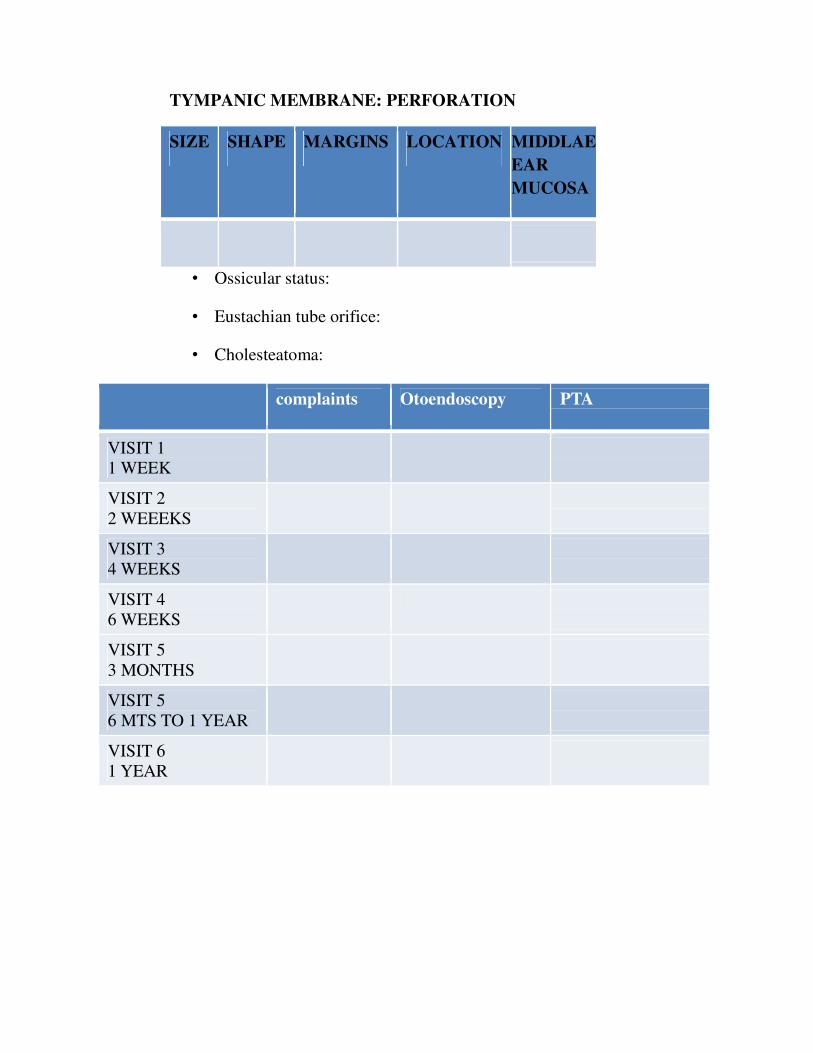
TYMPANIC MEMBRANE: PERFORATION
SIZE SHAPE MARGINS LOCATION MIDDLAE EAR MUCOSA
• Ossicular status:
• Eustachian tube orifice:
• Cholesteatoma:
complaints Otoendoscopy PTA
VISIT 1 1 WEEK
VISIT 2 2 WEEEKS
VISIT 3 4 WEEKS
VISIT 4 6 WEEKS
VISIT 5 3 MONTHS
VISIT 5 6 MTS TO 1 YEAR
VISIT 6 1 YEAR
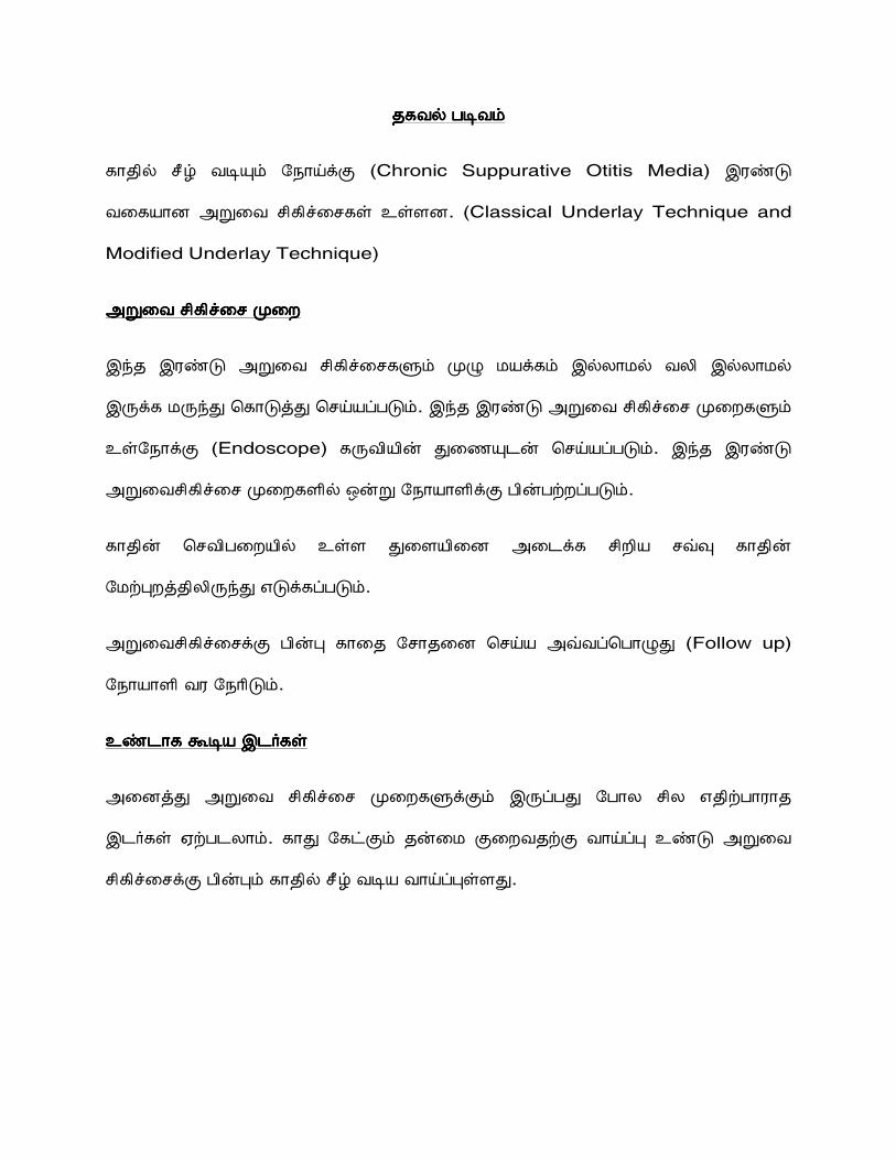
����������������������������������������
���� �� � ��� �� ����� � �� ������� � ������ �!"� # � �$� % "&��'� ( )* + �
�, �-. � / 0 , �� ��1, ��2 � 3 2 4 . 5� �� 6�$$���6� 7 �&"�6�8� 9"����:�"� ��&�
% �&�;�"&�7 �&"�6�8�9"����:�"'�
/ 0 , ����1, ��< , =�/ 0 , ����1, ��< , =�/ 0 , ����1, ��< , =�/ 0 , ����1, ��< , =�����
( >�� ( )* + � / 0 , �� ��1, ��? �� < @ � A-���� ( �B A�� �B � ( �B A��
( C ���AC >D �E�+ FD �E��-G�+ �5�( >��( )* + �/ 0 , ����1, ��< , =�? ��
3 2 ���� � �H �&�$���"'� �C �-I � D , J � K I � E��-G�+ �5� ( >�� ( )* + �
/ 0 , ���1, ��< , =�4 ��L I 0 ���-4 �� ��I �M=G�+ �5�
��I � E���, =-�� 3 2 4 � D , 4 -, . � / , K ��� �=- � �N O � ��I �
�AMP=F�B C >D �Q+ ��G�+ �5��
/ 0 , ���1, ��� � �I P� �, �� ���, . � E��- � / N �GE�@ D � �R�66�S � ��'�
��-4 ��)���+ �5�
3 * K ��T �- �( K U�23 * K ��T �- �( K U�23 * K ��T �- �( K U�23 * K ��T �- �( K U�2 ����
/ , . FD � / 0 , �� ��1, �� < , =�? �� �� ( C G�D � ��B � �B � Q�M�)��
( K U�2 � V M�K B �5� �D � ��W � �� �I , A � � , =��M� � ��GP� 3 * + � / 0 , ��
��1, ��� ��I P�������� ���- ���GP2 4 D 5����
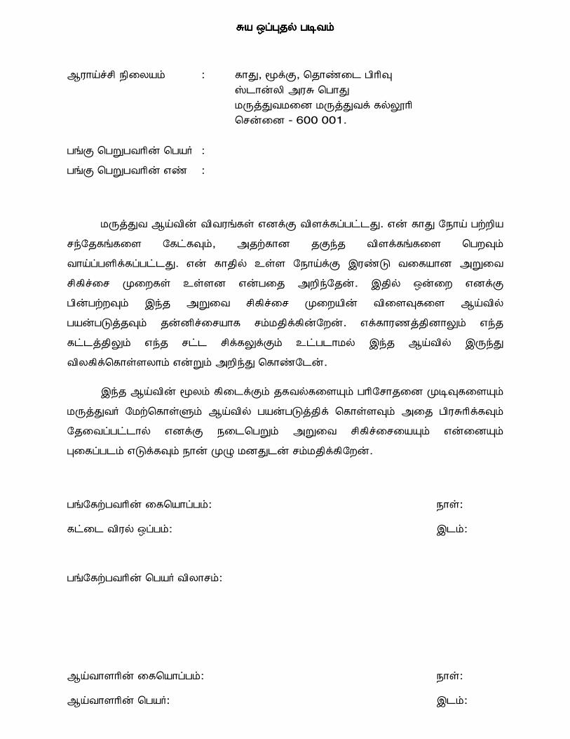
ÍÂ ´ôÒ¾ø ÀÊÅõ ÍÂ ´ôÒ¾ø ÀÊÅõ ÍÂ ´ôÒ¾ø ÀÊÅõ ÍÂ ´ôÒ¾ø ÀÊÅõ
¬Ã¡ö ¿¢¨ÄÂõ : ¸¡Ð, ãìÌ, ¦¾¡ñ¨¼ À¢¡¢× Š¼¡ýÄ¢ «ÃÍ ¦À¡Ð ÁÕòÐÅÁ¨É ÁÕòÐÅì ¸øæ¡¢ ¦ºý¨É - 600 001. ÀíÌ ¦ÀÚÀÅ¡¢ý ¦ÀÂ÷ :
ÀíÌ ¦ÀÚÀÅ¡¢ý ±ñ :
ÁÕòÐÅ ¬öÅ¢ý Å¢ÅÃí¸û ±ÉìÌ Å¢Çì¸ôÀð¼Ð. ±ý ¸¡Ð §¿¡ö ÀüÈ¢Â
ºó§¾¸í¸¨Ç §¸ð¸×õ, «¾ü¸¡É ¾Ìó¾ Å¢Çì¸í¸¨Ç ¦ÀÈ×õ
Å¡öôÀÇ¢ì¸ôÀð¼Ð. ±ý ¸¡¾¢ø ¯ûÇ §¿¡öìÌ þÃñΠŨ¸Â¡É «Ú¨Å
º¢¸¢î¨º Өȸû ¯ûÇÉ ±ýÀ¨¾ «È¢ó§¾ý. þ¾¢ø ´ý¨È ±ÉìÌ
À¢ýÀüÈ×õ þó¾ «Ú¨Å º¢¸¢î¨º ӨȢý Å¢¨Ç׸¨Ç ¬öÅ¢ø
ÀÂýÀÎò¾×õ ¾ýɢ¡¸ ºõÁ¾¢ì¸¢ý§Èý. ±ì¸¡Ã½ò¾¢É¡Öõ ±ó¾
¸ð¼ò¾¢Öõ ±ó¾ ºð¼ º¢ì¸ÖìÌõ ¯ðÀ¼¡Áø þó¾ ¬öÅ¢ø þÕóÐ
Ţĸ¢ì¦¸¡ûÇÄ¡õ ±ýÚõ «È¢óÐ ¦¸¡ñ§¼ý.
þó¾ ¬öÅ¢ý ãÄõ ¸¢¨¼ìÌõ ¾¸Åø¸¨ÇÔõ À¡¢§º¡¾¨É ÓÊ׸¨ÇÔõ
ÁÕòÐÅ÷ §Áü¦¸¡ûÙõ ¬öÅ¢ø ÀÂýÀÎò¾¢ì ¦¸¡ûÇ×õ «¨¾ À¢ÃÍ¡¢ì¸×õ
§¾¨ÅôÀð¼¡ø ±ÉìÌ ¿¨¼¦ÀÚõ «Ú¨Å º¢¸¢î¨º¨ÂÔõ ±ý¨ÉÔõ
Ò¨¸ôÀ¼õ ±Îì¸×õ ¿¡ý ÓØ ÁÉмý ºõÁ¾¢ì¸¢§Èý.
Àí§¸üÀÅ¡¢ý ¨¸¦Â¡ôÀõ: ¿¡û:
¸ð¨¼ Å¢Ãø ´ôÀõ: þ¼õ:
Àí§¸üÀÅ¡¢ý ¦ÀÂ÷ Ţġºõ:
¬öÅ¡Ç¡¢ý ¨¸¦Â¡ôÀõ: ¿¡û:
¬öÅ¡Ç¡¢ý ¦ÀÂ÷: þ¼õ:
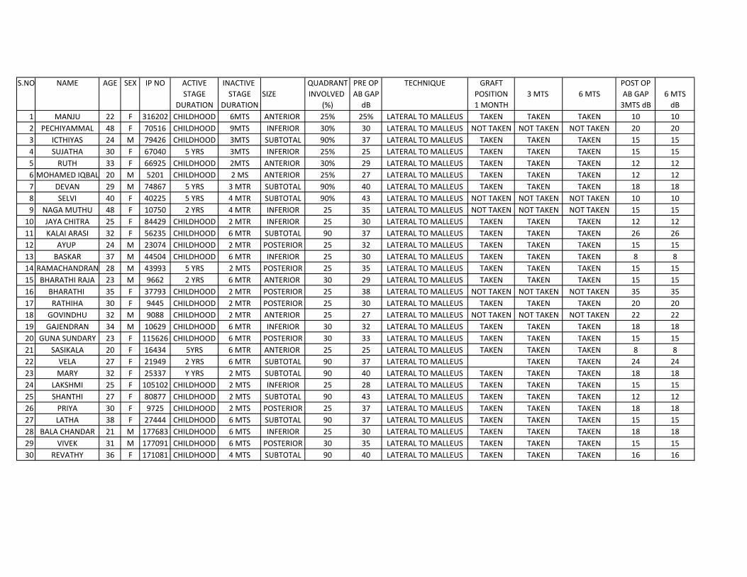
S.NO NAME AGE SEX IP NO ACTIVE INACTIVE QUADRANT PRE OP TECHNIQUE GRAFT POST OPSTAGE STAGE SIZE INVOLVED AB GAP POSITION 3 MTS 6 MTS AB GAP 6 MTS
DURATION DURATION (%) dB 1 MONTH 3MTS dB dB1 MANJU 22 F 316202 CHILDHOOD 6MTS ANTERIOR 25% 25% LATERAL TO MALLEUS TAKEN TAKEN TAKEN 10 102 PECHIYAMMAL 48 F 70516 CHILDHOOD 9MTS INFERIOR 30% 30 LATERAL TO MALLEUS NOT TAKEN NOT TAKEN NOT TAKEN 20 203 ICTHIYAS 24 M 79426 CHILDHOOD 3MTS SUBTOTAL 90% 37 LATERAL TO MALLEUS TAKEN TAKEN TAKEN 15 154 SUJATHA 30 F 67040 5 YRS 3MTS INFERIOR 25% 25 LATERAL TO MALLEUS TAKEN TAKEN TAKEN 15 155 RUTH 33 F 66925 CHILDHOOD 2MTS ANTERIOR 30% 29 LATERAL TO MALLEUS TAKEN TAKEN TAKEN 12 126 MOHAMED IQBAL 20 M 5201 CHILDHOOD 2 MS ANTERIOR 25% 27 LATERAL TO MALLEUS TAKEN TAKEN TAKEN 12 127 DEVAN 29 M 74867 5 YRS 3 MTR SUBTOTAL 90% 40 LATERAL TO MALLEUS TAKEN TAKEN TAKEN 18 188 SELVI 40 F 40225 5 YRS 4 MTR SUBTOTAL 90% 43 LATERAL TO MALLEUS NOT TAKEN NOT TAKEN NOT TAKEN 10 109 NAGA MUTHU 48 F 10750 2 YRS 4 MTR INFERIOR 25 35 LATERAL TO MALLEUS NOT TAKEN NOT TAKEN NOT TAKEN 15 15
10 JAYA CHITRA 25 F 84429 CHILDHOOD 2 MTR INFERIOR 25 30 LATERAL TO MALLEUS TAKEN TAKEN TAKEN 12 1211 KALAI ARASI 32 F 56235 CHILDHOOD 6 MTR SUBTOTAL 90 37 LATERAL TO MALLEUS TAKEN TAKEN TAKEN 26 2612 AYUP 24 M 23074 CHILDHOOD 2 MTR POSTERIOR 25 32 LATERAL TO MALLEUS TAKEN TAKEN TAKEN 15 1513 BASKAR 37 M 44504 CHILDHOOD 6 MTR INFERIOR 25 30 LATERAL TO MALLEUS TAKEN TAKEN TAKEN 8 814 RAMACHANDRAN 28 M 43993 5 YRS 2 MTS POSTERIOR 25 35 LATERAL TO MALLEUS TAKEN TAKEN TAKEN 15 1515 BHARATHI RAJA 23 M 9662 2 YRS 6 MTR ANTERIOR 30 29 LATERAL TO MALLEUS TAKEN TAKEN TAKEN 15 1516 BHARATHI 35 F 37793 CHILDHOOD 2 MTR POSTERIOR 25 38 LATERAL TO MALLEUS NOT TAKEN NOT TAKEN NOT TAKEN 35 3517 RATHIHA 30 F 9445 CHILDHOOD 2 MTR POSTERIOR 25 30 LATERAL TO MALLEUS TAKEN TAKEN TAKEN 20 2018 GOVINDHU 32 M 9088 CHILDHOOD 2 MTR ANTERIOR 25 27 LATERAL TO MALLEUS NOT TAKEN NOT TAKEN NOT TAKEN 22 2219 GAJENDRAN 34 M 10629 CHILDHOOD 6 MTR INFERIOR 30 32 LATERAL TO MALLEUS TAKEN TAKEN TAKEN 18 1820 GUNA SUNDARY 23 F 115626 CHILDHOOD 6 MTR POSTERIOR 30 33 LATERAL TO MALLEUS TAKEN TAKEN TAKEN 15 1521 SASIKALA 20 F 16434 5YRS 6 MTR ANTERIOR 25 25 LATERAL TO MALLEUS TAKEN TAKEN TAKEN 8 822 VELA 27 F 21949 2 YRS 6 MTR SUBTOTAL 90 37 LATERAL TO MALLEUS TAKEN TAKEN 24 2423 MARY 32 F 25337 Y YRS 2 MTS SUBTOTAL 90 40 LATERAL TO MALLEUS TAKEN TAKEN TAKEN 18 1824 LAKSHMI 25 F 105102 CHILDHOOD 2 MTS INFERIOR 25 28 LATERAL TO MALLEUS TAKEN TAKEN TAKEN 15 1525 SHANTHI 27 F 80877 CHILDHOOD 2 MTS SUBTOTAL 90 43 LATERAL TO MALLEUS TAKEN TAKEN TAKEN 12 1226 PRIYA 30 F 9725 CHILDHOOD 2 MTS POSTERIOR 25 37 LATERAL TO MALLEUS TAKEN TAKEN TAKEN 18 1827 LATHA 38 F 27444 CHILDHOOD 6 MTS SUBTOTAL 90 37 LATERAL TO MALLEUS TAKEN TAKEN TAKEN 15 1528 BALA CHANDAR 21 M 177683 CHILDHOOD 6 MTS INFERIOR 25 30 LATERAL TO MALLEUS TAKEN TAKEN TAKEN 18 1829 VIVEK 31 M 177091 CHILDHOOD 6 MTS POSTERIOR 30 35 LATERAL TO MALLEUS TAKEN TAKEN TAKEN 15 1530 REVATHY 36 F 171081 CHILDHOOD 4 MTS SUBTOTAL 90 40 LATERAL TO MALLEUS TAKEN TAKEN TAKEN 16 16
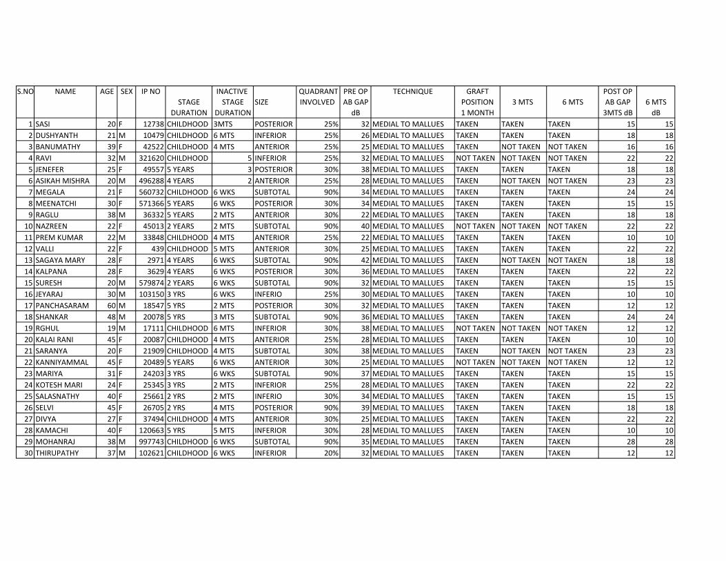
S.NO NAME AGE SEX IP NO INACTIVE QUADRANT PRE OP TECHNIQUE GRAFT POST OPSTAGE STAGE SIZE INVOLVED AB GAP POSITION 3 MTS 6 MTS AB GAP 6 MTS
DURATION DURATION dB 1 MONTH 3MTS dB dB1 SASI 20 F 12738 CHILDHOOD 3MTS POSTERIOR 25% 32 MEDIAL TO MALLUES TAKEN TAKEN TAKEN 15 152 DUSHYANTH 21 M 10479 CHILDHOOD 6 MTS INFERIOR 25% 26 MEDIAL TO MALLUES TAKEN TAKEN TAKEN 18 183 BANUMATHY 39 F 42522 CHILDHOOD 4 MTS ANTERIOR 25% 25 MEDIAL TO MALLUES TAKEN NOT TAKEN NOT TAKEN 16 164 RAVI 32 M 321620 CHILDHOOD 5 INFERIOR 25% 32 MEDIAL TO MALLUES NOT TAKEN NOT TAKEN NOT TAKEN 22 225 JENEFER 25 F 49557 5 YEARS 3 POSTERIOR 30% 38 MEDIAL TO MALLUES TAKEN TAKEN TAKEN 18 186 ASIKAH MISHRA 20 M 496288 4 YEARS 2 ANTERIOR 25% 28 MEDIAL TO MALLUES TAKEN NOT TAKEN NOT TAKEN 23 237 MEGALA 21 F 560732 CHILDHOOD 6 WKS SUBTOTAL 90% 34 MEDIAL TO MALLUES TAKEN TAKEN TAKEN 24 248 MEENATCHI 30 F 571366 5 YEARS 6 WKS POSTERIOR 30% 34 MEDIAL TO MALLUES TAKEN TAKEN TAKEN 15 159 RAGLU 38 M 36332 5 YEARS 2 MTS ANTERIOR 30% 22 MEDIAL TO MALLUES TAKEN TAKEN TAKEN 18 1810 NAZREEN 22 F 45013 2 YEARS 2 MTS SUBTOTAL 90% 40 MEDIAL TO MALLUES NOT TAKEN NOT TAKEN NOT TAKEN 22 2211 PREM KUMAR 22 M 33848 CHILDHOOD 4 MTS ANTERIOR 25% 22 MEDIAL TO MALLUES TAKEN TAKEN TAKEN 10 1012 VALLI 22 F 439 CHILDHOOD 5 MTS ANTERIOR 30% 25 MEDIAL TO MALLUES TAKEN TAKEN TAKEN 22 2213 SAGAYA MARY 28 F 2971 4 YEARS 6 WKS SUBTOTAL 90% 42 MEDIAL TO MALLUES TAKEN NOT TAKEN NOT TAKEN 18 1814 KALPANA 28 F 3629 4 YEARS 6 WKS POSTERIOR 30% 36 MEDIAL TO MALLUES TAKEN TAKEN TAKEN 22 2215 SURESH 20 M 579874 2 YEARS 6 WKS SUBTOTAL 90% 32 MEDIAL TO MALLUES TAKEN TAKEN TAKEN 15 1516 JEYARAJ 30 M 103150 3 YRS 6 WKS INFERIO 25% 30 MEDIAL TO MALLUES TAKEN TAKEN TAKEN 10 1017 PANCHASARAM 60 M 18547 5 YRS 2 MTS POSTERIOR 30% 32 MEDIAL TO MALLUES TAKEN TAKEN TAKEN 12 1218 SHANKAR 48 M 20078 5 YRS 3 MTS SUBTOTAL 90% 36 MEDIAL TO MALLUES TAKEN TAKEN TAKEN 24 2419 RGHUL 19 M 17111 CHILDHOOD 6 MTS INFERIOR 30% 38 MEDIAL TO MALLUES NOT TAKEN NOT TAKEN NOT TAKEN 12 1220 KALAI RANI 45 F 20087 CHILDHOOD 4 MTS ANTERIOR 25% 28 MEDIAL TO MALLUES TAKEN TAKEN TAKEN 10 1021 SARANYA 20 F 21909 CHILDHOOD 4 MTS SUBTOTAL 30% 38 MEDIAL TO MALLUES TAKEN NOT TAKEN NOT TAKEN 23 2322 KANNIYAMMAL 45 F 20489 5 YEARS 6 WKS ANTERIOR 30% 25 MEDIAL TO MALLUES NOT TAKEN NOT TAKEN NOT TAKEN 12 1223 MARIYA 31 F 24203 3 YRS 6 WKS SUBTOTAL 90% 37 MEDIAL TO MALLUES TAKEN TAKEN TAKEN 15 1524 KOTESH MARI 24 F 25345 3 YRS 2 MTS INFERIOR 25% 28 MEDIAL TO MALLUES TAKEN TAKEN TAKEN 22 2225 SALASNATHY 40 F 25661 2 YRS 2 MTS INFERIO 30% 34 MEDIAL TO MALLUES TAKEN TAKEN TAKEN 15 1526 SELVI 45 F 26705 2 YRS 4 MTS POSTERIOR 90% 39 MEDIAL TO MALLUES TAKEN TAKEN TAKEN 18 1827 DIVYA 27 F 37494 CHILDHOOD 4 MTS ANTERIOR 30% 25 MEDIAL TO MALLUES TAKEN TAKEN TAKEN 22 2228 KAMACHI 40 F 120663 5 YRS 5 MTS INFERIOR 30% 28 MEDIAL TO MALLUES TAKEN TAKEN TAKEN 10 1029 MOHANRAJ 38 M 997743 CHILDHOOD 6 WKS SUBTOTAL 90% 35 MEDIAL TO MALLUES TAKEN TAKEN TAKEN 28 2830 THIRUPATHY 37 M 102621 CHILDHOOD 6 WKS INFERIOR 20% 32 MEDIAL TO MALLUES TAKEN TAKEN TAKEN 12 12