Embed Size (px)
Citation preview

A CELLULAR AUTOMATA MODEL FOR BIOFILM GROWTH
D. Rodriguez1, A. Carpio2, B. Einarsson3
1 Department of Applied Mathematics, Facultad de Matematicas at the Universidad Com-plutense de Madrid ([email protected])
2 Department of Applied Mathematics, Facultad de Matematicas at the Universidad Com-plutense de Madrid ([email protected])
3 Center for Complex and Nonlinear Science, University of California at Santa Barbara ([email protected])
Abstract. Biofilms are aggregates of bacteria attached to surfaces, which are very adaptableto changes in the environment and survive under extreme conditions. These living structuresare behind many problems in research and industry. Therefore there is an increasing inter-est in improving our understanding on biofilms to be able to control them, either designingprotocols to destroy them when harmful or promoting their growth when beneficial.
A bidimensional cellular automata model for biofilm development is proposed to studythe biofilm behaviour as its key growth parameters vary. The model includes several metabolicand spreading mechanisms typical of bacteria: cell division and spreading, detachment mech-anisms adapted to the flow and probabilistic rules for EPS matrix generation.
Numerical simulations of the model reproduce a number of biofilm patterns observedin real experiments: ripples, streamers, mushroom networks and patches. The influence ofthe nutrient concentration and the type of flow on the evolution of the bacterial community ismonitorized.
Biofilm tends to cover the whole surface when enough nutrients are available. Erosionenhances the creation of holes in this cover and promotes a variety of geometric patterns. Thesurvival of the colony and its final shape will depend on the balance between the main growthparameters.
Large Reynolds numbers and poor nutrient sources promote the formation of flat, andthin biofilms. Decreasing the Reynolds number or increasing the nutrient and oxygen concen-tration enhance pattern formation.
Keywords: Biofilms, cellular automata, probabilistic models, erosion, EPS Matrix.
1. INTRODUCTION
Nowadays, the study of biofilms is a promising area of research because its presencein many industrial and medical problems contexts. They can be defined as an aggregate ofbacteria embedded into a self-generated polymeric substance which can attach onto a widevariety of supports, from any kind of inorganic surfaces until living tissues, being extremely
Blucher Mechanical Engineering ProceedingsMay 2014, vol. 1 , num. 1www.proceedings.blucher.com.br/evento/10wccm

resistant to physical or chemical aggressions. They develop and reproduce trying to spreadinto the environment using many different strategies [11] that are not fully understood.
These living organisms take part in many critical processes of great importance intheir respective fields of study. Examples are the biofilms formed in the lungs, which areresponsible for deadly cystic fibrosis, or biofilm impact on surgery, when pacemakers andartificial joints are implanted and may induce chronic and acute infections [20,3]. However,biofilms can be used to our advantage to achieve different purposes if they are properly con-trolled. Biofilms that become a problem in drinking water systems [13] can be exploited forbiodegradation of pollutants [20] in soil or water. They can also be used to detect chemicalsand pathogens, or to measure variables in MEMS [20,8]. For technological applications, aprecise control of growth processes is required to produce biofilms with a specific structure.
In order to achieve this last objective, a deep understanding of biofilm developmentand spreading mechanisms is essential. A good biofilm model should take into account thenature of the considered bacteria, the environment in which the biofilm is formed, the param-eters that have to be fitted with experiments, and the predictions to be made [23,12,10].
There are many of models trying to explain how biofilms behave under different ex-ternal and internal variables which affect them. These models can be classified into threedifferent categories [23,12,10]: continuum, individual based (IbM) or cellular automata (CA)models.
Continuum models treat the biofilm as a continuum solid with similar properties, typ-ically a gel, polymer or viscous fluid [9]. They are often two-phase models which considera fluid phase containing a mixture of nutrients and the biofilm phase [4]. IbM models are adiscrete approach in which microbes are seen as spherical particles which evolve accordingto reaction-diffusion equations for nutrients and oxygen coupled with bacterial growth andspreading of biomass [15]. There are several variants which try to include a greater degree ofcomplexity observed in real biofilms, like the EPS matrix [1]. CA models propose a simpleset of evolution rules [7,16] in a bounded discrete domain for a system filled with biomassand fluid which will evolve in time. They can include basic metabolic mechanisms easily,but more complex behaviours such as microbe attachment to surfaces [6], quorum sensing toform biofilms [2,25], generation of EPS matrix [22,24] or interaction with the surroundingflow [21,5,14] are more difficult to implement. The stochastic approach of these type of mod-els allow them to reduce assumptions and hypothesis about the dynamic of the system to aminimum. CA models are usually less costly than solving classical PDE models.
In this paper we develop a cellular automata model to obtain a qualitative understand-ing of biofilm evolution in a rectangular pipe at increasing Reynolds numbers. The paper isorganized as follows. Section 2 and 3 describes the model and the probabilistic rules for allmetabolic processes that participate in biofilm dynamics. An asymmetric erosion mechanismadapted to flows moving parallel to the surface is proposed, together with simple probabilisticrules accounting for EPS matrix generation and its influence on biofilm cohesion. Section 4illustrates several results found in the performed simulations. Finally, section 5 summarizesthe conclusions.

2. OVERALL APPROACH
3. Overall approach
Our model attempts to describe the formation and evolution of a biofilm, which isbelieved to be as follows [11]. Once planktonic cells have reach the substratum, they attachto it and start reproducing if conditions are favorable (enough nutrients, not very agressiveexternal conditions, etc.). First, microcolonies are formed as a prelude of an exponentialgrowth. During the growth fase, the bacteria forming the biofilm suffer physiological changeslike the loss of their flagella, promoting the expression of new metabolic behaviours (EPSsecretation). EPS generation enhances biofilm growth on vertical direction, looking for richercarbon and oxygen concentrations. Macrocolonies are formed. These mature objects willbe eroded by the external hydrodynamic forces, resulting on a rich variety of morphologies[17] (mushrooms, ripples, streamers, etc.). After some time and once a critical bacterialpopulation has been reached, quorum sensing mechanisms activate spreading mechanisms(rolling, rippling, darting, etc., see figure 2). The main objective of these mechanisms is tocolonize new surface and ensure the survival of the colony. The biofilm life cycle is closedthen. A schematic figure can be seen in Figure 1.
Figure 1. Schematic development of a biofilm.
The CA model will be developed in a 2D grid (see [7,16] and references therein),corresponding to a longitudinal section of a rectangular pipe (x=length,z=height), see Figure3. Grid tiles can be occupied by water, biomass or substratum material (three possible states),and have the size of one bacteria in order to consider them as individual units.
The pipe is filled with a mixture of water and nutrients that flows at a certain velocity.The Reynolds number (computed using the hydraulic diameter) will be used as a measurementof the shear stress produced by the flow. A biofilm seed is already attached to the bottom ofthe pipe at the beginning of each simulation.
The model will evolve with time, simulating a dynamic system where grid tiles changetheir state according to a set of sub-models which govern the metabolic activities associated to
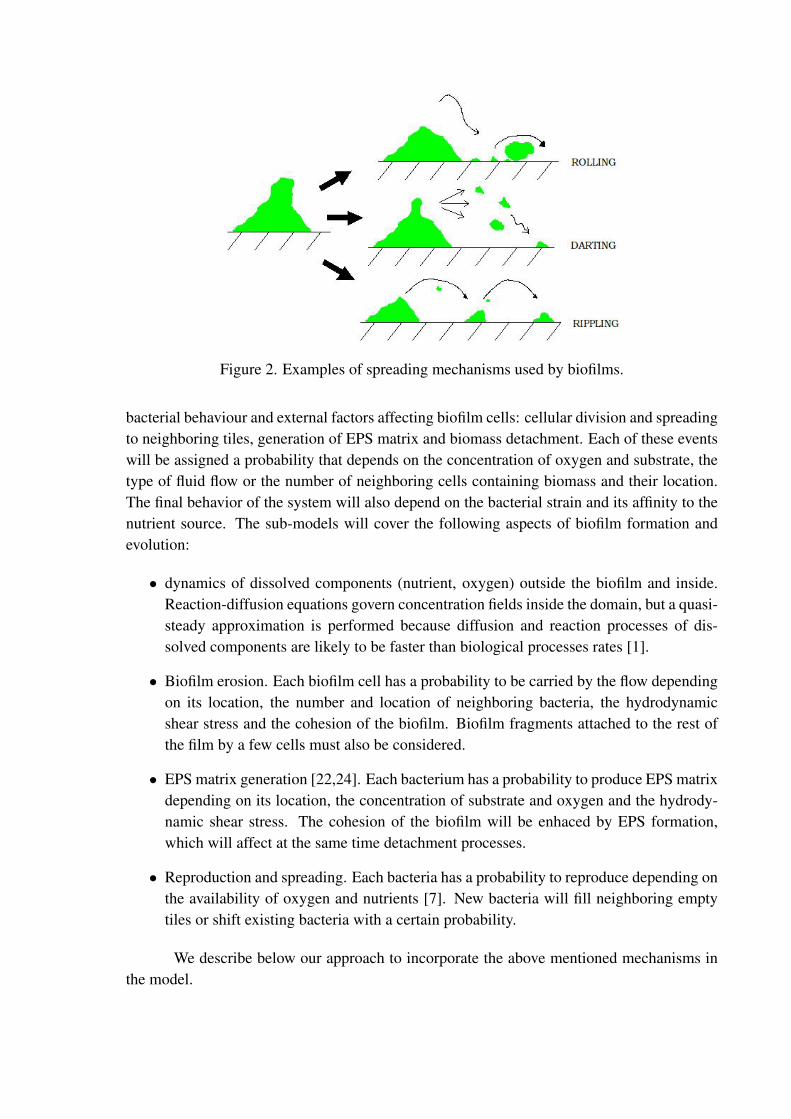
Figure 2. Examples of spreading mechanisms used by biofilms.
bacterial behaviour and external factors affecting biofilm cells: cellular division and spreadingto neighboring tiles, generation of EPS matrix and biomass detachment. Each of these eventswill be assigned a probability that depends on the concentration of oxygen and substrate, thetype of fluid flow or the number of neighboring cells containing biomass and their location.The final behavior of the system will also depend on the bacterial strain and its affinity to thenutrient source. The sub-models will cover the following aspects of biofilm formation andevolution:
• dynamics of dissolved components (nutrient, oxygen) outside the biofilm and inside.Reaction-diffusion equations govern concentration fields inside the domain, but a quasi-steady approximation is performed because diffusion and reaction processes of dis-solved components are likely to be faster than biological processes rates [1].
• Biofilm erosion. Each biofilm cell has a probability to be carried by the flow dependingon its location, the number and location of neighboring bacteria, the hydrodynamicshear stress and the cohesion of the biofilm. Biofilm fragments attached to the rest ofthe film by a few cells must also be considered.
• EPS matrix generation [22,24]. Each bacterium has a probability to produce EPS matrixdepending on its location, the concentration of substrate and oxygen and the hydrody-namic shear stress. The cohesion of the biofilm will be enhaced by EPS formation,which will affect at the same time detachment processes.
• Reproduction and spreading. Each bacteria has a probability to reproduce depending onthe availability of oxygen and nutrients [7]. New bacteria will fill neighboring emptytiles or shift existing bacteria with a certain probability.
We describe below our approach to incorporate the above mentioned mechanisms inthe model.

Figure 3. Geometry for the cellular automata description.
4. Implemented metabolic mechanisms
Our CA model defines certain rules for the basic metabolic activities of bacteria. Assaid in the previous section, the bacterial behaviours taken into account are: biofilm surfaceerosion, limiting concentration mass transfer, cellular division, spreading and generation ofEPS matrix.
4.1. Dynamics of concentration fields
The evolution of a biofilm depends on the availability of carbon sources and oxygen,which will be governed by how concentration fields change in both phases. One of themwill become the limiting concentration, that is, the one that penetrates less deeper into thebiofilm. In the fluid phase, the concentration field is governed by convection-diffusion equa-tions coupled to the fluid. Inside the biofilm, we have a reaction-diffusion equation. Themodel is completed with boundary conditions at the walls of the pipe and the fluid/biofilminterface. As said previously, diffusion and reaction rates are faster than biological processesrates [1], allowing to simplify the complete set of diffusion-convection-reaction equations to aquasi-steady approximation. In experiments, the concentration is usually kept almost constantwithin the fluid. We approximate the solution of the outer convection-diffusion equations bya constant outside a boundary layer of thickness dB. In practice, this thickness is controlledby the velocity of the flow. In our framework, it becomes a model parameter, following [7].Choosing the concentration value at the bulk/boundary layer interface C as control parame-

Figure 4. Neighbor location in the cellular automata description.
ters, the concentrations inside the region containing the biofilm and the boundary layer aregoverned by:
−D∆2c = kc
c+K, (1)
with zero flux conditions at the substratum. The diffusion constants are assumed to be thesame for the biomass and the boundary layer. The right hand side in the equation representsthe nutrient uptake kinetics. Here, k is the uptake rate for the limiting concentration and K itsMonod half-saturation coefficient. The values of all these parameters depend on the bacteriaspecies forming the biofilm.
Performing a zero-order uptake kinetics approximation (the right hand side is replacedby just a constant k) an explicit approximation to the solution is found [7]:
c(cell) =
C1/2−
√√√√ k
2D
[1
8
8∑i=1
1
di(cell)2
]−1
2
, (2)
where di(cell) are distances between the cell and the bulk/boundary layer interface in thedirections joining the cell with its eight neighbors.
4.2. Erosion and detachment
Surface cells are exposed to shear forces exerted by the flow, straining them and fi-nally leading them to fracture into small pieces of biofilm which will be carried away by theflow. Cells covered by other cells are assumed to be protected from erosion. Exposed cellswill detach with a probability which depends on the number and location of their neighborsrelative to the motion of the fluid, the biofilm cohesion (which is controlled by EPS matrixgeneration) and the hydrodynamic shear stress (which is a function of the Reynolds numberand the position of the cell).
The hydrodynamic stress on a cell acts in the x direction (see Figure 3). In each cell,a balance between this stress and mechanical support against the flow given by its neigh-bours will be performed. This mechanical support will depend on factors like the number

of neighbours or their specific spatial distribution around the considered cell. Notice thateach neighbour will contribute with a specific support. Let us number the eight neighboringtiles around the considered biomass tile clockwise, starting with the western direction, so thatn1, n2, n3, n4, n5, n6, n7, n8 denote the neighbors located to the west, northwest, north, north-east, east, southeast, south, and southwest, respectively. Figure 4 illustrates the location ofpossible neighbours. There are several considerations about them: If cell n1 is occupied, con-sidered cell is unlikely to be carried away. The strongest support against the flow is exertedby the neighbor in position n5. Next in magnitude are positions n4 and n6, and next n3, n7.Neighbors n2, n8 add a small contribution.
A dimensionless normalized parameter representing the total force on each cell rang-ing from zero to one is introduced:
τ(cell) = R(Re)(1− χ1(cell))(1−8∑i=2
eiχi(cell)). (3)
R(Re) ∈ (0, 1) is an increasing function of the Reynolds number. The functions χi(cell)take the value one whenever the cell has a neighbor located at the position ni, and vanishotherwise. The weights and other parameters have been chosen taking into account that thefluid flows in the x direction and satisfy ei ∈ (0, 1),
∑8i=2 ei = 1. They represent the added
resistance against the flow due to neighboring cells depending on their position.A probability law for cell erosion can be defined following [7]:
Pe(cell) =1
1 + σ(cell)τ(cell)
. (4)
Whenever τ(cell) = 0, we set Pe(cell) = 0. Here, τ(cell) is given by (3), and σ(cell)
represents the biofilm strength. In practice, the cohesion of a biofilm is governed by thegeneration of EPS matrix. A cohesion parameter σ(cell) varying in accordance with the localEPS generation production is introduced in Section 4.3.
At each time step and for each cell, we generate a random number r ∈ (0, 1). Whenr < Pe(cell), the cell detaches from the biofilm. Erosion due to the flow may occur asdetachment of single cells or of whole fragments of biofilm, depending on the geometry.
4.3. EPS matrix generation
In small colonies, bacteria try to reproduce at the maximum possible rate by consum-ing all available resources in the environment. As the size and thickness of the biofilm grow,nutrients and oxygen become scarce inside due to the high bacterial population. A strategydeveloped by bacteria to keep a constant supply of nutrients and oxygen is to invest part oftheir energy on generating EPS matrix, allowing the bacterial colony to expand their masstransfer surface with the environment by growing mainly in vertical direction. This grants aneasier access to nutrients and oxygen supply [22,24]. In our model, each cell has a probabilityto produce EPS matrix [11], see Figure 1. The EPS matrix also spreads over the neighbouringbacteria making their reproduction harder. As bacteria fall deeper in the biofilm, their chancesto produce EPS increase.

EPS generation is also affected by the shear stress acting on biofilm surface. Highshear forces will lead to stronger EPS matrix structures. Biofilms grown at low Reynoldsnumbers tend to be carried away with the flow as the Reynolds number is increased. How-ever, biofilms grown at large Reynolds numbers in turbulent regimes are difficult to detachfrom surfaces, and expand easily when hydrodynamic conditions become less agressive withbiofilm surface or the availability of carbon improves (switching to a richer source or increas-ing the nutrient concentration) [3].
In our model, EPS matrix production has a probability of happening that depends onthe availability of nutrients and oxygen at the cell position and the shear exerted by the flow.The EPS matrix is generated with the following probability law:
Peps(cell) = R(Re)
(1− c(cell)
c(cell) +K
), (5)
where c represents the limiting concentration (nutrient or oxygen) computed as described inSection 4.1. The parameter R(Re) ∈ (0, 1) has been introduced in section 4.2.
At each time step and for each cell, a random number r ∈ (0, 1) is generated: Ifr < Peps(cell), the cell will generate EPS instead of reproducing, as it requires less externalchemicals and energy than reproduction.
The amount of EPS matrix produced controls the cohesion (strength) of the biofilm.We propose a local measure σ of the biofilm cohesion which takes into account the number ofneighbors and whether they generate EPS matrix or not:
σ(cell) =σ08
8∑i=1
σi(cell), (6)
where
σi(cell) =
0 if cell ni is not presentα if cell ni is present, but does
not produce EPS matrix1 if cell ni produces EPS matrix
(7)
and n1(cell), n2(cell),...,n8(cell) denote the eight neighbour locations for the cell under study(see Figure 4) and σ0, α ∈ (0, 1). These parameters represent the strength of the EPS matrixgenerated by the bacteria and the strength of the attachment between standard bacteria. Wehave selected σ0 = 1, α = 1
2in our computer experiments, but they should be fitted according
with the type of bacteria considered in the simulation, being necessary real experiments withselected bacteria. Cohesion increases with the Reynolds number, since the probability togenerate EPS matrix is larger for large Reynolds number.
4.4. Reproduction
The cell reproduction mechanism is similar to that proposed in [7], but we considerthat EPS producers do not participate in the reproduction stage. At each time step, and dis-carding cells producing EPS matrix, the remaining cells will divide with probability:
Pd(cell) =c(cell)
cl(cell) +K, (8)

where c denotes the limiting concentration and K its saturation coefficient in the Monod law.The evolution of the concentration is described in Section 4.1. We are neglecting changes inconcentration due to newborn cell consumption or cell switching to EPS generation.
At each time step, and for each cell not generating EPS matrix, we compute a randomnumber r ∈ (0, 1). If r < Pd(cell), the cell will divide. Spreading of newborn cells isperformed by considering neighbouring grid tiles: if some of them are empty the daughter cellis placed in any of them with equal probability. If reproducing cell is completely surrounded,newborn cell will shift the neighbour cell which offer the minimal mechanical resistance,considering this criteria as the minimum distance from the considered cell to the biofilm outerborder.
4.5. Nondimensionalization and parameters
Once the model has been proposed, nondimensionalization of the variables is per-formed by identifying the minimum number of independent parameters and considering thedifferent magnitude order between terms. Because of all probabilities and controlling param-eters concerning the erosion and cohesion intensities are dimensionless, only concentrationfields, length and time scale introduce dimensions in the model:
• Time: In the model, time is not given explicitly. It appears in the number of time stepscarried out at each simulation. An upper bound for the time step, which allows to relatecomputational and experimental times is:
t =ln(2)
µmax,
where µmax is the growth rate.
• Length: The basic distance considered in the model is the size of a bacteria a, about 1
or 2 micrometers.
• Concentration: The concentration field is calculated using expression (2) which de-pends on distances. It involves a number of constants with their units that must benondimensionalized. Making the changes of variables:
c=c
K, C=
C
K, F =
ka2
2DK, δB =
dBa, δi=
dia,
we get:
c =
C0.5 −
F [1
8
8∑i=1
1
δ2i
]−10.52
, (9)
which gives a dimensionless expression for the concentration.
The four parameters a, k, D, K are reduced to one: F . The controlling parameters aretherefore F , C and δB for the concentration, plus the additional parameters R(Re), σ0, α, thatappear in the probability laws. Once a specific bacteria species is selected, σ0, α are fixed.The remaining parameters depend on the type of nutrient and flow.

Figure 5. Differences in biofilm geometric patterns if different shear stress values are applied.The same initial conditions for all cases leads to different final morphologies.
5. Numerical results and discussion
In this section, we illustrate the evolution of several initial configurations under differ-ent conditions, fixing parameter values that actually do not correspond to any specific bacteriaspecies, since a way to calibrate a number of parameters has yet to be devised.
Figure 5 shows the effect of Reynolds number in the biofilm spreading for two dif-ferent cases. At low Reynolds numbers, biofilms generate tower-like structures. If the flowstrength is increased, this configuration evolves into different shapes which are already re-ported in the experimental literature: streamers (typical shape in biofilms similar to flagsmoved by the wind), ripples and finally flat layers.
Figure 6 shows the effect of nutrient concentration in biofilm growth: greater quan-tities produce an increased growth rate of the biofilm even in high shear stress conditions,because the bacterial colonies sustain the needed growth rate to substitute eroded cells fornewborn ones.
In Figure 7, a biofilm patch is eroded by the flow until is completely wiped out. It canbe also seen that biofilm initial patch is dragged downstream along the x axis.
Figure 8 shows the effect of the EPS matrix in the biofilm structure. The higher cohe-sion produced by the effect of the EPS matrix promotes a bigger and more compact biofilmcolony if we compare the evolution of the same initial seed using the same Reynolds number.

Figure 6. Evolution of a biofilm using a high shear stress flow but different concentrationvalues. High concentration values produce significative growth even though the shear stressis large.
Figure 7. Into a high shear stress conditions, biofilm is dragged downstream and finally beingwiped out.
6. Conclusions
We have presented a CA model that reproduces biofilm behavior and patterns exper-imentally observed. The model describes the evolution of an initial seed of biofilm attachedto the surface inside a duct carrying a flow of nutrients and water. Space is discretized ina grid of square tiles. Each tile can have three states: water, biomass or surface. The rulesdescribing numerically reproduction, spreading, EPS matrix generation, erosion and dynamicof dissolved components have been detailed. At each time step, each cell present in the do-main has a chance to perform the different programmed metabolic activities, being the systemcompletely stochastic.
The results of the simulations show that the strength of the flow shapes the biofilmstructure, giving rise to different morphologies already known in experimental literature:
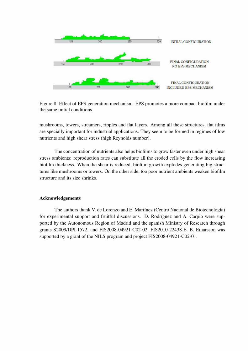
Figure 8. Effect of EPS generation mechanism. EPS promotes a more compact biofilm underthe same initial conditions.
mushrooms, towers, streamers, ripples and flat layers. Among all these structures, flat filmsare specially important for industrial applications. They seem to be formed in regimes of lownutrients and high shear stress (high Reynolds number).
The concentration of nutrients also helps biofilms to grow faster even under high shearstress ambients: reproduction rates can substitute all the eroded cells by the flow increasingbiofilm thickness. When the shear is reduced, biofilm growth explodes generating big struc-tures like mushrooms or towers. On the other side, too poor nutrient ambients weaken biofilmstructure and its size shrinks.
Acknowledgements
The authors thank V. de Lorenzo and E. Martınez (Centro Nacional de Biotecnologıa)for experimental support and fruitful discussions. D. Rodrıguez and A. Carpio were sup-ported by the Autonomous Region of Madrid and the spanish Ministry of Research throughgrants S2009/DPI-1572, and FIS2008-04921-C02-02, FIS2010-22438-E. B. Einarsson wassupported by a grant of the NILS program and project FIS2008-04921-C02-01.

7. REFERENCES
[1] E. Alpkvist, C. Picioreanu, M.C.M. Loosdrecht, and A. Heyden, ”Three-dimensionalbiofilm model with individual cells and continuum eps matrix”, Biotechnology and Bio-engineering, vol. 94, no. 5, pp. 961-979, 2006.
[2] K. Anguige, J.R. King, and J.P. Ward, ”A multiphase mathematical model of quorumsensing in maturing Pseudonomas aeruginosa biofilm”, Math. Biosciences, vol. 203, pp.240-276, 2006.
[3] R.M. Donlan and J.W. Costerton, ”Biofilms: survival mechanisms of clinically relevantmicroorganisms”, Clinical Microbiology Reviews, vol. 15, pp.167-193, 2002.
[4] D. Duddu, S. Boradas, D. Chopp, and B. Moran, ”A combined extended finite elementand level set method for biofilm growth”, Int. J. for Num. Meth. in Eng, vol. 74, no. 5,pp. 848-870, 2008.
[5] H.J. Eberl, C. Picioreanu, J.J. Heijnen, and M.C.M. van Loosdrecht, ”A three dimensionalnumerical study on the correlation of spatial structure, hydrodynamic conditions, andmass transfer and conversion in biofilms”, Chem. Eng. Sc., vol. 55, pp. 6209-6222, 2000.
[6] B. Gottenbos, H.C. van der Mei, and H.J. Busscher, ”Models for studying initial adhesionand surface growth in biofilm formation on surfaces”, Meth. in Enzymol., vol. 310, pp.523-533, 1999.
[7] S.W. Hermanovicz, ”A simple 2D biofilm model yields a variety of morphological fea-tures”, Math. Biosciences, vol. 169, pp. 1-14, 2001.
[8] E. Humanes, Desarrollo de microbiosensores para aplicaciones aeroespaciales, B. Eng.final project, UPM, 2008.
[9] V. Korstgens, H.C. Flemming, J. Wingender, and W. Borchard, ”Uniaxial compressionmeasurement device for investigation of mechanical stability of biofilms”, J. Microbiol.Meth., vol. 46, pp. 9-17, 2001.
[10] M.C.M. Loosdrecht, J.J. Heijnen, H. Eberl, J. Kreft, and C. Piciorenau, ”Mathematicalmodelling of biofilm structures”, Antonie van Leeuwenhoek, vol. 81, pp. 245-256, 2002.
[11] R.D. Monds and G.A. O’Toole, ”The developmental model of microbial biofilms: tenyears of a paradigm up to review”, Trends in Microbiology, vol. 17, no. 2, pp. 73-87.2009.
[12] E. Morgenroth, M.C.M. Van Loosdrecht, and O. Wanner, ”Biofilm models for the practi-cioner”, Water Science and Technology, vol. 41, no. 4-5, pp. 509-512, 2000.
[13] C.M. Manuel, O.C. Nunes, and L.F. Melo, ”Dynamics of drinking water biofilm inflow/non flow conditions”, Water Research, vol. 41, pp. 551-562, 2007.
[14] C. Picioreanu, M.C.M van Loosdrecht, J.J. Heijnen, ”Two dimensional model of biofilmdetachment caused by internal stress from liquid flow”, Biotechnology and Bioengineer-ing, vol. 72, no. 2, pp. 205-218, 2001.
[15] C. Picioreanu, J.U. Kreft, and M.C.M. Van Loosdrecht, ”Particle-based multidimensionalmultiespecies biofilm model”, Appl. and Env. Microbiology, vol. 70, no. 5, pp. 3024-3040, 2004.
[16] G. Pizarro, D. Griffeath, and D.R. Noguera, ”Quantitative cellular automaton model forbiofilms”, J. Environ. Engineering, vol. 127, no. 9, pp. 782-789, 2001.
[17] B. Purevdorj-Gage, ”Pseudomonas aeruginosa biofilm structure, behavior and hydrody-namics”, PhD Thesis, Montana State University, 2004.
[18] D. Rodriguez, ”Elaboracion de una base de datos experimental para el modeladomatematico de un microsensor fluido-termico basado en biologıa sintetica”, Master The-sis, UCM, 2010.
[19] D. Rodriguez et al, ”Pseudomonas putida biofilm growth”, preprint, 2011.[20] B. Schachter, ”Slimy business the biotechnology of biofilms”, Nature biotechnology, vol.
21, pp. 361-365, 2003.[21] P. Stoodley, I. Dodds, J.D. Boyle, and H.M. Lappin-Scott, ”Influence of hydrodynamics
and nutrients on biofilm structure”, J. Appl. Microb. Symposium. Supplement, vol. 85,pp. 19S-28S, 1999.
[22] I.W. Sutherland, ”The biofilm matrix an inmmobilized but dynamic microbial environ-ment”, Trends in Microbiology, vol. 9, no. 5, pp. 222-227, 2001.
[23] O. Wanner, H. Eberl, E. Morgenroth, D. Noguera, C. Picioreanu, B. Rittmann, andM.C.M. van Loosdrecht, Mathematical modeling of biofilms, IWA Publishing, LondonUK, 2006.
[24] J.B. Xavier and K.R. Foster, ”Cooperation and conflict in microbial biofilms”, Proc. Natl.Acad. Sci. USA, vol. 104, no. 3, pp. 876-881, 2007.
[25] E. Yoruk, M.F. Ochs, D. German, and L. Younes, ”A comprehensive statistical model forcell signaling”, IEEE/ACS Transactions on Computational Biology and Bioinformatics,vol. 8, no. 3, pp. 592-606, 2011.