Embed Size (px)
Citation preview

Neoplasia v Vol. 1, No. 1, April 1999, pp. 63–70 63
Available On-line at http://www.neoplasia.org
A Caspase-Resistant Form of Bcl-X , but Not Wild Type Bcl-X ,L L
Promotes Clonogenic Survival After Ionizing Radiation
Alnawaz Rehemtulla ) , A. Christin Hamilton ) , Neelam Taneja ) , Jordan Fridman a, Todd S.C. Juan n,Jonathan Maybaum ) ,a and Arul Chinnaiyan †
Departments of )Radiation Oncology, †Pathology and aPharmacology, University of Michigan Medical School,Ann Arbor, MI; nAmgen Inc. Thousand Oaks, CA
AbstractBcl-2 and Bcl-X belong to a family of proteins overex-L
pressed in a variety of human cancers which inhibitapoptosis in response to a number of stimuli includingchemotherapeutic agents and ionizing radiation. To bet-ter understand the role of these polypeptides in modu-lating the response of cancer cells to ionizing radiationwe used cell lines that were engineered to overexpressthe two polypeptides. Although Bcl-2 and Bcl-X over-L
expression resulted in inhibition of radiation-inducedapoptosis, it did not result in enhanced clonogenicsurvival. Consistent with this was the observation thatBcl-2 and Bcl-X protected cells from DNA fragmenta-L
tion, loss of mitochondrial membrane potential, andcaspase activation for up to 72 hours after irradiation.Beyond 72 hours, there was a rapid loss in the ability ofBcl-2 and Bcl-X to inhibit these markers of apoptosis.L
When Bcl-X was analyzed at 72 hours after irradiationL
and beyond, a rapid accumulation of a 16-kDa form ofBcl-X was observed. To test the hypothesis that cleav-L
age of the 29-kDa form of Bcl-X by caspases to aL
16-kDa polypeptide results in its inability to inhibitapoptosis beyond 72 hours, we constructed a cell linethat overexpressed a caspase-resistant form of Bcl-XL( )Bcl-X -Dloop . Cells overexpressing Bcl-X -Dloop wereL L
resistant to apoptosis beyond 72 hours after irradiationand did not contain the 16-kDa form at these timepoints. In addition, Bcl-X -Dloop overexpression re-L
sulted in enhanced clonogenic survival compared withcontrol or Bcl-X overexpressing cells. These resultsL
provide a molecular basis for the observation that ex-pression of Bcl-2 or Bcl-X is not a prognostic markerL
of tumor response to cancer therapy.
Keywords: apoptosis, ionizing radiation, Bcl-X, caspase, survival.
IntroductionEukaryotic cells respond to ionizing radiation by undergoingcell cycle arrest to allow for DNA repair. In the event ofirreparable damage, irradiated cells can undergo pro-grammed cell death or apoptosis. Our understanding of themolecular events that lead to activation of the apoptoticpathway in response to ionizing radiation is limited, although
numerous studies have demonstrated a requirement for p53[ ]for efficient apoptosis 1,2 and the activation of zymogen
caspases as an essential step in radiation-induced apopto-[ ]sis 3 . In addition, consistent with their function in regulating
apoptosis in response to various insults, overexpression of[ ] [ ]Bcl-2 4 and Bcl-X 5 results in inhibition of radiation-in-L
duced apoptosis. Two primary activities have been as-signed to Bcl-2 and Bcl-X in their function as inhibitors ofL
apoptosis. First, Bcl-2 and Bcl-X have been shown to beL
[ ]able to inhibit the activation of zymogen caspases 6,7 , andsecond, they are able to protect the loss of mitochondrial
[ ]membrane potential 8 in response to various apoptoticstimuli. Although apoptosis has been identified as an impor-tant determinant of tumor growth as well as of response to
[ ]cancer therapy 9 , the role of Bcl-2 and Bcl-X expressionL
levels as predictive markers of clinical responsiveness isunclear. Reports that Bcl-2 expression is indicative of a poorprognosis have been contradicted by those that suggest animproved prognosis by the presence of Bcl-2 overexpres-
[ ]sion 10–12 . Additionally, in-vitro studies on the role ofBcl-2 in regulating sensitivity of cell lines to apoptosis in-duced by chemotherapeutic agents as well as ionizing radi-ation confirm the ability of Bcl-2 to inhibit apoptosis but failto demonstrate enhanced long-term survival of such cell
[ ]lines 13–15 .To better understand the role of Bcl-2 and Bcl-X inL
cancer therapy, we used lymphoid as well as breast carci-noma cells that were engineered to overexpress these twoproteins. Overexpression of Bcl-2 and Bcl-X resulted inL
inhibition of apoptosis induced by irradiation, but failed topromote clonogenic survival. These results were consistentwith the observation that Bcl-2 and Bcl-X only inhibitedL
apoptosis for up to 72 hours after irradiation, after whichthere was a time-dependent loss in protection from apopto-sis. Coincident with the inability of Bcl-X to protect cellsL
from apoptosis, there was an appearance of active caspase(3 and cleavage of Bcl-X to a 16-kDa form. Bcl-X -Dloop aL L
)caspase-resistant form of Bcl-X , on the other hand, pro-L
tected cells from apoptosis beyond 72 hours and was alsoable to enhance clonogenic survival. These studies provide
Address correspondence to: Alnawaz Rehemtulla, PhD, Department of RadiationOncology, University of Michigan Medical School, 1331 East Ann St, Rm 4111, AnnArbor, MI 48109. E-mail: [email protected] 29 January 1999; Accepted 9 February 1999.
Copyright 1999 Stockton Press. All rights reserved 1522-8002/99/$12.00

A Caspase-Resistant Form of Bcl-X and Clonogenic Survival Rehemtulla et al.L64
a molecular explanation for the often made observation thatBcl-2 and Bcl-X only delay the onset of apoptosis.L
Materials and Methods
Cell Lines and Culture Conditions(The MCF7 and Jurkat stable transfected cell lines Bcl2
)and Bcl-X used in this study have been described previ-L
[ ]ously 16 . The cells were maintained in RPMI 1640 contain-ing 10% heat-inactivated fetal bovine serum, 1% L-gluta-mine, 100 U/mL penicillin, and 100 mg/mL streptomycin at378 in an atmosphere of 5% carbon dioxide. The transfected
( )Jurkat cell lines Bcl2 and Bcl-X were grown in the pres-L
(ence of 0.4 mg/mL Hygromycin Boehringer Mannheim,)Indianapolis, IN . Wild-type Jurkat cells were treated with 25
(mmol/L ZVAD-fmk Enzyme System Products, Livermore,)CA 24 hours before irradiation and again after it.
IrradiationsFreshly seeded Jurkat cells were irradiated at room tem-
perature with a 60Co source at 1 to 2 Gy/minutes. Dosit-metry was carried out with an ionization chamber connectedto an electrometer system that was directly traceable to aNIST standard. After irradiation, the cells were incubated forthe indicated time.
Cell Survival AssayCell survival was assessed with a standard clonogenic
assay. The survival curves were fitted by using alinear-quadratic equation. The surviving fraction was calcu-lated as the ratio of the mean inactivation dose undercontrol conditions divided by the mean inactivation doseafter radiation exposure.
Trypan Blue AssayAcute viability was assessed by using trypan blue solu-
( ) ( )tion 0.4% Sigma, St. Louis, MO diluted in phos-( ) ( )phate-buffered solution PBS after irradiation 10 Gy at the
indicated timepoints. A minimum of 300 cells were countedfor each sample, and the results were based on 4 indepen-dent experiments.
Figure 1. Overexpression of Bcl - 2 and Bcl - X and ZVAD - fmk treat-Lment promotes cell viability 48 hours after irradiation but fails to en-
( ) ( )hance clonogenic survival. A Bcl- X circles or Bcl- 2 triangles over-L(expressing Jurkat cells, as well as ZVAD-fmk–treated control di-
) ( )amonds and untreated control squares Jurkat cells were treated withvarious doses of ionizing radiation. The fraction of cells surviving thetreatment was determined by plating a fixed number of cells onto dishesto allow for colony formation. The data presented are derived from asingle experiment. Similar results were obtained from at least 3 indepen-
( )dent experiments. B Wild-type MCF-7 squares cells or Bcl- X over-L( )expressing MCF-7 cells circles were irradiated with various doses of
ionizing radiation, and the surviving fraction of cells was determined byplating a fixed number of cells onto dishes to allow for colony formation.
( ) (C Wild-type Jurkat cells closed squares , as well as Bcl- 2 open) ( )circles or Bcl- X open squares overexpressing and ZVAD-fmk–L
( )treated Jurkat cells closed circles were treated with various doses ofionizing radiation, and cell viability was determined by trypan blueexclusion 48 hours after irradiation.
Morphological AnalysisApoptotic morphology was assessed by using a DNA
( 6)staining dye. Jurkat cells 1-2=10 were stained with( )propidium iodide Sigma, St. Louis, MO . After irradiation
both treated and control cells were incubated for either 72hours or 168 hours before staining. Cells were rinsed 2times with PBS, fixed in 4% paraformaldehyde at roomtemperature for 30 minutes, rinsed with PBS, and stained atroom temperature for 30 minutes in a 50 mg/mL solution ofpropidium iodide in PBS. After staining, the cells wererinsed 3 times with PBS, resuspended in a small volume ofPBS, 15 mL aliquots were transferred to glass slides, and
Neoplasia v Vol. 1, No. 1, April 1999
A Caspase-Resistant Form of Bcl-X and Clonogenic Survival Rehemtulla et al.L 65
cells were mounted by using Vectashield mounting medium( )Vector Laboratories, Buringame, CA . Slides were exam-ined immediately after staining with a Leitz Laborlux Smicroscope. Apoptotic cells were quantitated based on nu-clear morphology by using fluorescence microscopy, andthe percentage of apoptotic cells was calculated. A mini-mum of 200 cells was counted for each sample, and eachexperiment was done at least in duplicate.
Flow CytometryJurkat cells were irradiated with 10 Gy of gamma radia-
tion. At the indicated time points, 2=106 cells were har-vested, washed with PBS, fixed by dropwise addition of icecold 70% ethanol, and stored at 4 degrees until the day ofanalysis. The cells were pelleted, washed with PBS, and
(stained with propidium iodide final concentration 20 ug/ml)PI and 40 ug/ml Ribonuclease A in PBS . The samples
(were analyzed on a Coulter XL flow cytometer Coulter)Electronics, Hialeah, FL . Trout erythrocyte nuclei were used( )as an internal standard Biosure .
Mitochondrial Membrane Potential AssayJurkat cells 5=106 were either left untreated or treated
with 10 Gy of gamma radiation each day for the indicated( )time points starting with the longest incubation 168 hours .
On the day of analysis, 2=106 cells for each sample wereremoved, pelleted, and resuspended in 1 to 2 mL of PBS
(buffer containing 5 mg/mL rhodamine 123 Molecular)Probes, Eugene, OR . Cells were incubated for 30 min at
378, washed with PBS, and resuspended in 0.5 mL of PBSbuffer containing 2 mg/mL propidium iodide. The sampleswere analyzed immediately on a Coulter XL flow cytometer.
Bcl-X ImmunoblottingL
In each sample, 15=106 cells were either left untreatedor treated with 10 Gy of gamma radiation. At the indicated
( )timepoints 72, 144, and 192 hours cells were pelleted,washed with PBS, and lysed with NP-40 lysis buffer contain-
(ing a protease inhibitor mixture Boehringer Mannheim, Indi-)anapolis, IN . The lysates were assayed for protein concen-
(trations the Bio-Rad D protein assay kit Bio-Rad Laborato-c
)ries, Hercules, CA . After quantitation, an equal volume ofLaemmli buffer was added to each sample. The extractswere then boiled for 5 minutes and 100 mg of protein wasloaded and resolved on a 15% sodium dodecyl sulfatepolyacrylamide gel electrophoresis. The resolved samples
(were transferred onto a nitrocellulose membrane Gelman)Sciences, Ann Arbor, MI for Western blot analysis. The
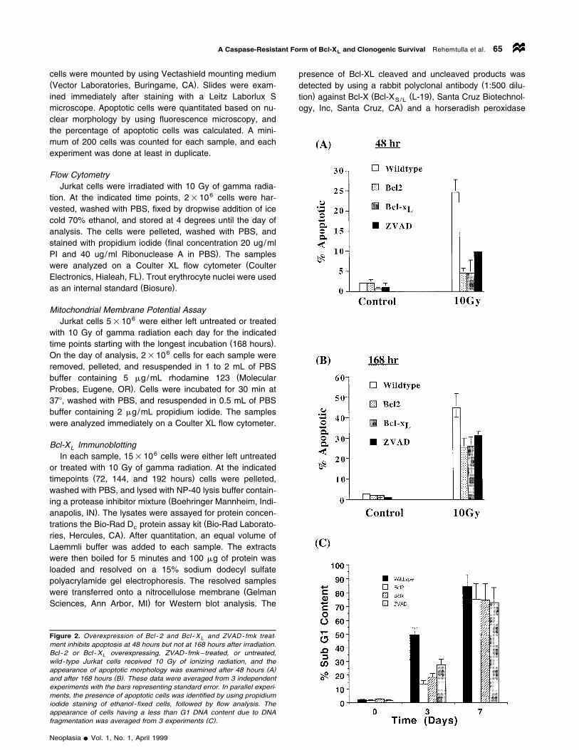
Figure 2. Overexpression of Bcl - 2 and Bcl - X and ZVAD - fmk treat-Lment inhibits apoptosis at 48 hours but not at 168 hours after irradiation.Bcl- 2 or Bcl- X overexpressing, ZVAD-fmk–treated, or untreated,Lwild-type Jurkat cells received 10 Gy of ionizing radiation, and the
( )appearance of apoptotic morphology was examined after 48 hours A( )and after 168 hours B . These data were averaged from 3 independent
experiments with the bars representing standard error. In parallel experi-ments, the presence of apoptotic cells was identified by using propidiumiodide staining of ethanol- fixed cells, followed by flow analysis. Theappearance of cells having a less than G1 DNA content due to DNA
( )fragmentation was averaged from 3 experiments C .
presence of Bcl-XL cleaved and uncleaved products was(detected by using a rabbit polyclonal antibody 1:500 dilu-
) ( ( )tion against Bcl-X Bcl-X L-19 , Santa Cruz Biotechnol-S/L
)ogy, Inc, Santa Cruz, CA and a horseradish peroxidase
Neoplasia v Vol. 1, No. 1, April 1999

A Caspase-Resistant Form of Bcl-X and Clonogenic Survival Rehemtulla et al.L66
Figure 3. Bcl-2 and Bcl-X expression protects loss of mitochondrial membrane potential at 3 but not at 6 days afterLionizing radiation. Bcl-X overexpressing or wild-type Jurkat cells were irradiated with 10 Gy of ionizing radiation, afterLwhich the cells were stained with Rh123 and propidium iodide and analyzed by flow. A. A decrease in Rh123 fluorescence( )leftward shift on the X-axis is indicative of loss of mitochondrial membrane potential, whereas an increase in propidium
( )iodide staining upward shift on the Y-axis is indicative of loss of cell membrane integrity and hence viability. B. Data from( )3 independent experiments were averaged "standard error . Wild-type Jurkat cells are shown by solid bars, Bcl-2
overexpressing cells with open bars, Bcl-X expressing cells with cross bars, and ZVAD-treated wild-type cells with doubleLbars.
Neoplasia v Vol. 1, No. 1, April 1999

A Caspase-Resistant Form of Bcl-X and Clonogenic Survival Rehemtulla et al.L 67
( )HRP –conjugated secondary antibody. The blots were de-(veloped by using a CL-HRP substrate system Pierce,
)Rockford, IL .
Results
Bcl-2 and Bcl-X Inhibit Radiation-Induced Apoptosis butL
Fail to Promote Long-Term SurvivalTo understand the contribution of apoptosis in
radiation-induced cell death we first investigated the impactof Bcl-2 and Bcl-X expression on clonogenic survival ofL
Jurkat cells after ionizing radiation. In contrast to our initialprediction, expression of Bcl-2 or Bcl-X did not significantlyL
( )alter the clonogenic survival of Jurkat cells Figure 1A .Similarly, treatment of Jurkat cells with the caspase inhibitorZVAD-fmk also had no significant impact on clonogenic
( )survival Figure 1A compared with untreated Jurkat cells.To examine if this was a cell line–specific phenomenon, westudied clonogenic survival of MCF-7 cells in the presenceor absence of Bcl-X expression. As shown in Figure 1B,L
Bcl-X expression did not protect MCF-7 cells from radia-L
tion-induced clonogenic death. To test if the failure to en-hance clonogenic survival under these conditions was dueto failure of Bcl-2, Bcl-X , and ZVAD-fmk to inhibitL
radiation-induced cell death, we performed a short-termviability experiment. Control Jurkat cells, Bcl-2 or Bcl-XL
expressing Jurkat cells, and ZVAD-fmk–treated Jurkat cellswere irradiated, after which cell viability was measured bytrypan blue exclusion. Expression of Bcl-2 or Bcl-X , as wellL
as treatment with the caspase inhibitor ZVAD-fmk, resultedin significant protection from radiation-induced cell death ina dose-dependent manner, compared with untreated cells( )Figure 1C .
We hypothesized that the discrepancy in these two re-sults was due to the clonogenic survival assay requirementof a 10 to 14 day period, in which a surviving cell has toform a colony. However, in the viability assay, survival isdetermined immediately by using trypan blue exclusion. Tofurther examine if Bcl-X and Bcl-2 are able to inhibitL
apoptosis in the short term but not in the long term, cellswere irradiated with 10 Gy, and the fraction of cells appear-
(ing morphologically apoptotic as visualized by the presence)of condensed chromatin and pyknotic nuclei after 72 hours
or 168 hours was counted. As shown in Figure 2A, 22% ofJurkat cells appeared morphologically apoptotic 72 hoursafter irradiation, whereas Jurkat/Bcl-2 and Jurkat/Bcl-XL
cells appeared 4% and 3% apoptotic, respectively.ZVAD-fmk treated cells were 10% apoptotic 72 hours afterirradiation. When the same cultures were analyzed for mor-
( )phological apoptosis 168 hours after irradiation Figure 2B ,Jurkat cells appeared 45% apoptotic, whereas Bcl-2 and
(Bcl-X overexpressing cells appeared 25% apoptotic Fig-L
)ure 2B and ZVAD-fmk treated cells were 30% apoptotic.The ability of Bcl-2 and Bcl-X to inhibit apoptosis byL
greater than 5-fold at the 72-hour timepoint and by less than2-fold at the 168-hour timepoint is consistent with our hy-pothesis that Bcl-2 and Bcl-X inhibit apoptosis in the shortL
term but not in the long term.
Bcl-2 and Bcl-X Delay but Do Not Prevent DNA Fragmen-L
tation, Loss of Mitochondrial Membrane Potential, and Cas-pase-3 Activation
The results just discussed were substantiated when hy-( )podiploidy was used as a measure of apoptosis Figure 2C .
Bcl-2 and Bcl-X overexpression as well as ZVAD-fmkL
(treatment resulted in inhibition of DNA fragmentation asdetermined by the presence of cells having a less than G1
)content of DNA , 3 but not 7 days after irradiation. Bcl-XL
and Bcl-2 mediate their antiapoptotic effect by 2 mecha-nisms. First, Bcl-X protects the loss of mitochondrial mem-L
brane potential, and second, Bcl-X inhibits activation ofL
caspases. To test if the loss of Bcl-X ’s ability to inhibitL
radiation-induced apoptosis at day 7 was consistent with theloss of these functions, we performed the following experi-ments. Using rhodamine 123 fluorescence as a marker formitochondrial membrane potential and propidium iodide
(staining as marker for cell membrane integrity and there-)fore cell viability , we demonstrated that 45% of Jurkat cells
(lost their mitochondrial membrane potential as evidenced)by a decrease in Rh123 fluorescence and were nonviable
( )as evidenced by increased propidium iodide staining 72( )hours after irradiation Figure 3A and B . Bcl-X overex-L
pression resulted in only 17% of Jurkat/Bcl-X cells losingL
(their mitochondrial membrane potential at day 3 Figure 3A)and B . Similarly, Bcl-2 expression also inhibited
radiation-induced loss of mitochondrial membrane otentialand apoptosis 3 days after irradiation. The ability of Bcl-2and Bcl-X to protect mitochondrial dysfunction was lostL
Figure 4. Cleavage of Bcl - X and caspase 3 activation is initiated 72Lhours after irradiation. Bcl- X overexpressing Jurkat cells were irradi-Lated with 10 Gy of ionizing radiation, and cell extracts were prepared atvarious times and analyzed by Western blot for the presence of Bcl- XL
( ) ( )cleavage A and caspase 3 activation B .
Neoplasia v Vol. 1, No. 1, April 1999

A Caspase-Resistant Form of Bcl-X and Clonogenic Survival Rehemtulla et al.L68
beyond day 6. ZVAD-fmk treatment also resulted in inhibi-tion of loss of mitochondrial membrane potential, althoughto a much lesser extent. Interestingly, Bcl-2 inhibited apop-tosis as late as day 5, at which point Bcl-X and ZVAD-fmkL
( )had lost their inhibitory capacity Figure 3B .Recent reports have suggested that cleavage of Bcl-2, as
well as Bcl-X , by caspases can result in loss of functionL
[ ]17–19 . To examine if loss of Bcl-X function at the laterL
timepoints in response to ionizing radiation correlated withits cleavage, we performed Western blot analysis of Jurkat/Bcl-X cells. Untreated cells or irradiated cells were har-L
vested at the indicated timepoints and analyzed. As shownin Figure 4A, 72 hours after irradiation an additional Bcl-XL
immunoreactive band was detected at 16 kDa. This form of( )Bcl-X was undetectable in untreated cells Figure 4A , asL
Figure 5. Bcl - X -D loop but not Bcl - X is resistant to cleavage byL Lcaspases and is able to inhibit apoptosis in the short - and long-termassays. Jurkat cells overexpressing Bcl- X or Bcl- X -D loop were irra-L Ldiated with 10 Gy of ionizing radiation, and at various times cells werecollected for the preparation of cell extracts for Western blot analysis
( )with a Bcl- X –specific antibody A . At these times cells were alsoL( )collected for determination of cell viability with trypan blue exclusion B .
Control cultures that were mock irradiated were also analyzed in paral-( )lel. Data presented in B are an average of 4 experiments, with bars
representing standard error.
Figure 6. Bcl - X -D loop– expressing Jurkat cells have an improvedLclonogenic survival in response to ionizing radiation, compared with
( )Jurkat cells or Jurkat/Bcl- X cells. Jurkat cells squares , Jurkat/Bcl- XL L( ) ( )cells circles , or Jurkat/Bcl- X -D loop cells triangles were irradiatedL
with various doses of ionizing radiation and immediately plated ontodishes for the determination of the surviving fraction. Bcl- X -D loop–Loverexpressing cells were much more resistant to ionizing radiation andresulted in a much larger fraction of surviving cells at all doses ofradiation. Similar results were obtained from 3 independent experi-ments, and the data presented are from a representative experiment.
(well as 24 hours and 48 hours after irradiation data not)shown . The 16-kDa band accumulated in a time-dependent
manner. Interestingly, detection of the 16-kDa form of Bcl-XL
(coincided with the appearance of active caspase 3 Figure)4B . Caspase 3 is a zymogen that is cleaved to a 2-chain
polypeptide during apoptosis, resulting in its functional acti-vation. The cleaved form of caspase 3 was undetectable in
( )nonirradiated cells Figure 4B or at 24 and 48 hours after( )irradiation of Jurkat/Bcl-X cells data not shown . Cleaved,L
active caspase was readily detectable 72 hours after irradia-tion and accumulated at 144 and 192 hours. The appear-ance of the 16-kDa form of Bcl-X mirrored the activation ofL
caspase 3, suggesting that activation of caspase 3 resultedin cleavage of Bcl-X to a 16-kDa protein. Taken together,L
these results indicate that expression of Bcl-X results inL
inhibition of radiation-induced apoptosis in the short term( )up to 72 hours , but subsequent cleavage and inactivationof Bcl-X results in loss of protection from apoptosis.L
Role of Caspases in the Cleavage and Inactivation of Bcl-XL
To directly test the role of caspases in the inactivation ofBcl-X , we used a mutant of Bcl-X that lacks the caspaseL L
( ) [ ]recognition sequence Bcl-X -Dloop 20 . Bcl-X -DloopL L
cannot be cleaved but still retains antiapoptotic function.Jurkat cells were stably transfected with the expressionvector for Bcl-X -Dloop to derive the Jurkat/Bcl-X -DloopL L
cell line. When cell extracts from irradiated Jurkat/Bcl-XL
and Jurkat/Bcl-X -Dloop cells were analyzed by WesternL
blot, Bcl-X was detected as a 29-kDa polypeptide at theL
( )24-and 48-hour timepoints data not shown . As before,Bcl-X was also detected as a 16-kDa polypeptide 72 andL
(144 hours after irradiation but not in the absence of irradia-
Neoplasia v Vol. 1, No. 1, April 1999

A Caspase-Resistant Form of Bcl-X and Clonogenic Survival Rehemtulla et al.L 69
)tion , whereas Bcl-X -Dloop was not cleaved to the 16-kDaL
( )species at these times after irradiation Figure 5A . Next, weexamined the ability of Bcl-X -Dloop to protect cells fromL
cell death at different times after irradiation compared towild-type Bcl-X . As shown in Figure 5B, Bcl-X -loop, unlikeL L
wild type Bcl-X , was able to protect cells from radiation-in-L
( )duced cell death at early 24 and 48 hours as well as late( )timepoints beyond 72 hours . The ability of Bcl-X -Dloop toL
protect cells from radiation-induced cell death also resultedin enhanced clonogenic survival of cells expressing thecaspase-resistant mutant of Bcl-X , compared with cellsL
expressing the wild-type molecule or cells that were trans-( )fected with the vector only Figure 6 .
DiscussionOur studies on understanding the role Bcl-2 and Bcl-X playL
in determining the radiosensitivity of tumor cells led to theobservation that expression of these antiapoptotic proteinsresults in inhibition of apoptosis when assays that measureapoptosis within 72 hours after irradiation are used. Incontrast, when apoptosis was measured 96 hours or moreafter irradiation or with either a clonogenic assay, a morpho-logical assay, or a cell viability assay, the ability of Bcl-2 andBcl-X to inhibit radiation-induced apoptosis was greatlyL
diminished. Inability of these proteins to block apoptosis inthe long term has been observed in response to many
[ ]stimuli. Bissonnette et al. 21 observed that in Chinesehamster ovary cells undergoing c-myc–induced apoptosis,Bcl-2 expression resulted in a temporary delay but did not
[ ]inhibit apoptosis. Yin and Schimke 14 also observed thatBcl-2 expression inhibited apoptosis in response to colec-imid, aphidicolin, and trimetrexate treatment when short-termassays of apoptosis were used, such as determination ofnuclear morphology after propidium iodide staining and vitaldye exclusion. In contrast, when a clonogenic assay wasused to measure cell viability, Bcl-2 expression did notappear to protect cells from these insults. Kyprianou et al.[ ]13 also observed that Bcl-2 expression rendered prostatecancer cells resistant to radiation-induced apoptosis butfailed to enhance clonogenic survival. A number of possibleexplanations for the discrepancy in the role of Bcl-2 ininhibiting apoptosis by clonogenic assays compared withshort-term assays have been put forward. Yin and Schimke[ ] [ ]14 , as well as Kyprianu et al. 13 , suggested that thisinconsistency could be due to the fact that Bcl-2 may not
[ ]inhibit apoptosis but simply delay it. Milner et al. 22 con-cluded that Bcl-2 expression results in inhibition of apopto-sis in irradiated B-lymphoma–derived cells when short-termassays but not long-term assays are used, because Bcl-2can also promote apoptosis through its ability promote
[ ]growth arrest. Lock and Stribinskiene 15 proposed that theability of Bcl-2 to prevent apoptosis may not translate intoincreased survival in response to etoposide treatment be-cause Hela cells respond to etoposide either by apoptosisor by mitotic catastrophe. Cells that are protected fromapoptosis by Bcl-2 expression fail to survive in the long termowing to mitotic death.
To understand the molecular basis of the failure of Bcl-2and Bcl-X to protect cells from undergoing apoptosis atL
( )later timepoints beyond 96 hours , we examined the keyactivities of these proteins over time. Two primary activitieshave been ascribed to Bcl-2 and its homologues, such asBcl-X . First, in an Apaf-1–dependent manner, Bcl-2–likeL
[ ]proteins can inhibit activation of caspases 23 . Second, theability of Bcl-2–like proteins to inhibit loss of mitochondrial
[ ]membrane potential is also key to their function 23 . Ourresults indicate that despite the presence of Bcl-X , irradia-L
tion of Jurkat cells resulted in activation of caspase 3 at 72hours. In addition, despite the presence of Bcl-2 and Bcl-X ,L
irradiation of Jurkat cells resulted in the loss of mitochon-drial membrane potential, although in a much more delayedtime course compared with control cells. Inability of Bcl-2 orBcl-X to protect cells from apoptosis in the long term isL
consistent with the inability of these proteins to completelyinhibit caspase activation or to protect the loss of mitochon-drial membrane potential in the long term. This, therefore,suggested to us that Bcl-2 and Bcl-X must be nonfunc-L
tional at these timepoints. Based on previous observations[ ]17–19 that both these proteins can be cleaved and inacti-vated during apoptosis, we investigated whether during radi-ation-induced apoptosis, cleavage of Bcl-X was occurring.L
Indeed, approximately 72 hours after irradiation, a 16-kDaform of Bcl-X was detected in cell extracts, and this contin-L
ued to accumulate with time. Analysis of Bcl-2 also revealedthe appearance of an approximately 22-kDa polypeptide at
( )96 hours after irradiation data not shown . Appearance ofthe cleaved form of Bcl-X and Bcl-2 coincided with theirL
inability to protect cells from radiation-induced apoptosis. Inaddition, cleavage of Bcl-X was coincident with the pres-L
ence of active caspase 3 at 72 hours. This is consistent withpublished results that the 16-kDa form of Bcl-X appearsL
upon cleavage of Bcl-X at an aspartic acid residue atL
[ ]position 61 17 . The report also demonstrated that the16-kDa form of the protein, which lacks the BH4 domain,has proapoptotic activity rather than antiapoptotic activity,similar to Bcl-X . We believe that in our system, in re-S
sponse to the appearance and accumulation of the cleavedprotein, there was a corresponding increase in the rate ofapoptosis.
Recent reports that the cleavage of Bcl-2 by caspases[ ]also results in a polypeptide with proapoptotic activity 18
further validates our hypothesis that failure to protect cellsfrom radiation-induced apoptosis in the long term is due tocleavage of antiapoptotic proteins such as Bcl-2 and Bcl-X .L
To test the role of caspases in inactivating Bcl-X , weL
irradiated Bcl-X expressing and nonexpressing Jurkat cellsL
and monitored cell viability over time in the presence orabsence of caspase inhibitors. Although the expression ofBcl-X protected Jurkat cells from cell death over the first 72L
hours, we consistently observed an acceleration in the rateof apoptosis beyond this time, which we propose is due toinactivation of Bcl-X by caspases. This acceleration in theL
rate of apoptosis was greatly inhibited in the presence of( )caspase inhibitors ZVAD-fmk, data not shown , suggesting
that inhibition of caspases prevented the cleavage of Bcl-X ,L
Neoplasia v Vol. 1, No. 1, April 1999

A Caspase-Resistant Form of Bcl-X and Clonogenic Survival Rehemtulla et al.L70
which therefore retained its antiapoptotic function. To di-rectly test the role of caspases in the cleavage and inactiva-tion of Bcl-X , a deletion mutant that lacks a caspaseL
recognition sequence and hence a noncleavable form of( ) [ ]Bcl-X Bcl-X -Dloop was used 20 . Expression ofL L
Bcl-X -Dloop resulted in protection of cells from apoptosis inL
the short term as well as long term and also enhancedclonogenic survival. These studies provide a definitive ex-planation for a number of previous observations that indi-cated that the antiapoptotic proteins Bcl-2 and Bcl-X sim-L
ply delay the induction of apoptosis.We believe our results also provide a molecular explana-
tion for numerous studies that failed to demonstrate a corre-lation between expression of antiapoptic proteins such asBcl-2 in tumors and a poor clinical prognosis. In somereports, expression of Bcl-2 was associated with improved
[ ]prognosis 10–12 . Based on previous reports that the cas-pase cleaved form of Bcl-2 has proapoptotic activity andbased on results presented here, it is not difficult to imaginehow a Bcl-2 overexpressing tumor would respond better totherapy. Further studies to test the impact of Bcl-2, Bcl-X ,L
and Bcl-X -Dloop expression on tumor response duringL
radiation treatment as well as during chemotherapy are inprogress and will enable the evaluation of the role of Bcl-XL
cleavage in vivo.
AcknowledgementsWe thank Amy Pace for the preparation of the figures, MaryDavis for her support throughout these studies. This work
( )was supported by NIH grants CA78041 A.R. and CA56663( )J.M. J.F. is supported by a Giffum Upjohn Fellowship andtraining grant GM07767.
References[ ]1 Lowe SW, Schmitt EM, Smith SW, Osborne BA, and Jacks T
( )1993 . p53 is required for radiation-induced apoptosis in mousethymocytes. Nature 62, 847–849.
[ ]2 Clarke AR, Purdie CA, Harrison DJ, Morris RG, Bird CC, Hooper( )MI, and Wyllie AH 1993 . Thymocyte apoptosis induced by
p53-dependent and independent pathways. Nature 362, 849–852.[ ]3 Datta R, Kojima H, Banach D, Bump NJ, Talanian RV, Alnemri ES,
( )Weichselbaum RR, Wong WW, and Kufe DW 1997 . Activation ofa CrmA-insensitive, p35-sensitive pathway in ionizing radiation-in-duced apoptosis. J Biol Chem 272, 1965–1969.
[ ]4 Sentman CL, Shutter JR, Hockenbery D, Kanagawa O, and Ko-( )rsmeyer SJ 1991 . Bcl-2 inhibits multiple forms of apoptosis but
not negative selection in thymocytes. Cell 67, 879–888.[ ]5 Datta R, Manome Y, Taneja N, Boise LH, Weichselbaum R,
( )Thompson CB, Slapak CA, and Kufe D 1995 . Overexpression ofBcl-XL by cytotoxic drug exposure confers resistance to ionizing
radiation-induced internucleosomal DNA fragmentation. Cell GrowthDiffer 6, 363–370.
[ ]6 Emoto Y, Manome Y, Meinhardt G, Kisaki H, Kharbanda S, Robert-son M, Ghayur T, Wong WW, Kamen R, Weichselbaum R, and
( )Kufe D 1995 . Proteolytic activation of protein kinase C delta by anICE-like protease in apoptotic cells. EMBO J 14, 6148–6156.
[ ] ( )7 Pan G, O’Rourke K, and Dixit VM 1998 . Caspase 9, Bcl-XL andApaf-1 form a ternary complex. J Biol Chem 273, 5841–5845.
[ ]8 Vander Heiden MG, Chandel NS, Williamson EK, Schumacker PT,( )and Thompson CB 1997 . Bcl-X regulates the membrane poten-L
tial and volume homeostasis of mitochondria. Cell 91, 627–637.[ ] ( )9 Thompson CB 1995 . Apoptosis in pathogenesis and treatment of
disease. Science 267, 1456–1462.[ ]10 Pezzella F, Jones M, Ralfkiaer E, Ersboll J, Gatter KC, and Mason
( )DY 1993 . Evaluation of bcl-2 protein expression and 14;18translocation as prognostic markers in follicular lymphoma. Br JCancer 65, 87–89.
[ ]11 Piris MA, Pezzella F, Martinez-Montero JC, Orradre JL, Villuendas( )R, Sanchez-Beato M, Cuena R, Cruz MA, Martinez B, et al. 1994 .
p53 and bcl-2 expression in high-grade B-cell lymphomas: correla-tion with survival time. Br J Cancer 69, 337–341.
[ ]12 Tjalma W, Weyler J, Goovaerts G, De Pooter C, Van Marck E, and( )van Dam P 1997 . Prognostic value of bcl-2 expression in patients
with operable carcinoma of the uterine cervix. J Clin Pathol 50,33–36.
[ ] ( )13 Kyprianou N, King ED, Bradbury D, and Rhee JG 1997 . bcl-2over-expression delays radiation-induced apoptosis without affect-ing the clonogenic survival of human prostate cancer. Int J Cancer70, 341–348.
[ ] ( )14 Yin DX, and Schimke RT 1995 . BCL-2 expression delays drug-in-duced apoptosis but does not increase clonogenic survival afterdrug treatment in HeLa cells. Cancer Res Nov 55, 4922–4928.
[ ] ( )15 Lock RB, and Stribinskiene L 1996 . Dual modes of death inducedby etoposide in human epithelial tumor cells allow Bcl-2 to inhibitapoptosis without affecting clonogenic survival. Cancer Res 56,4006–4012.
[ ]16 Chinnaiyan AM, Orth K, O’Rourke K, Duan H, Poirier GG, and Dixit( )VM 1996 . Molecular ordering of the cell death pathway. Bcl-2 and
Bcl-xL function upstream of the CED-3-like apoptotic proteases. JBiol Chem 271, 4573–4576.
[ ]17 Clem RJ, Cheng EH, Karp CL, Kirsch DG, Ueno K, Takahashi A,Kastan MB, Griffin DE, Earnshaw WC, Veliuona MA, and Hardwick
( )JM 1997 . Modulation of cell death by Bcl-XL through caspaseinteraction. Proc Natl Acad Sci USA 95, 554–559.
[ ]18 Cheng EH, Kirsch DG, Clem RJ, Ravi R, Kastan MB, Bedi A, Ueno( )K, and Hardwick JM 1997 . Conversion of Bcl-2 to a Bax-like
death effector by caspases. Science 1997 278, 1966–1968.[ ]19 Grandgirard D, Studer E, Monney L, Belser T, Fellay I, Borner C,
( )and Michel MR 1998 . Alphaviruses induce apoptosis inBcl-2-overexpressing cells: Evidence for a caspase-mediated, pro-teolytic inactivation of Bcl-2. EMBO J 17, 1268–1278.
[ ]20 Chang BS, Minn AJ, Muchmore SW, Fesik SW, and Thompson CB( ) ( )1997 . Identification of a novel regulatory domain in Bcl-X L andBcl-2. EMBO J 16, 968–977.
[ ] ( )21 Bissonnette RP, Echeverri F, Mahboubi A, and Green DR 1992 .Apoptotic cell death induced by c-myc is inhibited by bcl-2. Nature59, 552–554.
[ ]22 Milner AE, Grand RJ, Vaughan AT, Armitage RJ, and Gregory CD( )1997 . Differential effects of BCL-2 on survival and proliferation ofhuman B- lymphoma cells following gamma-irradiation. Oncogene15, 1815–1822.
[ ] ( )23 Adams JM, and Cory S 1998 . The Bcl-2 protein family: Arbiters ofcell survival. Science 28, 11322–11326.
Neoplasia v Vol. 1, No. 1, April 1999
![New Method to Quantitate Clonogenic Tumor Cells in the ......[CANCER RESEARCH 43, 5451-5455, November 1983] New Method to Quantitate Clonogenic Tumor Cells in the Blood Circulation](https://img.pdfslide.us/doc/110x75/6068d20ce566193e3e18220a/new-method-to-quantitate-clonogenic-tumor-cells-in-the-cancer-research.jpg)


![BM-1197: A Novel and Specific Bcl-2/Bcl-xL Inhibitor ...€¦ · i values of ,1 nM [16]. BM-1074 inhibits cancer cell growth with IC 50 values of 1–2 nM in four small-cell lung](https://img.pdfslide.us/doc/110x75/603fc99e43de8d135c2cb7b1/bm-1197-a-novel-and-specific-bcl-2bcl-xl-inhibitor-i-values-of-1-nm-16.jpg)