Embed Size (px)
Citation preview
55
Received:March 26, 2015, Revised:April 16, 2015, Accepted:April 20, 2015
Corresponding to:Jin-Wuk Hur, Division of Rheumatology, Department of Internal Medicine, Eulji General Hospital, Eulji University College of Medicine, 68 Hangeulbiseok-ro, Nowon-gu, Seoul 01830, Korea. E-mail: [email protected]
pISSN: 2093-940X, eISSN: 2233-4718Copyright ⓒ 2016 by The Korean College of Rheumatology. All rights reserved.This is a Free Access article, which permits unrestricted non-commerical use, distribution, and reproduction in any medium, provided the original work is properly cited.
Case ReportJournal of Rheumatic Diseases Vol. 23, No. 1, February, 2016http://dx.doi.org/10.4078/jrd.2016.23.1.55
A Case of Sepsis Caused by Cellulitis in a Patient with Rheumatoid Arthritis after Tocilizumab Treatment
Min Seok Yoo, Ji Sang Park, Yoon Suk Park, Hye Won Kim, Jin-Wuk HurDivision of Rheumatology, Department of Internal Medicine, Eulji General Hospital, Eulji University College of Medicine, Seoul, Korea
Tocilizumab, a humanized monoclonal antibody against the interleukin-6 receptor, is therapeutically effective in patients diag-nosed with rheumatoid arthritis (RA) compared with placebo. However patients treated with tocilizumab are at increased risk of several adverse effects including anaphylaxis and serious infections that may lead to hospitalization or death. Therefore, the risks and benefits of treatment with tocilizumab should be considered carefully and close monitoring of patients for develop-ment of signs and symptoms of side effects is required during and after treatment. Here, we report on a rare case of anaphylaxis and severe sepsis caused by cellulitis in a patient with RA after tocilizumab treatment. (J Rheum Dis 2016;23:55-60)
Key Words. Tocilizumab, Anaphylaxis, Cellulitis, Sepsis
INTRODUCTION
The recent development of biological agents has brought about many changes in the treatment of rheuma-toid arthritis (RA). Despite the positive effects of the new biological agents on the treatment for patients with RA in clinical settings, they are reportedly more likely to cause adverse effects such as serious infections than the exist-ing classical disease-modifying antirheumatic drugs (DMARDs). In particular, a strong correlation was con-firmed between biological agents and increased tuber-culosis infection and infections by Streptococcus pneumo-niae and Listeria monocytogenes [1]. In addition, a tumor ne-crosis factor (TNF) inhibitor has been reported to in-crease the risk of musculocutaneous infection [2,3]. However, in Korea, cellulitis has not been reported to de-velop after treatment with tocilizumab, a biological agent. We encountered a patient with RA who was treated with tocilizumab and experienced anaphylaxis accompanied by cellulitis in both the lower limbs and secondary sepsis. We hereby report this case along with a literature review.
CASE REPORT
A 57-year-old man was admitted to our hospital with complains of edema, ecchymosis in the lower limbs and blisters on the left dorsal foot. Nine years ago he visited with a chief complaint of pain and edema that had per-sisted for more than 3 months in the right elbow and the proximal interphalangeal joints of fingers on both hands. He was diagnosed with RA based on the following find-ings: anti-cyclic citrullinated peptide antibody ≥100 IU/mL; increased rheumatoid factor level (186.6 IU/mL), erythrocyte sedimentation rate (ESR; 38 mm/h), and C-reactive protein (CRP) level (2.81 mg/dL); and evidence of bone erosion in both elbows on radiography. Since then, oral methotrexate (MTX; 12.5 mg/wk) and oral methylprednisolone (2 mg/d) had been continuously prescribed. However, his symptoms did not improve with medication from 24 months prior to admission, so tacro-limus (2 mg/d) was prescribed for oral administration. However, his symptoms worsened again and the disease activity score in 28 joints increased to 5.24. Thus, intra-venous administration of tocilizumab was planned be-
Min Seok Yoo et al.
56 J Rheum Dis Vol. 23, No. 1, February, 2016
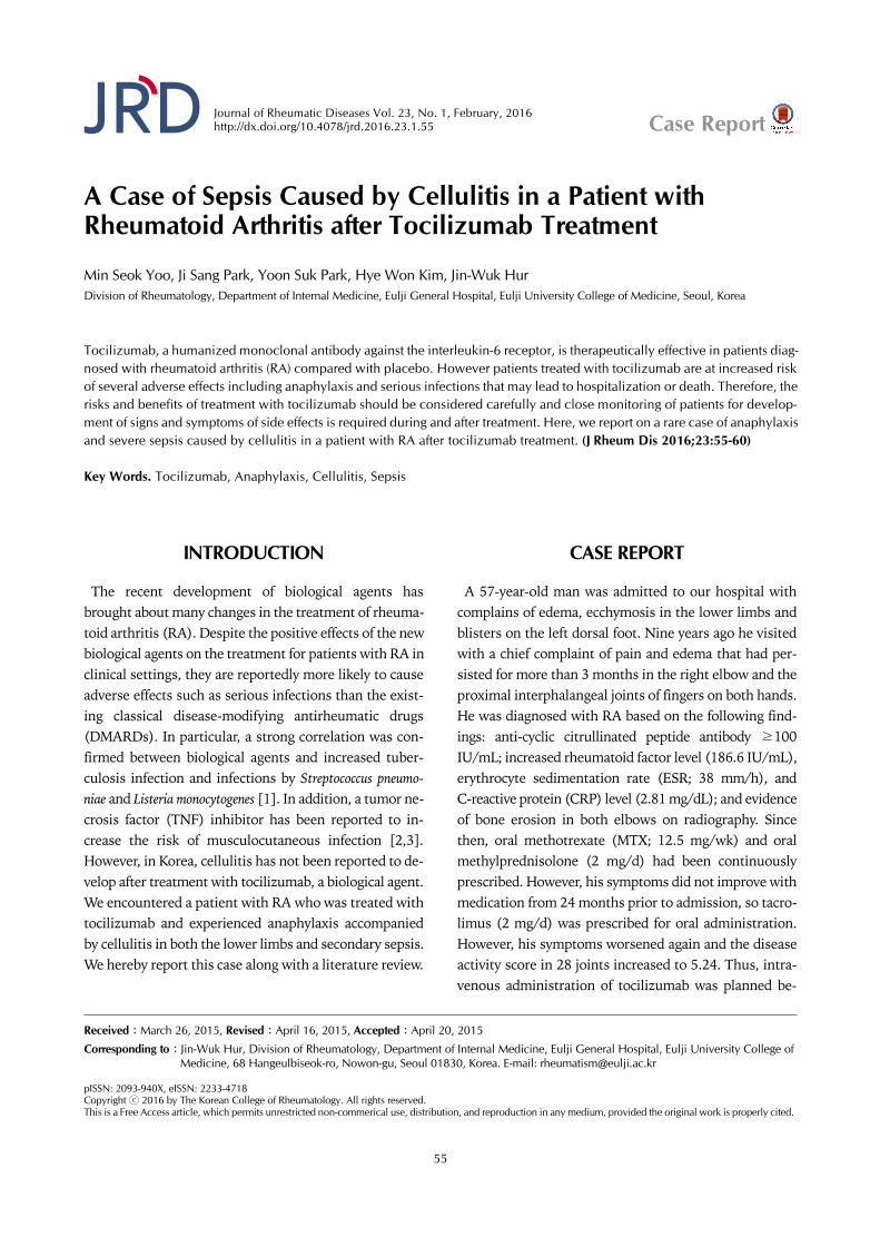
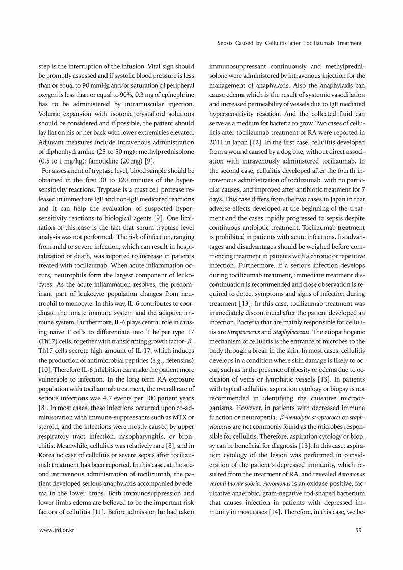
Figure 1. Four days after the second intravenous administra-tion of tocilizumab, edema and ecchymosis developed in the both lower limbs (A) and hem-orrhagic blisters developed on the left dorsal foot (B).
cause of persisting symptoms despite treatment with more than two types of DMARDs including MTX. Prior to the commencement of treatment, he did not receive other medications, including oriental medicine, and he had no history of fish and shellfish intake or travel. He was diag-nosed with tuberculosis 40 years prior and was cured af-ter treatment and he had histories of social drinking and 40 years of smoking. He had no history of allergic disease or drug hypersensitivity reactions.Four weeks before admission, 480 mg of tocilizumab
was first administered by intravenous injection after con-firmation of the negative result of intradermal skin test. Thirty minutes after the injection, the patient complained of mild generalized rash and itchiness but did not com-plain of edema or dyspnea, and his vital signs were normal. His symptoms improved with administration of antihistamine by intravenous injection, so he was dis-charged after the tocilizumab injection. When he visited the hospital for the second round of treatment with tocili-zumab, an antihistamine was administered by intra-venous injection prior to the intravenous administration of tocilizumab (480 mg). However, the patient developed generalized rash and itchiness that were more severe than before. He complained of dyspnea and generalized edema over the face, neck, and limbs, which was considered as anaphylaxis. Vital signs, including oxygen saturation lev-el, were normal, and intravenous administration of tocili-zumab was immediately discontinued. Antihistamine and methylprednisolone were administered by intra-
venous injection and thereafter, the itchiness and dysp-nea improved but the generalized rash and edema persisted. The edema in both lower limbs was particularly severe, confirmed as a 2+ pitting edema on physical examination. Four days later, ecchymosis and blisters de-veloped in the lower limbs and on the left dorsal foot, respectively.At admission, the initial vital signs may be summarized
as follows: body temperature, 37.6oC; pulse rate, 98 beats/min; blood pressure, 140/80 mmHg; and respira-tory rate, 22 breaths/min. Edema was accompanied by pain, hot flushes, redness, and petechia in the lower limbs, and 4×4-cm hemorrhagic blisters were observed on the left dorsal foot (Figure 1). The peripheral blood smear showed the following values: leukocyte count, 16,950/mm3; hemoglobin level, 13 g/dL; and platelet count, 41,000/mm3. The blood chemistry values were as follows: sodium, 132 mMol/L; potassium, 3.3 mMol/L; chloride, 101 mMol/L; total protein, 5.7 g/dL; albumin, 2.5 g/dL; aspartate aminotransferase, 64 U/L; alanine aminotransferase, 15 U/L; alkaline phosphatase, 154 U/L; total-bilirubin, 1.7 mg/dL; direct-bilirubin, 0.6 mg/dL; gamma-glutamyl transferase, 30 mg/dL; pro-thrombin time, 13.7 s; international normalized ratio, 1.29; ESR, 32 mm/h; and CRP, 4.84 mg/dL. The uri-nalysis results were normal. A virus antibody test was performed again after admission, revealing HBs Ag (−), HBs Ab (−), HCV Ab (−), and AFP 14.1 ng/mL. Abdominal ultrasonography revealed mild liver cirrhosis
Sepsis Caused by Cellulitis after Tocilizumab Treatment
www.jrd.or.kr 57
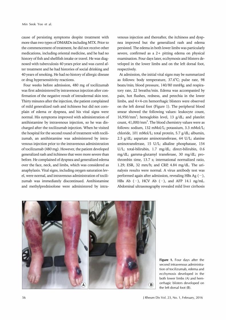
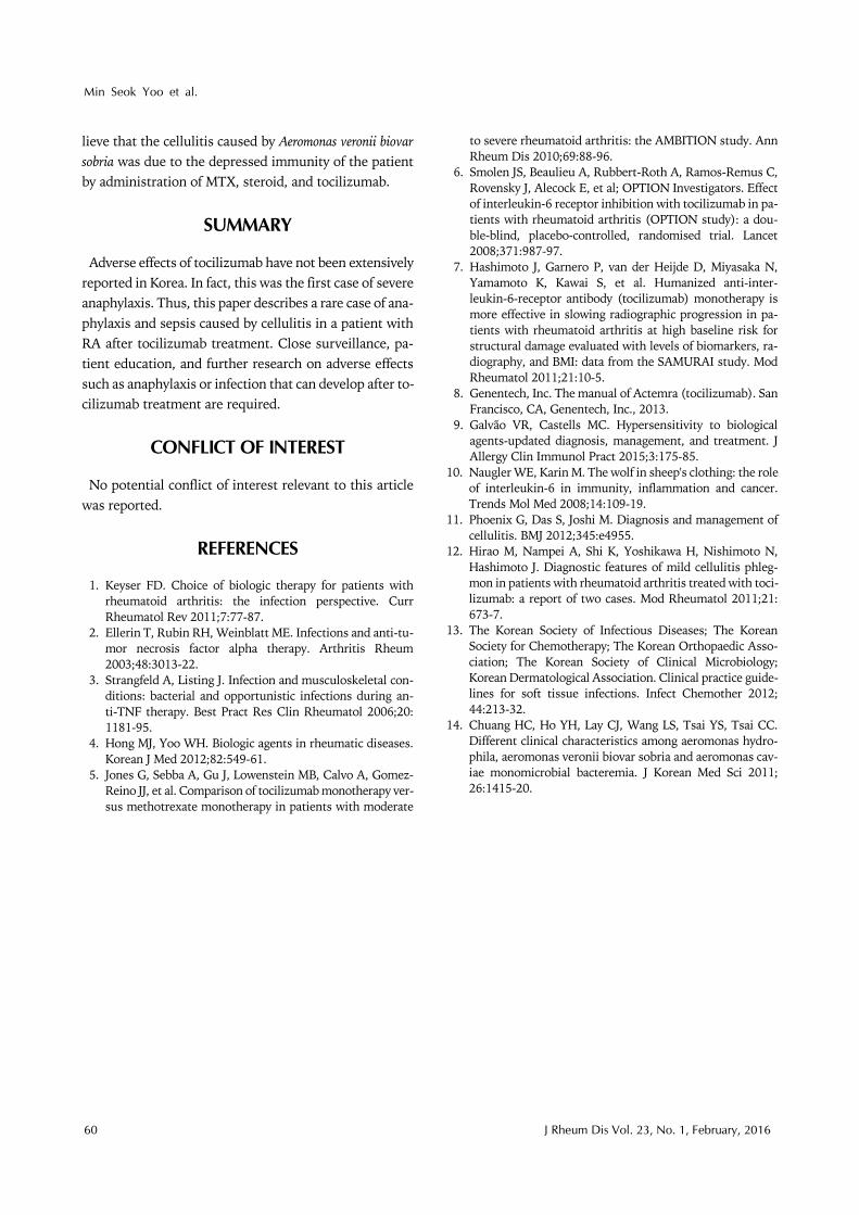
Figure 2. (A) Axial T2-weighted magnetic resonance (MR), left lower leg. (B) Axial T1-weighted MR, left lower leg. (C) Axial T1-weighted post contrast MR, left lower leg. (D) Axial T2-weighted MR, left lower leg. (E) Axial T1-weighted MR, right lower leg.(F) Axial T1-weighted post contrast MR, right lower leg. Subcutaneous thickening with fluid collections (arrows) was revealed onT2-weighted images and subcutaneous tissue and superficial fascia (arrowheads) showed contrast enhancement.
and splenomegaly but no signs of active hepatitis. In magnetic resonance imaging (MRI) performed for both lower limbs, subcutaneous thickening with fluid collec-tions was revealed on T2-weighted images and subcuta-neous tissue and superficial fascia showed contrast enhancement. Additional involvement of deep fasciae with fluid collections, thickening, and enhancement after contrast administration were not seen. MRI confirmed cellulitis but no indication of necrotizing fasciitis (Figure 2).After a lesion culture was performed, an empirical anti-
biotic therapy was commenced to treat the cellulitis. Ceftazidime, levofloxacin, and doxycycline were pre-scribed because a possibility of cellulitis caused by Vibrio vulnificus could not be excluded. Tocilizumab, methyl-prednisolone, and MTX were discontinued. On a blister lesion culture performed on the ninth day of admission, Aeromonas veronii biovar sobria was identified. Thus, dox-ycycline was discontinued and ceftazidime and levo-
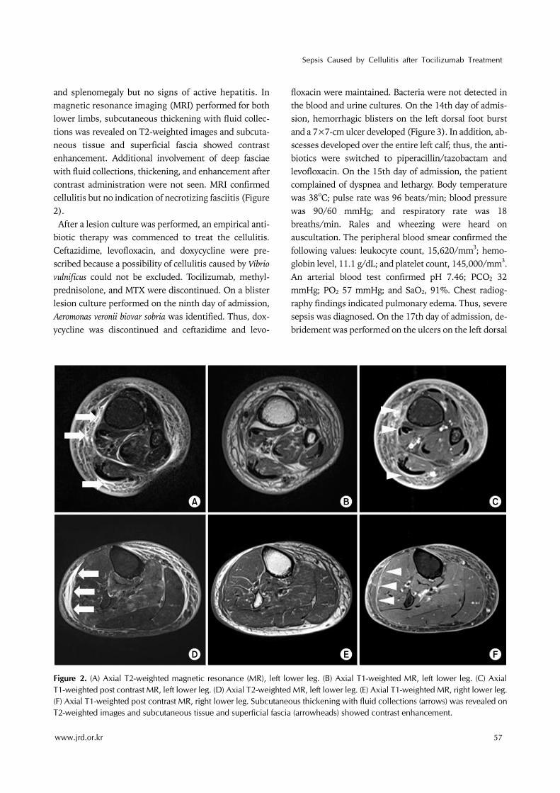
floxacin were maintained. Bacteria were not detected in the blood and urine cultures. On the 14th day of admis-sion, hemorrhagic blisters on the left dorsal foot burst and a 7×7-cm ulcer developed (Figure 3). In addition, ab-scesses developed over the entire left calf; thus, the anti-biotics were switched to piperacillin/tazobactam and levofloxacin. On the 15th day of admission, the patient complained of dyspnea and lethargy. Body temperature was 38oC; pulse rate was 96 beats/min; blood pressure was 90/60 mmHg; and respiratory rate was 18 breaths/min. Rales and wheezing were heard on auscultation. The peripheral blood smear confirmed the following values: leukocyte count, 15,620/mm3; hemo-globin level, 11.1 g/dL; and platelet count, 145,000/mm3. An arterial blood test confirmed pH 7.46; PCO2 32 mmHg; PO2 57 mmHg; and SaO2, 91%. Chest radiog-raphy findings indicated pulmonary edema. Thus, severe sepsis was diagnosed. On the 17th day of admission, de-bridement was performed on the ulcers on the left dorsal
Min Seok Yoo et al.
58 J Rheum Dis Vol. 23, No. 1, February, 2016
Figure 3. On the 14th day of admission, hemorrhagic blisters on the left dorsal foot burst and a 7×7-cm ulcer developed.
foot and abscesses on the left calf. From the day after the operation, the fever improved and the vital signs re-mained stable. On the 20th day of admission, abscesses developed on the outer side of the right calf; thus, de-bridement was performed. Loss of soft tissue in the ulcers on the left dorsal foot did not improve, so a split-thick-ness skin graft was performed. The patient was dis-charged when the skin lesions on both lower limbs improved. During hospitalization, the patient com-plained of multiple arthralgia and morning stiffness, which were his previously experienced symptoms of RA. These symptoms were controlled by oral administration of acetaminophen, tramadol. After discharge, oral admin-istration of MTX (12.5 mg/wk), methylprednisolone (4 mg/d), and tacrolimus (2 mg/d) was commenced, with the patient under outpatient follow-up observation.
DISCUSSION
RA is known to occur in individuals with a genetic pre-disposition due to abnormalities in immunologic toler-ance, which produce autoreactive cells and induce activa-tion of T and B lymphocytes, production of autoanti-bodies and secretion of various inflammatory cytokines, thus causing inflammation in synovial membranes, which in turn causes joint deformities. Cytokines asso-ciated with this occurrence of inflammation have been an important treatment target. The exact mechanism of ac-tion of the existing DMARDs, which are used in the clin-ical treatment of RA, on the immune system is not
known. However, they are known to suppress cytotoxic T cells. Hence, their use is considered disadvantageous ow-ing to the risk of serious adverse events. Therefore, se-lective suppression of the factors that play key roles in the development of diseases is important for a more effective and safer treatment. In recent years, biological agents have been widely used in clinical settings since TNF in-hibitors showed a positive effect on the treatment of RA. Moreover, other biological agents with various mecha-nisms have continued to be developed and are in use or under clinical testing. Biological agents used in the treat-ment of RA are generally classified into three types ac-cording to the following targeted biological processes: Inhibition of cytokine function, inhibition of secondary signals that stimulate T cells, and inhibition of function or elimination of B cells. Among the cytokines, interleukin (IL)-6 has various biological functions and plays an im-portant role in immune response, inflammation, acute phase response, and hematopoiesis. IL-6 induces differ-entiation of B lymphocytes into mature plasma cells that secrete immunoglobulin (Ig) and are involved in the growth and differentiation of T lymphocytes by inducing IL-2 and IL-2 receptors. In addition, IL-6 was identified to be associated with rheumatic diseases such as RA, juve-nile arthritis, Sjögren’s syndrome, and systemic lupus er-ythematosus [4].Tocilizumab is a humanized monoclonal antibody that
binds with naturally existing IL-6 receptors, inhibiting the effects of IL-6. It is created by fusing the binding sites of antibody against mouse-derived human IL-6 receptors with human IgG1. In the treatment of RA, tocilizumab alone or through a combined therapy with MTX showed more significant efficacy than MTX therapy alone [5,6]. In addition, it showed a more significant efficacy than a placebo in terms of physiological functions and fatigue in patients with severe RA who do not respond well to clas-sical DMARDs and TNF inhibitors [6]. Also, it inhibited radiological progression [7]. However, several adverse ef-fects including infection, gastrointestinal perforation, anaphylaxis, neutropenia, thrombocytopenia, and in-creased liver enzyme, total cholesterol, and triglyceride levels can occur [8].According to a controlled study with a 6-month intra-
venous administration of tocilizumab, anaphylaxis and other hypersensitivity reactions that require immediate cessation of treatment developed in 0.1% of the partic-ipants (3/2,644) [8]. In managing anaphylaxis and hyper-sensitivity reactions related to biological agents, the first
Sepsis Caused by Cellulitis after Tocilizumab Treatment
www.jrd.or.kr 59
step is the interruption of the infusion. Vital sign should be promptly assessed and if systolic blood pressure is less than or equal to 90 mmHg and/or saturation of peripheral oxygen is less than or equal to 90%, 0.3 mg of epinephrine has to be administered by intramuscular injection. Volume expansion with isotonic crystalloid solutions should be considered and if possible, the patient should lay flat on his or her back with lower extremities elevated. Adjuvant measures include intravenous administration of diphenhydramine (25 to 50 mg); methylprednisolone (0.5 to 1 mg/kg); famotidine (20 mg) [9]. For assessment of tryptase level, blood sample should be
obtained in the first 30 to 120 minutes of the hyper-sensitivity reactions. Tryptase is a mast cell protease re-leased in immediate IgE and non-IgE medicated reactions and it can help the evaluation of suspected hyper-sensitivity reactions to biological agents [9]. One limi-tation of this case is the fact that serum tryptase level analysis was not performed. The risk of infection, ranging from mild to severe infection, which can result in hospi-talization or death, was reported to increase in patients treated with tocilizumab. When acute inflammation oc-curs, neutrophils form the largest component of leuko-cytes. As the acute inflammation resolves, the predom-inant part of leukocyte population changes from neu-trophil to monocyte. In this way, IL-6 contributes to coor-dinate the innate immune system and the adaptive im-mune system. Furthermore, IL-6 plays central role in caus-ing naive T cells to differentiate into T helper type 17 (Th17) cells, together with transforming growth factor-β. Th17 cells secrete high amount of IL-17, which induces the production of antimicrobial peptides (e.g., defensins) [10]. Therefore IL-6 inhibition can make the patient more vulnerable to infection. In the long term RA exposure population with tocilizumab treatment, the overall rate of serious infections was 4.7 events per 100 patient years [8]. In most cases, these infections occurred upon co-ad-ministration with immune-suppressants such as MTX or steroid, and the infections were mostly caused by upper respiratory tract infection, nasopharyngitis, or bron-chitis. Meanwhile, cellulitis was relatively rare [8], and in Korea no case of cellulitis or severe sepsis after tocilizu-mab treatment has been reported. In this case, at the sec-ond intravenous administration of tocilizumab, the pa-tient developed serious anaphylaxis accompanied by ede-ma in the lower limbs. Both immunosuppression and lower limbs edema are believed to be the important risk factors of cellulitis [11]. Before admission he had taken
immunosuppressant continuously and methylpredni-solone were administered by intravenous injection for the management of anaphylaxis. Also the anaphylaxis can cause edema which is the result of systemic vasodilation and increased permeability of vessels due to IgE mediated hypersensitivity reaction. And the collected fluid can serve as a medium for bacteria to grow. Two cases of cellu-litis after tocilizumab treatment of RA were reported in 2011 in Japan [12]. In the first case, cellulitis developed from a wound caused by a dog bite, without direct associ-ation with intravenously administered tocilizumab. In the second case, cellulitis developed after the fourth in-travenous administration of tocilizumab, with no partic-ular causes, and improved after antibiotic treatment for 7 days. This case differs from the two cases in Japan in that adverse effects developed at the beginning of the treat-ment and the cases rapidly progressed to sepsis despite continuous antibiotic treatment. Tocilizumab treatment is prohibited in patients with acute infections. Its advan-tages and disadvantages should be weighed before com-mencing treatment in patients with a chronic or repetitive infection. Furthermore, if a serious infection develops during tocilizumab treatment, immediate treatment dis-continuation is recommended and close observation is re-quired to detect symptoms and signs of infection during treatment [13]. In this case, tocilizumab treatment was immediately discontinued after the patient developed an infection. Bacteria that are mainly responsible for celluli-tis are Streptococcus and Staphylococcus. The etiopathogenic mechanism of cellulitis is the entrance of microbes to the body through a break in the skin. In most cases, cellulitis develops in a condition where skin damage is likely to oc-cur, such as in the presence of obesity or edema due to oc-clusion of veins or lymphatic vessels [13]. In patients with typical cellulitis, aspiration cytology or biopsy is not recommended in identifying the causative microor-ganisms. However, in patients with decreased immune function or neutropenia, β-hemolytic streptococci or staph-ylococcus are not commonly found as the microbes respon-sible for cellulitis. Therefore, aspiration cytology or biop-sy can be beneficial for diagnosis [13]. In this case, aspira-tion cytology of the lesion was performed in consid-eration of the patient’s depressed immunity, which re-sulted from the treatment of RA, and revealed Aeromonas veronii biovar sobria. Aeromonas is an oxidase-positive, fac-ultative anaerobic, gram-negative rod-shaped bacterium that causes infection in patients with depressed im-munity in most cases [14]. Therefore, in this case, we be-
Min Seok Yoo et al.
60 J Rheum Dis Vol. 23, No. 1, February, 2016
lieve that the cellulitis caused by Aeromonas veronii biovar sobria was due to the depressed immunity of the patient by administration of MTX, steroid, and tocilizumab.
SUMMARY
Adverse effects of tocilizumab have not been extensively reported in Korea. In fact, this was the first case of severe anaphylaxis. Thus, this paper describes a rare case of ana-phylaxis and sepsis caused by cellulitis in a patient with RA after tocilizumab treatment. Close surveillance, pa-tient education, and further research on adverse effects such as anaphylaxis or infection that can develop after to-cilizumab treatment are required.
CONFLICT OF INTEREST
No potential conflict of interest relevant to this article was reported.
REFERENCES
1. Keyser FD. Choice of biologic therapy for patients with rheumatoid arthritis: the infection perspective. Curr Rheumatol Rev 2011;7:77-87.
2. Ellerin T, Rubin RH, Weinblatt ME. Infections and anti-tu-mor necrosis factor alpha therapy. Arthritis Rheum 2003;48:3013-22.
3. Strangfeld A, Listing J. Infection and musculoskeletal con-ditions: bacterial and opportunistic infections during an-ti-TNF therapy. Best Pract Res Clin Rheumatol 2006;20: 1181-95.
4. Hong MJ, Yoo WH. Biologic agents in rheumatic diseases. Korean J Med 2012;82:549-61.
5. Jones G, Sebba A, Gu J, Lowenstein MB, Calvo A, Gomez- Reino JJ, et al. Comparison of tocilizumab monotherapy ver-sus methotrexate monotherapy in patients with moderate
to severe rheumatoid arthritis: the AMBITION study. Ann Rheum Dis 2010;69:88-96.
6. Smolen JS, Beaulieu A, Rubbert-Roth A, Ramos-Remus C, Rovensky J, Alecock E, et al; OPTION Investigators. Effect of interleukin-6 receptor inhibition with tocilizumab in pa-tients with rheumatoid arthritis (OPTION study): a dou-ble-blind, placebo-controlled, randomised trial. Lancet 2008;371:987-97.
7. Hashimoto J, Garnero P, van der Heijde D, Miyasaka N, Yamamoto K, Kawai S, et al. Humanized anti-inter-leukin-6-receptor antibody (tocilizumab) monotherapy is more effective in slowing radiographic progression in pa-tients with rheumatoid arthritis at high baseline risk for structural damage evaluated with levels of biomarkers, ra-diography, and BMI: data from the SAMURAI study. Mod Rheumatol 2011;21:10-5.
8. Genentech, Inc. The manual of Actemra (tocilizumab). San Francisco, CA, Genentech, Inc., 2013.
9. Galvão VR, Castells MC. Hypersensitivity to biological agents-updated diagnosis, management, and treatment. J Allergy Clin Immunol Pract 2015;3:175-85.
10. Naugler WE, Karin M. The wolf in sheep's clothing: the role of interleukin-6 in immunity, inflammation and cancer. Trends Mol Med 2008;14:109-19.
11. Phoenix G, Das S, Joshi M. Diagnosis and management of cellulitis. BMJ 2012;345:e4955.
12. Hirao M, Nampei A, Shi K, Yoshikawa H, Nishimoto N, Hashimoto J. Diagnostic features of mild cellulitis phleg-mon in patients with rheumatoid arthritis treated with toci-lizumab: a report of two cases. Mod Rheumatol 2011;21: 673-7.
13. The Korean Society of Infectious Diseases; The Korean Society for Chemotherapy; The Korean Orthopaedic Asso-ciation; The Korean Society of Clinical Microbiology; Korean Dermatological Association. Clinical practice guide-lines for soft tissue infections. Infect Chemother 2012; 44:213-32.
14. Chuang HC, Ho YH, Lay CJ, Wang LS, Tsai YS, Tsai CC. Different clinical characteristics among aeromonas hydro-phila, aeromonas veronii biovar sobria and aeromonas cav-iae monomicrobial bacteremia. J Korean Med Sci 2011; 26:1415-20.


![Sepsis and Inflammasome · secondary sepsis can be caused via broken skin, mucosal barrier etc. It also occurs with food poisoning [5], addiction of drugs, and contact with environmental](https://img.pdfslide.us/doc/110x75/5ecbe720ee14fa2f87138908/sepsis-and-secondary-sepsis-can-be-caused-via-broken-skin-mucosal-barrier-etc.jpg)