Embed Size (px)
Citation preview

J. clin. Path. (1958), 11, 237.
A CASE OF CUSHING'S SYNDROME WITHPIGMENTATION AND SEVERE HYPOKALAEMIC
ALKALOSISBY
A. W. B. EDMUNDS, K. C. McKEOWN, AND P. N. COLEMANFrom the Friarage Hospital, Northallerton
(RECEIVED FOR PUBLICATION SEPTEMBER 18, 1957)
Hypokalaemia is the most important result ofthe adrenal hyperfunction seen in the recently de-scribed Conn's syndrome (Conn and Louis, 1956).In this syndrome aldosterone is produced exces-
sively, but evidence of the same excess of otherhormones is lacking and the excretion of 17-keto-steroids and 17-ketogenic steroids in the urineis normal. Hypokalaemia may also occur incases with a high urinary excretion of 17-keto-steroids and 17-ketogenic steroids, associated insome cases with the classical clinical features ofCushing's syndrome and in others with few or
none of these features (Sprague and Power, 1953;Brooks. McSwiney, Prunty, and Wood, 1957;Foye and Feichtmeir, 1955). In one of thesecases excess aldosterone was demonstrated(Brooks et al., 1957) and in another excess
mineralo-corticoid (Foye and Feichtmeir, 1955).The case to be described had some of the
features of Cushing's syndrome and presented a
hypokalaemic alkalosis more severe than has beendescribed in either Conn's or Cushing's syndrome.In addition there was a melanin pigmentation witha distribution and intensity similar to that seenin Addison's disease. Subtotal adrenalectomy hasbeen effective in reversing all the abnormalfeatures, including the pigmentation.
Case ReportThe patient, a married woman aged 42 years, was
admitted to the Friarage Hospital, Northallerton, onAugust 30, 1956. For a year she had felt irritableand depressed but did not become really ill until sixweeks before admission. During these six weeksshe developed increasing lassitude. She lost nearly a
stone in weight and transient swelling of the face andankles was seen. Increasing pigmentation was firstnoticed three weeks before admission and about thistime there was some dysuria and frequency and a
complaint of thirst. Just before admission thepatient became slightly confused mentally. Her loss
at each menstrual period had been normal, but duringthe year before admission the intervals between theperiods had increased.On admission she was lethargic and depressed, but
showed no gross muscular weakness. There wasmarked pigmentation of the face, at various pressuresites on the trunk (Fig. 1), on the areolae of thebreasts, and on small areas of the mucous membraneinside both cheeks. She was well nourished(10 st. 10 lb.) and showed no abnormal deposits offat, no kyphosis, and only one small cutaneous stria.The body hair was normal in amount and distribution,but she had a slight, and recent, growth of hair onthe upper lip. There was no acne. Blood pressurewas 140/80 mm. Hg. Clinical examination revealedno other abnormality. All the deep reflexes werepresent, and Chvostek's and Trousseau's signs were
FiG. 1.-The patient on admission. The configuration of the faceneck, and shoulder does not suggest Cushing's syndrome. Notepigmentation of face, exposed area of neck, and line of pressurefrom shoulder strap.
copyright. on F
ebruary 21, 2022 by guest. Protected by
http://jcp.bmj.com
/J C
lin Pathol: first published as 10.1136/jcp.11.3.237 on 1 M
ay 1958. Dow
nloaded from

A. W. B. EDMUNDS, K. C. MCKEOWN, and P. N. COLEMAN
AII
FIG. 2.-Electrocardiographs (a) on August 30 showing gross changes typical of potassium deficiency (serumpotassium 1.3 mEq. litre); (b) on September 2, when standard leads have become normal with theattainment of normal blood levels for potassium (serum potassium 5.6 mEq./litre) although it is highlyprobable that there was at this time a considerable deficiency of intracellular potassium.
negative. The urine contained sugar and albuminand a moderate number of pus cells and gave agrowth of Bact. coli on culture.
Addison's disease seemed the probable diagnosis onclinical grounds, but the laboratory findings on theday of admission, far from confirming this, gaveresults that suggested adrenal hyperfunction, and wereas follows: Serum potassium, 1.3 mEq./litre, serumsodium, 148 mEq. /litre, serum chloride, 83 mEq. /litre, serum bicarbonate, 52 mEq./litre. Leucocytes,11,000 per c.mm., blood sugar, 210 mg. %, bloodurea, 27 mg.%, Hb 75%. An E.C.G. showedchanges in keeping with gross potassium deficiency(Fig. 2).
Before investigating the case further, measures wereat once taken to correct the hypokalaemia. Potas-sium chloride by mouth, 2 g. two-hourly, was startedat 11 p.m. on the day of admission. Next day thepatient was very much brighter and the serum potas-sium level was 2 mEq. / litre. Treatment with evenlarger doses of potassium chloride continued andwithin four days the serum potassium had returnedto normal. Within the same period, without anyadministration of ammonium chloride, the severealkalosis observed on admission disappeared and theplasma bicarbonate and chloride levels becamenormal.A glucose tolerance test gave a diabetic type of
curve. There were no circulating eosinophils, and theexcretion of 17-ketosteroids was 36 mg. in 24 hours.Professor F. T. G. Prunty then arranged for the esti-mation of the output of other steroids, including
TABLESTEROID EXCRETION AND A.C.T.H. RESPONSE TEST1
Potassium Dose of 17-Keto- 17- Aldo-Dat Supplement A.C.T.H. steroids Ketogenic steroneate (mEq./ (units' (mg./ Steroids (mg.j
24 hr.) 24 hr.) 24 hr.) (mg.124 hr.)' 24 hr.)2Pre-operative
4'9i56 Nil Nil 30-0 -
5/9'56 445 35-4 9012/9156 Nil 34-2 11413/9'56 ,, 24-8 109 214/9'56 ,, 40 43-3 12615/9,156 ,, 40 43-7 68 -16!9156 40 37-6 100 _17/9,'56 1'61 40 37-0 118 _
Post-operative21111 56 Patienton.0-5mg.9a
fluorohydrocortisonedaily (no metaboliteslappear in urine) 6-8 0 5
OTHER URINARY STEROIDSChromatography3 (Bush C system) of glucuronidase-hydrolysed
urine extract indicated:Tetrahydrocortisone 5 mg./24 hr.Tetrahydrohydrocortisone -15 mg./24 hr.4Free cortisone 4 mg./24 hr.
Total 24 mg., which is greatly in excess of normal.
PRE-OPERATIVE PLASMA LEVELSIntermedin5 .. None found.(M.S.H.)
A.C.T.H.6 .. Significant amounts (more than normal).Hydrocortisone 12 pg./100 ml. (upper limit of normal 8 /pg.,
mean normal 5 pg.).Corticosterone .. Quantity present could not have exceeded
3 ,g. 100 ml.: none found.
Dr. B. W. Brooks. 2 Dr. I. H. Mills. 3 Bush (1952). 4 Nostandard was available as a check on identity. 5 Kindly carried outby Professor F. W. Landgrebe. 6Dr. Barbara E. Clayton.
238
.-Nmdmk-..- 1 1.1 Ir,-qww.10 00"o-0-
copyright. on F
ebruary 21, 2022 by guest. Protected by
http://jcp.bmj.com
/J C
lin Pathol: first published as 10.1136/jcp.11.3.237 on 1 M
ay 1958. Dow
nloaded from
CUSHING'S SYNDROME
aldosterone, and for the measurement of the effect onthe 17-ketosteroid and 17-ketogenic steroid output ongiving A.C.T.H. (Prunty, 1956). Professor Pruntysupplied a batch of A.C.T.H. of known potency forthis purpose, and at his suggestion potassium supple-ments were stopped during the period of the test toexclude any secondary effect on the aldosterone out-put from the administration of potassium in largequantities.The results (Table) were in keeping with Cushing's
syndrome. It was thought that the lack of signifi-cant response of the steroid excretion to A.C.T.H.was due to a tumour. On admission the onlyclassical clinical features of Cushing's syndrome hadbeen the presence of a little hair on the upper lip, butwithin a week this had noticeably increased, and aftera further fortnight there was an obviously abnormaldegree of facial hirsutism. At the same time, markedacne appeared. The blood pressure, however, alwaysremained normal.
Metabolic StudiesMeanwhile the study of the potassium and sodium
metabolism continued. The time before operationdivides into two periods (periods 1 and 3) when thepatient was receiving a large potassium supplement;and a middle period (2) when all extra potassium wasomitted to facilitate the study of the steroid excretion(Fig. 3a). During periods 1 and 3 theserum potassium, initially at a low level,rapidly rose to normal when a largepotassium supplement was given, only tofall equally rapidly when it was omittted. 7During this time a large proportion of the @ 6potassium supplement was lost in the
6
urine, but it is clear that some was 0 5retained. (During period 1, the average v 4potassium supplement was 254 mEq. a wday and the average urine output 201 E 3mEq. a day; during period 3, the supple- 2ment was 278 mEq. and the urine output194 mEq.). The difficulty experienced Iby the patient in retaining potassium was 0illustrated in period 2. No supplement .fi 400was given and the serum potassium at r 200once fell to 2 mEq./litre and remained LUat this level throughout the six-day E 0period, yet the patient continued to ex-crete potassium at an average rate of55 mEq. per day. At the same time her . 4clinical condition deteriorated and on . 3several occasions her legs gave way.The serum sodium was always normal, v 2
but during period 2 it is probable that Ethe patient was retaining sodium since
E
oFIG. 3.-Diagram showing daily potassium sup- X 4
plements and serum potassium levels (a) before * 200adrenalectomy, (b) following adrenalectomy. wNote that early in period 3 potassium was E 0given for four days as enteric-coated tablets inan effort to avoid gastric irritation, but was in-completely absorbed.
the average excretion on a normal ward diet was only44 mEq. per day. During periods 1 and 3, the averageexcretion of sodium was at a higher level (148 mEq.per day in period 1). In fact during the pre-operativeperiod there was a rough parallel between the sodiumand the potassium excretion. When the excretion ofpotassium rose as the result of the potassium supple-ment so also did the excretion of sodium. This effectwas not seen after adrenalectomy.
Studies were also made of the sodium/potassiumratio in saliva and sweat. The results will be referredto in the discussion.Towards the end of the first month it had become
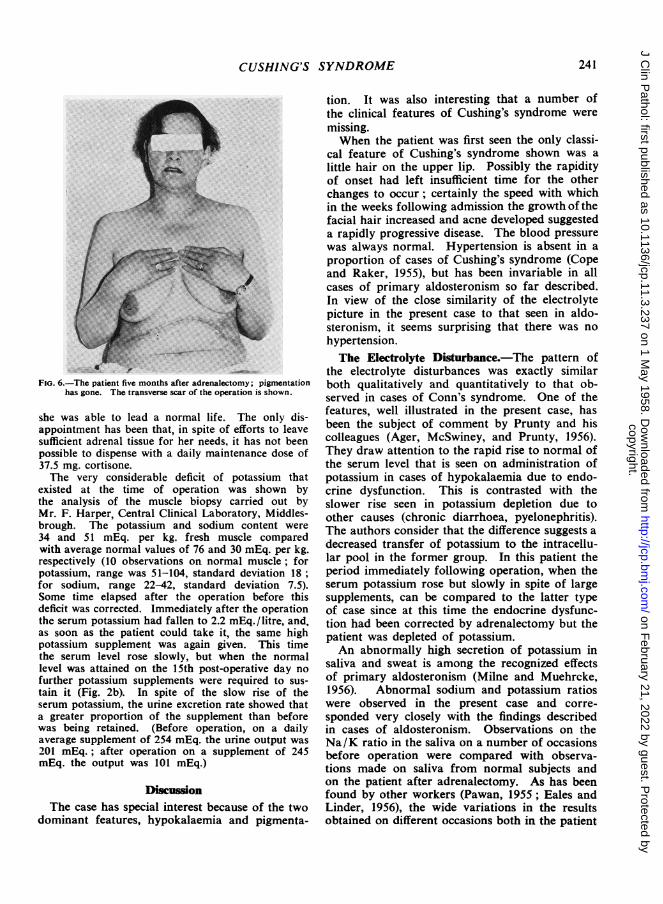
clear that an operation would be necessary. TheA.C.T.H. response test suggested that the patient hadan adrenal tumour, but tomograms taken after thepresacral insufflation of oxygen clearly demonstratedthe outlines of the adrenals and there seemed to beno tumour. An operation for the one-stage removalof the adrenals in cases of carcinoma of the breastthrough a semilunar incision placed transversely inthe epigastrium (Fig. 6) had recently been developedhere (McKeown and Ganguli, 1956). This operationallowed both adrenals to be inspected before actionwas taken, and meant that a pre-operative differentialdiagnosis between tumour and hyperplasia becameless important. The operation was carried out, withDr. K. H. Oldfield as the anaesthetist, on October 1.
Days
I
239
copyright. on F
ebruary 21, 2022 by guest. Protected by
http://jcp.bmj.com
/J C
lin Pathol: first published as 10.1136/jcp.11.3.237 on 1 M
ay 1958. Dow
nloaded from

A. W. B. EDMUNDS, K. C. McKEOWN, and P. N. COLEMAN
\~~PSr,1u,
0.5~~~~~~~~~~~
.... zI5L,E1 B. 31 | \ |r \ U|iEdTH
I; .L l.I;
;tIs ; k |Q t18 i
he -r h itlL *h; . i; l :1I L:- n th t thui'} 11;CIIc\eli LCL l A .'In
hft - k nc\i-n t iOiI ) itI' I ik cdl11111ufI]ttl1 'c TI Iru I1u1I
1 1i.l \ ci l cli2iicc tic r i hlp _*s, !l Iici|t \\ ci' f ;Ia; F) iit I 5 !i >C si:iS Lt
hii i p tiii t {it C hC I c\ i; hC 1 Il lllC( I ijIh
il,:ii:i hcicathl hu ca['ii i11 ililtlI:ccsi igt]Iit l ltI :i t h hL'1C I
;|s1ciNitf
cCfw-tl > Mit58[1 11ti .1 t iiw. cr1}5 L I 11
0II IL
i< i*iQ \il<
ti P.Ok:iz Ti 1L (
i( 1 Il1111t ]I a
L' Ad,11i d)'t;11 (i 1(;Li(11; I( hi,: li%C1-
ISI tI 1\1 *, L q"i5|tU'Li iC .1 co
C 1;1 I h ) |
.~~~ ~ ~ ~~~~~~~r C111, 3 I[ rt ztittf ;,5 !!1,.!..K, i|| f ,.l O1
Li iT rk Li Li
,11j; V L !roaa i}5( }I.l {i:
240
'.I
;.
K'w
b i
copyright. on F
ebruary 21, 2022 by guest. Protected by
http://jcp.bmj.com
/J C
lin Pathol: first published as 10.1136/jcp.11.3.237 on 1 M
ay 1958. Dow
nloaded from
CUSHING'S SYNDROME
FIG. 6.-The patient five months after adrenalectomy; pigmentationhas gone. The transverse scar of the operation is shown.
she was able to lead a normal life. The only dis-appointment has been that, in spite of efforts to leavesufficient adrenal tissue for her needs, it has not beenpossible to dispense with a daily maintenance dose of37.5 mg. cortisone.The very considerable deficit of potassium that
existed at the time of operation was shown bythe analysis of the muscle biopsy carried out byMr. F. Harper, Central Clinical Laboratory, Middles-brough. The potassium and sodium content were34 and 51 mEq. per kg. fresh muscle comparedwith average normal values of 76 and 30 mEq. per kg.respectively (10 observations on normal muscle; forpotassium, range was 51-104, standard deviation 18;for sodium, range 22-42, standard deviation 7.5).Some time elapsed after the operation before thisdeficit was corrected. Immediately after the operationthe serum potassium had fallen to 2.2 mEq./litre, and,as soon as the patient could take it, the same highpotassium supplement was again given. This timethe serum level rose slowly, but when the normallevel was attained on the 15th post-operative day no
further potassium supplements were required to sus-
tain it (Fig. 2b). In spite of the slow rise of theserum potassium, the urine excretion rate showed thata greater proportion of the supplement than beforewas being retained. (Before operation, on a dailyaverage supplement of 254 mEq. the urine output was
201 mEq.; after operation on a supplement of 245mEq. the output was 101 mEq.)
DiscussionThe case has special interest because of the two
dominant features, hypokalaemia and pigmenta-
tion. It was also interesting that a number ofthe clinical features of Cushing's syndrome weremissing.When the patient was first seen the only classi-
cal feature of Cushing's syndrome shown was alittle hair on the upper lip. Possibly the rapidityof onset had left insufficient time for the otherchanges to occur; certainly the speed with whichin the weeks following admission the growth of thefacial hair increased and acne developed suggesteda rapidly progressive disease. The blood pressurewas always normal. Hypertension is absent in aproportion of cases of Cushing's syndrome (Copeand Raker, 1955), but has been invariable in allcases of primary aldosteronism so far described.In view of the close similarity of the electrolytepicture in the present case to that seen in aldo-steronism, it seems surprising that there was nohypertension.The Electrolyte Disturbance.-The pattern of
the electrolyte disturbances was exactly similarboth qualitatively and quantitatively to that ob-served in cases of Conn's syndrome. One of thefeatures, well illustrated in the present case, hasbeen the subject of comment by Prunty and hiscolleagues (Ager, McSwiney, and Prunty, 1956).They draw attention to the rapid rise to normal ofthe serum level that is seen on administration ofpotassium in cases of hypokalaemia due to endo-crine dysfunction. This is contrasted with theslower rise seen in potassium depletion due toother causes (chronic diarrhoea, pyelonephritis).The authors consider that the difference suggests adecreased transfer of potassium to the intracellu-lar pool in the former group. In this patient theperiod immediately following operation, when theserum potassium rose but slowly in spite of largesupplements, can be compared to the latter typeof case since at this time the endocrine dysfunc-tion had been corrected by adrenalectomy but thepatient was depleted of potassium.An abnormally high secretion of potassium in
saliva and sweat is among the recognized effectsof primary aldosteronism (Milne and Muehrcke,1956). Abnormal sodium and potassium ratioswere observed in the present case and corre-sponded very closely with the findings describedin cases of aldosteronism. Observations on theNa/K ratio in the saliva on a number of occasionsbefore operation were compared with observa-tions made on saliva from normal subjects andon the patient after adrenalectomy. As has beenfound by other workers (Pawan, 1955; Eales andLinder, 1956), the wide variations in the resultsobtained on different occasions both in the patient
241
copyright. on F
ebruary 21, 2022 by guest. Protected by
http://jcp.bmj.com
/J C
lin Pathol: first published as 10.1136/jcp.11.3.237 on 1 M
ay 1958. Dow
nloaded from
A. W. B. EDMUNDS. K. C. McKEOWN, and P. N. COLEMAN
and in normal subjects limited the value of ob-servations on saliva as a method of demonstratingendocrine dysfunction. Nevertheless it was pos-sible to demonstrate that both the mean valuesand the range of the salivary Na/K ratios in thepatient before adrenalectomy were low comparedwith normal subjects and returned to normalfollowing adrenalectomy. The results obtainedwere as follows (10 observations in each series):Before adrenalectomy the mean Na/K ratio was0.27 with a range of 0.11 to 0.37, standard devia-tion 0.13, as compared with a mean for normalsubjects of 0.36, range 0.25 to 0.47, standard devia-tion 0.06. After adrenalectomy the mean valuefor the patient was 0.35, range 0.26 to 0.51, stan-dard deviation 0.06. (A simple technique for thedetermination of the Na /K ratio in sweat wasemployed, the sweat being collected on a filterpaper held in the axilla.) Two observations gaveNa/K ratios of I compared with normal valuesof 13-16 (Cantarow and Trumper, 1955). Afteradrenalectomy it was not possible to obtain asatisfactory measurement of the concentration ofpotassium by the simple technique in use.
It is of interest that, despite the close similaritybetween the electrolyte picture in the present casewith that of primary aldosteronism, no excessivesecretion of aldosterone was found in the urine.The unreliability of present methods of aldo-sterone estimation must, however, be recognizedand normal figures have been found in Conn'ssyndrome (Evans and Milne, 1954; Russell, Mar-shall, and Stanton, 1956). The question remains,therefore, whether the electrolyte changes wereproduced by aldosterone or by another steroidwith mineralo-corticoid effect, possibly cortico-sterone.
It was thought that the histological appearanceof the adrenals could throw light on this problem.There is evidence that in the normal adrenal aldo-sterone is formed in the glomerulosa (Bartter,1956), and in the present case the appearance ofthe glomerulosa did not suggest hyperfunction,rather the reverse. It is possible, however, thatin the abnormal adrenal aldosterone is also pro-duced in the fasciculata. This is the opinion ofvan Buchem, Doorenbos, and Elings (1956), whohave described the only case so far of primaryaldosteronism in which over-production of hor-mone was due not to tumour but to hyperplasia ofthe glands. By the courtesy of Dr. Arends andProfessor van Buchem the sections from theirpatient have been compared with those of ourown. Their sections differ in that the zonaglomerulosa can everywhere be readily made out,and in some few places is hyperplastic, but in the
sections from both cases a markedly hyperplasticzona fasciculata dominates the picture.Pigmentation.-Pigmentation is an occasional
feature of Cushing's syndrome. Simpson (1956)mentions pigmentation of the orbit and nipples,and Cope and Raker (1955) remark on it in a fewof their cases but do not mention its extent. Inthe present case the intensity and distribution ofthe pigmentation resembled that seen in Addison'sdisease.
Pigmentation in Addison's disease is nowthought to be due to excessive production ofmelanocyte-stimulating hormone by an overactivepituitary. Sulman (1952, 1956) was able todemonstrate excess in nearly all the samples ofplasma from cases of Addison's disease examined.He also found excess in the plasma in 30 out of45 cases of Cushing's syndrome. Sulman claimsthat excess of melanocyte-stimulating hormone isalways accompanied by excess of corticotropin andconsiders that by demonstrating an excess ofmelanocyte - stimulating hormone it is pos-sible to distinguish the " pituitary" from thepurely " adrenal " type of Cushing's syndrome.Geschwind, Li, and Barnafi (1956) have reportedthe isolation of pure melanocyte-stimulating hor-mone and find that it contains seven amino-acidswhich occur in an identical sequence in cortico-tropin. They suggest that this explains themelanocyte-stimulating activity of pure cortico-tropin.
It would seem probable, therefore, that the pig-mentation in the present case represented pituitaryoveractivity. Excess A.C.T.H. was found in theplasma and may have itself been the stimulus forthe pigmentation, since no excess of melanocyte-stimulating hormone was found in the singlesample of plasma examined. It is also probablethat pituitary overactivity was the reason for thefailure of the urinary steroid excretion to respondto test injections of A.C.T.H. The adrenals werealready responding maximally to endogenousA.C.T.H., and could do no more. An example ofa similar failure of the steroid excretion torespond to A.C.T.H. in a case of adrenal hyper-function not due to adrenal tumour has been re-ported by Rosenthal (1957). In this case alsothere was a melanin pigmentation with a distribu-tion characteristic of Addison's disease.The disappearance of the pigmentation after
adrenalectomy calls for an explanation, the moreso since many cases of Cushing's syndrome de-velop pigmentation after adrenalectomy (Sprague,Kvale, and Priestley, 1953; Hernberg, 1954). Theanswer may derive from the fact that in somerespects the actions of cortisone and aldosterone
242
copyright. on F
ebruary 21, 2022 by guest. Protected by
http://jcp.bmj.com
/J C
lin Pathol: first published as 10.1136/jcp.11.3.237 on 1 M
ay 1958. Dow
nloaded from

CUSHING'S SYNDROME
are antagonistic (Hetzel, McSwiney, Mills, andPrunty, 1956; Rosenbaum, Papper, and Ashley,1955). It may be that before the operation thepituitary was overacting in an attempt to stimulateproduction of more cortisone to counteract theexcessive mineralo-corticoid effect from other hor-mones. When the latter was removed by opera-tion this stimulus to A.C.T.H. production woulddisappear.
SummaryA woman of 42 years with adrenal hyperplasia
had pigmentation resembling that of Addison'sdisease, normal blood pressure, and severe hypo-kalaemic alkalosis. Subtotal adrenalectomy led tothe restoration of normal blood chemistry anddisappearance of the abnormal pigmentation.
Our thanks are due to Professor F. T. G. Pruntyfor his generous help and advice in the diagnosis ofthe case and in the preparation of this paper. Wewish to thank also, in addition to those mentioned inthe text. Dr. E. Gilbert for referring this patient, Dr.Simon Sevitt for his opinion on the histology, SisterCalvert, Sister Gibson, and Dr. E. R. L. Paton, of theFriarage Hospital, for their tireless efforts in caringfor this patient, Mr. S. Taylor for the photography,and Mrs. R. Carver for the biochemical estimations.
REFERENCESAger, J. A. M., McSwiney, R. D., and Prunty, F. T. G. (1956).
J. Endocr., 13, xxvi.Bartter, F. C. (1956). Metabolism, 5, 369.Brooks, R. V., McSwiney, R. R., Prunty, F. T. G., and Wood, R. J. Y.
(1957). Amer. J. Med., xxiii, 3, 391.Buchem, F. S. P. van, Doorenbos, H., and Elings, H. S. (1956).
Lancet, 2, 335.Bush, I. E. (1952). Biochem. J., 50, 370.Cantarow, A., and Trumper, M. (1955). Clinical Biochemistry,
p. 280. Saunders, Philadelphia and London.Chalmers, T. M., Fitzgerald, M. G., James, A. H., and Scarborough,
Harold (1956). Lancet, 1, 127.Conn, J. W., and Louis, L. H. (1956). Ann. intern. Med., 44, 1.Cope, O., and Raker, J. W. (1955). New Engi. J. Med., 253, 119
and 165.Eales, L., and Linder, G. C. (1956). Quart. J. Med., 25, 539.Evans, B. M., and Milne, M. D. (1954). Brit. med. J., 2, 1067.Foye, L. V., and Feichtmeir, T. V. (1955). Amer. J. Med., 19, 966.Geschwind, 1. 1., Li, C. H., and Barnafi, L. (1956). J. Amer. chem.
Soc., 73, 4494.Hernberg, C. A. (1954). Acta endocr. (Kbh.), 16, 309.Hetzel, B. S., McSwiney, R. R., Mills, 1. H., and Prunty, F. T. G.
(1956). J. Endocr., 13, 112.McKeown, K. C., and Ganguli, A. (1956). Brit. med. J., 1, 1466.Milne, M. D., and Muehrcke, R. C. (1956). Proc. roy. Soc. Med.,
43, 883.Pawan, G. L. S. (1955). Biochem. J., 60, xii.Prunty, F. T. G. (1956). Brit. med. J., 2, 615 and 673.Rosenbaum, J. D., Papper, S., and Ashley, M. M. (1955). J. clin.
Endocr., 15, 1459.Rosenthal, F. D. (1957). Brit. med. J., 2, 139.Russell, G. F. M., Marshall, J., and Stanton, J. B. (1956). Scot. med.
J., 1, 122.Simpson, S. L. (1956). In Price's Textbook of the Practice of Medi-
cine, ed. D. Hunter. Oxford lJniv. Press, London.Sprague, R. G., Kyale, W. F., and Priestley, J. T. (1953). J. Amer.
med. Ass., 151, 629.- and Power, M. H. (1953). J.-Lancet, 73, 217.Sulman, F. G. (1952). Acta endocr. (Kbh.), 10, 320.-(1956). J. clin. Endocr., 16, 755.
z
243
copyright. on F
ebruary 21, 2022 by guest. Protected by
http://jcp.bmj.com
/J C
lin Pathol: first published as 10.1136/jcp.11.3.237 on 1 M
ay 1958. Dow
nloaded from
![Introduction - UCL Computer Science€¦ · Web viewGuillain–Barré syndrome [Infectious polyneuritis] (Protein rises after 5–7 days) Cushing's disease Connective tissue disease](https://img.pdfslide.us/doc/110x75/5f9261a3478c3e103b2ba6ca/introduction-ucl-computer-web-view-guillainabarr-syndrome-infectious-polyneuritis.jpg)