Embed Size (px)
Citation preview

Cheng et al. Journal of Biomedical Science 2010, 17:34http://www.jbiomedsci.com/content/17/1/34
Open AccessR E S E A R C H
ResearchA biophysical elucidation for less toxicity of Agglutinin than Abrin-a from the Seeds of Abrus Precatorius in consequence of crystal structureJack Cheng1, Tian-Huey Lu*1, Chao-Lin Liu2 and Jung-Yaw Lin3
AbstractX-ray crystal structure determination of agglutinin from abrus precatorius in Taiwan is presented. The crystal structure of agglutinin, a type II ribosome-inactivating protein (RIP) from the seeds of Abrus precatorius in Taiwan, has been determined from a novel crystalline form by the molecular replacement method using the coordinates of abrin-a as the template. The structure has space group P41212 with Z = 8, and been refined at 2.6 Å to R-factor of 20.4%. The root-mean-square deviations of bond lengths and angles from the standard values are 0.009 Å and 1.3°. Primary, secondary, tertiary and quaternary structures of agglutinin have been described and compared with those of abrin-a to a certain extent. In subsequent docking research, we found that Asn200 of abrin-a may form a critical hydrogen bond with G4323 of 28SRNA, while corresponding Pro199 of agglutinin is a kink hydrophobic residue bound with the cleft in a more compact complementary relationship. This may explain the lower toxicity of agglutinin than abrin-a, despite of similarity in secondary structure and the activity cleft of two RIPs.
BackgroundRibosome inactivating proteins (RIPs) are enzymes thatcan inactivate ribosomes. The molecular mechanism ofinhibitory effect on protein synthesis has been shownthat RIPs act as a RNA N-glycosidase hydrolyzing the C-N glycosidic bond of the adenosine residue at position4324 in rat 28S rRNA [1,2]. They can cleave the syntheticRNA structure having a short double-helical stem and aloop containing a centered GAGA sequence, the first Abeing the cleavage site [3]. The depurination inactivatesthe ribosomes for binding to elongation factor 2 catalyz-ing GTP hydrolysis and translocation of peptidyl-tRNAto the P site [4], with a consequence inhibiting the proteinsynthesis. There are three categories of RIPs according tothe physical composition and characteristics. Most com-monly RIPs are type I RIPs, only single polypeptide chainproteins composed of the toxophoric A subunit with amolecular mass around 30 kDa [5-8] such as curcin [9]and trichomislin [10]. Some are type II RIPs consisting oftwo types of polypeptide subunits, A chain of homolo-
gous and functionally similar to type I RIPs and B chainwith a galactose-specific lectin domain that binds to cellsurfaces, such as ricin [11] abrin and abrus agglutinin(AAG) [12]. A chain and B chain are from one gene andlink through disulfide bond after post-translation modifi-cation [13]. Type III RIPs are derived from inactive pro-protein and activated after proteolysis [14]. The maturetype III RIPs are two polypeptide subunits acting as an N-glycosidase jointly.
Various RIPs can be isolated from the same plants[15,16]. Some type II RIPs have been isolated from thebeans of the tropical and subtropical leguminous plantAbrus precatorius, jequirity. They are lectins and have aninhibitory effect on the growth of experimental animaltumors [17,18]. They can be classified as abrins and AAGby oligomerization. Abrins are potent toxic heterodi-meric glycoproteins with an LD50 of 20 μg/kg bodyweight; while AAG is a relatively less toxic heterotetra-meric glycoprotein of which the LD50 is 5 mg/kg bodyweight [12]. But their therapeutics indexes are similar[18].
The primary structures of abrin-a and AAG were deter-mined [19-21]. AAG had high homology to the extremely
* Correspondence: [email protected] Department of Physics, National Tsing Hua University, Hsinchu 30013, TaiwanFull list of author information is available at the end of the article
BioMed Central© 2010 Cheng et al; licensee BioMed Central Ltd. This is an Open Access article distributed under the terms of the Creative CommonsAttribution License (http://creativecommons.org/licenses/by/2.0), which permits unrestricted use, distribution, and reproduction inany medium, provided the original work is properly cited.

Cheng et al. Journal of Biomedical Science 2010, 17:34http://www.jbiomedsci.com/content/17/1/34
Page 2 of 13
toxic ABRa, with 44 (8.0%) similar amino acid residuesand 382 (69.8%) invariant amino acid residues. In the Achain of AAG, the 13 amino acid residues with catalyticfunction among RIPs were completely conserved [21].The cDNAs of the RIPs isolated from Abrus precatoriushave been cloned and their A chains were expressed inEscherichia. coli [21-23]. The amino acid residues at pro-posed active sites and Pro199 of AAG, which correspond-ing to Asn200 of abrin-a, were analyzed with site-directedmutagenesis for studying the structure and function ofthese RIPs [21,23,24]. And the results showed that Pro199in A- (or C-) chain of AAG impair the activity of proteinsynthesis inhibition because of steric hindrance [21].According to the biochemical experiments, the mutationof Asn200 on abrin a-chain to Pro200 dramaticallydecreases the activity than other kind of mutation,including those residues without side-chain, such as Gly[23,24]. These peculiar results motivate us to crystallizeAAG, and make comparison with abrin, since both con-tains almost identical active pocket, and most importantof all, different at Asn200 (the corresponding residue ison AAG Pro199). Bagaria et al., [25] reported a 3.5 Å X-ray crystal structure, and proposed the less toxic nature isbecause of the fewer interactions involved with the sub-strate adenine.
Bagaria et al., [25] assigned their low resolution of AAGcrystal to belong to the space group of P42212, instead ofour present and previous P41212 [26], to analyze the crys-tal structure based on a mixture of indigenous and aliendata. They crystallized their Indian AAG material in acondition similar to, but different from ours [25,26].Strange to us, they did not determine their own IndianAAG amino acid sequence, but adopted the Taiwaneseprimary structure [21,25]. Indian AAG molecular pack-ing may be different from our Taiwanese that could man-ifest itself some way in different space group. Althoughthey published the controversial paper of 60 kDa struc-ture in advance [25], this detail worthwhile work of morecomplicated and precise 120 kDa heterotetramer aggluti-nin structure spurs the continuous study of our lastresearch [26].
MethodsPurificationAAG was isolated from the kernels of Abrus precatoriusseeds by chromatographies on a Sepharose 6B columnand a Sephadex G-100 column as described previously[12]. The flow rate of chromatography was 20 ml/hr andprotein concentration was determined by the bicinchonicacid method [27]. The kernels of 200 g were soaked in 5%cold acetic acid of 1 L overnight and homogenized. Aftercentrifuging at 10,000 g at 4°C for 15 mins, the superna-tant was collected for subsequently subjecting to the
ammonium sulfate fraction between 35 and 90 and thencentrifuging at 10,000 g at 4°C for 20 mins. The precipi-tate was collected for dialysis against cold 10 mM sodiumphosphate buffer, pH 8 at 4°C. The dialysis buffer waschanged every 8 hrs for more than 2 days. The superna-tant of dialysate was centrifuged at 17,800 g at 4°C for 20mins and then applied on a Sepharose 6B affinity column(3.0 × 50 cm) pre-equilibrated and washed with 10 mMsodium phosphate buffer, pH 8. The eluent constiting ofabrins and AAG were obtained with the elution buffer,the wash buffer containing 100 mM D-galactose. Thenthe precipitate was obtained from the eluent subjected to90% ammonium sulfate and dialyzed and centrifuged asmentioned above. The supernatant was loaded onto gelfiltration on Sephadex G-100 column (2.2 × 100 cm) with10 mM sodium phosphate buffer, pH 8. Two major peakscan be observed and the fractions of AAG, correspondingto the first peak, were pooled and lyophilized.
CrystallizationThe formula for crystallization was described in our pre-vious paper [26]. Crystals suitable for X-ray analysis wereobtained by the sitting drop vapor-diffusion method atroom temperature (297 (2) K) [28]. 8 μl of protein solu-tion at a concentration of 10 mg/ml prepared from lyo-philized protein was mixed with 8 μl of reservoir solutioncontaining PEG 8000; the precipitant condition was 0.1M Tris pH 7.5 with 6.5% PEG 8000 plus 1% sodium azideand crystals appeared after nearly four months.
Data CollectionX-ray Data were collected with a crystal of dimensions0.30 × 0.30 × 0.25 mm that was mounted in a cryo-loopmanufactured by Hampton Research. After immersed inthe cryo-protectant of 20% glycerol and 80% motherliquor for several seconds, the cryo-loop was mounted ongoniometer head inside liquid nitrogen stream at 100 K.X-ray diffraction was measured with CCD (ADSC Quan-tum-Q4R CCD Area Detector), on 1 D synchrotron radi-ation X-ray (SPring-8 Taiwan Contract Beam-lineBL12B2 of NSRRC). The crystal-to-detector distance was215 mm. The space group and unit-cell parameters weredetermined from the well resolved diffraction spots. Thedata were processed using the programs HKL2000 [29].The agglutinin crystal belongs to the tetragonal system,with unit-cell parameters a = b = 137.05, c = 214.42 Å, V= 4.0275 × 10⁶ Å3, Z = 8. A 99.1% complete dataset to 2.47Å resolution of 73,976 unique reflections was collectedwith averaged Rsym of 7.2%, averaged χ2 of 1.153, averagedI/σ of 11.89, and redundancy of 4.1.
Determination of space group and initial phaseThe systematic absences, l = 4n + 1, 2, 3 for 00l reflec-tions, and h = 2n + 1 for h00 reflections, indicate that

Cheng et al. Journal of Biomedical Science 2010, 17:34http://www.jbiomedsci.com/content/17/1/34
Page 3 of 13
there are two possible space groups, namely P41212 orP43212. The ambiguity of space group was solved togetherwith the initial phase problem by molecular replacementmethod using version 1.1 of CNS program [30] with thecoordinates of abrin-a [31] as model. An X-ray diffractiondata shell from 4 to 15 Å was used for the calculation ofthe cross rotation function with CNS program [32]. Thehighest two were corresponding to a rotation of themodel by the rotation angel of θ1 = 37.9E, θ2 = 39.6E, θ3= 342.1E, and θ1 = 358.1E, θ2 = -0.5E, θ3 = 2.4E in thespace group of P41212. After translation searches withCNS program [33] according to these two rotationangles, the initial model of AB- and CD-chains of aggluti-nin was established.
Crystallographic RefinementStructural refinement were performed in the followingiteration steps: rigid body refinement [34], simulatedannealing [35] of residue coordinates, group B factorrefinement [34], density modification [36], manualmanipulation using O program [37], and energy minimi-zation [38]. The crystal data and R factor are listed inTable 1. The final R factor using all reflections in the reso-lution range 2.6 to 30 Å is 20.4%, while Rfree using ran-domly selected 10% reflections which were excluded fromrefinement is 23.6%. The Ramachandran plot includingA-, B-, C-, and D-chains is acceptable as shown in Table 1.
DockingThe program SPHGEN identifies the active site, andother sites of interest, and generates the sphere centersthat fill the site. It has been described in the originalpaper [39]. The program GRID generates the scoringgrids [40,41]. Within the DOCK suite of programs, theprogram DOCK matches spheres (generated by SPH-GEN) with ligand atoms and uses scoring grids (fromGRID) to evaluate ligand orientations [38,39]. ProgramDOCK also minimizes energy based scores [42]. Parame-ters used in DOCK were modified from the paper of pro-tein docking and complementary principle [43].
The atomic coordinates of the refined agglutinin struc-ture and the reflection data have been deposited with theProtein Data Bank in Japan. The accession numbers forthese atomic coordinates are (PDB ID) 2ZR1and(RCSB ID) RCSB028317.
Results and DiscussionAs shown in figure 1, the AAG AB-chains are very similarto the abrin-a molecule, the structure of which has beendescribed in detail [31]. A conserved disulfide bondbetween Cys246 of A (or C)-chain and Cys8 of B (or D)-chain holds the two chains tightly as shown in figure 1.
Table 1: Crystal data and refinement statistics for AAG.
Crystal ID AAG
Agglutinin A-Chain Residues 1-250
Agglutinin B-Chain Residues 5-267
Agglutinin C-Chain Residues 1-250
Agglutinin D-Chain Residues 5-267
X-ray wavelength (Å) 1
Crystal system tetragonal
Space group name P41212
Cell length a (Å) 137.050
Cell length b (Å) 137.050
Cell length c (Å) 214.424
Cell volume (Å^3) 4027462.2
Cell formula units Z 16
Cell measurement temperature (K)
100
Crystal shape octahedron
Crystal color transparent
Crystal size (mm^3) 0.30 × 0.30 × 0.25
Colvent content (%) 72.33
Matthews coefficient (Å^3/Da)
4.45
Unique reflections 73976
Averaged R_sym (outer sell) 0.0727 (0.3600)
Averaged I/FI (outer sell) 11.9 (1.8)
Completeness (%) (outer sell) 99.1 (98.1)
Cheng et al. Journal of Biomedical Science 2010, 17:34http://www.jbiomedsci.com/content/17/1/34
Page 4 of 13
An asymmetric unit of AAG crystal contains four peptidechains, AB- and non-crystallographical-symmetricrelated CD-chains, as shown in figure 1. The two het-erodimers AB and CD are bonded together throughhydrogen bonds by using the water molecules betweenthem as intermediate bridges. They are identical excepttwo N-acetylglucosamines (NAGs) are found in AB-chains, and one in CD-chains. An AAG molecule is atetramer, consisting of AB (or CD) and symmetry-relatedA'B' (or C'D').
Structure of the AAG A(or C)-chainThe AAG A(or C)-chain was divided into three foldingdomains γ1,γ2, and γ3 by reference to the description ofthe abrin-a A-chain [31], and to the CATH database [44].Figure 2 shows the sequence and secondary structures,while figure 3 shows the cartoon of the three domains.Domain γ1 (figure 3(a)), composed of residues 1 to 111,consists of two β-sheets and two α-helices. The former β-sheets include six strands of adefgh (sheet 1) and twostrands of bc (sheet 2), while the latter α-helices includehelix A of residues 13 to 27, and helix B of residues 91 to96. The strands and helices alternate in the order aAb-cdefgBh. In sheet 1, the first strand, a, of the β-sheet 1 andthe last strand, h, lie parallel to the neighboring strands, dand g, respectively. The four central strands of the β-sheet 1, d to g, are anti-parallel. In sheet 2, strands b and care anti-parallel. The main differences between domainsγ1 of AAG and abrin-a occurred in N-terminal. The N-terminal of the AAG A-chain is one residue shorter thanthat of the abrin-a A-chain and the first five terminal resi-dues are different. Domain γ2, residues 112 to 195, isdominated by five helices (figure 3(b)), C to G. Helix C,composed of residues 112 to 119, D, residues 120 to 141,E, residues 147 to 166, F, residues 168 to 180, and G, resi-dues 188 to 194. Helix C is 3 residues longer than that ofabrin-a, due to replacement of Thr114 and Arg118 inabrin-a by Asp113 and Lys117 in AAG. Other secondarystructures in domain 2 are almost conserved in abrin-aand AAG. Domain γ3 (figure 3(c)), composed of residues198 to 250, contains two helices, H, residues 197 to 206and I, residues 234 to 238, and a β-sheet of two anti-par-allel strands, i and j, situated in the order HijI, and a ran-dom coil in the C terminal part. The last 8 residues in theC terminal of A-chain are severely disordered, and wecould not determine their structures by X-ray diffraction.
Structure of the AAG B (or D)-chainThe overall folding of the AAG B (or D)-chain and theabrin-a B-chain is very similar, as shown in figure 1, andthe disulfide bond connecting A- and B-chains is con-served. The α-carbon traces of their N terminal, residues1 to 12 differ significantly. The first four residues in the
Redundancy (outer sell) 4.1 (3.6)
Resolution range of collection (Å)
2.47 ~ 30.0
Resolution range of refinement (Å)
2.6 ~ 19.88
R_cryst (outer sell) 0.204 (0.211)
R_free (outer sell) 0.236 (0.256)
No. of protein atoms 8062
No. of water molecules 169
No. of NAG atoms 42
rms deviation from ideal bond length (Å)
0.009
rms deviation from ideal bond angle (º)
1.3
Isotropic thermal factor restraints
rms sigma
Main chain bond (Å^2)
1.87; 1.50
Main chain angle (Å^2)
2.84; 2.00
Side chain bond (Å^2) 2.87; 2.00
Side chain angle (Å^2)
3.90; 2.50
Ramachandran plot [50] (% of residues)
in the most favored regions (A, B, L)
81.7
in the additionally allowed regions (a, b, l, p)
18.3%
Table 1: Crystal data and refinement statistics for AAG.

Cheng et al. Journal of Biomedical Science 2010, 17:34http://www.jbiomedsci.com/content/17/1/34
Page 5 of 13
Figure 1 Comparison of AAG with abrin-a (green) molecule. The α-carbon backbone of abrin-a AB-chains are superimposed on that of the AAG molecule using least-squares analysis. A P41212 asymmetric unit of AAG contains an AB-chain and a CD-chain. Disulphide bonds are plotted as big yellow balls. This figure was generated by O program (Jones et al., 1991).
Figure 2 AAG A (or C)-chain sequence & secondary structures. The symbol of "arrow" represents a β-strand, "spiral" represents an α-helix, "dot" represents missing residues, and the alphabets a, b, A, etc, denote the corresponding secondary structures in figure 3.

Cheng et al. Journal of Biomedical Science 2010, 17:34http://www.jbiomedsci.com/content/17/1/34
Page 6 of 13
Figure 3 Three domains of AAG A (or C)-chain: (a) domain γ1, (b) domain γ2, (c) domain γ3. These figures were generated by O program (Jones et al., 1991).

Cheng et al. Journal of Biomedical Science 2010, 17:34http://www.jbiomedsci.com/content/17/1/34
Page 7 of 13
AAG B (or D)-chain are severely disordered, and wecould not determine their structures by X-ray diffraction.The AAG B-chain is composed of two homologousdomains, δ1 and δ2, mainly formed by β-sheets andloops. Figure 4 shows the sequence and secondary struc-tures, while figure 5 shows the cartoon of the twodomains. Domain δ1 (figure 5(a)), composed of residues5 to 140, consists of five anti-parallel β-sheets, one 4-stranded (of ijkl), one 3-stranded (of aef ), and three 2-stranded (strands bm, cd, and gh respectively), and one α-helix of residues 90 to 94. The strands and helices alter-nate in the order abcdefghAijklm. Domain δ2 (figure5(b)), composed of residues 141 to 267, consists of fouranti-parallel β-sheets, including two 4-stranded (strandsynqr and uvwx respectively), and two 2-stranded (strandsop and st) sheets.
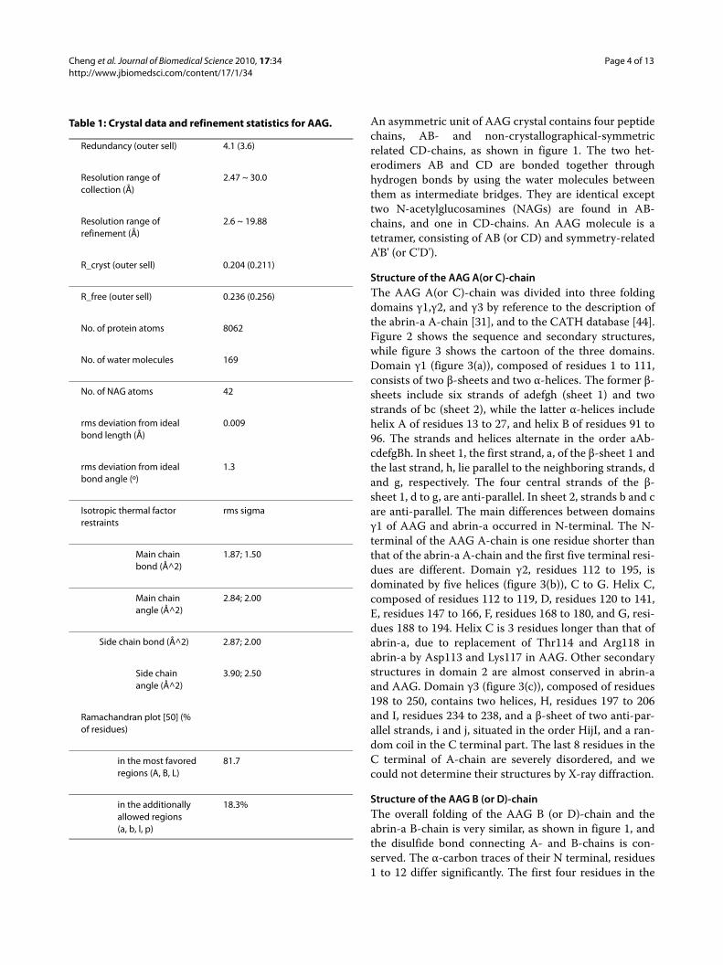
Each domain of δ1 and δ2 contains two intra-domaindisulfide bonds (Cys25-Cys44, Cys68-Cys85, Cys156-169,and Cys195-Cys212), which are conserved in abrin-a.Two NAGs are found in B-chain, but only one presents inD chain. The NAGs are bound to B-Asn100 (figure. 6), B-Asn140, and D-Asn140 respectively. The bond lengthbetween NAG and Asn140 is 1.45 Å.
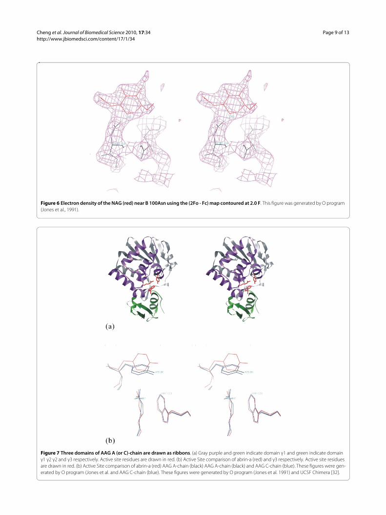
Structure of Active siteThe active site is exactly the cleft formed by the intersec-tion of all 3 domains in AAG A (or C)-chain. The locationof the active site region of the AAG A (or C)-chain isshown in figure 7(a), and enlarged in figure 7(b). Fiveinvariant residues (Tyr73, Tyr112, Glu163, Arg166 and
Trp197) and five conserved residues (Asn71, Arg123,Gln159, Glu194 and Asn195) are located in the active sitecleft. The alignment of the amino acid sequences showsthat all five invariant residues in the active site of abrin-aare absolutely conserved throughout the wide range ofribosome-inactivating proteins [19,45]. The similarity ofactive site structures between abrin-a and AAG shows infigure 7(b) that they may work in the same way, but couldnot explain the less than half biochemical activity ofAAG. We try to answer this question by the 28SRNAdocking study.
Quaternary Structure of AAGAn AAG molecule is a hetero-tetramer (as shown in fig-ure 8) contains two subunits, ABA'B' (or CDC'D'), stabi-lized by mainly hydrophilic and little hydrophobic forces.The two subunits are in equivalent positions of the spacegroup P41212. The transformation from AB to A'B' is (x, y,z) to (1-y, 1-x, 0.5-z), while CD to C'D' is (x, y, z) to (y, x,1-z). The hydrophilic interaction is dominated by inter-subunit hydrogen bonds, as listed in table 2. These hydro-gen bonds belong to residues of domains γ2 and γ2'. Sincethe γ2 domain is almost made up with α-helices, whichhydrophobic side-chains are buried inside, hydrophobicforces contribute little to the stabilization of quaternarystructure of AAG. The total buried surface area is 9360for ABA'B' and 9460 for CDC'D' interfaces. The gain inhydrophobic energy is -68 KCal/Mol for ABA'B' and -72KCal/Mol for CDC'D'. The buried surface and hydropho-bic energy are calculated by Protein interfaces, surfaces
Figure 4 AAG B (or D)-chain sequence & secondary structures. The symbol of "arrow" represents a β-strand, "spiral" represents an α-helix, "dot" represents missing residues, and the alphabets a, b, A, etc, denote the corresponding secondary structures in figure 5.

Cheng et al. Journal of Biomedical Science 2010, 17:34http://www.jbiomedsci.com/content/17/1/34
Page 8 of 13
Figure 5 Two domains of AAG B (or D)-chain: (a) domain δ1, and (b) domain δ2. These figures were generated by O program (Jones et al., 1991).
Cheng et al. Journal of Biomedical Science 2010, 17:34http://www.jbiomedsci.com/content/17/1/34
Page 9 of 13
Figure 6 Electron density of the NAG (red) near B 100Asn using the (2Fo - Fc) map contoured at 2.0 F. This figure was generated by O program (Jones et al., 1991).
Figure 7 Three domains of AAG A (or C)-chain are drawn as ribbons. (a) Gray purple and green indicate domain γ1 and green indicate domain γ1 γ2 γ2 and γ3 respectively. Active site residues are drawn in red. (b) Active Site comparison of abrin-a (red) and γ3 respectively. Active site residues are drawn in red. (b) Active Site comparison of abrin-a (red) AAG A-chain (black) AAG A-chain (black) and AAG C-chain (blue). These figures were gen-erated by O program (Jones et al. and AAG C-chain (blue). These figures were generated by O program (Jones et al. 1991) and UCSF Chimera [32].

Cheng et al. Journal of Biomedical Science 2010, 17:34http://www.jbiomedsci.com/content/17/1/34
Page 10 of 13
Figure 8 Ribbon presentation of AAG quaternary structure: Red residues indicate the active site location. Purple and green residues consti-tute inter-subunit hydrogen bonds. Domain γ2s are drawn in brown. This figure was generated by O program (Jones et al., 1991).
Table 2: Hydrogen bonds between inter-subunit with symmetry-related AA' and CC' chains.
Donor Acceptor D....A (A) Donor Acceptor D....A (A)
A Gln 121 NE2 A'Gln121 OE1 2.85 A Gln121 NE2 A'Gln121 OE1 2.85
A Arg 25 NH1 A'Glu148OE2 2.77 A Arg125NH1 A'Glu148OE2 2.77
A Leu 130 N A'Glu131OE2 3.15 A Leu 130 N A'Glu131OE2 3.15
A Arg 134 NH2 A'Asn 180 O 2.86 A Arg 134 NH2 A'Asn 180 O 2.86
A Arg 134 NE A'Asn 181 O 3.06 A Arg 134 NE A'Asn 181 O 3.06
A Gln 135 NE2 A'Ser 127 OG 3.07 A Gln 135 NE2 A'Ser 127 OG 3.07
C Gln 121 NE2 C'Ser 145 O 2.75 C Gln 121 NE2 C'Ser 145 O 2.75
C Ser 127 OG C'Gly 143 N 2.95 C Ser 127 OG C'Gly 143 N 2.95
C Arg 134 NR C'Tyr57 OH 2.64 C' Arg 134 NR C Tyr57 OH 2.64
C Gln 135 NE2 C'Gln135 O 2.94 C' Gln 135 NE2 C Gln135 O 2.94
C Ser 145 N C'Gln121OE1 2.59 C' Ser 145 N C Gln121 O 2.59
Cheng et al. Journal of Biomedical Science 2010, 17:34http://www.jbiomedsci.com/content/17/1/34
Page 11 of 13
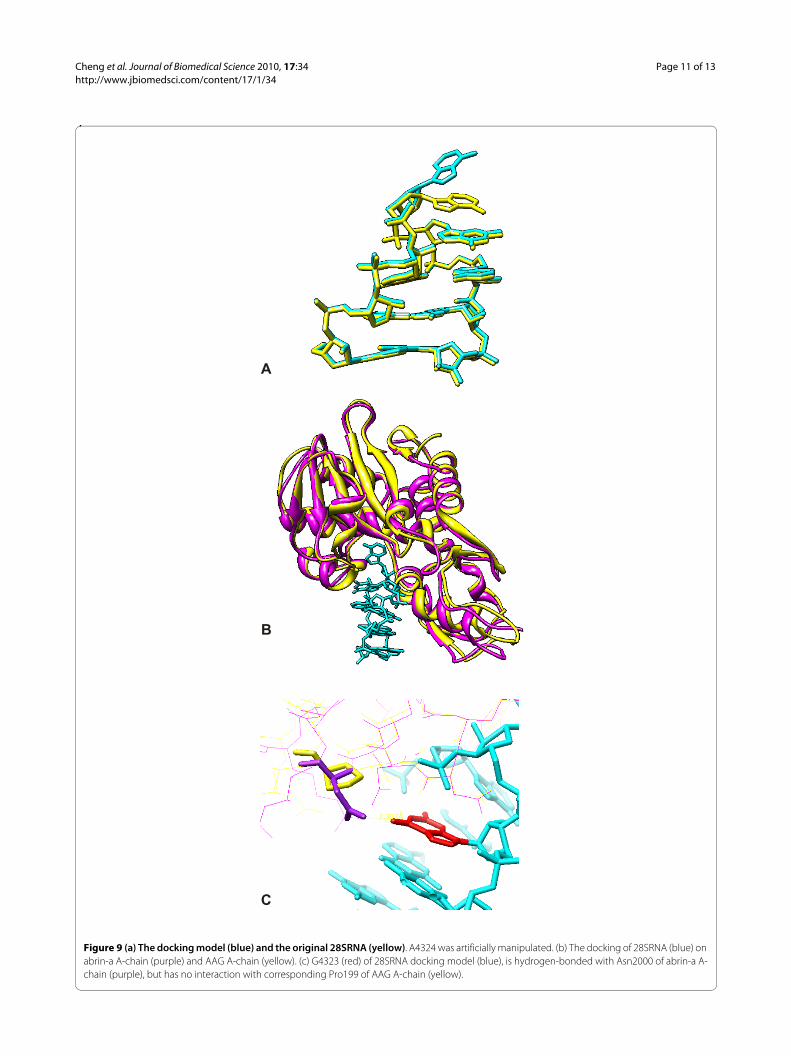
Figure 9 (a) The docking model (blue) and the original 28SRNA (yellow). A4324 was artificially manipulated. (b) The docking of 28SRNA (blue) on abrin-a A-chain (purple) and AAG A-chain (yellow). (c) G4323 (red) of 28SRNA docking model (blue), is hydrogen-bonded with Asn2000 of abrin-a A-chain (purple), but has no interaction with corresponding Pro199 of AAG A-chain (yellow).
A
B
C
Cheng et al. Journal of Biomedical Science 2010, 17:34http://www.jbiomedsci.com/content/17/1/34
Page 12 of 13
and assemblies service PISA at European BioinformaticsInstitute http://www.ebi.ac.uk/msd-srv/prot_int/pistart.html[46].
Docking of 28SRNA to AAG and Abrin-aAs pointed out in the mutagenesis study [21], Pro199 inA- (or C-) chain of AAG impair the activity of proteinsynthesis inhibition. Bagaria et al., [25] suggested that theless toxic nature is because of the fewer interactionsinvolved with the substrate adenine. From our dockingstudy, we found Asn200 of abrin-a may form a criticalhydrogen bond with G4323 of 28SRNA, while corre-sponding Pro199 of agglutinin is a non-extended residuebound with the cleft in a more compact complementaryrelationship as shown in figure 9(c). This may explain thelower toxicity of agglutinin, despite of similarity in sec-ondary structure and the activity cleft of two RIPs. Thedocking model of 28SRNA was artificially deformed atthe ribose sugar of A4324 from the X-ray crystal struc-ture 430D [47], as shown in figure 9(a), so that A4324 of28SRNA can overlap with the adenine of the ricin-ade-nine complex, 1IFS [48]. Figure 9(b) shows deformedmodel fit well in the active clefts of both abrin-a andAAG. Figures 9(a), 9(b) and 9(c) were generated withUCSF Chimera [49].
We want to explore whether the difference in the criti-cal residue, bring any change in structure. However, wecould not observe significant main-chain distortion dueto the difference in the 200 residue on higher resolutionstructure. Hence, the reason for the lower toxicity ofagglutinin than abrin-a might be due to the deformationfrom inactive to active state of abrin depends on themeta-stable huge helix, composed of helix h and helix g.The mutation of Asn200 to Pro200 destroys the mecha-nism.
ConclusionWe have successfully solved the structure of 120 kDa het-erotetramer agglutinin AB and CD chains, and reducedthe R-factor to 20.4% at 2.6 Å resolution data. Ten disul-fide bonds, three N-acetylglucosamines, and 169 watermolecules were found in the successive (2Fo-Fc) map. 22hydrogen bonds between A (or C)-chain and symmetry-related A' (or C') were found. Water molecules were notfound in the Bagaria's paper and no subsequent hydrogenbond lengths were listed based on their lower resolutionstructure [25]. Docking study revealed that due toPro199, agglutinin is unable to form a critical hydrogenbond with G4323 of 28SRNA, which is found in the dock-ing result of abrin-a. This may explain the lower toxicityof agglutinin than abrin-a, despite of similarity in second-ary structure and the activity cleft of two RIPs.
AbbreviationsRIP: ribosome-inactivating protein; AAG: agglutinin; NAG: N-acetylglu-cosamine; Fo: observed structure factor; Fc: calculated structure factor; PDB:protein data bank.
Competing interestsThe authors declare that they have no competing interests.
Authors' contributionsJC collected the X-ray diffraction data, analyzed the crystal structure and pre-pared the initial manuscript. LTH set up the laboratory of the crystal structuredetermination, screened the crystallization conditions and got the right one,and advised such studies. LJY and LCL purified the material of the agglutinin.
AcknowledgementsThe authors thank Mr Shyh-Ming Chen for setting up the computing pro-grams. They also thank to the National Science Council of Taiwan for financial support. They are indebted to SPring-8 and the National Synchrotron Radiation Research Center for data collection.
Author Details1Department of Physics, National Tsing Hua University, Hsinchu 30013, Taiwan, 2Graduate School of Biochemical Engineering, Ming Chi University of Technology, Taishan, Taipei, 24301, Taiwan and 3Institute of Biochemistry, College of Medicine, National Taiwan University, Taipei 10018, Taiwan
References1. Endo Y, Misui K, Motizuki K, Tsurugi K: The mechanism of action of ricin
and related toxic lectins on eukaryotic ribosomes. The site and the characteristics of the modification in 28 S ribosomal RNA caused by the toxins. J Biol Chem 1987, 262:5908-5912.
2. Endo Y, Tsurugi K: RNA N-glycosidase activity of ricin A-chain Mechanism of action of the toxic lectin ricin on eukaryotic ribosomes. J Biol Chem 1987, 262:8128-8130.
3. Endo Y, Gluck A, Wool IG: Ribosomal RNA identity elements for ricin A-chain recognition and catalysis. J Mol Biol 1991, 221:193-207.
4. Jimenez A, Vazquez DC: Plant and Fungal Protein and Glycoprotein Toxins Inhibiting Eukaryote Protein Synthesis. Annu Rev Microbiol 1985, 39:649-672.
5. Barbieri L, Stirpe F: Ribosome-inactivating proteins from plants: properties and possible uses. Cancer Surv 1982, 1:489-520.
6. Stirpe F, Barbieri L, Battelli MG, Soria M, Lappi DA: Ribosome-inactivating proteins leads to increased fungal protection in transgenic tobacco plants. Bio-Technology 1992, 10:405-412.
7. Hartley MR, Lord JM: Cytotoxic ribosome-inactivating lectins from plants. Biochim Biophys Acta 2004, 1701:1-14.
8. Olsnes S: The history of ricin abrin and related toxins. Toxicon 2004, 44:361-370.
9. Lin J, Chen Y, Xu Y, Yan F, Tang L, Chen F: Cloning and expression of curcin a ribosome inactivating protein from the seeds of jatropha curcas. Acta Botanica Sinica 2003, 45:858-863.
10. Mi SL, An CC, Wang Y, Chen JY, Che NY, Gao Y, Chen ZL: Trichomislin a novel ribosome-inactivating protein a novel ribosome-inactivating protein induces apoptosis that involves mitochondria and caspase-3. Archives of Biochemistry and Biophysics 2005, 434:258-265.
11. Olsnes S, Phil A: Different biological properties of the two constituent peptide chains of ricin a toxic protein inhibiting protein synthesis. Biochemistry 1973, 12:3121-3126.
12. Lin JY, Lee TC, Hsu ST, Tung TC: Isolation of four isotoxic proteins and one agglutinin from jequiriti bean (Abrus precatorius). Toxicon 1981, 19:41-51.
13. Lord JM: Synthesis and intracellular transport of lectin and storage protein precursors in endosperm from castor bean. Eur J Biochem 1985, 146:403-409.
14. Mundy J, Leah R, Boston R, Endo Y, Stirpe F: Genes encoding ribosome-inactivating proteins. Plant Mol Biol Rep 1994, 12:60-62.
Received: 4 January 2010 Accepted: 30 April 2010 Published: 30 April 2010This article is available from: http://www.jbiomedsci.com/content/17/1/34© 2010 Cheng et al; licensee BioMed Central Ltd. This is an Open Access article distributed under the terms of the Creative Commons Attribution License (http://creativecommons.org/licenses/by/2.0), which permits unrestricted use, distribution, and reproduction in any medium, provided the original work is properly cited.Journal of Biomedical Science 2010, 17:34
Cheng et al. Journal of Biomedical Science 2010, 17:34http://www.jbiomedsci.com/content/17/1/34
Page 13 of 13
15. Leah R, Tommerup H, Svendsen I, Mundy J: Biochemical and molecular characterization of three barley seed proteins with antifungal properties. J Biol Chem 1991, 266:1564-1573.
16. Desvoyes B, Poyet JL, Schlick JL, Adami P, Jouvenot M, Dulieu P: Identification of a biological inactive complex form of pokeweed antiviral protein. FEBS Lett 1997, 410:303-308.
17. Olsnes S, Phil A: In Receptors and Recognition Series: The Specificity and Action of Animal. In Bacterial and Plant Toxins Edited by: Cuatrecasas P. Chapman and Hall, London; 1982:31-131.
18. Lin JY, Li JS, Tung TC: Lectin Derivatives of Methotrexate and Chlorambucil as Chemotherapeutic Agents. J Natl Cancer Inst 1981, 66:523-528.
19. Funatsu G, Taguchi Y, Kamenosno M, Yanaka M: The complete amino acid sequence of the A-chain of abrin-a a toxic protein from the seeds of Abrus precatorius. Agric Biol Chem 1988, 52:1095-1097.
20. Chen YL, Chow LP, Tsugita A, Lin JY: The complete primary structure of abrin-a B chain. FEBS Lett 1992, 309:115-118.
21. Liu CL, Tsai CC, Lin SC, Wang LI, Hsu CI, Hwang MJ, Lin JY: Primary Structure and Function Analysis of the Abrus precatorius Agglutinin A Chain by Site-directed Mutagenesis. J Biol Chem 2000, 275:1897-1901.
22. Hung CH, Lee MC, Lee TC, Lin JY: Primary Structure of Three Distinct Isoabrins Determined by cDNA Sequencing: Conservation and Significance. J Mol Biol 1993, 229:263-267.
23. Hung CH, Lee MC, Chen JK, Lin JY: Cloning and expression of three abrin A-chains and their mutants derived by site-specific mutagenesisin Escherichia coli. Eur J Biochem 1994, 219:83-87.
24. Chen JK, Hung CH, Liaw YC, Lin JY: Identification of amino acid residues of abrin-a A chain is essential for catalysis and reassociation with abrin-a B chain by site-directed mutagenesis. Protein Engineering 1997, 10:827-833.
25. Bagaria A, Surendranath K, Ramagopal UA, Ramakumar S, Karande AA: Structure-Function Analysis and Insights into the Reduced Toxicity of Abrus precatorius Agglutinin I in Relation to Abrin. J Biol Chem 2006, 281:34465-34474.
26. Panneerselvam K, Lin SC, Liu CL, Liaw YC, Lin JY, Lu TH: Crystallization of agglutinin from the seeds of Abrus precatorius. Acta Cryst 2000, D56:898-899.
27. Smith PK, Krohn RI, Hermanson GT, Mallia AK, Gartner FH, Provenzano MD, Fujimoto EK, Goeke NM, Olson BJ, Klenk DC: Measurement of protein using bicinchoninic acid. Anal Biochem 1985, 150:76-85.
28. McPherson A: Preparation and Analysis of Protein Crystals. John Wiley & Sons John Wiley & Sons New York USA; 1982:94-96. 115
29. Otwinowski Z, Minor W: Processing of X-ray Diffraction Data Collected in Oscillation Mode. Methods in Enzymology. Macromolecular Crystallography part A 1997, 276:307-326.
30. Brunger AT, Adams PD, Clore GM, Delano WL, Gros P, Grosse-Kunstleve RW, Jiang JS, Kuszewski J, Nilges M, Pannu NS, Read RJ, Rice LM, Simonson T, Warren GL: Crystallography & NMR system: A new software system for macromolecular structure determination. Acta Cryst 1998, D54:905-921.
31. Tahirov TH, Lu TH, Liaw YC, Chen YL, Lin JY: Crystal Structure of Abrin-a at 2.14 D. J Mol Biol 1995, 250:354-367.
32. DeLano WL, Brunger AT: The Direct Rotation Function: Rotational Patterson Correlation Search Applied to Molecular Replacement. Acta Cryst 1995, D51:740-748.
33. Brunger AT: Extension of molecular replacement: A new search strategy based on Patterson correlation refinement. Acta Cryst 1990, A46:46-57.
34. Brunger AT: The Free R Value: a Novel Statistical Quantity for Assessing the Accuracy of Crystal Structures. Nature 1992, 355:472-474.
35. Brunger AT, Krukowski A, Erickson J: Slow-Cooling Protocols for Crystallographic Refinement by Simulated Annealing. Acta Cryst 1990, A46:585-593.
36. Abrahams JP, Leslie AG W: Methods used in the structure determination of bovine mitochondrial F1 ATPase. Acta Cryst 1996, D52:30-42.
37. Jones TA, Zou JY, Cowan SW, Kjeldgaard M: Improved methods for the building of protein models in electron density maps and the location of errors in these models. Acta Cryst 1991, A47:110-119.
38. Adams PD, Pannu NS, Read RJ, Brunger AT: Cross-validated Maximum Likelihood Enhances Crystallographic Simulated Annealing Refinement. Proc Natl Acad Sci USA 1997, 94:5018-5023.
39. Kuntz ID, Blaney JM, Oatley SJ, Langridge R, Ferrin TE: A geometric approach to macromolecule-ligand interactions. J Mol Biol 1982, 161:269-288.
40. Shoichet BK, Bodian DL, Kuntz ID: Molecular docking using shape descriptors. J Comp Chem 1992, 13:380-397.
41. Meng EC, Shoichet BK, Kuntz ID: Automated docking with grid-based energy evaluation. J Comp Chem 1992, 13:505-524.
42. Meng EC, Gschwend DA, Blaney JM, Kuntz ID: Orientational sampling and rigid-body minimization in molecular docking. Proteins 1993, 17:266-278.
43. Shoichet BK, Kuntz ID: Protein docking and complementarity. J Mol Biol 1991, 221:327-346.
44. Pearl F, Todd A, Sillitoe I, Dibley M, Redfern O, Lewis T, Bennett C, Marsden R, Grant A, Lee D, Akpor A, Maibaum M, Harrison A, Dallman T, Reeves G, Diboun I, Addou S, Lise S, Johnston C, Sillero A, Thornton J, Orengo C: The CATH Domain Structure Database and related resources Gene3D and DHS provide comprehensive domain family information for genome analysis. Nucl Acids Res 2005:D247-D251.
45. Rutenber E, Robertus JD: Structure of ricin B-chain at 2.5 Å resolution. Proteins Struct Func Genet 1991, 10:260-269.
46. Krissinel E, Henrick K: Inference of macromolecular assemblies from crystalline state. J Mol Biol 2007, 372:774-797.
47. Correll CC, Munishkin A, Chan YL, Ren Z, Wool IG, Steitz TA: Crystal structure of the ribosomal RNA domain essential for binding elongation factors. Proc Natl Acad Sci USA 1998, 95:13436-13441.
48. Weston SA, Tucker AD, Thatcher DR, Derbyshire DJ, Pauptit RA: X-ray structure of recombinant ricin A-chain at 1.8 Å resolution. J Mol Biol 1994, 244:410-422.
49. Pettersen EF, Goddard TD, Huang CC, Couch GS, Greenblatt DM, Meng EC, Ferrin TE: UCSF Chimera - A Visualization System for Exploratory Research and Analysis. J Comput Chem 2004, 25:1605-1612.
50. Laskowski RA, MacArthur MW, Moss DS, Thornton JM: PROCHECK: a program to check the stereochemical quality of protein structures. J App Cryst 1993, 26:283-291.
doi: 10.1186/1423-0127-17-34Cite this article as: Cheng et al., A biophysical elucidation for less toxicity of Agglutinin than Abrin-a from the Seeds of Abrus Precatorius in consequence of crystal structure Journal of Biomedical Science 2010, 17:34