Embed Size (px)
Citation preview
ISOLATION AND CHARACTERIZATION OF AGGLUTININ
AND RICIN FROM RICINUS COMMUNIS
THESIS SUBMITTED TO
NATIONAL INSTITUTE OF TECHNOLOGY, ROURKELA
FOR THE PARTIAL FULFILMENT
OF THE MASTER OF SCIENCE DEGREE IN LIFE SCIENCE
Submitted by
NITU MAJHI
ROLL NO – 410LS2056
Under the guidance of
Dr. SUJIT KUMAR BHUTIA
ASSISTANT PROFESSOR
DEPARTMENT OF LIFE SCIENCE
NATIONAL INSTITUTE OF TECHNOLOGY
ROURKELA-769008, ODISHA
2011-2012
DEPARTMENT OF LIFE SCIENCE
NATIONAL INSTITUTE OF TECHNOLOGY,
ROURKELA-769008
Dr. Sujit Kumar Bhutia Ref. No.
Assistant Professor. Date: ............................
CERTIFICATE
This is to certify that the thesis entitled “Isolation and Characterization of
agglutinin and ricin from Ricinus communis” which is being submitted by
Miss.Nitu Majhi, Roll No. 410LS2056, for the award of the degree of Master
of Science from National Institute of Technology, Rourkela, is a record of
bonafide research work, carried out by her under my supervision. The results
embodied in this thesis are new and have not been submitted to any other
university or institution for the award of any degree or diploma.
Dr. Sujit KumarBhutia
Assistant Professor
Department of Life Science
National Institute of Technology
Rourkela – 769008, Odisha, India.
Phone no: 91-661-2462686
Email:[email protected]

ACKNOWLEDGEMENT
This project is by far the most significant accomplishment in my life and it would not
have been impossible without people who supported me and believed in my calibre.
I would like to extend my gratitude and sincere thanks to my honourable supervisor
Dr. Sujit Kumar Bhutia, Assistant Professor, Department of Life Science. He is not only a
great lecturer with deep vision but also most importantly a kind person. I sincerely thank for
his exemplary guidance and encouragement. His trust and support inspired me in the most
important moments of making right decisions and I am glad to work under his supervision.
I express my sincere thanks to Head of the Dept. Dr. S.K Patra, and all other faculties of
Department of Life Science , NIT Rourkela for showing sustained interest and providing help
throughout the period of my work.
I express my heartfelt thanks to PhD scholars for their active cooperation and sincere help.
I am genuinely appreciative of all my batch mates for their suggestions and moral support
during my work.
Last, but not the least, I would thank the Almighty and my parents, whose dedicated and
untiring efforts towards me has brought me at this stage of my life.
Nitu Majhi
410LS2056
DECLARATION
I do hereby declare that the Project Work entitled “Isolation and characterization of
agglutinin and ricin from Ricinus communis”, submitted to the Department of Life
Science, National Institute of Technology, Rourkela is a faithful record of bonafide
and original research work carried out by me under the guidance and supervision
of Dr. Sujit Kumar Bhutia, Asst. Professor, Department of Life Science, National Institute
of Technology, Rourkela, Odisha.
Date:
Place: Nitu Majhi
Contents
1. Abstract
2. Introduction...................................................1
3. Review of literature......................................3
4. Materials and methods...................................8
5. Result.............................................................15
6. Discussion......................................................19
7. Conclusion.....................................................20
8. References.....................................................21
Abstract
Lectins exhibit a promising diagonistic and therapeutic approach for treating cancer . Lectins
found, almost in all organisms. On the surface of the cell lectins are attached with their sugar
binding site. Lectins are found to be toxic, as it is poisonous, it results in the death of cells
both in vivo and in vitro. Leguminous plant serve as the main source of lectin, most of the
lectins are derived from seed, present in cytoplasm of the seed. Ricin is the toxic lectin
which is derieved from the castor bean plant Ricinus communis belong to the family of
Euphorbiaceae. The toxicity of ricin is bound with their two chain A and B chain. The A and
B chains of ricin, more toxic to mammalian cells. The study of this project encompasses the
purification of ricin through affinity chromatography, characterization of ricin through
haemagglutination assay and SDS-PAGE. B chain of ricin bind to the sugar residue on the
cell surface of red blood cell .The cytotoxicity on cancer cells is yet to be explored in order to
showcase its anti-cancer property.
1
INTRODUCTION
Lectin is a sugar binding protein, found almost in all organisms mainly in plants; it may be in
the solubilised form or membrane-bound widely distributed in nature. The binding specificity
of lectins allows them to serve as recognition molecules within a cell, between the cells and
between the organisms. The name "lectin" is derived from the Latin word legere, meaning,
"to select" and derived the term lectin (Kocoureket et al., 1983). Although lectins occur in
living organisms, plant lectins were the first proteins that can be studied. Plant lectins are
found in root, stem and leaves and it is also present in seed cotyledon especially in cytoplasm.
Lectins comprise up to 3% of the weight of a mature seed. Lectins are carbohydrate-binding
proteins of non-immune origin that agglutinate cells and glycoconjugates and are capable of
specific recognition and reversible binding to carbohydrate and sugar containing substances,
without altering covalent structure of any glycosyl ligands (Goldstein et al., 1980).
Lectin protein binds carbohydrate with high specificity and they have special binding sites for
monosaccharide and oligosaccharides. Lectin obtained particularly from the seeds of
leguminous plant. Some plant lectins are classified as toxins, and they are the most poisonous
proteins on our planet it can result in death of the cells in vivo and in vitro.
Lectins vary in their composition, molecular weight, subunit structure and the number of
sugar binding sites per molecule. They are used as histology and blood transfusion reagents
but lectins may be toxic, inflammatory, resistant to digestive enzymes, and are found in much
of our foods proteins. Lectins agglutinate animal cells and precipitated in glycoconjugates.
Plant lectins are relatively soluble and it can be easily extracted. The extraction procedures
are laborious for the lectins obtain from stems and barks. Lectins can be purified to
homogeneity on appropriate immobilized carbohydrate matrices. The lectin activity is usually
measured by an agglutination assay that uses red blood cell. Other biological assays are also
performed for lectin activity.
2
Types of lectin: Four types of mature lectins are derived according to their structure,
a)Merolectin, b)Hololectins, c) Chimerolectins, d) Superlectins
Based on the sequence of amino acids, plant lectins are sub-divided into 3 different types.
These include:
a) Legume lectins,
b) Monocot mannose-binding lectins,
c) Chitin-binding lectins,
Many seeds contain a remarkable amount of lectin. For example, soyabean, castor seed,
peanut, abrus agglutinin constitutes 10–15% of the total protein content of the seed. However,
not all the seeds contain lectins. For example, “tomato” the tomato lectin occurs in a soluble
form in the locular fluid within the tomato fruit and not in tomato seeds. leguminous plant
are the main source of lectins isolated and characterized (Van Damme et al., 1998).
Leguminaceae the family of leguminous plant, within this family the greater number of the
lectins with their three-dimensional structures have been observed (Mourey et al.1998).
Lectin according to their small carbohydrate binding recognize as two type of binding
mannose-binding lectins, galactose-binding lectins or GlcNAc-binding lectins.
3
REVIEW OF LITERATURE
Many plants contain „lectins‟ or agglutinins in their seed. The first lectin was discoverd by
Stillmark in 1888. He found a proteinaceous hemagglutinating factor in castor beans (ricinus
communis). As several hundred lectins or hemaagglutinins have been isolated and studied at
biochemical or physiochemical level.
Lectins defined as proteins which interact non-covalently with carbohydrate moieties,
showing high affinity and specificity for their ligands. Large numbers of lectins have been
isolated at present with established biochemical characteristics. Recently by x-ray diffraction
number of three-dimensional structures of plant lectins is observed (Bourne et al., 1990).
Lectins are carbohydrate-binding proteins of non-immune origin that agglutinate cells and
glycoconjugates, capable of specific recognition and reversible binding to carbohydrate and
sugar containing substances, without altering covalent structure of any glycosyl ligands
(Goldstein et al.1980)
In glycoconjugates research, plant lectins have been use as analytical tool, these applications
is extended and refined by understanding structure and specificity functional relationship of
different groups.
A lectin researcher, Irwin J. Goldstein introduced affinity chromatography for the isolation
of lectin and this was published in the Biochemical Journal. He described the purification
process of concanavalin A by affinity chromatography. For the isolation of specific lectin
sephadex (a polymer of dextran) is used (Moreira et al., 1983).
Mostly all lectins isolated and characterized from Leguminoseae ,serve as a main source,
(Van Damme et al., 1998). Large number of the lectins with their three-dimensional
structures have been described within this family (Mourey et al., 1998). Different
monosaccharide binding specificities is exhibited by these proteins, belonging to the same
family with their common biochemical features (Debray et al., 1981).
4
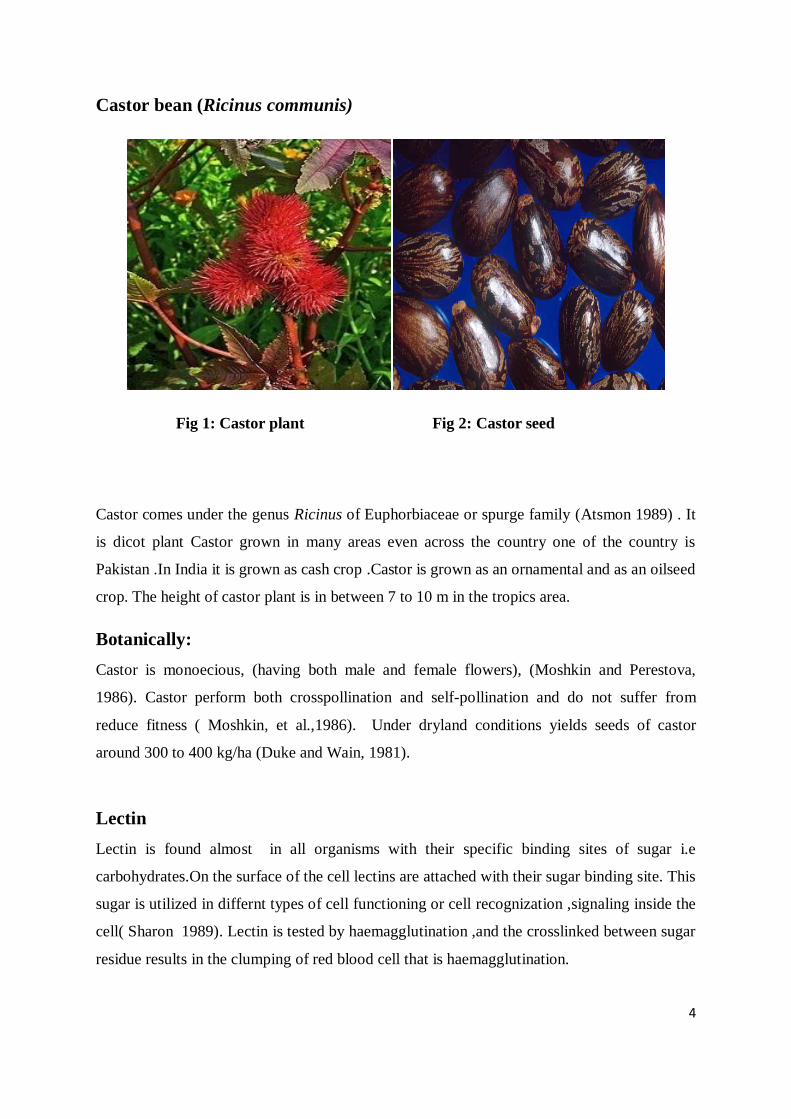
Castor bean (Ricinus communis)
Fig 1: Castor plant Fig 2: Castor seed
Castor comes under the genus Ricinus of Euphorbiaceae or spurge family (Atsmon 1989) . It
is dicot plant Castor grown in many areas even across the country one of the country is
Pakistan .In India it is grown as cash crop .Castor is grown as an ornamental and as an oilseed
crop. The height of castor plant is in between 7 to 10 m in the tropics area.
Botanically:
Castor is monoecious, (having both male and female flowers), (Moshkin and Perestova,
1986). Castor perform both crosspollination and self-pollination and do not suffer from
reduce fitness ( Moshkin, et al.,1986). Under dryland conditions yields seeds of castor
around 300 to 400 kg/ha (Duke and Wain, 1981).
Lectin
Lectin is found almost in all organisms with their specific binding sites of sugar i.e
carbohydrates.On the surface of the cell lectins are attached with their sugar binding site. This
sugar is utilized in differnt types of cell functioning or cell recognization ,signaling inside the
cell( Sharon 1989). Lectin is tested by haemagglutination ,and the crosslinked between sugar
residue results in the clumping of red blood cell that is haemagglutination.
5
Classification of plant lectin:
Plant lectins classified into small numbers of families:
Seven lectin families were distinguished:
i. Legume lectin.
ii.Mmonocot mannose-binding lectin.
iii.Chitin-binding protein containing domain.
iv.The type 2 ribosome inactivating protein.
v. Cucurbitaceae phloem lectins.
vi.Jacalin family.
vii.Amaranthaceae lectins.
Toxin from Castor Bean Plant, Ricinus communis the most natural poisonous substance is
ricin they are poisonous to animal, plants, etc. Stillmark in 1888 tested the beans extract on
red blood cell and found their agglutination activity, from there it came to be known that
agglutination was due to activity of another protein i.e., RCA (Ricinus communis
agglutinin). Ricin is catagorized under cytotoxin and weak hemagglutinin whereas RCA
cartagorised as weak cytotoxin and powerful hemagglutinin.lectins can be isolated (Nicolsan
et al .,1974).
Ricin:
Ricin is the protein component of Ricinus communis .Castor seeds contain two proteins which
are highly toxic (Lord et al., 1994). R. communis agglutinin (RCA) and ricin having 102 kD
and 65 kDa each. Ricin is a cytotoxic lectin. Toxic to the mammalian cells. In the late
nineteenth century H. Stillmark, in 1888 firstly introduced these two proteins in castor
seeds. Two polypeptides are found from ricin they are A chain and B chain. After that
agglutinin protein has found with their four polypeptides, and they are linked by disulfide
bonds (Butterworth and Lord, 1983). Toxicity is measured by agglutinate red blood cell.
6
Ricin A Chain and B Chain: four polypeptides chains are similar to the two chains of ricin.
A chain is similar with the two chain of agglutinin and other two chains of agglutin are also
similar to the B chain of ricin. The A and B chains of ricin, more toxic to mammalian cells,
while the R. Communis agglutinin shows low cytoxicity to the cell.
Ribosome-inactivating protein is the part of A chain (Lord et al., 1994). Having its
molecular weight 32 kDa . protein synthesis is inhibited by alteration of the ribosomal RNA
subunits that involved in the translation. 28S ribosomal subunits is binded with the A chain
causing in its structural change, without the help of B chain A chain is not able to enter into
the cell. A chain and B chain both are heterodimeric toxins. On the cell surface terminating in
galactose or N-acetylgalactosamine Ricin B chain completly binds with the glycoprotein and
glycolipids (Lord et al., 1994). The galactose/N-acetylgalactosaminebinding activity is
initiated by four disulphide bond of B chain. Bilobed structure is arises when bond breaks
between N-terminal and C-terminal then two disulfide bonds aligned , this bilobed structure
introduced galactose-binding sites halves of the B chain, contain This bilobal structure allows
for two galactose binding sites. Endocytosis is initiated by the binding of mannose residue to
the mannose receptor (Montfort et al., 1987).
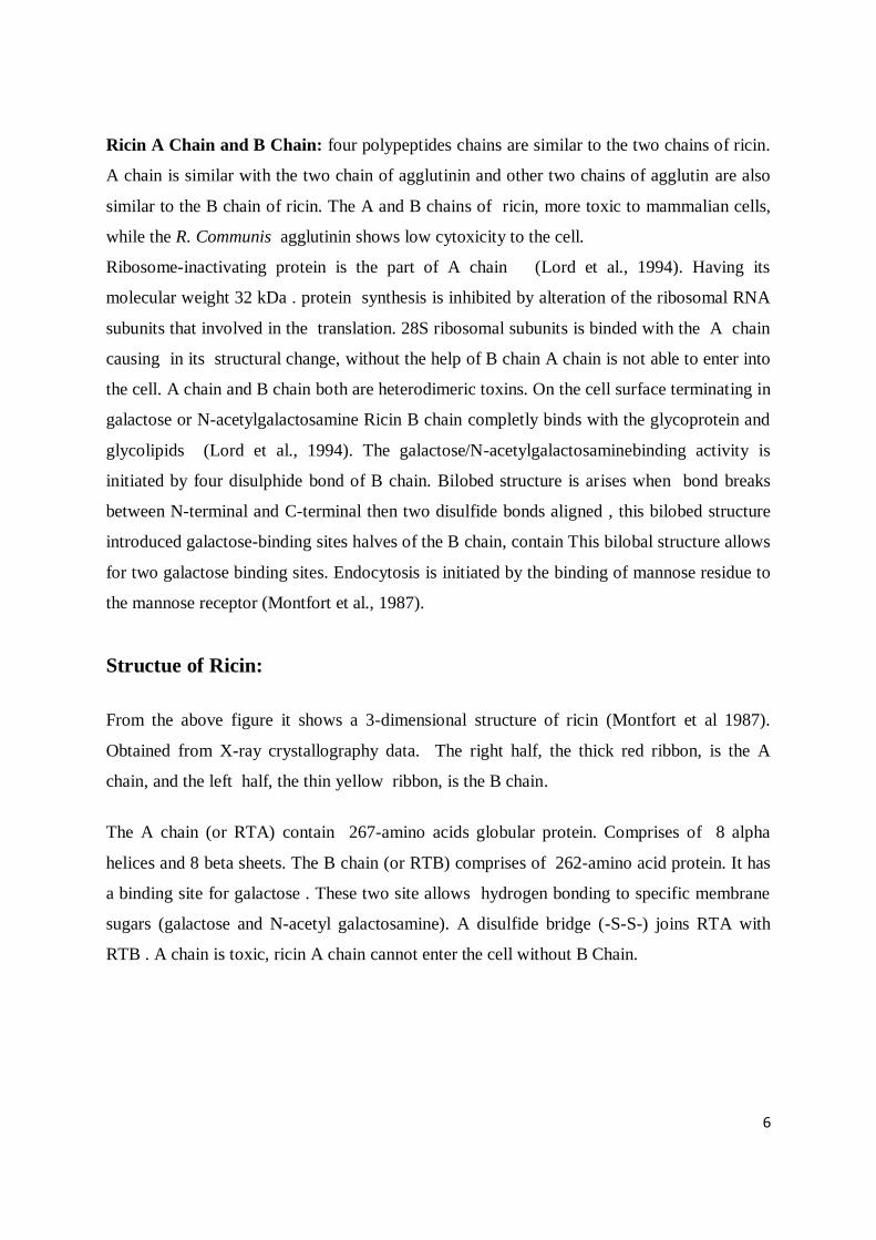
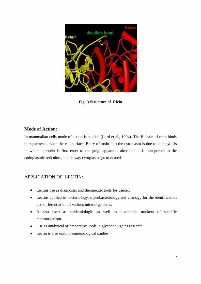
Structue of Ricin:
From the above figure it shows a 3-dimensional structure of ricin (Montfort et al 1987).
Obtained from X-ray crystallography data. The right half, the thick red ribbon, is the A
chain, and the left half, the thin yellow ribbon, is the B chain.
The A chain (or RTA) contain 267-amino acids globular protein. Comprises of 8 alpha
helices and 8 beta sheets. The B chain (or RTB) comprises of 262-amino acid protein. It has
a binding site for galactose . These two site allows hydrogen bonding to specific membrane
sugars (galactose and N-acetyl galactosamine). A disulfide bridge (-S-S-) joins RTA with
RTB . A chain is toxic, ricin A chain cannot enter the cell without B Chain.
7
Fig: 3 Structure of Ricin
Mode of Action:
In mammalian cells mode of action is studied (Lord et al., 1994). The B chain of ricin binds
to sugar residues on the cell surface. Entry of toxin into the cytoplasm is due to endocytosis
in which protein is first enter to the golgi apparatus after that it is transported to the
endoplasmic reticulum, in this way cytoplasm got toxicated.
APPLICATION OF LECTIN:
Lectins use as diagnostic and therapeutic tools for cancer.
Lectins applied in bacteriology, mycobacteriology,and virology for the identification
and differentiation of various microorganisms.
It also used as epidemiologic as well as taxonomic markers of specific
microorganism.
Use as analytical or preparative tools in glycoconjugates research.
Lectin is also used in immunological studies.
8
MATERIALS AND METHODS :
CHEMICALS :
Sodium hydroxide (NaOH), Sodium carbonate (Na2CO3), glycine,Cuppersulphate( CuSO4) ,
Potassium sodium tartarate (KNaC4H4O6) were purchased from SRL, Sisco Research
laboratories Pvt. Ltd., Mumbai. Acrylamide, bisacrylamide, Ammonium persulphate (APS),
Sodium dodecyl sulphate (SDS), N,N,N‟,N
‟-tetramethylenediamine ( TEMED), Bovine
serum albumin( BSA), Tris were purchased from Sigma Aldrich, USA. Folin-Ciocalteau
phenol reagent, Potassium Dihydrogen Phosphate (KH2PO4), Potassium hydrogen phosphate
(K2HPO4) were purchased from S.D. fine chem. Ltd., Mumbai. Acetic acid, Bromophenol
blue, agarose were purchased from Himedia, Mumbai. Glycerol was purchased from Rankem
Pvt Ltd. Ethanol purchased from Trimurty Chemicals, India. Pre stained molecular weight
marker was purchased from Bio-Rad, India. Methanol, Silver nitrate, Sodium thiosulphate
were purchased from Nice chemicals Pvt.Ltd. India.
Sample Collection:
The Castor Seeds (Ricinus communis) were collected for isolation and purification of Lectins
from the Department of Biotechnology , Indian Institute of Technology, Kharagpur and 1 ml
of blood was collected from CWS Hospital , Rourkela.
Seed Coat Removal:
Castor seeds were taken and grinded in a mixer for removal of sead coats and 45gms of
uncoated seed were taken for the study. The uncoated seeds were deeped in PBS of 100 ml
for one day.Then the seeds are grinded with PBS and the pastes were collected in 50 ml
Centrifuge tubes and the weights were made equal by measuring the weights by the electronic
weight balance. Then the samples were centrifuged by the Eppendorf centifuge with
7500rpm, at 40 c for 20 mins. The supernatant were taken after centrifuge and measured by a
measuring cylinder. Some supernatant were stored in an eppendorf tube as crude at 40 C and
the remaining were taken as salting out process.
9
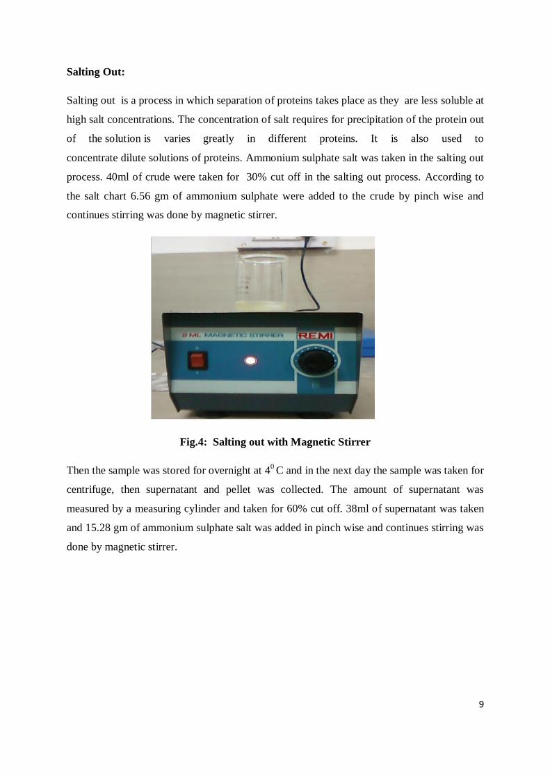
Salting Out:
Salting out is a process in which separation of proteins takes place as they are less soluble at
high salt concentrations. The concentration of salt requires for precipitation of the protein out
of the solution is varies greatly in different proteins. It is also used to
concentrate dilute solutions of proteins. Ammonium sulphate salt was taken in the salting out
process. 40ml of crude were taken for 30% cut off in the salting out process. According to
the salt chart 6.56 gm of ammonium sulphate were added to the crude by pinch wise and
continues stirring was done by magnetic stirrer.
Fig.4: Salting out with Magnetic Stirrer
Then the sample was stored for overnight at 40 C and in the next day the sample was taken for
centrifuge, then supernatant and pellet was collected. The amount of supernatant was
measured by a measuring cylinder and taken for 60% cut off. 38ml of supernatant was taken
and 15.28 gm of ammonium sulphate salt was added in pinch wise and continues stirring was
done by magnetic stirrer.
10
Preparation of Lactamyl Sepharose 4B affinity matrix
4gm of lactamyl Sepharose 4B matrix was washed with 6ml distill water and mixed with
2.6ml of 2N NaOH and 0.66ml epichlorohydrin were added so that the final
concentration of the various components were 30% v/v sepharose, 5% epichlorohydrin,
0.4 M NaOH. It was cover with aluminum foil and incubated at 400c for 2h with
shaking. It was then transferred to a glass filter funnel and the gel was washed with 500
ml of distilled water.
Preparation of Amino Sepharose 4B
Epoxy activated sepharose 4B was suspended in 1.5 volume of concentrated ammonia
solution i.e. 6 ml. The suspension was incubated at 40 c for one and half hour. It was
then again transferred to a glass filter funnel and the gel was washed with distilled water.
Coupling of Lactose with Amino Sepharose 4B:
4 gms of Suction dried Amino Sepharose 4B was suspended in 3ml of 0.2M K2HPO4
buffer, which was contain 51mg NaCNBH3 and 104 mg of Lactose. The Suspension was
incubated at room temperature for 10 days with occasionally shaking. The free amino
groups which remained in the gel were acetylated by adding 2 ml of acetic anhydride.
The suspension was incubated in the room temperature for 1 hour. The Lactamyl
sepharose 4B thus obtained was subsequently washed with distilled water, 0.1 M NaOH,
distilled water and 10 mM PBS subsequently. It was stored in distilled water with traces
of sodium azide at 4C.
11
Affinity chromatography:
The lactamyl sepharose column washed by PBS solution (pH7.2) and O.D of the
washed PBS was measured at 280nm. When the OD value decreases and tend to zero
then the protein sample of 60% cut off was passed through lactamyl sepharose beads
and the elute sample was collected and its O.D was determined at 280nmLactatamyl
sepharose beads were again washed with PBS solution (pH7.2) and the O.D of the
washed PBS was measured at 280nm. When the OD value decreases and tend to zero
then 20ml lactose solution was loaded on lactose sepharose beads and O.D of the eluent
was measured at 280nm. The eluent was collected for dialysis in PBS (pH 7.2) and
stored at 40c for 1day. Same procedure was followed for the 90% cut off. In 90% cut off
30 ml of Lactose solution was passed through the Lactose sepharose 4B column.
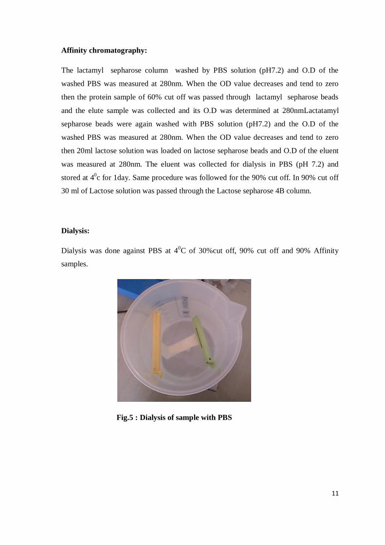
Dialysis:
Dialysis was done against PBS at 40C of 30%cut off, 90% cut off and 90% Affinity
samples.
Fig.5 : Dialysis of sample with PBS
12
Determination of concentration of protein:
The concentration of crude, 30% cut, 90% cut, and 90% affinity were measured by Lowry
Method.
Lowry’s Method:
REAGENT A=Sodium hydroxide(0.5%)
Sodium carbonate(2%) make it upto 1 litre
REAGENT B1=1% Copper sulphate
REAGENT B2=2% Sodium potassium tartarate
REAGENT C=A:B1:B2=100:1:1
BSA STANDARD=1mg/ml
Folinciocalteau‟s reagent=1N (5 ml solution +5 ml distill water)
Take different concentration of BSA solution from stock solution and add distill water to it
and made up to 2ml.Ricin protein taken unknown quantity dissolved in 1ml distill water ,and
add reagent C of 5 ml and protein of 0.5ml. Mixed properly and incubate for 10 mins. Then
0.5 ml of Folin reagent was added and incubate for 30min. Take OD at 750nm.
Preparation of Human Erythrocyte:
Healthy human venous blood was collected by a syringes and poured into a 15 ml tube to
which the anticoagulant EDTA was previously added. . In my experiment EDTA was added
as anti-coagulant.
Haemagglutination Assay:
1ml blood sample was Centrifuged in 2ml microtube at 1000 rpm for 5min at room
temperature by Eppendrof mini spin. The the pellet was collected and was added 10ml of
PBS. The mixture of blood and PBS was centrifuged at 1000 rpm for 5min at room
temperature. After centrifuge the Pellets were collected and c 100l of pellet was added
to10ml of PBS solution (pH 7.2). The Haemaglutination activity of Soyabean lectin was
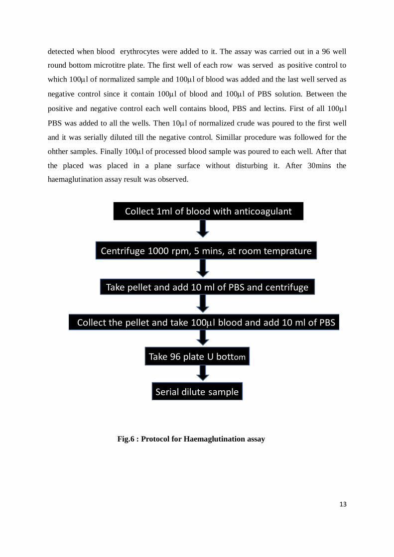
13
detected when blood erythrocytes were added to it. The assay was carried out in a 96 well
round bottom microtitre plate. The first well of each row was served as positive control to
which 100l of normalized sample and 100l of blood was added and the last well served as
negative control since it contain 100l of blood and 100l of PBS solution. Between the
positive and negative control each well contains blood, PBS and lectins. First of all 100l
PBS was added to all the wells. Then 10l of normalized crude was poured to the first well
and it was serially diluted till the negative control. Simillar procedure was followed for the
ohther samples. Finally 100l of processed blood sample was poured to each well. After that
the placed was placed in a plane surface without disturbing it. After 30mins the
haemaglutination assay result was observed.
Fig.6 : Protocol for Haemaglutination assay
Centrifuge 1000 rpm, 5 mins, at room temprature
Take pellet and add 10 ml of PBS and centrifuge
Collect the pellet and take 100l blood and add 10 ml of PBS
Take 96 plate U bottom
Serial dilute sample
Collect 1ml of blood with anticoagulant
14
SDS-PAGE:
The molecular mass of the subunits of the lectins was estimated by SDS-PAGE. The poly
acrylamide gel electrophoresis was done according to the protocol given in the Book
“Molecular Cloning” by Sambrook & Russell on a 12% gel. For the native 12%
polyacrylamide was employed and SDS along with B- mercaptoethanol was not added. The
mixture of 10l of sample, 10l of Sample loading buffer and 5l of Coomassie Brilliant
Blue were added to the well. In my experiment crude, 60%, 90%, 60% affinity and 90%
affinity was added with sample loading buffer and Coomassie Brilliant Blue. The gel was
again stained with Silver salts. Silver nitrate was used in the preparation of silver staining.
15
RESULT
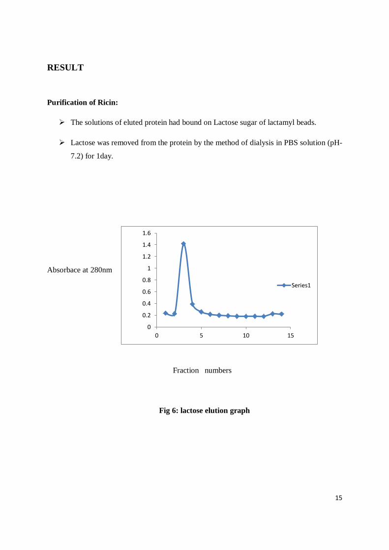
Purification of Ricin:
The solutions of eluted protein had bound on Lactose sugar of lactamyl beads.
Lactose was removed from the protein by the method of dialysis in PBS solution (pH-
7.2) for 1day.
Absorbace at 280nm
Fraction numbers
Fig 6: lactose elution graph
0
0.2
0.4
0.6
0.8
1
1.2
1.4
1.6
0 5 10 15
Series1
16
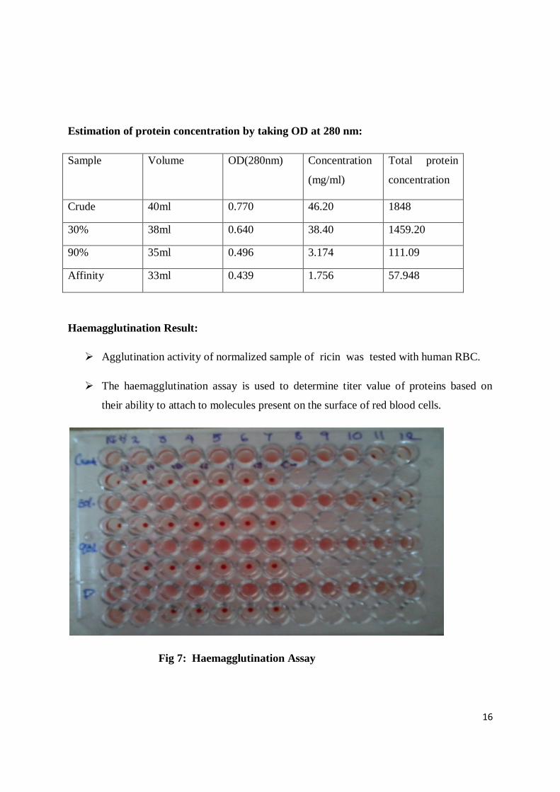
Estimation of protein concentration by taking OD at 280 nm:
Sample Volume OD(280nm) Concentration
(mg/ml)
Total protein
concentration
Crude 40ml 0.770 46.20 1848
30% 38ml 0.640 38.40 1459.20
90% 35ml 0.496 3.174 111.09
Affinity 33ml 0.439 1.756 57.948
Haemagglutination Result:
Agglutination activity of normalized sample of ricin was tested with human RBC.
The haemagglutination assay is used to determine titer value of proteins based on
their ability to attach to molecules present on the surface of red blood cells.
Fig 7: Haemagglutination Assay
17
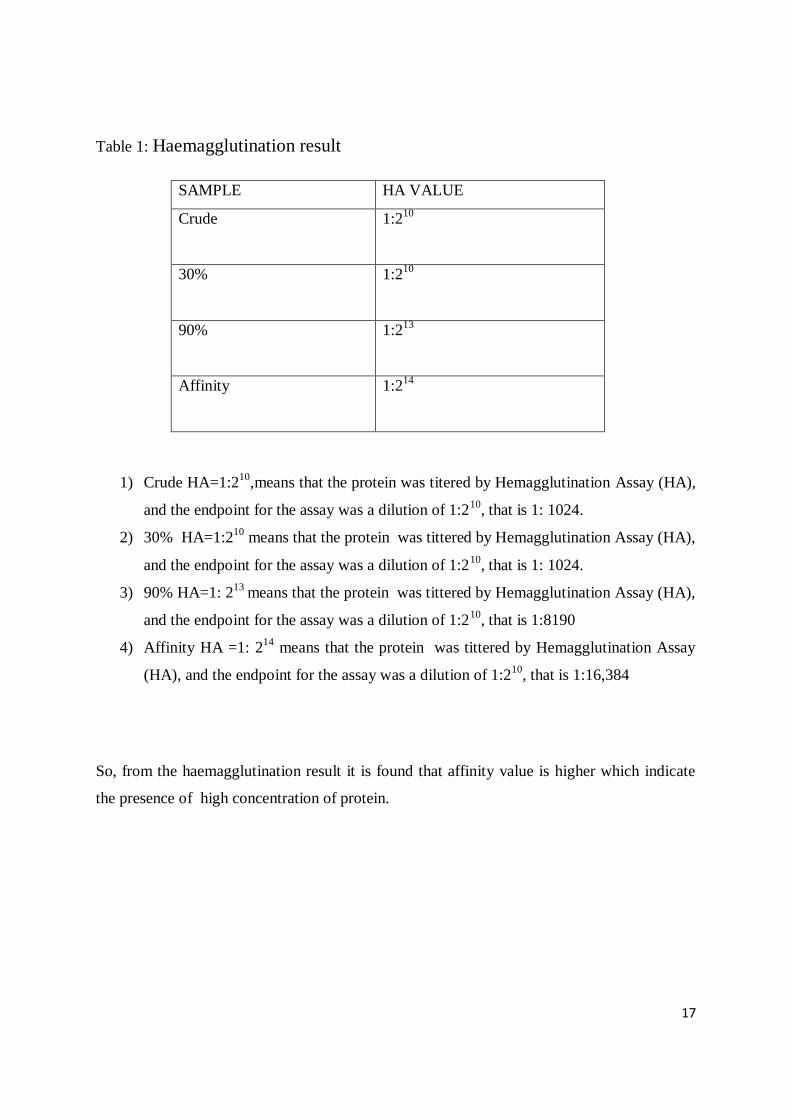
Table 1: Haemagglutination result
SAMPLE HA VALUE
Crude 1:210
30% 1:210
90% 1:213
Affinity 1:214
1) Crude HA=1:210
,means that the protein was titered by Hemagglutination Assay (HA),
and the endpoint for the assay was a dilution of 1:210
, that is 1: 1024.
2) 30% HA=1:210
means that the protein was tittered by Hemagglutination Assay (HA),
and the endpoint for the assay was a dilution of 1:210
, that is 1: 1024.
3) 90% HA=1: 213
means that the protein was tittered by Hemagglutination Assay (HA),
and the endpoint for the assay was a dilution of 1:210
, that is 1:8190
4) Affinity HA =1: 214
means that the protein was tittered by Hemagglutination Assay
(HA), and the endpoint for the assay was a dilution of 1:210
, that is 1:16,384
So, from the haemagglutination result it is found that affinity value is higher which indicate
the presence of high concentration of protein.
18
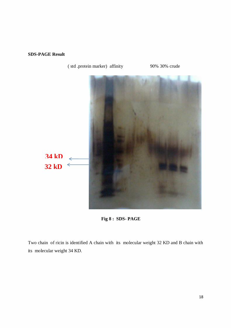
SDS-PAGE Result
( std .protein marker) affinity 90% 30% crude
Fig 8 : SDS- PAGE
Two chain of ricin is identified A chain with its molecular weight 32 KD and B chain with
its molecular weight 34 KD.
34 kD
32 kD
19
DISCUSSION
The hemagglutination assay is used to determine titre value of proteins based on their ability
to attach to molecules present on the surface of red blood cells. The red blood cells are
agglutinated by protein suspension which ensures there is no settling of RBCs out of
suspension. Serial dilution of a protein is performed in a 96-well plate and with consistent
addition of red blood cells, an estimation of the amount of protein in crude, 30 %, 90 % and
purified protein present is estimated.
Positive control are seen with a uniform film with indistinctive shape covering the bottom of
the tube. Negative control is seen perfectly outlined with round "button" of cells settled at the
bottom of the tube. Irregular clumps of cells are specified for the intermediately positive
results and are seen at the bottom of the tube.
The highest dilution of protein suspension that produces a positive result is termed as the end
point . HA (hemagglutination assay) is where the protein is mixed with diluted red blood
cells, the protein forms a network (lattice formation) with the red blood cells. The red blood
cells spread out as these "lattice" formations settle to the bottom of the tubes. If the protein is
absent, then the red blood cells are unable to form the lattice, and they settle down at the
bottom of the tube as a condensed button; interpreting the immunological property of ricin,
which is a weak agglutinin.
SDS PAGE : The purpose of SDS-PAGE is to separate proteins according to their size. The
molecular weight of desired protein after affinity was determined with respect to the
corresponding molecular weight of standard protein marker. We found out that Ricin
comprises of two chains A chain : 32 KD and B chain : 34 KD.
20
CONCLUSION:
SDS-PAGE enhanced our measurement of protein of interest, RICIN. Significantly, which
are of 32kD and 34 KD, characterised as A-Chain and B- chain.
Ricin is a weak agglutinin. As it is toxic, its cytotoxicity on normal cells and cancer cells
should be further explored to quote its anti-cancer property. Further research are required
for its application in different cancer cell lines to see whether it has inhibitory action on
proliferation.
21
References :
1. Atsmon, D. 1989. Castor. In. Oil crops of the World, (eds) GerhardRobbelen, R.K.
Downey, and A. Ashri. McGraw Hill Pub. Co., NewYork. pp. 438-447.
2. Bourne, Y.; Abergel, C.; Cambillau, C.; Frey, M.; Rougé, P. & Fontecilla-Camps,
(1990a), X-ray crystal structure determination and refinement at 1.9 Ao resolution of
isolectin from the seeds of Lathyrus ochrus I. J. Mol. Biol., 214: 571-584
3. Butterworth, Andrew G. and J. M. Lord. 1983. Ricin and Ricinus communis
agglutinin subunits are all derived from a single-siz polypeptide precursor. Eur. J.
Biochem. 137: 57-65
4. .Duke, J.A. and K.K. Wain. 1981. Medicinal Plants of the World.Computer index
with more than 85,000 entries. Vol. 3. University of Iowa. Ames , la.
5. Debray, Η., Decout, D., Strecker, G., Spik, G. and Montreuil, J. (1981) Eur.
Biochem., 117, 41.
6. .Lord, Michael J., L. M. Roberts, and J. D. Robertus. 1994. Ricin: structure, mode of
action, and some current applications. FASEB J. 8: 201-208.
7. Goldstein, I.J Hughes, R.C.;Monsignly , M.;Ozawa,T. & Sharon, N.;1980 Nature of
lectin .. Kocourek, J & Horejsi.V.,1983 recent discussion of definition of the
term”lectin”.
8. Goldstein, I.j. & Hayes, C.E., 1978. The lectins: carbohydrate binding proteins of
plant and animals.Adv. Carbohydr. Chem. Biochem., 35:127.340.
9. Montfort, William, J.E. Villafranca, A.F. Monzingo, S.R. Ernst, B. Katzin, E.
Rutenber, N.H. Xuong, R. Hamlin, and J.D. Robertus. 1987. The Three-dimensional
Structure of ricin at 2.8 A. J. of Biol. Chem. 262: 5398-5403.
10. Montfort, William, J.E. Villafranca, A.F. Monzingo, S.R. Ernst, B. Katzin,E.
Rutenber, N.H. Xuong, R. Hamlin, and J.D. Robertus. 1987. The Three-dimensional
Structure of ricin at 2.8 A. J. of Biol. Chem. 262:5398-5403.
11. Moreira, R.A.; Barros, A.C.H.; Stewart, J.C & Pusztai, A.,1983. Isolation and
characterization of a lectin from the seeds 158: 63-69.
22
12. Moshkin, V.A. and T.A.. Perestova. 1986. Morphology and Anatomy. In Castor. Ed.
V.A. Moshkin. Amerind Publ. Co., New Delhi, pp. 28-33.
13. Moshkin, V.A. Flowering and Pollination. 1986. In Castor. Ed. V.A.Moshkin.
Amerind Publ. Co., New Delhi, pp. 43-49. 14. Mourey L, Pédelacq J-D, Birck C, Fabre C, Rougé P, Samama J-P (1998)Crystal
structure of the arcelin-1 dimer from Phaseolus vulgaris at 1.9-Å resolution. J Biol
Chem 273:12914–12922.
15. Nicholsan,G.L.;Blaustein, J & Etzler, M.E.,1974 Characterization of two plant lectins
from Ricinus communis .
16. Sharon,N and Lis, H. (1989) Lectins as cell recognition molecules.Science 246, 227–
234
17. Van Damme EJM, Willy JP, Annick B, Pierre R (1998). Plant lectins: acomposite of
several distinct families of structurally and evolutionaryrelated proteins with diverse
biological roles. Crit. Rev. Plant Sci. 17:575-692.


![Isolation and characterization of D- Isolation and ... · ricin (RCA) and abrin (APA) as potential therapies for human cancer treatment [6,7]. Viscum album L. (Loranthaceae) has been](https://img.pdfslide.us/doc/110x75/6106caa300cacf06716cd083/isolation-and-characterization-of-d-isolation-and-ricin-rca-and-abrin-apa.jpg)