Embed Size (px)
Citation preview
Contents lists available at ScienceDirect
Clinical Biomechanics
journal homepage: www.elsevier.com/locate/clinbiomech
A biomechanical comparison of two plating techniques in lateral claviclefractures
Cyrill Sutera,⁎,1, Martina von Rohra,1, Martin Majewskia, Lutz Dürselenb, Daniela Warneckeb,Natalie Schildb, Andrej Maria Nowakowskia,c
aUniversity of Basel, Medical Faculty, Klingelbergstrasse 61, 4056 Basel, Switzerlandb Institute of Orthopedic Research and Biomechanics, Trauma Research Center Ulm, Ulm University - Medical Center, Helmholtzstr. 14, 89081 Ulm, GermanycOrthopedic Department, Spital Uster, Brunnenstrasse 42, 8610 Uster, Switzerland
A R T I C L E I N F O
Keywords:Lateral clavicle fractureBiomechanical testingPlate fixationSingle plateDouble plateSurgical treatment
A B S T R A C T
Background: Neer Type IIb lateral clavicle fractures typically lead to dislocation of the medial fragment.Therefore, most surgeons recommend surgical treatment for such a fracture pattern. The use of a lockingcompression plate with a lateral extension has produced satisfactory results in various studies over recent years.Double-plate fixation is a common technique in the treatment of complex distal radius fractures. The authors usethis technique as a routine procedure in the treatment of Neer type IIb fractures. In this biomechanical testingstudy, the mechanical properties of the two techniques were compared.Methods: On 20 clavicles from fresh frozen cadavers a Neer Type IIb fracture-like osteotomy was performed. Acyclic loading test followed by a load-to-failure test was carried out. Parameters for statistical evaluation werethe stiffness at cycles 1, 100 and 17,500 as well as the ultimate tensile load and the deformation at the point offailure.Findings: All specimens withstood the cyclic loading test without any noticeable damage. At cycles 100 and17,500, the double-plate technique was less stiff. Failure loads were not significantly different from each other,but deformation at the point of failure was significantly greater for the double-plate technique.Interpretation: Both techniques provided sufficient fixation to the fracture site to endure the cyclic loading test,which is supposed to simulate an incident-free week postoperatively. In summary, the double-plate techniqueoffers biomechanically a feasible alternative to the single-plate technique in lateral clavicle fractures of NeerType IIb.
1. Introduction
Clavicle fractures, in general, are common, particularly amongadolescents and young, active adults. They occur with the greatest in-cidence in the second and third decades, with an overall incidence of 64per 100,000 (Donnelly et al., 2013; Postacchini et al., 2002; Zlowodzkiet al., 2005). Lateral clavicle fractures account for approximately 18%of all clavicle fractures (Nowak et al., 2000). Finding the best treatmentof clavicle fractures remains a greatly debated topic in orthopedicpractice. This also extends to lateral clavicle fractures. Historically,clavicle fractures have mostly been treated conservatively with accep-table results (Donnelly et al., 2013). However, to determine whether alateral clavicle fracture requires surgical treatment, a precise diagnosticworkup is crucial, because up to 25% of all cases are unstable (Rieser
et al., 2013; Robinson, 1998). Even when the lateral fragment remainsin its anatomical position, whether the fragment will shift away or notdepends on the forces applied by the arm and on the integrity of thecoracoclavicular (CC) ligaments (Bishop et al., 2013). The functionalityof the CC ligaments is critical for the outcome of lateral clavicle frac-tures. Neer Type IIb fracture is believed to occur somewhere in betweenthe CC ligaments, which typically leads to a torn conoid ligament, anintact trapezoid ligament and thus to a displacement of the medialfragment (Neer, 1963).
When such an unstable fracture pattern like the Neer Type IIb ispresent, surgical treatment is indicated, because up to 28% of suchconservatively treated cases result in bony non-union and surgicaltreatment can lead to a union rate of up to 95% (Good et al., 2012;Schliemann et al., 2013). In addition, delayed surgical procedure can
https://doi.org/10.1016/j.clinbiomech.2019.05.001Received 16 September 2018; Accepted 1 May 2019
⁎ Corresponding author.E-mail address: [email protected] (C. Suter).
1 Contributed equally.
Clinical Biomechanics 67 (2019) 78–84
0268-0033/ © 2019 Published by Elsevier Ltd.
T
lead to a higher complication rate (Klein et al., 2010).When surgical therapy is indicated, numerous therapeutic options
come into consideration, including Kirschner-wire fixation, CC screws,plate fixation, hook plate fixation and CC sling procedures (Andersenet al., 2011; Badhe et al., 2007; Hessmann et al., 1996; Kalamaras et al.,2008; Neer, 1963; Nourissat et al., 2007; van der Meijden et al., 2012).Many of these lead to high complication rates, including implantfailure, non-union and subacromial impingement, or necessitate asecond operation for the removal of a hook plate (Carofino andMazzocca, 2010; Flinkkilä et al., 2002; Kashii et al., 2006; Lyons andRockwood, 1990; van der Meijden et al., 2012). In recent years, severalstudies have shown promising results using a precountered, superiorclavicle locking compression plate (LCP) with a lateral extension whenperforming internal fixation of lateral clavicle fractures (Beirer et al.,2014; Schliemann et al., 2013; Tiren and Vroemen, 2013; van derMeijden et al., 2012). In 2010, Kaipel et al. introduced another, newstrategy to treat lateral clavicle fractures by using a double-LCP tech-nique similar to the double-plate technique used in fractures of thedistal radius (Babst et al., 2003; Kaipel et al., 2010). The plates used forthe double-plate technique are significantly smaller than the one usedfor the single-plate technique, which makes smaller fracture fragmentsmore accessible for adequate plating. This advantage becomes moredistinctive in multifragmentary fractures of the lateral clavicle withsmaller fragments. Two other aspects that support the use of the double-plate technique are a) plate removal is required less frequently com-pared to the single-plate technique according to the authors experienceand b) the single-plate technique will not infrequently, with its quitebulky plate, visibly mark the treated clavicle, whereas the double-platetechnique offers a more discreet solution to this potential problem.
In the retrospective study of Kaipel et al., stable fixation using thedouble-plate of all treated fractures (n=11), even in small and com-minuted distal fragments, was reported (Kaipel et al., 2010). In additionto these clinically satisfying results, it is of interest to compare thebiomechanical behaviour of the newly introduced double-plate tech-nique to a time-tested plate-technique. Accordingly, the purpose of thisstudy was to compare the biomechanical properties of the double-platetechnique with anteriorly and superiorly administered, not pre-countered LCP to the single-plate technique, with a superiorly ad-ministered, precountered LCP, in lateral clavicle fractures of Neer TypeIIb.
2. Methods
2.1. Specimen and preparation
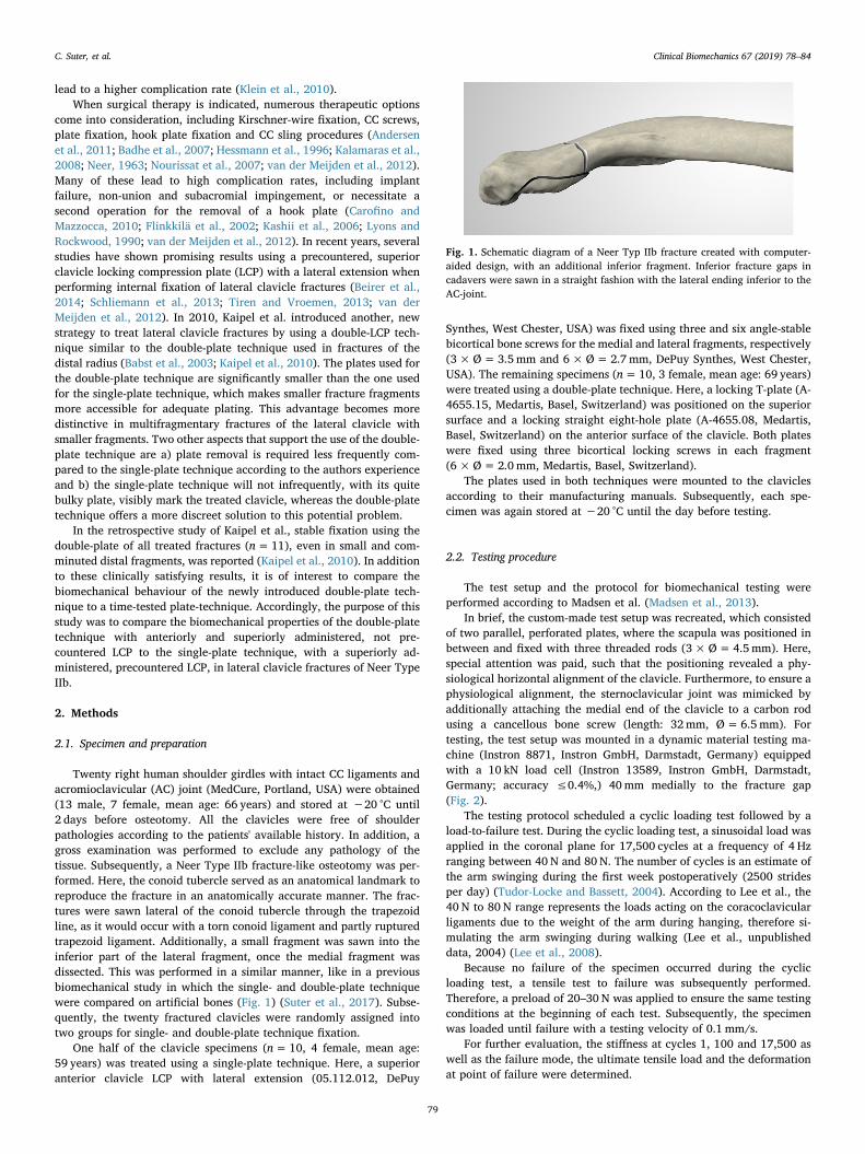
Twenty right human shoulder girdles with intact CC ligaments andacromioclavicular (AC) joint (MedCure, Portland, USA) were obtained(13 male, 7 female, mean age: 66 years) and stored at −20 °C until2 days before osteotomy. All the clavicles were free of shoulderpathologies according to the patients' available history. In addition, agross examination was performed to exclude any pathology of thetissue. Subsequently, a Neer Type IIb fracture-like osteotomy was per-formed. Here, the conoid tubercle served as an anatomical landmark toreproduce the fracture in an anatomically accurate manner. The frac-tures were sawn lateral of the conoid tubercle through the trapezoidline, as it would occur with a torn conoid ligament and partly rupturedtrapezoid ligament. Additionally, a small fragment was sawn into theinferior part of the lateral fragment, once the medial fragment wasdissected. This was performed in a similar manner, like in a previousbiomechanical study in which the single- and double-plate techniquewere compared on artificial bones (Fig. 1) (Suter et al., 2017). Subse-quently, the twenty fractured clavicles were randomly assigned intotwo groups for single- and double-plate technique fixation.
One half of the clavicle specimens (n=10, 4 female, mean age:59 years) was treated using a single-plate technique. Here, a superioranterior clavicle LCP with lateral extension (05.112.012, DePuy
Synthes, West Chester, USA) was fixed using three and six angle-stablebicortical bone screws for the medial and lateral fragments, respectively(3×Ø=3.5mm and 6×Ø=2.7mm, DePuy Synthes, West Chester,USA). The remaining specimens (n=10, 3 female, mean age: 69 years)were treated using a double-plate technique. Here, a locking T-plate (A-4655.15, Medartis, Basel, Switzerland) was positioned on the superiorsurface and a locking straight eight-hole plate (A-4655.08, Medartis,Basel, Switzerland) on the anterior surface of the clavicle. Both plateswere fixed using three bicortical locking screws in each fragment(6×Ø=2.0mm, Medartis, Basel, Switzerland).
The plates used in both techniques were mounted to the claviclesaccording to their manufacturing manuals. Subsequently, each spe-cimen was again stored at −20 °C until the day before testing.
2.2. Testing procedure
The test setup and the protocol for biomechanical testing wereperformed according to Madsen et al. (Madsen et al., 2013).
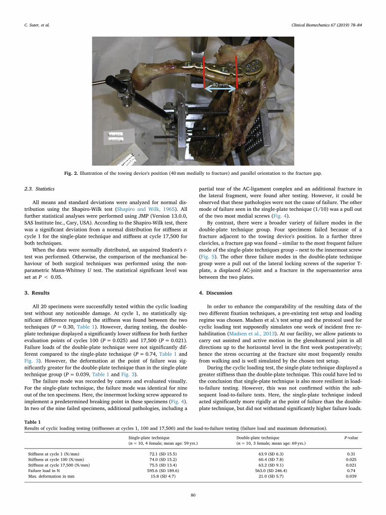
In brief, the custom-made test setup was recreated, which consistedof two parallel, perforated plates, where the scapula was positioned inbetween and fixed with three threaded rods (3×Ø=4.5mm). Here,special attention was paid, such that the positioning revealed a phy-siological horizontal alignment of the clavicle. Furthermore, to ensure aphysiological alignment, the sternoclavicular joint was mimicked byadditionally attaching the medial end of the clavicle to a carbon rodusing a cancellous bone screw (length: 32mm, Ø=6.5mm). Fortesting, the test setup was mounted in a dynamic material testing ma-chine (Instron 8871, Instron GmbH, Darmstadt, Germany) equippedwith a 10 kN load cell (Instron 13589, Instron GmbH, Darmstadt,Germany; accuracy ≤0.4%,) 40mm medially to the fracture gap(Fig. 2).
The testing protocol scheduled a cyclic loading test followed by aload-to-failure test. During the cyclic loading test, a sinusoidal load wasapplied in the coronal plane for 17,500 cycles at a frequency of 4 Hzranging between 40 N and 80 N. The number of cycles is an estimate ofthe arm swinging during the first week postoperatively (2500 stridesper day) (Tudor-Locke and Bassett, 2004). According to Lee et al., the40 N to 80 N range represents the loads acting on the coracoclavicularligaments due to the weight of the arm during hanging, therefore si-mulating the arm swinging during walking (Lee et al., unpublisheddata, 2004) (Lee et al., 2008).
Because no failure of the specimen occurred during the cyclicloading test, a tensile test to failure was subsequently performed.Therefore, a preload of 20–30 N was applied to ensure the same testingconditions at the beginning of each test. Subsequently, the specimenwas loaded until failure with a testing velocity of 0.1 mm/s.
For further evaluation, the stiffness at cycles 1, 100 and 17,500 aswell as the failure mode, the ultimate tensile load and the deformationat point of failure were determined.
Fig. 1. Schematic diagram of a Neer Typ IIb fracture created with computer-aided design, with an additional inferior fragment. Inferior fracture gaps incadavers were sawn in a straight fashion with the lateral ending inferior to theAC-joint.
C. Suter, et al. Clinical Biomechanics 67 (2019) 78–84
79
2.3. Statistics
All means and standard deviations were analyzed for normal dis-tribution using the Shapiro-Wilk test (Shapiro and Wilk, 1965). Allfurther statistical analyses were performed using JMP (Version 13.0.0,SAS Institute Inc., Cary, USA). According to the Shapiro-Wilk test, therewas a significant deviation from a normal distribution for stiffness atcycle 1 for the single-plate technique and stiffness at cycle 17,500 forboth techniques.
When the data were normally distributed, an unpaired Student's t-test was performed. Otherwise, the comparison of the mechanical be-haviour of both surgical techniques was performed using the non-parametric Mann-Whitney U test. The statistical significant level wasset at P < 0.05.
3. Results
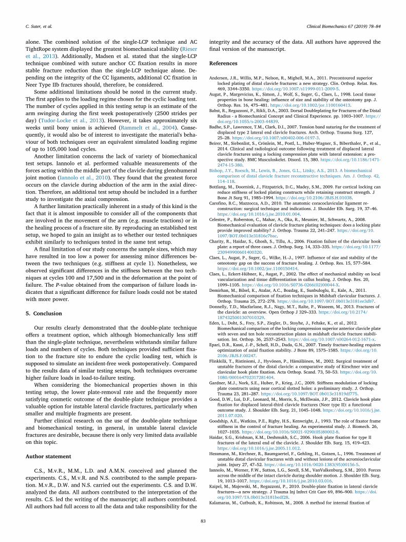
All 20 specimens were successfully tested within the cyclic loadingtest without any noticeable damage. At cycle 1, no statistically sig-nificant difference regarding the stiffness was found between the twotechniques (P=0.30, Table 1). However, during testing, the double-plate technique displayed a significantly lower stiffness for both furtherevaluation points of cycles 100 (P=0.025) and 17,500 (P=0.021).Failure loads of the double-plate technique were not significantly dif-ferent compared to the single-plate technique (P=0.74, Table 1 andFig. 3). However, the deformation at the point of failure was sig-nificantly greater for the double-plate technique than in the single-platetechnique group (P=0.039, Table 1 and Fig. 3).
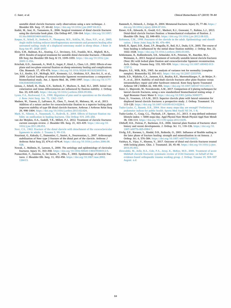
The failure mode was recorded by camera and evaluated visually.For the single-plate technique, the failure mode was identical for nineout of the ten specimens. Here, the innermost locking screw appeared toimplement a predetermined breaking point in these specimens (Fig. 4).In two of the nine failed specimens, additional pathologies, including a
partial tear of the AC-ligament complex and an additional fracture inthe lateral fragment, were found after testing. However, it could beobserved that these pathologies were not the cause of failure. The othermode of failure seen in the single-plate technique (1/10) was a pull outof the two most medial screws (Fig. 4).
By contrast, there were a broader variety of failure modes in thedouble-plate technique group. Four specimens failed because of afracture adjacent to the towing device's position. In a further threeclavicles, a fracture gap was found – similar to the most frequent failuremode of the single-plate techniques group – next to the innermost screw(Fig. 5). The other three failure modes in the double-plate techniquegroup were a pull out of the lateral locking screws of the superior T-plate, a displaced AC-joint and a fracture in the superoanterior areabetween the two plates.
4. Discussion
In order to enhance the comparability of the resulting data of thetwo different fixation techniques, a pre-existing test setup and loadingregime was chosen. Madsen et al.'s test setup and the protocol used forcyclic loading test supposedly simulates one week of incident free re-habilitation (Madsen et al., 2013). At our facility, we allow patients tocarry out assisted and active motion in the glenohumeral joint in alldirections up to the horizontal level in the first week postoperatively;hence the stress occurring at the fracture site most frequently resultsfrom walking and is well simulated by the chosen test setup.
During the cyclic loading test, the single-plate technique displayed agreater stiffness than the double-plate technique. This could have led tothe conclusion that single-plate technique is also more resilient in load-to-failure testing. However, this was not confirmed within the sub-sequent load-to-failure tests. Here, the single-plate technique indeedacted significantly more rigidly at the point of failure than the double-plate technique, but did not withstand significantly higher failure loads.
Fig. 2. Illustration of the towing device's position (40mm medially to fracture) and parallel orientation to the fracture gap.
Table 1Results of cyclic loading testing (stiffnesses at cycles 1, 100 and 17,500) and the load-to-failure testing (failure load and maximum deformation).
Single-plate technique(n=10, 4 female; mean age: 59 yrs.)
Double-plate technique(n=10, 3 female; mean age: 69 yrs.)
P-value
Stiffness at cycle 1 (N/mm) 72.1 (SD 15.5) 63.9 (SD 6.3) 0.31Stiffness at cycle 100 (N/mm) 74.0 (SD 15.2) 60.4 (SD 7.8) 0.025Stiffness at cycle 17,500 (N/mm) 75.5 (SD 13.4) 63.2 (SD 9.1) 0.021Failure load in N 595.6 (SD 189.6) 563.0 (SD 246.4) 0.74Max. deformation in mm 15.8 (SD 4.7) 21.0 (SD 5.7) 0.039
C. Suter, et al. Clinical Biomechanics 67 (2019) 78–84
80

Therefore, it can be assumed that both techniques provide a stablefixation for Neer Typ IIb fractures during the first week after surgery, ifno incident (e.g. a fall) occurs. As seen in the load-to-failure testing, alsowhen an incident, which was similar to our testing set up (e.g. pullingforce on the arm), occurs, both techniques presented similar resistancetowards failure.
Stiffness at the fracture site is one of many components, which canalter bone healing. It is generally accepted that extremely flexiblefixation techniques can lead to a delay in bone healing, compared torigid fixation. However, a perfect rigidity without movement at thefracture site is hard to achieve (Epari et al., 2007; Mark et al., 2004;Utvåg et al., 2001). A certain amount of motion is additionally helpfulfor stimulating bone healing.
Nevertheless, the ideal amount of motion (or stiffness respectively)as well as how much motion at what time of fracture healing has notbeen conclusively identified yet (Augat et al., 1998; Claes et al., 1997;
Claes et al., 2002; Epari et al., 2007; Goodship et al., 1993; Kaspar et al.,2005; Krischak et al., 2002; Lienau et al., 2005; Schell et al., 2005). Thesingle-plate technique tested in the current study is a time-tested de-vice, which leads to bony non-union very rarely, as we have found onlyone case of bony non-union in the literature (Vaishya et al., 2017). Withregard to the literature, we feel confident stating that double-platetechnique, the more flexible construct, also leads to sufficient stability,because a) constructs with locking plates are believed to lead morelikely to too stiff fracture fixation (Bottlang et al., 2009; Gardner et al.,2009; Uhthoff et al., 2006) and b) has shown to achieve regular osseousunion in a case series (Kaipel et al., 2010).
However, since we did not measure the motion in the fracture gapduring testing, we cannot conclude which fixation technique leads tomost adequate stabilisation for bone healing – single or double platetechnique. Nevertheless, both techniques lead to a high rate of bonyunion, which may lead to the assumption that their fixation stiffness
Fig. 3. Box-plots for comparison of “Deformation at the point of failure” and “Load to failure” values.
Fig. 4. Modes of failure of the single-plate technique. A) fracture adjacent to the innermost screw (9/10), B) pull out of the two most medial screws (1/10).
C. Suter, et al. Clinical Biomechanics 67 (2019) 78–84
81

does not influence bone healing to a clinically significant degree andplate removal shouldn't be performed until.
The failure mode of the single-plate technique can be considered asa limitation of this study. The most frequent failure mode (9/10), aperiprosthetic failure, is rarely seen in clinical situations. However, thisclinically, rather uncommon failure mode appears to be a well-knownproblem in the biomechanical testing of clavicle fractures, because itsimilarly occurred in various other studies (Bishop et al., 2013; Celestreet al., 2008; Demirhan et al., 2011; Eden et al., 2012; Smith et al.,2014). Additionally, the aforementioned failure mode has been re-ported in two case reports of hook plate fixation (Charity et al., 2006;Haidar et al., 2006). The two most frequent fracture modes of thedouble-plate technique were fracture adjacent to the traction deviceposition (4/10) and fracture adjacent to the innermost screw (3/10).Because no data were found for the failure modes of the double-platetechnique, it could not be determined whether or not the fracturesadjacent to the most medial screw represent a typical clinical situation.
Both techniques sufficiently stabilized the actual fracture site, be-cause the most frequent modes of failure did not occur directly at thefracture site (18/20). To be more precise, in the single-plate techniquegroup, all ten specimens failed medial to the fracture site. In the double-plate technique group, seven out of the ten specimens failed medial tofracture site and one lateral to fracture site (displaced AC-joint), whichwas the clavicle with highest failure load in this group (1027 N).
When combining both techniques, almost all failures occurred at thesame site (17/20), which underlines a good reproducibility of thetesting setup.
Comparing our test results with those of two studies using a similartest setup, we can conclude that the results of the current study forfailure loads and deformation at the point of failure are not inferior,although our specimens displayed less stiffness (Bishop et al., 2013;
Madsen et al., 2013). For example, in the study of Madsen et al., thesuperior LCP failed at a mean load of 401.3 (SD 172 N), resulting in amean maximal deformation of 7.6 (SD 2.8mm) and displayed a stiffnessof 80.9 (SD 7.8 N/mm). Bishop et al. compared the superior LCP withthe hook plate technique, and no significant difference in the bio-mechanical strength could be determined between these two groups.The superior LCP group withstood a mean of 487.8 (SD 230 N) (Bishopet al., 2013).
Within a previous biomechanical study, the mechanical propertiesof these two techniques – single and double plate - were compared. Incontrast to the current study, testing was carried out using a cantileverbending test and Neer Type IIb fractures were produced on artificialbones. The failure loads seen in the previous testing were substantiallylower to the ones in this test series, which might be due to the differ-ence in the test-setup. Double plate technique withstood a failure loadof 134.6 (SD 9.45 N), whereas single plate technique failed at a load of112.1 (SD 12.39 SD). The main findings were a) double plate techniqueoffered a slightly more stable fixation to the fracture as it withstoodhigher failure loads and more cycles in cyclic loading test and b) also inthis test setup single plate technique acted more rigid (Suter et al.,2017).
We are aware that such comparisons only allow very restrictedconclusions, because small incongruities (e.g. in length of the leverarm) can already lead to noticeable discrepancies between two ob-served values. Nonetheless, it is the only way to receive feedback aboutthe validation of the biomechanical performance of a new fixationtechnique.
An aspect not addressed in this study concerns the fixation of the CCligaments. In the study of Rieser et al., the single-LCP technique wascompared to the single-LCP technique combined with the AC TightRopesystem for coraclavicular fixation and to the AC TightRope system
Fig. 5. Most frequent modes of failure of the double-plate technique. A) fracture adjacent to the traction device position (4/10), B) fracture adjacent to the innermostscrew (3/10).
C. Suter, et al. Clinical Biomechanics 67 (2019) 78–84
82
alone. The combined solution of the single-LCP technique and ACTightRope system displayed the greatest biomechanical stability (Rieseret al., 2013). Additionally, Madsen et al. stated that the single-LCPtechnique combined with suture anchor CC fixation results in morestable fracture reduction than the single-LCP technique alone. De-pending on the integrity of the CC ligaments, additional CC fixation inNeer Type IIb fractures should, therefore, be considered.
Some additional limitations should be noted in the current study.The first applies to the loading regime chosen for the cyclic loading test.The number of cycles applied in this testing setup is an estimate of thearm swinging during the first week postoperatively (2500 strides perday) (Tudor-Locke et al., 2013). However, it takes approximately sixweeks until bony union is achieved (Rammelt et al., 2004). Conse-quently, it would also be of interest to investigate the material's beha-viour of both techniques over an equivalent simulated loading regimeof up to 105,000 load cycles.
Another limitation concerns the lack of variety of biomechanicaltest setups. Iannolo et al. performed valuable measurements of theforces acting within the middle part of the clavicle during glenohumeraljoint motion (Iannolo et al., 2010). They found that the greatest forceoccurs on the clavicle during abduction of the arm in the axial direc-tion. Therefore, an additional test setup should be included in a furtherstudy to investigate the axial compression.
A further limitation practically inherent in a study of this kind is thefact that it is almost impossible to consider all of the components thatare involved in the movement of the arm (e.g. muscle tractions) or inthe healing process of a fracture site. By reproducing an established testsetup, we hoped to gain an insight as to whether our tested techniquesexhibit similarly to techniques tested in the same test setup.
A final limitation of our study concerns the sample sizes, which mayhave resulted in too low a power for assessing minor differences be-tween the two techniques (e.g. stiffness at cycle 1). Nonetheless, weobserved significant differences in the stiffness between the two tech-niques at cycles 100 and 17,500 and in the deformation at the point offailure. The P-value obtained from the comparison of failure loads in-dicates that a significant difference for failure loads could not be statedwith more power.
5. Conclusion
Our results clearly demonstrated that the double-plate techniqueoffers a treatment option, which although biomechanically less stiffthan the single-plate technique, nevertheless withstands similar failureloads and numbers of cycles. Both techniques provided sufficient fixa-tion to the fracture site to endure the cyclic loading test, which issupposed to simulate an incident-free week postoperatively. Comparedto the results data of similar testing setups, both techniques overcamehigher failure loads in load-to-failure testing.
When considering the biomechanical properties shown in thistesting setup, the lower plate-removal rate and the frequently moresatisfying cosmetic outcome of the double-plate technique provides avaluable option for instable lateral clavicle fractures, particularly whensmaller and multiple fragments are present.
Further clinical research on the use of the double-plate techniqueand biomechanical testing, in general, in unstable lateral claviclefractures are desirable, because there is only very limited data availableon this topic.
Author statement
C.S., M.v.R., M.M., L.D. and A.M.N. conceived and planned theexperiments. C.S., M.v.R. and N.S. contributed to the sample prepara-tion. M.v.R., D.W. and N.S. carried out the experiments. C.S. and D.W.analyzed the data. All authors contributed to the interpretation of theresults. C.S. led the writing of the manuscript; all authors contributed.All authors had full access to all the data and take responsibility for the
integrity and the accuracy of the data. All authors have approved thefinal version of the manuscript.
References
Andersen, J.R., Willis, M.P., Nelson, R., Mighell, M.A., 2011. Precontoured superiorlocked plating of distal clavicle fractures: a new strategy. Clin. Orthop. Relat. Res.469, 3344–3350. https://doi.org/10.1007/s11999-011-2009-5.
Augat, P., Margevicius, K., Simon, J., Wolf, S., Suger, G., Claes, L., 1998. Local tissueproperties in bone healing: influence of size and stability of the osteotomy gap. J.Orthop. Res. 16, 475–481. https://doi.org/10.1002/jor.1100160413.
Babst, R., Regazzoni, P., Rikli, D.A., 2003. Dorsal Doubleplating for Fractures of the DistalRadius - a Biomechanical Concept and Clinical Experience. pp. 1003–1007. https://doi.org/10.1055/s-2003-44839.
Badhe, S.P., Lawrence, T.M., Clark, D.I., 2007. Tension band suturing for the treatment ofdisplaced type 2 lateral end clavicle fractures. Arch. Orthop. Trauma Surg. 127,25–28. https://doi.org/10.1007/s00402-006-0197-3.
Beirer, M., Siebenlist, S., Crönlein, M., Postl, L., Huber-Wagner, S., Biberthaler, P., et al.,2014. Clinical and radiological outcome following treatment of displaced lateralclavicle fractures using a locking compression plate with lateral extension: a pro-spective study. BMC Musculoskelet. Disord. 15, 380. https://doi.org/10.1186/1471-2474-15-380.
Bishop, J.Y., Roesch, M., Lewis, B., Jones, G.L., Litsky, A.S., 2013. A biomechanicalcomparison of distal clavicle fracture reconstructive techniques. Am. J. Orthop. 42,114–118.
Bottlang, M., Doornink, J., Fitzpatrick, D.C., Madey, S.M., 2009. Far cortical locking canreduce stiffness of locked plating constructs while retaining construct strength. JBone Jt Surg 91, 1985–1994. https://doi.org/10.2106/JBJS.H.01038.
Carofino, B.C., Mazzocca, A.D., 2010. The anatomic coracoclavicular ligament re-construction: surgical technique and indications. J. Shoulder Elb. Surg. 19, 37–46.https://doi.org/10.1016/j.jse.2010.01.004.
Celestre, P., Roberston, C., Mahar, A., Oka, R., Meunier, M., Schwartz, A., 2008.Biomechanical evaluation of clavicle fracture plating techniques: does a locking plateprovide improved stability? J. Orthop. Trauma 22, 241–247. https://doi.org/10.1097/BOT.0b013e31816c7bac.
Charity, R., Haidar, S., Ghosh, S., Tillu, A., 2006. Fixation failure of the clavicular hookplate: a report of three cases. J. Orthop. Surg. 14, 333–335. https://doi.org/10.1177/230949900601400320.
Claes, L., Augat, P., Suger, G., Wilke, H.-J., 1997. Influence of size and stability of theosteotomy gap on the success of fracture healing. J. Orthop. Res. 15, 577–584.https://doi.org/10.1002/jor.1100150414.
Claes, L., Eckert-Hübner, K., Augat, P., 2002. The effect of mechanical stability on localvascularization and tissue differentiation in callus healing. J. Orthop. Res. 20,1099–1105. https://doi.org/10.1016/S0736-0266(02)00044-X.
Demirhan, M., Bilsel, K., Atalar, A.C., Bozdag, E., Sunbuloglu, E., Kale, A., 2011.Biomechanical comparison of fixation techniques in Midshaft clavicular fractures. J.Orthop. Trauma 25, 272–278. https://doi.org/10.1097/BOT.0b013e3181ee3db7.
Donnelly, T.D., Macfarlane, R.J., Nagy, M.T., Ralte, P., Waseem, M., 2013. Fractures ofthe clavicle: an overview. Open Orthop J 329–333. https://doi.org/10.2174/1874325001307010329.
Eden, L., Doht, S., Frey, S.P., Ziegler, D., Stoyhe, J., Fehske, K., et al., 2012.Biomechanical comparison of the locking compression superior anterior clavicle platewith seven and ten hole reconstruction plates in midshaft clavicle fracture stabili-sation. Int. Orthop. 36, 2537–2543. https://doi.org/10.1007/s00264-012-1671-x.
Epari, D.R., Kassi, J.-P., Schell, H.D., Duda, G.N., 2007. Timely fracture-healing requiresoptimization of axial fixation stability. J Bone 89, 1575–1585. https://doi.org/10.2106/JBJS.F.00247.
Flinkkilä, T., Ristiniemi, J., Hyvönen, P., Hämäläinen, M., 2002. Surgical treatment ofunstable fractures of the distal clavicle: a comparative study of Kirschner wire andclavicular hook plate fixation. Acta Orthop. Scand. 73, 50–53. https://doi.org/10.1080/000164702317281404.
Gardner, M.J., Nork, S.E., Huber, P., Krieg, J.C., 2009. Stiffness modulation of lockingplate constructs using near cortical slotted holes: a preliminary study. J. Orthop.Trauma 23, 281–287. https://doi.org/10.1097/BOT.0b013e31819df775.
Good, D.W., Lui, D.F., Leonard, M., Morris, S., McElwain, J.P., 2012. Clavicle hook platefixation for displaced lateral-third clavicle fractures (Neer type II): a functionaloutcome study. J. Shoulder Elb. Surg. 21, 1045–1048. https://doi.org/10.1016/j.jse.2011.07.020.
Goodship, A.E., Watkins, P.E., Rigby, H.S., Kenwright, J., 1993. The role of fixator framestiffness in the control of fracture healing. An experimental study. J. Biomech. 26,1027–1035. https://doi.org/10.1016/S0021-9290(05)80002-8.
Haidar, S.G., Krishnan, K.M., Deshmukh, S.C., 2006. Hook plate fixation for type IIfractures of the lateral end of the clavicle. J. Shoulder Elb. Surg. 15, 419–423.https://doi.org/10.1016/j.jse.2005.11.012.
Hessmann, M., Kirchner, R., Baumgaertel, F., Gehling, H., Gotzen, L., 1996. Treatment ofunstable distal clavicular fractures with and without lesions of the acromioclavicularjoint. Injury 27, 47–52. https://doi.org/10.1016/0020-1383(95)00156-5.
Iannolo, M., Werner, F.W., Sutton, L.G., Serell, S.M., VanValkenburg, S.M., 2010. Forcesacross the middle of the intact clavicle during shoulder motion. J. Shoulder Elb. Surg.19, 1013–1017. https://doi.org/10.1016/j.jse.2010.03.016.
Kaipel, M., Majewski, M., Regazzoni, P., 2010. Double-plate fixation in lateral claviclefractures—a new strategy. J Trauma Inj Infect Crit Care 69, 896–900. https://doi.org/10.1097/TA.0b013e3181bedf28.
Kalamaras, M., Cutbush, K., Robinson, M., 2008. A method for internal fixation of
C. Suter, et al. Clinical Biomechanics 67 (2019) 78–84
83
unstable distal clavicle fractures: early observations using a new technique. J.Shoulder Elb. Surg. 17, 60–62. https://doi.org/10.1016/j.jse.2007.04.012.
Kashii, M., Inui, H., Yamamoto, K., 2006. Surgical treatment of distal clavicle fracturesusing the clavicular hook plate. Clin Orthop 447, 158–164. https://doi.org/10.1097/01.blo.0000203469.66055.6a.
Kaspar, K., Schell, H., Seebeck, P., Thompson, M.S., SchÜtz, M., Haas, N.P., et al., 2005.Angle stable locking reduces interfragmentary movements and promotes healing afterunreamed nailing: study of a displaced osteotomy model in sheep tibiae. J Bone JtSurg-Am 87, 2028–2037.
Klein, S.M., Badman, B.L., Keating, C.J., Devinney, D.S., Frankle, M.A., Mighell, M.A.,2010. Results of surgical treatment for unstable distal clavicular fractures. J ShoulderElb Surg Am Shoulder Elb Surg Al 19, 1049–1055. https://doi.org/10.1016/j.jse.2009.11.056.
Krischak, G.D., Janousek, A., Wolf, S., Augat, P., Kinzl, L., Claes, L.E., 2002. Effects of one-plane and two-plane external fixation on sheep osteotomy healing and complications.Clin. Biomech. 17, 470–476. https://doi.org/10.1016/S0268-0033(02)00039-6.
Lee, S.J., Keefer, E.P., McHugh, M.P., Kremenic, I.J., Orishimo, K.F., Ben-Avi, S., et al.,2008. Cyclical loading of coracoclavicular ligament reconstructions: a comparativebiomechanical study. Am. J. Sports Med. 36, 1990–1997. https://doi.org/10.1177/0363546508324284.
Lienau, J., Schell, H., Duda, G.N., Seebeck, P., Muchow, S., Bail, H.J., 2005. Initial vas-cularization and tissue differentiation are influenced by fixation stability. J. Orthop.Res. 23, 639–645. https://doi.org/10.1016/j.orthres.2004.09.006.
Lyons, F.A., Rockwood, C.A., 1990. Migration of pins used in operations on the shoulder.J. Bone Joint Surg. Am. 72, 1262–1267.
Madsen, W., Yaseen, Z., LaFrance, R., Chen, T., Awad, H., Maloney, M., et al., 2013.Addition of a suture anchor for coracoclavicular fixation to a superior locking plateimproves stability of type IIB distal clavicle fractures. Arthrosc J Arthrosc Relat Surg29, 998–1004. https://doi.org/10.1016/j.arthro.2013.02.024.
Mark, H., Nilsson, A., Nannmark, U., Rydevik, B., 2004. Effects of fracture fixation sta-bility on ossification in healing fractures. Clin Orthop 419, 245–250.
van der Meijden, O.A., Gaskill, T.R., Millett, P.J., 2012. Treatment of clavicle fractures:current concepts review. J. Shoulder Elb. Surg. 21, 423–429. https://doi.org/10.1016/j.jse.2011.08.053.
Neer, C.S., 1963. Fracture of the distal clavicle with detachment of the coracoclavicularligaments in adults. J. Trauma 3, 99–110.
Nourissat, G., Kakuda, C., Dumontier, C., Sautet, A., Doursounian, L., 2007. Arthroscopicstabilization of Neer type 2 fracture of the distal part of the clavicle. Arthrosc JArthrosc Relat Surg 23, 674.e1–674.e4. https://doi.org/10.1016/j.arthro.2006.08.028.
Nowak, J., Mallmin, H., Larsson, S., 2000. The aetiology and epidemiology of clavicularfractures. Injury 31, 353–358. https://doi.org/10.1016/S0020-1383(99)00312-5.
Postacchini, F., Gumina, S., De Santis, P., Albo, F., 2002. Epidemiology of clavicle frac-tures. J. Shoulder Elb. Surg. 11, 452–456. https://doi.org/10.1067/mse.2002.126613.
Rammelt, S., Heineck, J., Zwipp, H., 2004. Metatarsal fractures. Injury 35, 77–86. https://doi.org/10.1016/j.injury.2004.07.016.
Rieser, G.R., Edwards, K., Gould, G.C., Markert, R.J., Goswami, T., Rubino, L.J., 2013.Distal-third clavicle fracture fixation: a biomechanical evaluation of fixation. J.Shoulder Elb. Surg. 22, 848–855. https://doi.org/10.1016/j.jse.2012.08.022.
Robinson, C.M., 1998. Fractures of the clavicle in the adult. Epidemiology and classifi-cation. J Bone Joint Surg Br 80, 476–484.
Schell, H., Epari, D.R., Kassi, J.P., Bragulla, H., Bail, H.J., Duda, G.N., 2005. The course ofbone healing is influenced by the initial shear fixation stability. J. Orthop. Res. 23,1022–1028. https://doi.org/10.1016/j.orthres.2005.03.005.
Schliemann, B., Roßlenbroich, S.B., Schneider, K.N., Petersen, W., Raschke, M.J.,Weimann, A., 2013. Surgical treatment of vertically unstable lateral clavicle fractures(Neer 2b) with locked plate fixation and coracoclavicular ligament reconstruction.Arch. Orthop. Trauma Surg. 133, 935–939. https://doi.org/10.1007/s00402-013-1737-2.
Shapiro, S.S., Wilk, M.B., 1965. An analysis of variance test for normality (completesamples). Biometrika 52, 591–611. https://doi.org/10.2307/2333709.
Smith, S.D., Wijdicks, C.A., Jansson, K.S., Boykin, R.E., Martetschlaeger, F., de Meijer, P.-P., et al., 2014. Stability of mid-shaft clavicle fractures after plate fixation versusintramedullary repair and after hardware removal. Knee Surg Sports TraumatolArthrosc Off J ESSKA 22, 448–455. https://doi.org/10.1007/s00167-013-2411-5.
Suter, C., Majewski, M., Nowakowski, A.M., 2017. Comparison of 2 plating techniques forlateral clavicle fractures, using a new standardized biomechanical testing setup. JAppl Biomater Funct Mater 0. https://doi.org/10.5301/jabfm.5000377.
Tiren, D., Vroemen, J.P.A.M., 2013. Superior clavicle plate with lateral extension fordisplaced lateral clavicle fractures: a prospective study. J. Orthop. Traumatol. 14,115–120. https://doi.org/10.1007/s10195-013-0228-0.
Tudor-Locke, C., Bassett, D.R., 2004. How many steps/day are enough? Preliminarypedometer indices for public health. Sports Med Auckl NZ 34, 1–8.
Tudor-Locke, C., Craig, C.L., Thyfault, J.P., Spence, J.C., 2013. A step-defined sedentarylifestyle index:< 5000 steps/day. Appl Physiol Nutr Metab Physiol Appl Nutr Metab38, 100–114. https://doi.org/10.1139/apnm-2012-0235.
Uhthoff, H.K., Poitras, P., Backman, D.S., 2006. Internal plate fixation of fractures: shorthistory and recent developments. J. Orthop. Sci. 11, 118–126. https://doi.org/10.1007/s00776-005-0984-7.
Utvåg, S.E., Korsnes, L., Rindal, D.B., Reikerås, O., 2001. Influence of flexible nailing inthe later phase of fracture healing: strength and mineralization in rat femora. J.Orthop. Sci. 6, 576–584. https://doi.org/10.1007/s007760100015.
Vaishya, R., Vijay, V., Khanna, V., 2017. Outcome of distal end clavicle fractures treatedwith locking plates. Chin. J. Traumatol. 20, 45–48. https://doi.org/10.1016/j.cjtee.2016.05.003.
Zlowodzki, M., Zelle, B.A., Cole, P.A., Jeray, K., McKee, M.D., 2005. Treatment of acuteMidshaft clavicle fractures: systematic review of 2144 fractures: on behalf of theevidence-based orthopaedic trauma working group. J. Orthop. Trauma 19, 504–507August. n.d.
C. Suter, et al. Clinical Biomechanics 67 (2019) 78–84
84