Embed Size (px)
Citation preview
Journal of Clinical Pathology, 1979, 32, 35-39
A new antigen on the epithelial membrane: itsimmunoperoxidase localization in normal andneoplastic tissueEADIE HEYDERMAN', KATE STEELE2, AND MICHAEL G. ORMEROD2
From the I Unit ofHuman Cancer Biology, London Branch, Ludwig Institute for Cancer Research, RoyalMarsden Hospital, and the institute of Cancer Research, Haddow Laboratories, Clifton Avenue, Sutton,Surrey SM2 5PX, UK
SUMMARY Using antisera raised against defatted human cream we have demonstrated a new
antigen on the epithelial membrane. An indirect immunoperoxidase technique on routinely processedhistological sections was used, and we describe results which show that this antigen has a wide-spread but highly selective distribution, being apparently localised to membranes that have a
secretary function. The antigen is carried by a variety of adenocarcinomas, suggesting that itslocalisation may have a role in the diagnosis and differential diagnosis of malignant disease.
The breast is a highly differentiated organ, and itseems likely that the surface of the mammaryepithelium carries unique determinants. An anti-serum to such a determinant might be of consider-able value in the diagnosis and differential diagnosisof malignant disease. It could also elucidate aspectsof cell differentiation in vitro as well as in vivo,and it might add a functional dimension to thepresent purely morphological classification of breastdisease. The only convincing evidence for a tissue-specific antigen on the mammary membrane comesfrom the work of Ceriani et al. (1977), who describean antiserum to defatted human cream, which isapparently specific to mammary epithelium, al-though comprehensive immunocytochemicalexperiments have yet to be published.We have been raising antisera to elements of the
human breast. Some of these sera define an antigenon the mammary membrane which, while it is notspecific to breast, does show specificity for secretaryepithelia. In this paper we describe a study of thedistribution of this epithelial membrane antigenusing immunoperoxidase staining of histologicalsections.
Material and methods
ANTISERA
While several antisera obtained by injecting rabbits
Received for publication 3 July 1978
with material from hepatic metastases of mammarycarcinomas reacted with the epithelial membrane,the most effective were obtained using defattedcream (Ceriani et al., 1977), prepared for us by Mr A.Imam by extracting washed cream twice with twovolumes of chloroform and twice with one volume ofether. This yielded membranes of the fat globules,which are a convenient antigen since they ateformed from the plasma membrane of breast epi-thelial cells (Soake and Heald, 1974). Membraneswere suspended in water and an emulsion wasprepared with complete Freund's adjuvant. Rabbitswere injected subcutaneously at monthly intervalsalternately in the nuchal area and in the rump.Antisera of high titre, as judged by immunoperoxi-dase staining, were obtained two weeks after thefourth injection.The antisera were absorbed with plasma, extracts
of kidney and liver (made by extraction of tissue with3 mol/l KCI), non-specific cross-reacting antigen(Von Kleist et al., 1972), and lactoferrin. Theabsorbents were immobilised by reaction withcyanogen bromide-activated Sepharose so that anyantigen-antibody complexes formed could be re-moved from the serum by centrifugation.
Sheep anti-rabbit-y-globulin serum was preparedby injecting rabbit IgG with the same schedule asthat used for rasing antisera in rabbits. Two weeksafter the fourth injection the sheep was exsanguinatedand the serum was harvested.
Antisera to carcinoembryonic antigen, a-lactalbu-35
on 10 May 2019 by guest. P
rotected by copyright.http://jcp.bm
j.com/
J Clin P
athol: first published as 10.1136/jcp.32.1.35 on 1 January 1979. Dow
nloaded from
Eadie Heyderman, Kate Steele, and Michael G. Ormerod
min, and ferritin were prepared in rabbits; those tolactoferrin, secretary piece of IgA, and muramidasewere purchased from Dakopatts Ltd. Other anti-sera were generous gifts: normal cross-reactingantigen from Dr E. Engvall, 3-oncofetal antigenfrom Dr J. P. Mach, casein from Dr R. Woods,pregnancy specific 91-glycoprotein from Dr H.Bohn, and cystic disease fluid protein from DrHaagensen.
IMMUNOPEROXIDASE STAINING
Our indirect immunoperoxidase technique, includingour method for blocking endogenous peroxidase,has been described in detail elsewhere (Heydermanand Neville, 1976; Heyderman, 1978). Specific sheepanti-rabbit-y-globulin antibodies were purified byaffinity chromatography, and peroxidase conjugateswere prepared by a modification of Nakane's method(Nakane and Kawaoi, 1974; Heyderman, 1978).To ensure uniformity, each batch of conjugate wastested for optimum working dilution and storedfrozen, diluted in aliquots until required.We have established that the most valid negative
immunocytochemical control is the complete extinc-tion of staining when the antiserum is absorbed withthe relevant antigen. In this case, however, since theantigen has not been characterised and purified, weabsorbed the antiserum with a fraction preparedfrom human milk (see below). To check whether theabolition of staining was non-specific, we used thisfraction to absorb inappropriate antisera. Thestaining of malignant syncytiotrophoblast with anti-human chorionic gonadotrophin and of carcinomaof the colon with anti-carcinoembryonic antigenwas unaffected.
TISSUESThe sections were cut at 3-5 pum from mainlyformalin-fixed, routinely prepared, paraffin-embed-ded blocks. Most of the material was from surgicalbiopsies while some was obtained at necropsy;sections of marrow prepared from bone trephineswere decalcified in dilute nitric acid. Cells in tissueculture were grown in monolayers; the cells wereharvested either mechanically or by treatment withtrypsin and a pellet fixed for one hour in Bouin'ssolution (our preferred fixative for immunocyto-chemistry). Alternatively, cells were grown oncoverslips and fixed as monolayers in Bouin'ssolution for five minutes.
PREPARATION OF ACTIVE FRACTION FROMHUMAN MILKWe found that if the antisera were absorbed withhuman milk, the immunoperoxidase staining wasabolished. Using this as a test system, we are trying
to isolate soluble epithelial membrane antigen frommilk; this work is still at a preliminary state. Inorder to absorb antisera for controls, we used apartially purified fraction of epithelial membraneantigen from milk.Skimmed human milk was centrifuged at 4 x 10⁴
rev/min (105 g) for five hours to remove fragments ofmembranes and casein micelles. Ammonium sulphatewas added to the supernatant to 40 % saturation, theresulting precipitate was discarded, and the solutionwas brought up to 60% saturation. The precipitatewas collected by centrifugation, dissolved in water,dialysed against 0-1 mol/l phosphate, 0 25 mol/lNaCI, 1 m mol/l EDTA, pH 8-0, and applied to acolumn of Sepharose 6B. The active fraction elutedfrom the column with a molecular weight of about106 daltons was dialysed against 0*01 mol/l tris/HCI,0-1 mol/l NaCI, pH 8-5, and applied to a column ofDEAE Sephadex (A50) in this buffer. The fractioneluted in the same buffer contained epithelial mem-brane antigen and was used to absorb antisera.
Results
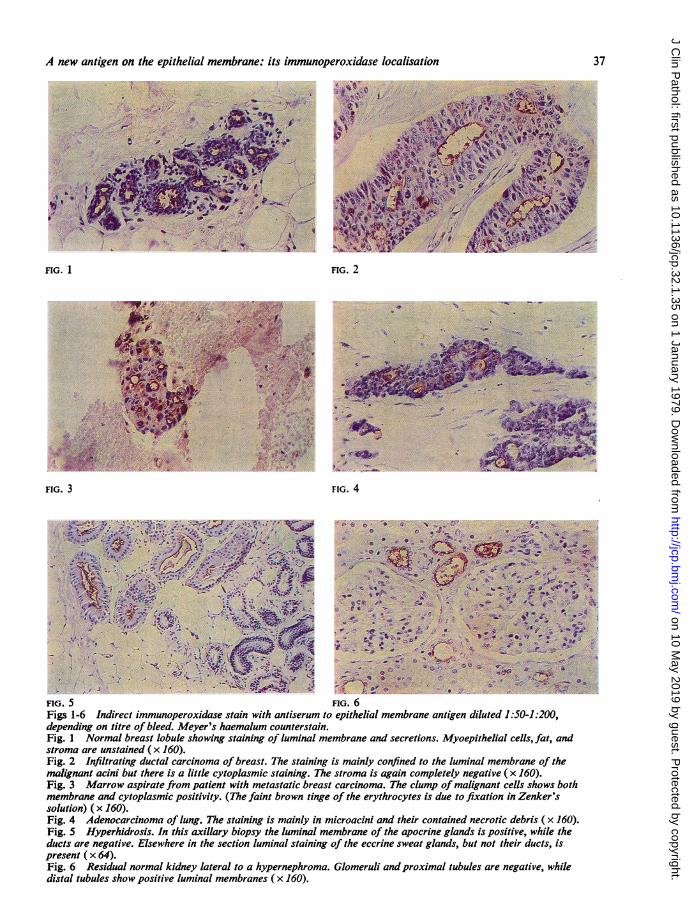
BREASTOn an immunodiffusion plate the antisera raisedagainst defatted cream demonstrated a multi-plicity of precipitin lines against milk and differentextracts of the milk fat globule membrane or cream.After absorption with plasma, extracts of kidney andliver, normal cross-reacting antigen, and lactoferrin,precipitin lines could no longer be detected byimmunodiffusion. In contrast, sections of normalor malignant breast were still stained by the immuno-peroxidase technique. The antisera reacted with allthe mammary tissues studied-lactating, benign,and malignant. The most striking feature was thelocalisation of staining on the surface of the luminalmembrane (see Fig. 1). It is this that leads us toconclude that the antisera detect an epithelialmembrane antigen.
In moderately or well differentiated mammarycarcinomas the antigen was mainly concentrated onthe luminal border of the malignant acini (Fig. 2).In poorly differentiated adenocarcinomas the antigenwas also found in the cytoplasm; a similar findinghas been reported for the colonic membrane antigen,carcinoembryonic antigen (Heyderman and Neville,1976; Pascal et al., 1977).The presence of epithelial membrane antigen in
the cytoplasm allowed the observation of mammarycarcinoma cells infiltrating bone marrow; in largerclumps the luminal membranes of small acini werepicked out (Fig. 3). A xenograft of a primary breastcarcinoma in nude mice (prepared for us by MsS. I. Detre) continued to express epithelial membrane,
36
on 10 May 2019 by guest. P
rotected by copyright.http://jcp.bm
j.com/
J Clin P
athol: first published as 10.1136/jcp.32.1.35 on 1 January 1979. Dow
nloaded from
A new antigen on the epithelial memnbrane: its immunoperoxidase localisation
FIG. 1 FIG. 2
$?.2IG.-P 3 G 4
.Q ~ ~ '.
FIG. 3 FIG. 4
139k~~~~~~~~~~~~~~~~u % I' ItFig. 1 Noa bO
Fg3Marro aprat from pain wit 61ati bras cacnm.Tecupoalgatclssosbt
membrane andcyto&*tv .(ThefainV tbrw t o'!
soluion ( ~ .rxr~'* 160). .A..
-irecy .wDc#;72:,
Figs 1- InderirectimmuInopero xidase stinpswtheatsermitnpihalmembraneoth pciegantgndsiluostede1 hlSO he20Fig.t1 NormabrgtieasElobulee soingtescio ustaining ofluialmmbae and seceatiolnds.Myoepitthelia cells,fatanstroman ar untie (x460)maignan acnbeiutl therea kisnea lttleractoplasi staining.Thestomais agand roxmpleteblysarnegative,(xh160
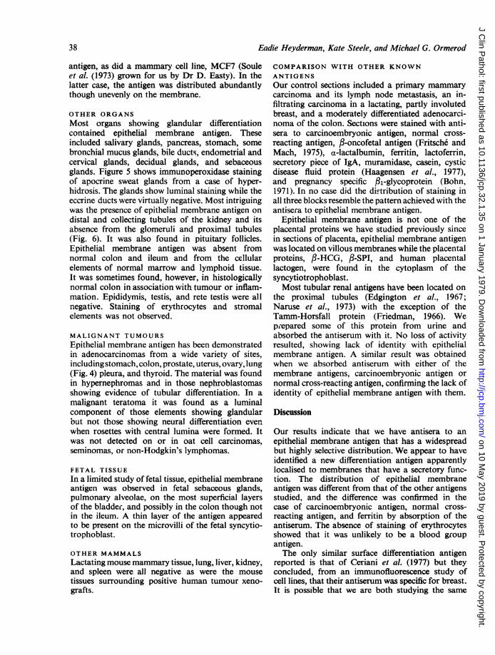
Fig.a5 HypuerhidrposisInthisvxlayipyteluminal membranes of th apcin6lnd0spoiivwil.h
ducs ae ngatve Elewhre n te ecton umial tanin oftheeccin swat lans, utnotther dcts i
prsn (x 64).Fig. 16 Residual normalkinoeylateraStoai wihypeneproma Glomeruliandpemroximalntubuesarlued negative0,whldist1oraltubulesthowbpostivehluing tiigoualmembranesad(xeins 160).lilcel, a, n
37
on 10 May 2019 by guest. P
rotected by copyright.http://jcp.bm
j.com/
J Clin P
athol: first published as 10.1136/jcp.32.1.35 on 1 January 1979. Dow
nloaded from
Eadie Heyderman, Kate Steele, and Michael G. Ormerod
antigen, as did a mammary cell line, MCF7 (Souleet al. (1973) grown for us by Dr D. Easty). In thelatter case, the antigen was distributed abundantlythough unevenly on the membrane.
OTHER ORGANSMost organs showing glandular differentiationcontained epithelial membrane antigen. Theseincluded salivary glands, pancreas, stomach, somebronchial mucus glands, bile ducts, endometrial andcervical glands, decidual glands, and sebaceousglands. Figure 5 shows immunoperoxidase stainingof apocrine sweat glands from a case of hyper-hidrosis. The glands show luminal staining while theeccrine ducts were virtually negative. Most intriguingwas the presence of epithelial membrane antigen ondistal and collecting tubules of the kidney and itsabsence from the glomeruli and proximal tubules(Fig. 6). It was also found in pituitary follicles.Epithelial membrane antigen was absent fromnormal colon and ileum and from the cellularelements of normal marrow and lymphoid tissue.It was sometimes found, however, in histologicallynormal colon in association with tumour or inflam-mation. Epididymis, testis, and rete testis were allnegative. Staining of erythrocytes and stromalelements was not observed.
MALIGNANT TUMOURSEpithelial membrane antigen has been demonstratedin adenocarcinomas from a wide variety of sites,including stomach, colon, prostate, uterus, ovary, lung(Fig. 4) pleura, and thyroid. The material was foundin hypernephromas and in those nephroblastomasshowing evidence of tubular differentiation. In amalignant teratoma it was found as a luminalcomponent of those elements showing glandularout not those showing neural differentiation evenwhen rosettes with central lumina were formed. Itwas not detected on or in oat cell carcinomas,seminomas, or non-Hodgkin's lymphomas.
FETAL TISSUEIn a limited study of fetal tissue, epithelial membraneantigen was observed in fetal sebaceous glands,pulmonary alveolae, on the most superficial layersof the bladder, and possibly in the colon though notin the ileum. A thin layer of the antigen appearedto be present on the microvilli of the fetal syncytio-trophoblast.
OTHER MAMMALSLactating mouse mammary tissue, lung, liver, kidney,and spleen were all negative as were the mousetissues surrounding positive human tumour xeno-grafts.
COMPARISON WITH OTHER KNOWNANTIGENSOur control sections included a primary mammarycarcinoma and its lymph node metastasis, an in-filtrating carcinoma in a lactating, partly involutedbreast, and a moderately differentiated adenocarci-noma of the colon. Sections were stained with anti-sera to carcinoembryonic antigen, normal cross-reacting antigen, ,3-oncofetal antigen (Fritsche andMach, 1975), a-lactalbumin, ferritin, lactoferrin,secretary piece of IgA, muramidase, casein, cysticdisease fluid protein (Haagensen et al., 1977),and pregnancy specific /1-glycoprotein (Bohn,1971). In no case did the distribution of staining inall three blocks resemble the pattern achieved with theantisera to epithelial membrane antigen.
Epithelial membrane antigen is not one of theplacental proteins we have studied previously sincein sections of placenta, epithelial membrane antigenwas located on villous membranes while the placentalproteins, 3-HCG, f3-SPI, and human placentallactogen, were found in the cytoplasm of thesyncytiotrophoblast.Most tubular renal antigens have been located on
the proximal tubules (Edgington et al., 1967;Naruse et al., 1973) with the exception of theTamm-Horsfall protein (Friedman, 1966). Weprepared some of this protein from urine andabsorbed the antiserum with it. No loss of activityresulted, showing lack of identity with epithelialmembrane antigen. A similar result was obtainedwhen we absorbed antiserum with either of themembrane antigens, carcinoembryonic antigen ornormal cross-reacting antigen, confirming the lack ofidentity of epithelial membrane antigen with them.
Discussion
Our results indicate that we have antisera to anepithelial membrane antigen that has a widespreadbut highly selective distribution. We appear to haveidentified a new differentiation antigen apparentlylocalised to membranes that have a secretary func-tion. The distribution of epithelial membraneantigen was different from that of the other antigensstudied, and the difference was confirmed in thecase of carcinoembryonic antigen, normal cross-reacting antigen, and ferritin by absorption of theantiserum. The absence of staining of erythrocytesshowed that it was unlikely to be a blood groupantigen.The only similar surface differentiation antigen
reported is that of Ceriani et al. (1977) but theyconcluded, from an immunofluorescence study ofcell lines, that their antiserum was specific for breast.It is possible that we are both studying the same
38
on 10 May 2019 by guest. P
rotected by copyright.http://jcp.bm
j.com/
J Clin P
athol: first published as 10.1136/jcp.32.1.35 on 1 January 1979. Dow
nloaded from

A new antigen on the epithelial membrane: its immunoperoxidase localisation 39
antigen and that the differences in specificity aredue to differences in the method of study. Thisquestion will be resolved by an exchange of reagents,which is being arranged.We have assumed that, after absorption, our
antisera can detect only one component on theepithelial membrane. Our assumption is probablyjustified since a fraction from milk-containingpartially purified epithelial membrane antigen re-moved all remaining activity from the antisera (seeMaterial and methods). Conclusive proof will besupplied only when the antigen has been isolated andcharacterised.
Purification of epithelial membrane antigen isclearly a priority. While human milk offers a con-venient source, our early results indicate that thephysical properties of the soluble antigen depend onthe treatment of the sample of milk.The yield appearsto be rather low, and we might have to use an alterna-tive source of material-possibly a large metastaticdeposit ofmammary carcinoma obtained at necropsy.We speculate that the antigen has a specific
function related directly or indirectly to the secretoryprocess. When we have succeeded in isolating themolecule, a detailed characterisation might yieldclues as to its role, although this is far from assured.Our data on the immunoperoxidase localisation of
epithelial membrane antigen already suggest a rolefor these antisera in the diagnosis and differentialdiagnosis of malignant disease. Several such aspectsare under active investigation.
We thank Professor A. M. Neville for most valuablesupport and guidance, Ms P. Davies for excellenttechnical assistance, and the pathologists whogenerously gave us access to their histologicalmaterial.
References
Bohn, H. (1971). Nachweis und Charakterisierung vonSchwangerschafts-proteinen in der menschlichenPlacenta, sowie ihre quantitative immunologischeBestimmung im Serum schwangerer Frauen. Archivfur Gynaekologie, 210, 440-457.
Ceriani, R. L., Thompson, K., Peterson, J. A., andAbraham, S. (1977). Suiface differentiation antigens ofhuman mammary epithelial cells carried on the humanmilk fat globule. Proceedings of the National Academyof Sciences ofthe United States ofAmerica, 74, 582-586.
Edgington, T. S., Glassock, R. J., Watson, J. I., andDixon, F. J. (1967). Characterisation and isolationi ofspecific renal tubular epithelial antigens. Journal ofImmunology, 99, 1199-1210.
Friedman, T. (1966). Immunofluorescent localisation ofTamm-Horsfall mucoprotein. Experientia, 22, 624-625.
Fritsche, R., and Mach, J. P. (1975). Identification of anew oncofoetal antigen associated with several typesof human carcinomas. Nature, 258, 734-737.
Haagensen, D. E., Jr., Mazoujian, G., Holder, W. D.,Jr., Kister, S. J., and Wells, S. A., Jr. (1977). Evalua-tion of a breast cyst fluid protein detectable in theplasma of breast carcinoma patients. Annals ofSurgery, 185, 279-285.
Heyderman, E. (1978). Multiple tissue markers in humanmalignant testicular tumours. 5th Meeting of the Inter-national Research Group for CarcinoembryonicProteins, Copenhagen. Scandinavian Journal ofImnmun-ology, 7/2, Supplement 8. (In press.)
Heyderman, E., and Neville, A. M. (1976). A shorterimmunoperoxidase technique for the demonstration ofcarcinoembryonic antigen and other cell products.Journal of Clinical Pathology, 30, 138-140.
Nakane, P. K., and Kawaoi, A. (1974). Peroxidase-labeled antibody: a new method of conjugation.Journal of Histochemistry and Cytochemistry, 22,1084-1091.
Naruse, T., Kitamura, K., Miyakawa, Y., and Shibita,S. (1973). Deposition of renal tubular epithelialantigen along the glomerular capillary walls of patientswith membranous glomerulonephritis. Journal ofImmunology, 110, 1163-1166.
Pascal, R. R., Mesa-Tejada, R., Bennett, S. J., Garces,A., and Fenoglio, C. M. (1977). Carcinoembryonicantigen. ArchivesofPathologyandLaboratory Medicines,101, 568-571.
Soake, R. G., and Heald, C. W. (1974). In Lactation,edited by B. L. Lasson and V. R. Smith, Volume 2,pp. 147-187. Academic Press, New York.
Soule, H. D., Vazquez, J., Long, A., Albert, S., andBrennan, M. (1973). A human cell line from a pleuraleffusion derived from a breast carcinoma. Journal ofthe National Cancer Institute, 51, 1409-1413.
Von Kleist, S., Chavanel, G., and Burtin, P. (1972).Identification of an antigen from normal humantissue that cross-reacts with the carcinoembryonicantigen. Proceedings of the National Academy ofSciences of the United States of America, 69, 2492-2494.
Requests for reprints to: Dr E. Heyderman, Unit ofHuman Cancer Biology, Ludwig Institute for CancerResearch, The Haddow Laboratories, Clifton Avenue,Sutton, Surrey SM2 5PX,UK
on 10 May 2019 by guest. P
rotected by copyright.http://jcp.bm
j.com/
J Clin P
athol: first published as 10.1136/jcp.32.1.35 on 1 January 1979. Dow
nloaded from