Embed Size (px)
Citation preview

3,350+OPEN ACCESS BOOKS
108,000+INTERNATIONAL
AUTHORS AND EDITORS115+ MILLION
DOWNLOADS
BOOKSDELIVERED TO
151 COUNTRIES
AUTHORS AMONG
TOP 1%MOST CITED SCIENTIST
12.2%AUTHORS AND EDITORS
FROM TOP 500 UNIVERSITIES
Selection of our books indexed in theBook Citation Index in Web of Science™
Core Collection (BKCI)
Chapter from the book Herpesviridae - A Look Into This Unique Family of VirusesDownloaded from: http://www.intechopen.com/books/herpesviridae-a-look-into-this-unique-family-of-viruses
PUBLISHED BY
World's largest Science,Technology & Medicine
Open Access book publisher
Interested in publishing with IntechOpen?Contact us at [email protected]

4
Contributions of the EBNA1 Protein of Epstein-Barr Virus Toward B-Cell
Immortalization and Lymphomagenesis
Amber T. Washington and Ashok Aiyar LSU Health Sciences Center, New Orleans
United States of America
1. Introduction
Epstein-Barr Virus (EBV) is a human herpesvirus, infecting 95% of humans, that is causally
associated with the benign B-cell proliferative disorder, infectious mononucleosis. EBV has
also been etiologically associated with several cancers, such as Burkitt's lymphoma, AIDS-
related immunoblastic lymphomas, post-transplant lymphomas, nasopharyngeal carcinoma,
and gastric carcinoma. Very few viral genes are expressed in these malignancies and
infectious virus is rarely released, defining a state of infection termed latency. Many EBV-
associated malignancies respond poorly to treatment with large variability in success rates
dependent on disease stage, viral gene expression patterns, and concurrent
immunosuppressive therapy. Developing broadly effective strategies to suppress cell
proliferation induced by EBV requires careful consideration of viral factors common to
many malignancy types. One interesting viral therapeutic target is the viral protein Epstein-
Barr nuclear antigen 1 (EBNA1), which is expressed in all EBV-associated malignancies and
has been extensively characterized. Upon association with a region on EBV's genome,
termed oriP, EBNA1 facilitates the licensed replication and mitotic segregation of EBV
genomes in proliferating tumor cells. EBNA1 bound to oriP also activates transcription from
two major EBV promoters. Finally, EBNA1 is known to suppress apoptosis of EBV-positive
tumor cells. In this review, we have described the molecular mechanisms by which EBNA1's
functions are operant.
2. Malignancies associated with Epstein-Barr virus (EBV)
EBV is a double-stranded lymphotropic gammaherpesvirus with a 172-kb linear DNA
genome that is widely distributed in the human population (Kieff and Rickinson 2007;
Rickinson and Kieff 2007). EBV was discovered through its association with B-cell
lymphomas in children and young adults in sub-Saharan Africa. These tumors, now termed
Burkitt's lymphoma, were recognized by Denis Burkitt, a British surgeon, as occurring at an
unusually high rate in regions of Africa where malaria was endemic. Postulating that an
infectious agent was the cause, Burkitt provided tumor biopsies to Anthony Epstein and
Yvonne Barr, who screened them for the presence of viral-like particles using electron
www.intechopen.com

Herpesviridae – A Look into This Unique Family of Viruses
66
microscopy (Epstein, Achong et al. 1964). EBV subsequently became the first virus to be
isolated from human cancer (Henle, Henle et al. 1968). Soon after this discovery,
epidemiological studies revealed that a primary acute infection with EBV was causally
associated with the development of infectious mononucleosis in adolescence and adulthood
(Niederman, McCollum et al. 1968). Similar studies revealed a causal association between
EBV and Burkitt's lymphoma (de-The, Geser et al. 1978). Molecular studies conducted since
1975 have revealed EBV genomes and expressed genes to be present in several other
malignancies.
EBV is currently associated with Hodgkin's lymphoma, non-Hodgkin's lymphoma, AIDS-
related immunoblastic lymphomas, primary effusion lymphomas, post-transplant
lymphomas, CNS lymphomas, gastric carcinoma, and nasopharyngeal carcinoma
(Crawford, Thomas et al. 1980; Purtilo and Klein 1981; Ernberg and Altiok 1989; Glaser,
Lin et al. 1997; Dockrell, Strickler et al. 1998; Taylor, Marcus et al. 2005; Navarro and
Kaplan 2006). Indeed, together with Kaposi's sarcoma, lymphomas caused by EBV were
the first malignancies identified as AIDS-defining clinical conditions (1987). In Figure 1,
we have depicted a brief overview of the EBV life-cycle. During primary infection, EBV is
transmitted by saliva to oral epithelial cells in which it replicates lytically (Kieff and
Rickinson 2007; Rickinson and Kieff 2007). Released virus infects circulating B-
lymphocytes using the B-cell surface proteins MHC class II and complement receptor 2
(CR2/CD21) as viral receptors (Fingeroth, Weis et al. 1984; Li, Spriggs et al. 1997). A small
subset of viral genes, including EBNA1, are expressed in these infected B-cells that
concurrently home to the closest lymphoid tissue, such as the Waldeyer's tonsillar ring,
where they proliferate (Laichalk, Hochberg et al. 2002). These latently infected B-cells
proliferate rapidly, a process driven by the expression of viral proteins that are described
in the next section.
It is pertinent to note that these infected B-cells are latently infected; while they express viral
genes that drive cell proliferation, they do not express the large number of viral genes
required for production of infectious virus. Cytokines released by rapidly proliferating
infected B-cells promote a strong primary CTL response that suppresses their proliferation
(Rickinson, Lee et al. 1996; Steven, Leese et al. 1996). Infected B-cells that are not deleted by
the CTL response exist in peripheral circulation as quiescent memory B-cells in which
EBNA1 is typically the only viral protein expressed (Babcock, Decker et al. 1999). With a
very low frequency, EBV's lytic replication is activated in these latently infected cells,
releasing virus that is ultimately transmitted through oral mucosa (Laichalk, Hochberg et al.
2002). If EBV-positive memory B-cells transit to a regional lymph node or back to the tonsils,
they can return to a highly proliferative state (ibid). When this occurs, a strong secondary
CTL response limits their proliferation.
Under immunosuppressive conditions, such as a prior malarial infection, HIV-disease, or
post-transplantation immunosuppressive therapy, an impaired CTL response permits the
unimpeded proliferation of EBV-infected cells (Rickinson, Lee et al. 1996; Kieff and
Rickinson 2007). Mutations in cellular genes caused by errors in DNA replication initially
result in oligoclonal proliferative disorders, such as post-transplant lymphoproliferative
disease; the progressive acquisition of additional mutations ultimately results in clonal
malignancies, such as Burkitt's lymphoma (Hammerschmidt 2011; Vereide and Sugden
2011)
www.intechopen.com
Contributions of the EBNA1 Protein of Epstein-Barr Virus Toward B-Cell Immortalization and Lymphomagenesis
67
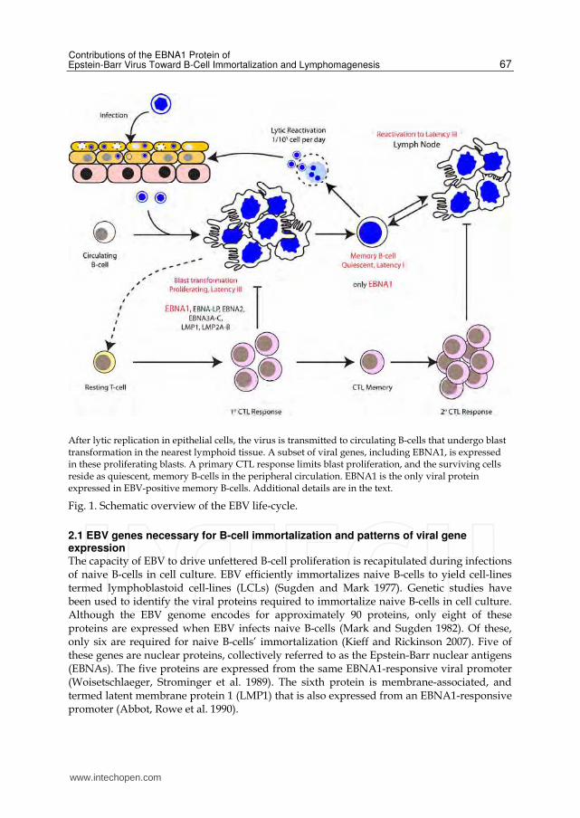
After lytic replication in epithelial cells, the virus is transmitted to circulating B-cells that undergo blast transformation in the nearest lymphoid tissue. A subset of viral genes, including EBNA1, is expressed in these proliferating blasts. A primary CTL response limits blast proliferation, and the surviving cells reside as quiescent, memory B-cells in the peripheral circulation. EBNA1 is the only viral protein expressed in EBV-positive memory B-cells. Additional details are in the text.
Fig. 1. Schematic overview of the EBV life-cycle.
2.1 EBV genes necessary for B-cell immortalization and patterns of viral gene expression The capacity of EBV to drive unfettered B-cell proliferation is recapitulated during infections of naive B-cells in cell culture. EBV efficiently immortalizes naive B-cells to yield cell-lines termed lymphoblastoid cell-lines (LCLs) (Sugden and Mark 1977). Genetic studies have been used to identify the viral proteins required to immortalize naive B-cells in cell culture. Although the EBV genome encodes for approximately 90 proteins, only eight of these proteins are expressed when EBV infects naive B-cells (Mark and Sugden 1982). Of these, only six are required for naive B-cells’ immortalization (Kieff and Rickinson 2007). Five of these genes are nuclear proteins, collectively referred to as the Epstein-Barr nuclear antigens (EBNAs). The five proteins are expressed from the same EBNA1-responsive viral promoter (Woisetschlaeger, Strominger et al. 1989). The sixth protein is membrane-associated, and termed latent membrane protein 1 (LMP1) that is also expressed from an EBNA1-responsive promoter (Abbot, Rowe et al. 1990).
www.intechopen.com
Herpesviridae – A Look into This Unique Family of Viruses
68
The EBV proteins expressed during immortalization have numerous functions that are
described briefly here. LMP1 is a homolog of the B-cell membrane protein CD40. While
CD40 requires a ligand to be activated, LMP1 is constitutively active (Kaye, Izumi et al.
1993; Zimber-Strobl, Kempkes et al. 1996; Gires, Zimber-Strobl et al. 1997). Akin to
activated CD40, it activates NFκB, AP-1, and JNK pathways, and their cellular targets, to
sustain B-cell proliferation (Laherty, Hu et al. 1992; Mosialos, Birkenbach et al. 1995;
Kieser, Kaiser et al. 1999). The five EBNA proteins have distinct, as well as some
overlapping, functions. EBNA2 subverts the cellular Notch pathway (Grossman,
Johannsen et al. 1994; Henkel, Ling et al. 1994). Like the intracellular domain of Notch,
Notch-IC, EBNA2 associates with the transcription repressor human suppressor of
hairless (hSH) converting it into an activator. This complex activates transcription from
the same two viral promoters activated by EBNA1, and the cellular Enhancer of Split
complex genes. While EBNA-LP greatly augments the efficiency with which EBV
immortalizes naive B-cells, it is not required for immortalization (Hammerschmidt and
Sugden 1989). In either event, it acts in concert with EBNA2 at specific viral promoters
(Harada and Kieff 1997; Ling, Peng et al. 2005; Peng, Moses et al. 2005). EBNA3A and
EBNA3C are similar in sequence and have some overlapping functions. They can both act
to modulate the activation of hSH-responsive genes by EBNA2 by interacting with hSH
(Robertson, Lin et al. 1996; Zhao, Marshall et al. 1996; Dalbies-Tran, Stigger-Rosser et al.
2001). However, only EBNA3C alters the expression of the metastatic suppressor Nm23-
H1 (Murakami, Kaul et al. 2009). Finally, recent evidence indicates that EBNA3A and
EBNA3C can individually, and cooperatively, down-modulate the expression of the pro-
apoptotic cellular protein Bim (Paschos, Smith et al. 2009).
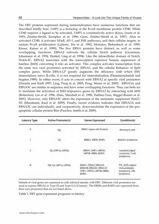
Subsets of viral genes are expressed in cells infected latently with EBV. Different viral promoters are used to express EBNA1 in Type III and Type 0/I/II latency. The EBERs and BARTs are expressed from their own promoters that are not listed above.
Table 1. EBV gene expression programs in latency.
www.intechopen.com
Contributions of the EBNA1 Protein of Epstein-Barr Virus Toward B-Cell Immortalization and Lymphomagenesis
69
In addition to the five EBNA proteins and LMP1, there are three other EBV proteins expressed
during latent infection. These proteins are EBNA3B, LMP2A and LMP2B (Kieff and Rickinson
2007; Rickinson and Kieff 2007). Two non-coding RNAs, termed EBER1 and EBER2, as well as
a variable number of viral microRNAs, are also expressed during latency (ibid). The six
proteins required for immortalization (EBNA1, EBNA2, EBNA3A, EBNA3C, EBNA-LP,
LMP1) are expressed in four distinct programs termed Latency types 0, I, II, and III (ibid).
These programs, indicated in Table I, are associated with specific cellular phenotypes. All six
proteins are expressed in type III latency, which is a gene expression pattern observed in
lymphoblastoid cell lines immortalized by EBV. Type III latency is also observed in post-
transplant lymphomas and AIDS-related immunoblastic lymphomas. A more restricted
pattern of gene expression is observed during the other latency types. Only EBNA1 is
expressed in most Burkitt's lymphoma cells, which is a pattern termed type I latency. Latency
type 0 is observed in infected memory B-cells where only EBNA1 expression is detected, and
that too only when cells divide. No other viral proteins are expressed during latency type 0.
Finally, latency type II is observed in rare T-cell lymphomas and EBV-associated carcinomas.
In this pattern, the expression of EBNA1, LMP1, LMP2A, and LMP2B is detected.
The differential expression of the EBNA proteins in these latency types results from the use
of two different promoters for the expression of EBNA1. During latency type III, the
chromatin conformation and epigenetic markup of the viral genome favors the activation of
the viral BamHI-C promoter (BamHI-Cp) by EBNA1 and EBNA2 (Day, Chau et al. 2007;
Tempera, Klichinsky et al. 2011). All the six EBNA proteins are expressed from spliced
transcripts that originate at BamHI-Cp. During latency type I, an alternative conformation
and markup represses BamHI-Cp (ibid). When this occurs, the viral BamHI-Q promoter
(BamHI-Qp) is activated; splicing of BamHI-Qp transcripts permits the expression of
EBNA1 but not the other EBNAs (ibid). Irrespective of the promoter used, EBNA1 is the only
viral protein expressed in all four latency types, rendering it an excellent target for therapies
directed against EBV-infected cells.
3. EBNA1, its domains, and their functions
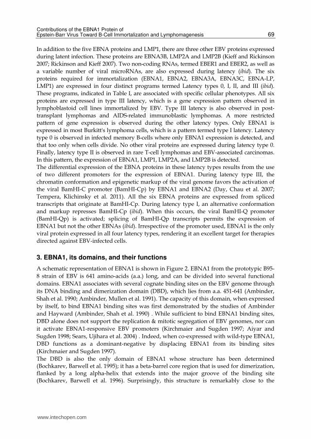
A schematic representation of EBNA1 is shown in Figure 2. EBNA1 from the prototypic B95-
8 strain of EBV is 641 amino-acids (a.a.) long, and can be divided into several functional
domains. EBNA1 associates with several cognate binding sites on the EBV genome through
its DNA binding and dimerization domain (DBD), which lies from a.a. 451-641 (Ambinder,
Shah et al. 1990; Ambinder, Mullen et al. 1991). The capacity of this domain, when expressed
by itself, to bind EBNA1 binding sites was first demonstrated by the studies of Ambinder
and Hayward (Ambinder, Shah et al. 1990) . While sufficient to bind EBNA1 binding sites,
DBD alone does not support the replication & mitotic segregation of EBV genomes, nor can
it activate EBNA1-responsive EBV promoters (Kirchmaier and Sugden 1997; Aiyar and
Sugden 1998; Sears, Ujihara et al. 2004) . Indeed, when co-expressed with wild-type EBNA1,
DBD functions as a dominant-negative by displacing EBNA1 from its binding sites
(Kirchmaier and Sugden 1997).
The DBD is also the only domain of EBNA1 whose structure has been determined (Bochkarev, Barwell et al. 1995); it has a beta-barrel core region that is used for dimerization, flanked by a long alpha-helix that extends into the major groove of the binding site (Bochkarev, Barwell et al. 1996). Surprisingly, this structure is remarkably close to the
www.intechopen.com
Herpesviridae – A Look into This Unique Family of Viruses
70
structure of the DNA binding domain of the E2 protein (E2DBD) from bovine papilomavirus (BPV) (Hegde, Grossman et al. 1992), despite a lack of sequence conservation. In this context, it should be noted that BPV, a small DNA virus, is very distantly related to EBV, and the related structure of DBD and E2DBD is proposed to result from convergent evolution (Grossman and Laimins 1996). The modular nature of the DBD is reflected by several observations. Sugden and co-workers demonstrated that the DBD could be substituted by DNA-binding domain from the yeast GAL4 protein (Mackey, Middleton et al. 1995; Mackey and Sugden 1997). The chimeric protein displayed several biochemical properties of EBNA1 when bound to GAL4 binding sites. In a similar observation, we have shown that a chimeric protein in which a.a. 1-450 of EBNA1 was fused to E2DBD activated transcription from a cluster of E2 binding sites in a manner similar to WT EBNA1 (Aras, Singh et al. 2009). We have also demonstrated that a chimeric protein in which a strong heterologous acidic activation domain was fused to DBD activated transcription with the characteristics of the heterologous activation domain (ibid).
(A) EBNA1 is 641 a.a. in length. ATH1 and ATH2 are two AT-hooks, and UR1 is a domain necessary for transactivation. The GAr is a repeat of glycine and alanine, which can vary in length between EBV isolates. The DBD is used to bind defined sites on EBV's genome. N represents EBNA1's NLS. (B) ATH1 and ATH2 are highly conserved, 80% and 77% respectively, in EBNA1 orthologs from other EBV-like gammaherpesviruses. These regions have a repeated sequence of glycine and arginine. A portion of UR1 is also highly conserved in EBNA1 orthologs. UR1 contains a conserved cys-x-x-cys motif. (C) In addition to DBD, genome replication and segregation requires ATH1 and ATH2. For transactivation, EBNA1 needs both AT-hooks and UR1.
Fig. 2. Schematic diagram of EBNA1 and its domains.
www.intechopen.com
Contributions of the EBNA1 Protein of Epstein-Barr Virus Toward B-Cell Immortalization and Lymphomagenesis
71
EBNA1 contains at least one nuclear localization sequence (NLS) between a.a. 379-386. When fused to a cytoplasmic protein, the NLS is sufficient to render it nuclear (Ambinder, Mullen et al. 1991). Consistent with its function, the NLS interacts with the nuclear transporters karyopherin alpha1 and alpha2 (Fischer, Kremmer et al. 1997; Kim, Maher et al. 1997). However, an EBNA1 NLS mutant that is substantially reduced in its association with the karyopherins remains nuclear and is functional (Kim, Maher et al. 1997). Therefore, it is likely that EBNA1 has additional NLS signals that are currently unknown. While B95-8 EBNA1 is 641 a.a. long, EBNA1 proteins from other EBV strains and isolates
vary in size. This difference in size arises from differences in the length of a central glycine-
alanine repeat region (GAr) (Falk, Gratama et al. 1995). Reductions in the length of the GAr
to just 15 a.a. have no effect on the efficiency of B-cell immortalization by EBV (Lee,
Diamond et al. 1999), EBNA1's capacity to activate transcription (Aiyar and Sugden 1998), or
its ability to support replication and mitotic segregation of EBV genomes (Yates, Warren et
al. 1985). It is now appreciated that the GAr reduces the efficiency with which EBNA1
epitopes are presented on the surface of EBV-infected cells. This reduction in efficiency is
either a consequence of the GAr affecting processing by the proteosome (Levitskaya,
Sharipo et al. 1997), or by reducing the efficiency with which EBNA1 is translated (Yin,
Manoury et al. 2003; Apcher, Komarova et al. 2009; Apcher, Daskalogianni et al. 2010).
Peculiarly, while cell-culture studies indicate that long GAr sequences substantially reduce
proteosome processing and EBNA1 translation, the presence of a long GAr reduces the
recognition of EBV-infected cells by EBNA1-specific CTLs by 50% or less (Lee, Brooks et al.
2004). Therefore, the conservation of long GArs in various EBV isolates may reflect other
GAr functions, in addition to reduced epitope presentation, that are not recapitulated in cell
culture.
The GAr is flanked by positively charged domains. These domains were originally termed
linking regions 1 and 2 (LR1/LR2) (Figure 2) because they had the capacity to link DNAs
bound by EBNA1 into large multimeric complexes (Mackey, Middleton et al. 1995; Mackey
and Sugden 1997; Mackey and Sugden 1999) . Linking is dependent on the capacity of LR1
and LR2 to directly bind nucleic acids. LR1 and LR2 contain within them repeats of glycine
and arginine (GR repeats) that can associate specifically with AT-rich DNA and G-
quadruplex RNA (Sears, Ujihara et al. 2004; Norseen, Thomae et al. 2008) . These nucleic
acid binding properties are observed for cellular AT-hook proteins that also contain GR
repeats (Huth, Bewley et al. 1997; Reeves 2001; Norseen, Thomae et al. 2008). For this reason,
the GR repeats of EBNA1 are referred to as AT-hook 1 and 2 (ATH1, ATH2) in this review.
Deletion of ATH1 or ATH2 reduces the capacity of EBNA1 to transactivate EBV promoters
and to support genome replication/segregation (Sears, Ujihara et al. 2004; Singh, Aras et al.
2009). Deletion of both ATH1 and ATH2 eliminates both functions (Sears, Kolman et al.
2003). A chimeric protein in which ATH1 and ATH2 are replaced by the cellular AT-hook
protein HMGA1 supports transactivation and genome replication/segregation when bound
to EBNA1 binding sites (Hung, Kang et al. 2001; Sears, Kolman et al. 2003). In contrast, a
chimeric protein in which EBNA1's AT-hooks were replaced by a non-sequence specific
cellular DNA binding protein, HMG1, does not support either function (Sears, Kolman et al.
2003). Therefore, an association between EBNA1's AT-hooks and specific nucleic acids, such
as AT-rich DNA or G-quadruplex RNA, is necessary for EBNA1's functions.
While initially considered to be a single positively-charged domain, it is now appreciated that LR1 contains two distinct domains: 1) ATH1; and 2) A short unique region (UR1) that
www.intechopen.com
Herpesviridae – A Look into This Unique Family of Viruses
72
lies between ATH1 and GAr (Figure 2) (Kennedy and Sugden 2003; Singh, Aras et al. 2009). Deletion mutagenesis has revealed that UR1 is essential for EBNA1 to transactivate, but is not required for EBNA1 to support genome replication/segregation (Kennedy and Sugden 2003). Recent studies have revealed that UR1 contains a short sequence with a cys-x-x-cys motif that is conserved in the EBNA1 orthologs from other EBV-like primate gammaherpesviruses (Aras, Singh et al. 2009). Mutation of the conserved cysteines is sufficient to abrogate EBNA1's capacity to activate transcription, emphasizing the importance of the conserved cys-x-x-cys motif (ibid). EBNA1's ability to transactivate EBV promoters and to support EBV genome
replication/segregation is dependent upon its association with two clusters of cognate
binding sites on the EBV genome (Figure 3). The organization of these two clusters is critical
to EBNA1's EBV-specific functions and therefore is detailed below.
4. OriP, the family of repeats (FR), and the dyad symmetry element (DS)
Adams observed that akin to eukaryotic chromosomes, EBV genomes are replicated once
per cell-cycle during latency, by a process termed licensed DNA replication (Lindner and
Sugden 2007). This pattern of genome replication in which genomes are precisely duplicated
during S phase had not been observed previously for other DNA viruses such as
polyomaviruses, papilomaviruses, and alphaherpesviruses. EBV's unique mode of
replication, coupled with an efficient segregation mechanism, permits viral genomes to be
distributed equally to daughter cells when latently infected cells proliferate (Figure 4) (Sears,
Kolman et al. 2003; Sears, Ujihara et al. 2004; Nanbo, Sugden et al. 2007; Norseen, Thomae et
al. 2008). To identify EBV sequences necessary for licensed replication and mitotic
segregation, Yates and Sugden screened for EBV fragments that conferred these properties
to small plasmids introduced into EBV-positive cells (Yates, Warren et al. 1984). Their
screen identified a fragment with 24 similar sequences arranged in two clusters. This
fragment was termed oriP and is depicted in Figure 3. Upon determining oriP to be sufficient
for the licensed replication and mitotic segregation of plasmids in EBV-positive cells, these
investigators identified EBNA1 as the sole EBV protein necessary for licensed replication
and mitotic segregation of oriP-plasmids (Yates, Warren et al. 1985). Later, it was
determined that EBNA1 bound each of the repeated sequences within oriP as a dimer
(Rawlins, Milman et al. 1985; Ambinder, Shah et al. 1990; Ambinder, Mullen et al. 1991). The
repeats in oriP are arranged in two clusters (Figure 3): 1) A cluster with 20 EBNA1-binding
sites, termed the Family of Repeats (FR); and 2) A cluster with four EBNA1-binding sites
arranged as a dyad (DS) (Lupton and Levine 1985; Reisman, Yates et al. 1985).
Deletion and complementation experiments revealed that EBNA1 bound to FR and DS has distinct functions. Plasmids containing only DS undergo DNA replication in cells expressing EBNA1, but are mitotically unstable and lost within 1-2 cell-cycles. Plasmids containing FR alone are distributed as EBNA1-expressing cells divide, but do not replicate, and therefore ultimately diluted out of a proliferating culture (Yates, Camiolo et al. 2000). These studies indicate that DS functions primarily as a licensed replication origin, and FR functions as an element similar to a chromosomal centromere in that it permits newly-replicated oriP-plasmids to be mitotically stable and segregated (Wysokenski and Yates 1989; Aiyar, Tyree et al. 1998). The studies of Calos and co-workers reiterate these functional assignations (Krysan, Haase et al. 1989; Krysan and Calos 1993). Similar to DS-only plasmids, plasmids
www.intechopen.com
Contributions of the EBNA1 Protein of Epstein-Barr Virus Toward B-Cell Immortalization and Lymphomagenesis
73
containing putative chromosomal replication origins undergo licensed replication but are mitotically unstable and lost within 1-2 cell cycles. Introducing FR into these plasmids permitted them to undergo licensed replication and become mitotically stable in EBNA1-expressing cells (ibid).
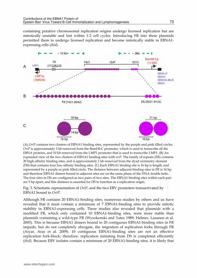
(A) OriP contains two clusters of EBNA1 binding sites, represented by the purple and pink filled circles.
OriP is approximately 3 kb removed from the BamHI-C promoter, which is used to transcribe all the
EBNA proteins, and 10 kb removed from the LMP1 promoter that is used to transcribe LMP1. (B) An
expanded view of the two clusters of EBNA1 binding sites with oriP. The family of repeats (FR) contains
20 high-affinity binding sites, and is approximately 1 kb removed from the dyad symmetry element
(DS) that contains four low-affinity binding sites. (C) Each EBNA1 binding site is 16 bp is length, and
represented by a purple or pink filled circle. The distance between adjacent binding sites in FR is 14 bp,
and therefore EBNA1 dimers bound to adjacent sites are on the same phase of the DNA double helix.
The four sites in DS are configured as two pairs of two sites. The EBNA1 binding sites within each pair
are 5 bp apart, and this distance is essential for DS to function as a replication origin.
Fig. 3. Schematic representation of OriP, and the two EBV promoters transactivated by EBNA1 bound to OriP.
Although FR contains 20 EBNA1-binding sites, numerous studies by others and us have revealed that it must contain a minimum of 7 EBNA1-binding sites to provide mitotic stability in EBNA1-expressing cells. These studies also revealed that plasmids with a modified FR, which only contained 10 EBNA1-binding sites, were more stable than plasmids containing a wild-type FR (Wysokenski and Yates 1989; Hebner, Lasanen et al. 2003). This is because EBNA1 dimers bound to 20 contiguous EBNA1-binding sites in FR impede, but do not completely abrogate, the migration of replication forks through FR (Aiyar, Aras et al. 2009). 10 contiguous EBNA1-binding sites are not an effective replication fork-block; therefore, replication initiating from DS is completed efficiently (ibid). Because EBV isolates contain a minimum of 20 EBNA1-binding sites, it is likely that
www.intechopen.com

Herpesviridae – A Look into This Unique Family of Viruses
74
the capacity of 20 or more contiguous binding sites to limit genome replication is of importance to EBV. The four EBNA1-binding sites in DS are of lower affinity than the binding sites in FR
(Ambinder, Shah et al. 1990). The four sites are arranged in two pairs in which the
distance between the centers of the two sites in each pair is 21 base-pairs (bp) (Figure 3).
Either pair is sufficient for DS to function as an origin, but alterations of the spacing
between them blocks origin function (Harrison, Fisenne et al. 1994; Bashaw and Yates
2001). When EBNA1 binds the sites in DS, it induces a large symmetrical bend in the DNA
and forms a structure necessary for the cellular licensed DNA replication machinery to
function at DS (Bashaw and Yates 2001). There is an additional sequence juxtaposed 3' to
DS, termed Rep*, which can associate with EBNA1 (Kirchmaier and Sugden 1998). The
non-canonical EBNA1 binding sites in Rep* are also 21 bp apart, and multiple copies of
Rep* can substitute for DS for the licensed replication of oriP-plasmids (Wang, Lindner et
al. 2006).
5. Contributions of EBNA1 to the licensed replication and segregation of oriP-plasmids
Although it bends DS, DBD alone is not sufficient to support replication from DS
(Kirchmaier and Sugden 1998). The latter requires contributions from other EBNA1
domains, particularly ATH1 and ATH2. Investigations into the mechanism by which DS
functions as a cell-cycle controlled licensed replication origin have revealed it is similar to
the mechanism that restricts chromosomal replication origins to "fire" only once per cell-
cycle (Lindner and Sugden 2007). During licensed replication of chromosomal DNA, the
cellular origin recognition complex (ORC) marks replication origins throughout the cell-
cycle. Late in mitosis or early in G1, the cellular minichromosome maintenance complex
(MCM) associates with ORC. Phosphorylation events at the G1/S boundary convert the
MCM complex into an active helicase that opens the replication origin and permits DNA
polymerase /primase to be recruited to the origin. MCM functions as the leading strand
helicase and can reassociate with ORC only at the end of mitosis. The inability for MCM to
be re-used during S phase prevents any single origin from being used more than once in a
single cell-cycle. Studies by the groups of Dutta, Lieberman, Schepers, and Yates have
provided insights into the mechanism which DS functions as an origin through (Chaudhuri,
Xu et al. 2001; Dhar, Yoshida et al. 2001; Schepers, Ritzi et al. 2001; Ritzi, Tillack et al. 2003;
Zhou, Chau et al. 2005; Norseen, Thomae et al. 2008). It is now clear that EBNA1 recruits
ORC to DS, with the subsequent cell-cycle dependent recruitment of MCM, thus ensuring
that oriP is subject to the same cell-cycle controlled replication as cellular chromosomes.
Studies from several groups, including ours, indicated that EBNA1's AT-hooks were
essential to recruit ORC to DS (Sears, Ujihara et al. 2004; Norseen, Thomae et al. 2008). This
conclusion has been reiterated by our observations that chimeric proteins in which ATH1
and ATH2 were substituted by cellular AT-hook proteins support licensed replication from
DS (Sears, Kolman et al. 2003; Kelly, Singh et al. 2011). Lieberman and co-workers have
demonstrated that EBNA1 uses its AT-hooks to recruit the ORC complex to DS via a G-
quadruplex RNA intermediate, underscoring a critical role for these domains in oriP-
replication (Norseen, Thomae et al. 2008).
www.intechopen.com
Contributions of the EBNA1 Protein of Epstein-Barr Virus Toward B-Cell Immortalization and Lymphomagenesis
75
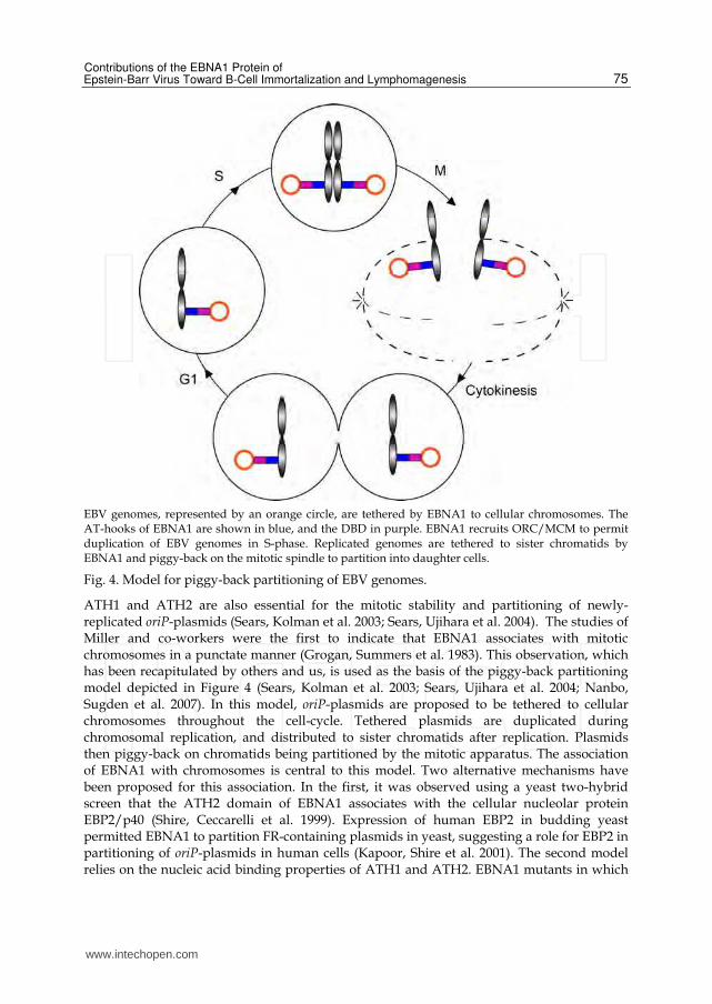
EBV genomes, represented by an orange circle, are tethered by EBNA1 to cellular chromosomes. The AT-hooks of EBNA1 are shown in blue, and the DBD in purple. EBNA1 recruits ORC/MCM to permit duplication of EBV genomes in S-phase. Replicated genomes are tethered to sister chromatids by EBNA1 and piggy-back on the mitotic spindle to partition into daughter cells.
Fig. 4. Model for piggy-back partitioning of EBV genomes.
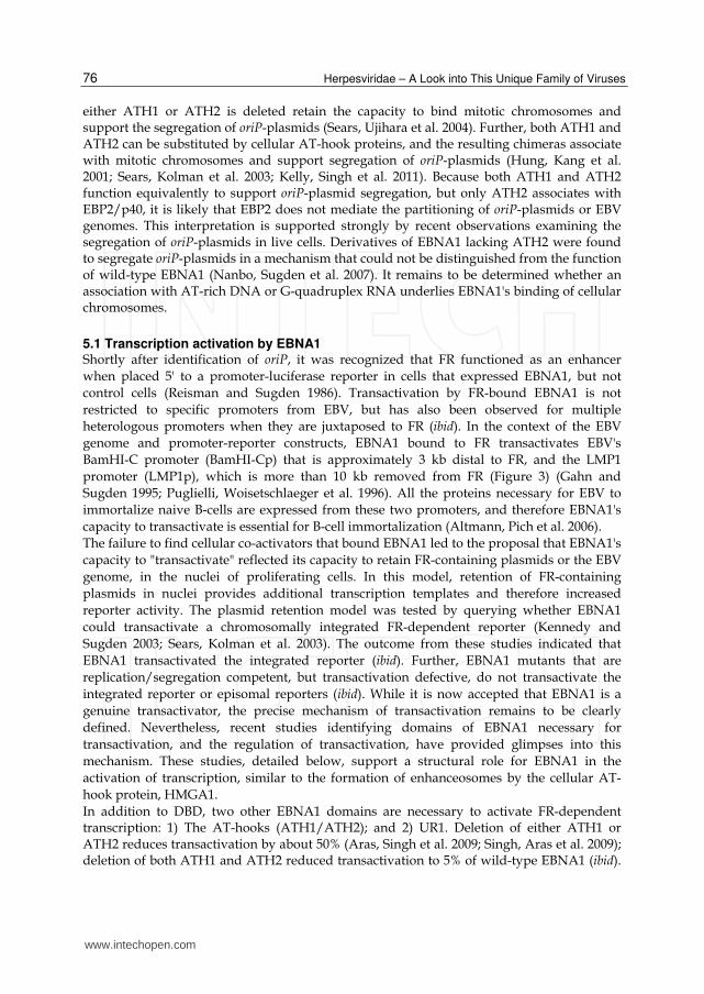
ATH1 and ATH2 are also essential for the mitotic stability and partitioning of newly-replicated oriP-plasmids (Sears, Kolman et al. 2003; Sears, Ujihara et al. 2004). The studies of Miller and co-workers were the first to indicate that EBNA1 associates with mitotic chromosomes in a punctate manner (Grogan, Summers et al. 1983). This observation, which has been recapitulated by others and us, is used as the basis of the piggy-back partitioning model depicted in Figure 4 (Sears, Kolman et al. 2003; Sears, Ujihara et al. 2004; Nanbo, Sugden et al. 2007). In this model, oriP-plasmids are proposed to be tethered to cellular chromosomes throughout the cell-cycle. Tethered plasmids are duplicated during chromosomal replication, and distributed to sister chromatids after replication. Plasmids then piggy-back on chromatids being partitioned by the mitotic apparatus. The association of EBNA1 with chromosomes is central to this model. Two alternative mechanisms have been proposed for this association. In the first, it was observed using a yeast two-hybrid screen that the ATH2 domain of EBNA1 associates with the cellular nucleolar protein EBP2/p40 (Shire, Ceccarelli et al. 1999). Expression of human EBP2 in budding yeast permitted EBNA1 to partition FR-containing plasmids in yeast, suggesting a role for EBP2 in partitioning of oriP-plasmids in human cells (Kapoor, Shire et al. 2001). The second model relies on the nucleic acid binding properties of ATH1 and ATH2. EBNA1 mutants in which
www.intechopen.com
Herpesviridae – A Look into This Unique Family of Viruses
76
either ATH1 or ATH2 is deleted retain the capacity to bind mitotic chromosomes and support the segregation of oriP-plasmids (Sears, Ujihara et al. 2004). Further, both ATH1 and ATH2 can be substituted by cellular AT-hook proteins, and the resulting chimeras associate with mitotic chromosomes and support segregation of oriP-plasmids (Hung, Kang et al. 2001; Sears, Kolman et al. 2003; Kelly, Singh et al. 2011). Because both ATH1 and ATH2 function equivalently to support oriP-plasmid segregation, but only ATH2 associates with EBP2/p40, it is likely that EBP2 does not mediate the partitioning of oriP-plasmids or EBV genomes. This interpretation is supported strongly by recent observations examining the segregation of oriP-plasmids in live cells. Derivatives of EBNA1 lacking ATH2 were found to segregate oriP-plasmids in a mechanism that could not be distinguished from the function of wild-type EBNA1 (Nanbo, Sugden et al. 2007). It remains to be determined whether an association with AT-rich DNA or G-quadruplex RNA underlies EBNA1's binding of cellular chromosomes.
5.1 Transcription activation by EBNA1 Shortly after identification of oriP, it was recognized that FR functioned as an enhancer
when placed 5' to a promoter-luciferase reporter in cells that expressed EBNA1, but not
control cells (Reisman and Sugden 1986). Transactivation by FR-bound EBNA1 is not
restricted to specific promoters from EBV, but has also been observed for multiple
heterologous promoters when they are juxtaposed to FR (ibid). In the context of the EBV
genome and promoter-reporter constructs, EBNA1 bound to FR transactivates EBV's
BamHI-C promoter (BamHI-Cp) that is approximately 3 kb distal to FR, and the LMP1
promoter (LMP1p), which is more than 10 kb removed from FR (Figure 3) (Gahn and
Sugden 1995; Puglielli, Woisetschlaeger et al. 1996). All the proteins necessary for EBV to
immortalize naive B-cells are expressed from these two promoters, and therefore EBNA1's
capacity to transactivate is essential for B-cell immortalization (Altmann, Pich et al. 2006).
The failure to find cellular co-activators that bound EBNA1 led to the proposal that EBNA1's
capacity to "transactivate" reflected its capacity to retain FR-containing plasmids or the EBV
genome, in the nuclei of proliferating cells. In this model, retention of FR-containing
plasmids in nuclei provides additional transcription templates and therefore increased
reporter activity. The plasmid retention model was tested by querying whether EBNA1
could transactivate a chromosomally integrated FR-dependent reporter (Kennedy and
Sugden 2003; Sears, Kolman et al. 2003). The outcome from these studies indicated that
EBNA1 transactivated the integrated reporter (ibid). Further, EBNA1 mutants that are
replication/segregation competent, but transactivation defective, do not transactivate the
integrated reporter or episomal reporters (ibid). While it is now accepted that EBNA1 is a
genuine transactivator, the precise mechanism of transactivation remains to be clearly
defined. Nevertheless, recent studies identifying domains of EBNA1 necessary for
transactivation, and the regulation of transactivation, have provided glimpses into this
mechanism. These studies, detailed below, support a structural role for EBNA1 in the
activation of transcription, similar to the formation of enhanceosomes by the cellular AT-
hook protein, HMGA1.
In addition to DBD, two other EBNA1 domains are necessary to activate FR-dependent transcription: 1) The AT-hooks (ATH1/ATH2); and 2) UR1. Deletion of either ATH1 or ATH2 reduces transactivation by about 50% (Aras, Singh et al. 2009; Singh, Aras et al. 2009); deletion of both ATH1 and ATH2 reduced transactivation to 5% of wild-type EBNA1 (ibid).
www.intechopen.com
Contributions of the EBNA1 Protein of Epstein-Barr Virus Toward B-Cell Immortalization and Lymphomagenesis
77
Similarly, deletion of UR1 reduced transactivation to 5% of WT EBNA1 (Kennedy and Sugden 2003; Aras, Singh et al. 2009; Singh, Aras et al. 2009). These studies also indicate that UR1 and the AT-hooks are individually insufficient to activate transcription; deletion of either domain eliminates the capacity of the remaining domain to promote transactivation (Singh, Aras et al. 2009). Studies using chimeric proteins indicate the nucleic-acid binding properties of ATH1/ATH2 are critical for EBNA1 to transactivate. The chimeric protein HMGA1-UR1-DBD, in which ATH1/ATH2 are replaced by the cellular AT-hook protein, HMGA1, can transactivate when bound to FR (Altmann, Pich et al. 2006). The UR1-deleted EBNA1 mutant was used to establish definitively that EBNA1's capacity to transactivate was necessary for EBV to immortalize naive B-cells. A recombinant EBV in which WT EBNA1 was replaced by this protein established latent infection in transformed B-cell lymphomas, but failed to immortalize naive B-cells (Altmann, Pich et al. 2006). Consistent with immortalization requiring both AT-hooks and UR1, a recombinant EBV in which WT EBNA1 was replaced by HMGA1-UR1-DBD was competent to immortalize naive B-cells (ibid).
5.2 AT-hooks and transactivation Cellular AT-hook proteins function as architectural proteins in transactivation. This
structural function was first revealed for the cellular AT-hook protein, HMGA1, at the -
interferon enhancer by the studies of Thanos and Maniatis (Kim and Maniatis 1997; Yie,
Liang et al. 1997; Yie, Merika et al. 1999). The β-interferon enhancer contains sites bound by
the transactivators ATF2, NFκB, and IRF1, and contains four short AT-rich sites bound by
HMGA1, previously referred to as HMG-I(Y) (Kim and Maniatis 1997; Yie, Merika et al.
1999). A basal level of transcription was observed during in vitro transcription reactions in
which ATF2, NFκB, and IRF1 were provided individually, or as a combination of all three.
Similarly, addition of HMGA1 alone also resulted in basal transcription. However, a
dramatic increase in transcription was observed when all four factors were provided at the
same time. DNA phasing experiments were used to establish a structural role for HMGA1 in
transactivation. A six bp deletion that changed the phasing between two HMGA1-binding
sites by a half-turn of DNA double helix decreased transactivation by 50%. In contrast, a 10
bp deletion, which restored the original DNA phasing, also restored transactivation (ibid).
On this basis, it was proposed that structural changes imposed in the enhancer by HMGA1-
induced DNA bending, and HMGA1 self-association, formed a transactivation complex
termed an "enhanceosome" (Maniatis, Falvo et al. 1998). Other proteins that bind the β-
interferon enhancer, namely ATF2, IRF1, and NFκB, recruited transcription co-activators
only in the context of this enhanceosome (Merika, Williams et al. 1998). The role of HMGA1
at the β-interferon enhanceosome has been recapitulated at other promoters, such as the
early promoter of human papilomavirus type 18 (Bouallaga, Massicard et al. 2000;
Bouallaga, Teissier et al. 2003). Six bp deletions or insertions between HMGA1 binding sites
at this promoter reduce transcription by about 50%. In contrast, no change in transcription is
observed consequent to 10 bp deletions or insertions (Bouallaga, Massicard et al. 2000). At
this enhanceosome as well, HMGA1 is necessary to recruit transcription co-activators
(Bouallaga, Teissier et al. 2003).
The unusual property of phasing dependent transactivation that was previously observed only for HMGA1 has been observed for EBNA1 (Hebner, Lasanen et al. 2003). The center-to-center distance between adjacent EBNA1-binding sites in FR is 30 bp, or three turns of the
www.intechopen.com
Herpesviridae – A Look into This Unique Family of Viruses
78
DNA double-helix (Figure 3). Reduction of this distance to 24 bp alters transactivation by 50% without affecting EBNA1's capacity to bind adjacent sites (ibid). EBNA1's phasing-dependent transactivation provided the first indication that it functions as an architectural transactivator. Studies with inhibitors indicate the AT-rich DNA binding property of ATH1/ATH2 to be critical for EBNA1 to transactivate. The peptidomimetic netropsin, which displaces AT-hook proteins from AT-rich DNA (Wartell, Larson et al. 1974; Freyer, Buscaglia et al. 2007), also reduces the capacity of EBNA1 to transactivate at BamHI-Cp (Sears, Ujihara et al. 2004). Deletion of ATH1 reduces transactivation to 50% of WT EBNA1, and the reduction is reversed when ATH1 is substituted by AT-hooks from HMGA1 (Singh, Aras et al. 2009). While it is apparent that EBNA1's AT-hooks are necessary for transactivation, how they function bears clarification. AT-hook proteins typically bind AT-rich sequences that are close to the transcription start-site. Although AT-rich sequences are found juxtaposed to BamHI-Cp, and increase BamHI-Cp activity in reporter assays (Walls and Perricaudet 1991), it is yet to be determined if this increase is EBNA1-dependent and if EBNA1 associates with these sequences.
5.3 The role of UR1 in transactivation Because the UR1-deleted EBNA1 mutant is transactivation defective, efforts have focused on cellular transcription co-activators that associate with UR1. However, several yeast two-hybrid screens (Fischer, Kremmer et al. 1997; Kim, Maher et al. 1997; Aiyar, Tyree et al. 1998), and proteomic analyses, have failed to identify co-activators that bind EBNA1. It has been reported recently that the chromatin remodeling protein Brd4 interacts with EBNA1 in yeast (Lin, Wang et al. 2008). While both over-expression and depletion of Brd4 reduced EBNA1's capacity to transactivate (ibid), these conditions also cause a striking reduction in cell viability (Schweiger, Ottinger et al. 2007). Therefore, it is currently not possible to assign a role for Brd4 in EBNA1's capacity to transactivate.
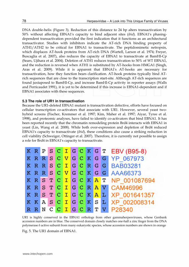
UR1 is highly conserved in the EBNA1 orthologs from other gammaherpesviruses, whose Genbank accession numbers are in blue. The conserved domain closely matches one-half a zinc finger from the DNA
polymerase active subunit from many eukaryotic species, whose accession numbers are shown in orange
Fig. 5. The UR1 domain of EBNA1.
www.intechopen.com
Contributions of the EBNA1 Protein of Epstein-Barr Virus Toward B-Cell Immortalization and Lymphomagenesis
79
During our studies to identify co-activators that associated with UR1, we observed that a sub-sequence within UR1 was highly conserved in the UR1 domains of EBNA1 orthologs from other EBV-like gammaherpesviruses (Aras, Singh et al. 2009). The conserved sequence, KRPSCIGCK, also resembles one-half of a C4 zinc-finger present in the catalytic subunit of
DNA polymerase from multiple eukaryotes (Figure 5). In zinc fingers, the group 12 metal zinc is coordinated between two cys-x-x-cys containing sequences of a protein, bringing these two segments together. Because EBNA1 and its orthologs do not contain another pair of cysteines in a cys-x-x-cys configuration, it is unlikely that zinc is coordinated by a single molecule of EBNA1. Sometimes, zinc coordination between dicysteine motifs in two separate proteins is used to mediate an association between the two proteins. This is
exemplified by the interaction between the C-terminal cytoplasmic tail of CD4 or CD8 with the Lck kinase (Huse, Eck et al. 1998; Lin, Rodriguez et al. 1998; Kim, Sun et al. 2003). Therefore, we postulated, and experimentally confirmed, that: 1) UR1 bound zinc; and 2) Zinc coordination resulted in UR1 self-association (Aras, Singh et al. 2009). The role of the cys-x-x-cys motif within UR1 for both properties was confirmed by replacing both cysteines with serines. This mutant UR1 did not bind zinc or self-associate (ibid). Zinc is essential for EBNA1 to transactivate. Chelation of zinc using the cell-permeable zinc
chelator N,N,N’,N’-tetrakis(2-pyridylmethyl)ethylenediamine (TPEN) severely reduced
EBNA1's capacity to transactivate without affecting levels of EBNA1 or cell viability (ibid).
The specific effect of TPEN on EBNA1-dependent transactivation was confirmed by
determining it had no effect on transcription from EBNA1-independent promoters, on the
function of zinc-independent transactivators. Consistent with the chelation studies, an
EBNA1 mutant in which both UR1 cysteines were altered to serine transactivates as poorly
as UR1-deleted EBNA1 (ibid). Biochemical studies indicate that zinc is coordinated
intermolecularly between adjacent EBNA1 dimers bound to adjacent sites in FR, rather than
intramolecularly within an EBNA1 dimer bound to a single site in FR (ibid). Intermolecular
coordination results in a large structured array of EBNA1 at FR that is essential for EBNA1
to transactivate cooperatively. Confirming this conclusion, the EBNA1 mutant in which UR1
cysteines were changed to serines does not transactivate cooperatively. On this basis, it is
proposed that a zinc-coordinated array of EBNA1 at FR creates a structured complex within
which EBNA1's AT-hooks can form an enhanceosome at promoter proximal sequences
(Aras, Singh et al. 2009; Singh, Aras et al. 2009). The model mechanistically explains the co-
dependent roles of UR1 and the AT-hooks in transactivation. In the absence of UR1,
EBNA1's AT-hooks cannot form a structured complex at promoter proximal AT-rich
sequences. In the absence of the AT-hooks, EBNA1 bound to FR lacks the domains necessary
to bind such sequences. This model also explains why alteration of phasing between
adjacent EBNA1-binding sites in FR affects transactivation. Adjacent EBNA1 dimers bound
to sites that lie on opposite sides of DNA will coordinate zinc with different kinetics than
dimers bound to adjacent sites on the same side of DNA. This in turn will alter the structure
necessary for effective enhanceosome formation, as observed previously for the function of
HMGA1 in enhanceosomes (Kim and Maniatis 1997; Bouallaga, Massicard et al. 2000).
5.4 Regulation of transactivation by phosphorylation EBNA1 contains multiple serine and threonine residues in sequence contexts that resemble recognition sites for protein kinases including cAMP-dependent protein kinase, protein kinase C, glycogen synthase kinase, and mitogen activated protein kinase (PKA)
www.intechopen.com
Herpesviridae – A Look into This Unique Family of Viruses
80
(Duellman, Thompson et al. 2009). Indeed, a sequence recognized by PKA, KRxS, is conserved in UR1 (Figure 5), and the conserved serine is phosphorylated in vivo (ibid). Because this sequence is juxtaposed to the critical cys-x-x-cys motif within UR1, we created three mutants of EBNA1 in which this serine was altered to alanine, aspartic acid, or threonine (Singh, Aras et al. 2009). These substitutions were chosen because alanine resembles an unphosphorylated serine, aspartic acid resembles a phosphorylated serine, and threonine restores a PKA recognition site. All three substitutions did not affect the half-life of EBNA1, its nuclear localization, or the efficiency of transactivation (ibid). Therefore, although this site is phosphorylated in vivo, phosphorylation does not affect any property or function of EBNA1. Subsequent to our analysis, Duellman and Burgess created a mutant derivative of EBNA1 in which all 10 serines known to be phosphorylated were simultaneously substituted by alanine (Duellman, Thompson et al. 2009). This mutant EBNA1 displayed no defects in half-life, expression level, or nuclear localization. There were minor reductions in the efficiency of replication/segregation of oriP-plasmids and transactivation (ibid). Therefore, it appears unlikely that phosphorylation is a major mechanism that regulates the activities of EBNA1. This conclusion was also drawn from studies in which activators and inhibitors of various kinases were observed to not affect EBNA1's capacity to transactivate.
5.5 Transactivation is sensitive to oxidative stress and regulated by cellular redox effectors Many viral and mammalian transactivators, including AP-1, NFκB, the Tat protein of
human immunodeficiency virus (HIV), and the E2 protein of papillomavirus, are regulated
by redox (Hutchison, Matic et al. 1991; Xanthoudakis and Curran 1992; Xanthoudakis, Miao
et al. 1992; Huang and Adamson 1993; Mitomo, Nakayama et al. 1994; Xanthoudakis and
Curran 1996; Jayaraman, Murthy et al. 1997; Okamoto, Tanaka et al. 1999; Kalantari,
Narayan et al. 2008; Wan, Ottinger et al. 2008; Washington, Singh et al. 2010). These proteins
contain one or several cysteine residues susceptible to oxidation, whose redox status
regulates the capacity to transactivate, due to the effect of oxidative stress on protein
structure. Upon exposure to superoxide and hydroxyl radicals, the thiol groups of cysteine
can either be oxidized to form a disulfide bond, or progressively oxidized to form sulfenic
acid, sulfinic acid, and finally sulfonic acid via the Fenton reaction (Held, Sylvester et al.
1996). Proteins containing zinc fingers essential for their function are especially sensitive to
oxidative stress because the thiol group, but not oxidized thiol adducts, can coordinate zinc
(Kiley and Storz 2004). In light of EBNA1's UR1 domain containing two conserved cysteines
that are critical for transactivation, we tested whether transactivation was susceptible to
oxidative stress or alterations in environmental oxygen tension (Aras, Singh et al. 2009;
Washington, Singh et al. 2010). Exposure of EBNA1-expressing cells to low levels of agents
that generate intracellular hydroxyl and superoxide radicals, such as menadione and
paraquat, dramatically reduced transactivation without substantially affecting cell survival
or proliferation. In contrast, lowering the oxygen tension by placing EBNA1 expressing cells
in hypoxic conditions extended the half-life of transactivation (ibid). The latter result
suggests that although EBNA1 is very stable, the critical cysteines within UR1 are subject to
intracellular oxidative stress, reducing EBNA1's capacity to transactivate over time. Hypoxic
conditions reduce the generation of intracellular oxidative radicals, and therefore prolong
the activity of EBNA1 as a transactivator (ibid).
www.intechopen.com
Contributions of the EBNA1 Protein of Epstein-Barr Virus Toward B-Cell Immortalization and Lymphomagenesis
81
Cellular and viral transactivators with redox-sensitive cysteines are often regulated by cellular redox effectors such as thioredoxin, thioredoxin reductase and the bifunctional enzyme AP-endonuclease 1 (APE1) (Hutchison, Matic et al. 1991; Xanthoudakis and Curran 1992; Xanthoudakis, Miao et al. 1992; Huang and Adamson 1993; Mitomo, Nakayama et al. 1994; Xanthoudakis and Curran 1996; Jayaraman, Murthy et al. 1997; Okamoto, Tanaka et al. 1999; Kalantari, Narayan et al. 2008; Wan, Ottinger et al. 2008; Washington, Singh et al. 2010). Thioredoxin and thioredoxin reductase can reduce disulfide bridges that result from cysteine oxidation, and therefore function similarly to small molecules such as b-mercaptoethanol (b-ME) and dithiothreitol (DTT) (Lothrop, Ruggles et al. 2009). APE1 is a bifunctional enzyme with two activities: 1) It is an AP-endonuclease that participates in DNA damage repair (Bhakat, Mantha et al. 2009); and 2) It can reduce the oxidized cysteine adduct sulfenic acid back to a thiol (ibid). Therefore, oxidized cysteine adducts reduced by APE1 are not acted on by b-ME and DTT. We determined that treating cells with b-ME or DTT did not alter EBNA1's ability to transactivate, or ameliorate the effect of oxidative stress on transactivation (Washington, Singh et al. 2010). Therefore, it is unlikely that oxidative stress reduces EBNA1's ability to transactivate by creating intra- or intermolecular disulfide bridges. Consistent with this, over-expression of thioredoxin or thioredoxin reductase also did not increase transactivation, or rescue transactivation from intracellular oxidative stress generated by menadione or paraquat exposure (ibid). In striking contrast, over-expression of APE1 increases EBNA1's ability to transactivate, and curtails the effect of oxidative stress on EBNA1's function as a transactivator (Aras, Singh et al. 2009; Washington, Singh et al. 2010). It is relevant to note that cysteine residues regulated by APE1 are often adjacent to arginine or lysine residues, as observed for the cysteines in UR1 (Figure 5).
6. Functions of EBNA1 in EBV-positive lymphomas
Two patterns of gene expression predominate in EBV-positive lymphomas: 1) The type III latency pattern of gene expression; and 2) The type I latency pattern of gene expression, in which EBNA1 is the only viral protein expressed. In type III lymphomas, EBNA1 drives the expression of other EBV genes necessary for cell proliferation and facilitates virus genome replication/segregation into daughter cells. It is not clear what functions are provided by EBNA1 in type I lymphomas. It is possible that EBNA1 itself provides functions necessary for cell survival and proliferation. One function indicated by the studies of Kennedy and Sugden is that EBNA1 can inhibit or reduce p53-dependent apoptosis (Kennedy, Komano et al. 2003). In addition, it is possible that by facilitating genome replication/segregation, EBNA1 permits the expression of EBV microRNAs in type I lymphoma cells (Cai, Schafer et al. 2006; Choy, Siu et al. 2008; Pratt, Kuzembayeva et al. 2009). Finally, as described in the next section, several studies indicate that EBNA1 can transactivate some cellular genes, and it is possible that one or a combination of these genes is necessary for lymphoma survival/proliferation (Wood, O'Neil et al. 2007; Baumforth, Birgersdotter et al. 2008; Flavell, Baumforth et al. 2008; O'Neil, Owen et al. 2008; Gruhne, Sompallae et al. 2009). In either event, it is of interest that introduction of a dominant-negative derivative of EBNA1 into EBV-positive Burkitt's lymphoma cells severely decreases their rate of proliferation, and dramatically increases their apoptosis, reiterating the utility of devising therapies against EBNA1 (Kennedy, Komano et al. 2003; Nasimuzzaman, Kuroda et al. 2005).
www.intechopen.com

Herpesviridae – A Look into This Unique Family of Viruses
82
6.1 Modulation of cellular gene expression by EBNA1 Recent studies show that EBNA1 alters the transcriptional pattern of several cellular genes (Dawson, Rickinson et al. 1990; Falk, Gratama et al. 1995; Wood, O'Neil et al. 2007; Baumforth, Birgersdotter et al. 2008; Flavell, Baumforth et al. 2008; O'Neil, Owen et al. 2008). These include increasing the expression of STAT1 and AP-1 family transcription factors c-Jun and ATF2. EBNA1 has been demonstrated to bind the promoters of the latter two genes in carcinoma cells (Wood, O'Neil et al. 2007; O'Neil, Owen et al. 2008). If these genes are also activated in the context of an EBV infection, they may contribute to the proliferation of cells transformed by EBV and the immortalization of naive B-cells by EBV. One pro-oncogenic function of EBNA1 is predicted by the recent observation that EBNA1 induces the expression of NOX2, resulting in the generation of reactive oxygen species (ROS) that cause genome instability (Gruhne, Sompallae et al. 2009). The cellular enzyme complex, nicotinamide adenine dinucleotide phosphate (NADPH) oxidase, produces ROS by transferring electrons from cytosolic NADPH to O2 (Geiszt and Leto 2004). NOX2 is the β-subunit of flavocytochrome b558, which forms the catalytic core of NADPH oxidase (D'Autreaux and Toledano 2007). While all the components of NADPH oxidase are expressed in B-cells, NOX2 is expressed at very low levels (Suzuki and Ono 1999). Therefore, by inducing NOX2 expression, EBNA1 causes a significant increase in ROS production, which in turn causes pro-oncogenic DNA damage (Gruhne, Sompallae et al. 2009; Gruhne, Sompallae et al. 2009). Several sequences bound by EBNA1's DBD have been identified in the human genome (Canaan, Haviv et al. 2009; Dresang, Vereide et al. 2009; Lu, Wikramasinghe et al. 2010), permitting the creation of a position-weighted matrix that has been used to predict additional binding sites (Dresang, Vereide et al. 2009). The relevance of EBNA1 binding to these sites is yet to be established because it does not transactivate any genes within 10 kb of the sites bound with highest affinity (ibid). Curiously, there are no sequences predicted to be recognized by EBNA1's DBD within the promoter/enhancer regions of genes that are transactivated by EBNA1, suggesting that EBNA1 may associate with these promoters without using its DBD. It is possible that EBNA1 associates with the promoters for these genes through its AT-hooks, similar to the association of HMGA1 with AT-rich sequences at several promoters (Skalnik and Neufeld 1992; Siddiqa, Sims-Mourtada et al. 2001; Martinez Hoyos, Fedele et al. 2004; Tesfaye, Di Cello et al. 2007; Henriksen, Stabell et al. 2010). In this context, it may be of relevance that HMGA1 binds to the NOX2 promoter (Skalnik and Neufeld 1992).
7. Conclusions
In this review, we have focused on the biological functions and biochemical properties of the EBNA1 protein of EBV. Genetic studies have indicated that EBNA1 is essential for EBV to immortalize naive B-cells, and EBNA1 is the only EBV protein that is expressed in all cells infected latently by EBV, including lymphomas. EBNA1 provides two major functions necessary for EBV to immortalize naive B-cells: 1) It permits replication/segregation of virus genomes; and 2) It activates transcription from two critical viral promoters. Current therapies against EBV have severe side-effects, and recurrent tumors are often resistant to therapy (O'Reilly, Connors et al. 1997; Khanna, Moss et al. 1999; Gottschalk, Rooney et al. 2005; Heslop 2005; Mounier, Spina et al. 2006; Mounier, Spina et al. 2007; Spina, Simonelli et al. 2007). It is therefore desirable to develop new therapies that are specific to EBV-positive
www.intechopen.com
Contributions of the EBNA1 Protein of Epstein-Barr Virus Toward B-Cell Immortalization and Lymphomagenesis
83
cells, and are highly effective at disrupting their proliferation and survival. Its unique expression pattern and critical functions render EBNA1 an excellent target for the development of such therapies. Molecular and biochemical analyses have provided insights into the mechanisms by which EBNA1 functions to support replication/segregation and transactivate viral promoters. For both replication and transactivation, EBNA1 needs to bind the oriP region of EBV's genome. Therefore, small molecules that interfere with the capacity of DBD to bind its cognate binding sites are predicted to block EBNA1's functions, and indeed, several candidate inhibitors have already been identified (Li, Thompson et al. 2010). EBNA1 needs its AT-hook domains for replication, segregation and transactivation, and therefore these domains are good targets for therapies against EBV-positive lymphomas. Laemmli and co-workers have designed peptidomimetics that interfere with the function of specific AT-hook proteins in Drosophila, with three desirable properties: 1) high specificity; 2) low toxicity; and 3) oral delivery (Janssen, Cuvier et al. 2000; Janssen, Durussel et al. 2000; Blattes, Monod et al. 2006). They also provide a framework for the development of small molecules that disrupt the functions of EBNA1's AT-hooks.
8. Acknowledgements
This work was supported in part by NIH awards CA112564 and AI055172. AW was supported by NIH award CA150442.
9. References
Abbot, S. D., M. Rowe, et al. (1990). "Epstein-Barr virus nuclear antigen 2 induces expression of the virus-encoded latent membrane protein." J Virol 64(5): 2126-34.
Aiyar, A., S. Aras, et al. (2009). "Epstein-Barr Nuclear Antigen 1 modulates replication of oriP-plasmids by impeding replication and transcription fork migration through the family of repeats." Virol J 6: 29.
Aiyar, A. and B. Sugden (1998). "Fusions between Epstein-Barr viral nuclear antigen-1 of Epstein-Barr virus and the large T-antigen of simian virus 40 replicate their cognate origins." J Biol Chem 273(49): 33073-81.
Aiyar, A., C. Tyree, et al. (1998). "The plasmid replicon of EBV consists of multiple cis-acting elements that facilitate DNA synthesis by the cell and a viral maintenance element." EMBO J 17(21): 6394-403.
Altmann, M., D. Pich, et al. (2006). "Transcriptional activation by EBV nuclear antigen 1 is essential for the expression of EBV's transforming genes." Proc Natl Acad Sci U S A 103(38): 14188-93.
Ambinder, R. F., M. A. Mullen, et al. (1991). "Functional domains of Epstein-Barr virus nuclear antigen EBNA-1." J Virol 65(3): 1466-78.
Ambinder, R. F., W. A. Shah, et al. (1990). "Definition of the sequence requirements for binding of the EBNA-1 protein to its palindromic target sites in Epstein-Barr virus DNA." J Virol 64(5): 2369-79.
Apcher, S., C. Daskalogianni, et al. (2010). "Epstein Barr virus-encoded EBNA1 interference with MHC class I antigen presentation reveals a close correlation between mRNA translation initiation and antigen presentation." PLoS Pathog 6(10): e1001151.
www.intechopen.com
Herpesviridae – A Look into This Unique Family of Viruses
84
Apcher, S., A. Komarova, et al. (2009). "mRNA translation regulation by the Gly-Ala repeat of Epstein-Barr virus nuclear antigen 1." J Virol 83(3): 1289-98.
Aras, S., G. Singh, et al. (2009). "Zinc coordination is required for and regulates transcription activation by Epstein-Barr nuclear antigen 1." PLoS Pathogens.
Babcock, G. J., L. L. Decker, et al. (1999). "Epstein-barr virus-infected resting memory B cells, not proliferating lymphoblasts, accumulate in the peripheral blood of immunosuppressed patients." J Exp Med 190(4): 567-76.
Bashaw, J. M. and J. L. Yates (2001). "Replication from oriP of Epstein-Barr virus requires exact spacing of two bound dimers of EBNA1 which bend DNA." J Virol 75(22): 10603-11.
Baumforth, K. R., A. Birgersdotter, et al. (2008). "Expression of the Epstein-Barr virus-encoded Epstein-Barr virus nuclear antigen 1 in Hodgkin's lymphoma cells mediates Up-regulation of CCL20 and the migration of regulatory T cells." Am J Pathol 173(1): 195-204.
Bhakat, K. K., A. K. Mantha, et al. (2009). "Transcriptional regulatory functions of mammalian AP-endonuclease (APE1/Ref-1), an essential multifunctional protein." Antioxid Redox Signal 11(3): 621-38.
Blattes, R., C. Monod, et al. (2006). "Displacement of D1, HP1 and topoisomerase II from satellite heterochromatin by a specific polyamide." EMBO J 25(11): 2397-408.
Bochkarev, A., J. A. Barwell, et al. (1996). "Crystal structure of the DNA-binding domain of the Epstein-Barr virus origin-binding protein, EBNA1, bound to DNA." Cell 84(5): 791-800.
Bochkarev, A., J. A. Barwell, et al. (1995). "Crystal structure of the DNA-binding domain of the Epstein-Barr virus origin-binding protein EBNA 1." Cell 83(1): 39-46.
Bouallaga, I., S. Massicard, et al. (2000). "An enhanceosome containing the Jun B/Fra-2 heterodimer and the HMG-I(Y) architectural protein controls HPV 18 transcription." EMBO Rep 1(5): 422-7.
Bouallaga, I., S. Teissier, et al. (2003). "HMG-I(Y) and the CBP/p300 coactivator are essential for human papillomavirus type 18 enhanceosome transcriptional activity." Mol Cell Biol 23(7): 2329-40.
Cai, X., A. Schafer, et al. (2006). "Epstein-Barr virus microRNAs are evolutionarily conserved and differentially expressed." PLoS Pathog 2(3): e23.
Canaan, A., I. Haviv, et al. (2009). "EBNA1 regulates cellular gene expression by binding cellular promoters." Proc Natl Acad Sci U S A 106(52): 22421-6.
Centers for Disease Control (1987). "Revision of the CDC surveillance case definition for acquired immunodeficiency syndrome. Council of State and Territorial Epidemiologists; AIDS Program, Center for Infectious Diseases." MMWR Morb Mortal Wkly Rep 36 Suppl 1: 1S-15S.
Chaudhuri, B., H. Xu, et al. (2001). "Human DNA replication initiation factors, ORC and MCM, associate with oriP of Epstein-Barr virus." Proc Natl Acad Sci U S A 98(18): 10085-9.
Choy, E. Y., K. L. Siu, et al. (2008). "An Epstein-Barr virus-encoded microRNA targets PUMA to promote host cell survival." J Exp Med 205(11): 2551-60.
Crawford, D. H., J. A. Thomas, et al. (1980). "Epstein Barr virus nuclear antigen positive lymphoma after cyclosporin A treatment in patient with renal allograft." Lancet 1(8182): 1355-6.
www.intechopen.com
Contributions of the EBNA1 Protein of Epstein-Barr Virus Toward B-Cell Immortalization and Lymphomagenesis
85
D'Autreaux, B. and M. B. Toledano (2007). "ROS as signalling molecules: mechanisms that generate specificity in ROS homeostasis." Nat Rev Mol Cell Biol 8(10): 813-24.
Dalbies-Tran, R., E. Stigger-Rosser, et al. (2001). "Amino acids of Epstein-Barr virus nuclear antigen 3A essential for repression of Jkappa-mediated transcription and their evolutionary conservation." J Virol 75(1): 90-9.
Dawson, C. W., A. B. Rickinson, et al. (1990). "Epstein-Barr virus latent membrane protein inhibits human epithelial cell differentiation." Nature 344(6268): 777-80.
Day, L., C. M. Chau, et al. (2007). "Chromatin profiling of Epstein-Barr virus latency control region." J Virol 81(12): 6389-401.
de-The, G., A. Geser, et al. (1978). "Epidemiological evidence for causal relationship between Epstein-Barr virus and Burkitt's lymphoma from Ugandan prospective study." Nature 274(5673): 756-61.
Dhar, S. K., K. Yoshida, et al. (2001). "Replication from oriP of Epstein-Barr virus requires human ORC and is inhibited by geminin." Cell 106(3): 287-96.
Dockrell, D. H., J. G. Strickler, et al. (1998). "Epstein-Barr virus-induced T cell lymphoma in solid organ transplant recipients." Clin Infect Dis 26(1): 180-2.
Dresang, L. R., D. T. Vereide, et al. (2009). "Identifying sites bound by Epstein-Barr virus nuclear antigen 1 (EBNA1) in the human genome: defining a position-weighted matrix to predict sites bound by EBNA1 in viral genomes." J Virol 83(7): 2930-40.
Duellman, S. J., K. L. Thompson, et al. (2009). "Phosphorylation sites of Epstein-Barr virus EBNA1 regulate its function." J Gen Virol 90(Pt 9): 2251-9.
Epstein, M. A., B. G. Achong, et al. (1964). "Virus Particles in Cultured Lymphoblasts from Burkitt's Lymphoma." Lancet 1(7335): 702-3.
Ernberg, I. and E. Altiok (1989). "The role of Epstein-Barr virus in lymphomas of HIV-carriers." APMIS Suppl 8: 58-61.
Falk, K., J. W. Gratama, et al. (1995). "The role of repetitive DNA sequences in the size variation of Epstein-Barr virus (EBV) nuclear antigens, and the identification of different EBV isolates using RFLP and PCR analysis." J Gen Virol 76 ( Pt 4): 779-90.
Fingeroth, J. D., J. J. Weis, et al. (1984). "Epstein-Barr virus receptor of human B lymphocytes is the C3d receptor CR2." Proc Natl Acad Sci U S A 81(14): 4510-4.
Fischer, N., E. Kremmer, et al. (1997). "Epstein-Barr virus nuclear antigen 1 forms a complex with the nuclear transporter karyopherin alpha2." J Biol Chem 272(7): 3999-4005.
Flavell, J. R., K. R. Baumforth, et al. (2008). "Down-regulation of the TGF-beta target gene, PTPRK, by the Epstein-Barr virus encoded EBNA1 contributes to the growth and survival of Hodgkin lymphoma cells." Blood 111(1): 292-301.
Freyer, M. W., R. Buscaglia, et al. (2007). "Binding of netropsin to several DNA constructs: evidence for at least two different 1:1 complexes formed from an -AATT-containing ds-DNA construct and a single minor groove binding ligand." Biophys Chem 126(1-3): 186-96.
Gahn, T. A. and B. Sugden (1995). "An EBNA-1-dependent enhancer acts from a distance of 10 kilobase pairs to increase expression of the Epstein-Barr virus LMP gene." J Virol 69(4): 2633-6.
Geiszt, M. and T. L. Leto (2004). "The Nox family of NAD(P)H oxidases: host defense and beyond." J Biol Chem 279(50): 51715-8.
Gires, O., U. Zimber-Strobl, et al. (1997). "Latent membrane protein 1 of Epstein-Barr virus mimics a constitutively active receptor molecule." EMBO J 16(20): 6131-40.
www.intechopen.com
Herpesviridae – A Look into This Unique Family of Viruses
86
Glaser, S. L., R. J. Lin, et al. (1997). "Epstein-Barr virus-associated Hodgkin's disease: epidemiologic characteristics in international data." Int J Cancer 70(4): 375-82.
Gottschalk, S., C. M. Rooney, et al. (2005). "Post-transplant lymphoproliferative disorders." Annu Rev Med 56: 29-44.
Grogan, E. A., W. P. Summers, et al. (1983). "Two Epstein-Barr viral nuclear neoantigens distinguished by gene transfer, serology, and chromosome binding." Proc Natl Acad Sci U S A 80(24): 7650-3.
Grossman, S. R., E. Johannsen, et al. (1994). "The Epstein-Barr virus nuclear antigen 2 transactivator is directed to response elements by the J kappa recombination signal binding protein." Proc Natl Acad Sci U S A 91(16): 7568-72.
Grossman, S. R. and L. A. Laimins (1996). "EBNA1 and E2: a new paradigm for origin-binding proteins?" Trends Microbiol 4(3): 87-9.
Gruhne, B., R. Sompallae, et al. (2009). "The Epstein-Barr virus nuclear antigen-1 promotes genomic instability via induction of reactive oxygen species." Proc Natl Acad Sci U S A 106(7): 2313-8.
Gruhne, B., R. Sompallae, et al. (2009). "Three Epstein-Barr virus latency proteins independently promote genomic instability by inducing DNA damage, inhibiting DNA repair and inactivating cell cycle checkpoints." Oncogene 28(45): 3997-4008.
Hammerschmidt, W. (2011). "What keeps the power on in lymphomas?" Blood 117(6): 1777-8. Hammerschmidt, W. and B. Sugden (1989). "Genetic analysis of immortalizing functions of
Epstein-Barr virus in human B lymphocytes." Nature 340(6232): 393-7. Harada, S. and E. Kieff (1997). "Epstein-Barr virus nuclear protein LP stimulates EBNA-2
acidic domain-mediated transcriptional activation." J Virol 71(9): 6611-8. Harrison, S., K. Fisenne, et al. (1994). "Sequence requirements of the Epstein-Barr virus latent
origin of DNA replication." J Virol 68(3): 1913-25. Hebner, C., J. Lasanen, et al. (2003). "The spacing between adjacent binding sites in the
family of repeats affects the functions of Epstein-Barr nuclear antigen 1 in transcription activation and stable plasmid maintenance." Virology 311(2): 263-74.
Hegde, R. S., S. R. Grossman, et al. (1992). "Crystal structure at 1.7 A of the bovine papillomavirus-1 E2 DNA-binding domain bound to its DNA target." Nature 359(6395): 505-12.
Held, K. D., F. C. Sylvester, et al. (1996). "Role of Fenton chemistry in thiol-induced toxicity and apoptosis." Radiat Res 145(5): 542-53.
Henkel, T., P. D. Ling, et al. (1994). "Mediation of Epstein-Barr virus EBNA2 transactivation by recombination signal-binding protein J kappa." Science 265(5168): 92-5.
Henle, G., W. Henle, et al. (1968). "Relation of Burkitt's tumor-associated herpes-ytpe virus to infectious mononucleosis." Proc Natl Acad Sci U S A 59(1): 94-101.
Henriksen, J., M. Stabell, et al. (2010). "Identification of target genes for wild type and truncated HMGA2 in mesenchymal stem-like cells." BMC Cancer 10(1): 329.
Heslop, H. E. (2005). "Biology and treatment of Epstein-Barr virus-associated non-Hodgkin lymphomas." Hematology Am Soc Hematol Educ Program: 260-6.
Huang, R. P. and E. D. Adamson (1993). "Characterization of the DNA-binding properties of the early growth response-1 (Egr-1) transcription factor: evidence for modulation by a redox mechanism." DNA Cell Biol 12(3): 265-73.
Hung, S. C., M. S. Kang, et al. (2001). "Maintenance of Epstein-Barr virus (EBV) oriP-based episomes requires EBV-encoded nuclear antigen-1 chromosome-binding domains,
www.intechopen.com
Contributions of the EBNA1 Protein of Epstein-Barr Virus Toward B-Cell Immortalization and Lymphomagenesis
87
which can be replaced by high-mobility group-I or histone H1." Proc Natl Acad Sci U S A 98(4): 1865-70.
Huse, M., M. J. Eck, et al. (1998). "A Zn2+ ion links the cytoplasmic tail of CD4 and the N-terminal region of Lck." J Biol Chem 273(30): 18729-33.
Hutchison, K. A., G. Matic, et al. (1991). "Redox manipulation of DNA binding activity and BuGR epitope reactivity of the glucocorticoid receptor." J Biol Chem 266(16): 10505-9.
Huth, J. R., C. A. Bewley, et al. (1997). "The solution structure of an HMG-I(Y)-DNA complex defines a new architectural minor groove binding motif." Nat Struct Biol 4(8): 657-65.
Janssen, S., O. Cuvier, et al. (2000). "Specific gain- and loss-of-function phenotypes induced by satellite-specific DNA-binding drugs fed to Drosophila melanogaster." Mol Cell 6(5): 1013-24.
Janssen, S., T. Durussel, et al. (2000). "Chromatin opening of DNA satellites by targeted sequence-specific drugs." Mol Cell 6(5): 999-1011.
Jayaraman, L., K. G. Murthy, et al. (1997). "Identification of redox/repair protein Ref-1 as a potent activator of p53." Genes Dev 11(5): 558-70.
Kalantari, P., V. Narayan, et al. (2008). "Thioredoxin reductase-1 negatively regulates HIV-1 transactivating protein Tat-dependent transcription in human macrophages." J Biol Chem 283(48): 33183-90.
Kapoor, P., K. Shire, et al. (2001). "Reconstitution of Epstein-Barr virus-based plasmid partitioning in budding yeast." EMBO J 20(1-2): 222-30.
Kaye, K. M., K. M. Izumi, et al. (1993). "Epstein-Barr virus latent membrane protein 1 is essential for B-lymphocyte growth transformation." Proc Natl Acad Sci U S A 90(19): 9150-4.
Kelly, B. L., G. Singh, et al. (2011). "Molecular and cellular characterization of an AT-hook protein from Leishmania." PLoS One 6(6): e21412.
Kennedy, G., J. Komano, et al. (2003). "Epstein-Barr virus provides a survival factor to Burkitt's lymphomas." Proc Natl Acad Sci U S A 100(24): 14269-74.
Kennedy, G. and B. Sugden (2003). "EBNA-1, a bifunctional transcriptional activator." Mol Cell Biol 23(19): 6901-8.
Khanna, R., D. J. Moss, et al. (1999). "Vaccine strategies against Epstein-Barr virus-associated diseases: lessons from studies on cytotoxic T-cell-mediated immune regulation." Immunol Rev 170: 49-64.
Kieff, E. D. and A. B. Rickinson (2007). Epstein-Barr Virus and Its Replication. Fields Virology. D. M. Knipe and P. M. Howley. 2: 2603-2653.
Kieser, A., C. Kaiser, et al. (1999). "LMP1 signal transduction differs substantially from TNF receptor 1 signaling in the molecular functions of TRADD and TRAF2." EMBO J 18(9): 2511-21.
Kiley, P. J. and G. Storz (2004). "Exploiting thiol modifications." PLoS Biol 2(11): e400. Kim, A. L., M. Maher, et al. (1997). "An imperfect correlation between DNA replication
activity of Epstein-Barr virus nuclear antigen 1 (EBNA1) and binding to the nuclear import receptor, Rch1/importin alpha." Virology 239(2): 340-51.
Kim, P. W., Z. Y. Sun, et al. (2003). "A zinc clasp structure tethers Lck to T cell coreceptors CD4 and CD8." Science 301(5640): 1725-8.
www.intechopen.com
Herpesviridae – A Look into This Unique Family of Viruses
88
Kim, T. K. and T. Maniatis (1997). "The mechanism of transcriptional synergy of an in vitro assembled interferon-beta enhanceosome." Mol Cell 1(1): 119-29.
Kirchmaier, A. L. and B. Sugden (1997). "Dominant-negative inhibitors of EBNA-1 of Epstein-Barr virus." J Virol 71(3): 1766-75.
Kirchmaier, A. L. and B. Sugden (1998). "Rep*: a viral element that can partially replace the origin of plasmid DNA synthesis of Epstein-Barr virus." J Virol 72(6): 4657-66.
Krysan, P. J. and M. P. Calos (1993). "Epstein-Barr virus-based vectors that replicate in rodent cells." Gene 136(1-2): 137-43.
Krysan, P. J., S. B. Haase, et al. (1989). "Isolation of human sequences that replicate autonomously in human cells." Mol Cell Biol 9(3): 1026-33.
Laherty, C. D., H. M. Hu, et al. (1992). "The Epstein-Barr virus LMP1 gene product induces A20 zinc finger protein expression by activating nuclear factor kappa B." J Biol Chem 267(34): 24157-60.
Laichalk, L. L., D. Hochberg, et al. (2002). "The dispersal of mucosal memory B cells: evidence from persistent EBV infection." Immunity 16(5): 745-54.
Lee, M. A., M. E. Diamond, et al. (1999). "Genetic evidence that EBNA-1 is needed for efficient, stable latent infection by Epstein-Barr virus." J Virol 73(4): 2974-82.
Lee, S. P., J. M. Brooks, et al. (2004). "CD8 T cell recognition of endogenously expressed epstein-barr virus nuclear antigen 1." J Exp Med 199(10): 1409-20.
Levitskaya, J., A. Sharipo, et al. (1997). "Inhibition of ubiquitin/proteasome-dependent protein degradation by the Gly-Ala repeat domain of the Epstein-Barr virus nuclear antigen 1." Proc Natl Acad Sci U S A 94(23): 12616-21.
Li, N., S. Thompson, et al. (2010). "Discovery of selective inhibitors against EBNA1 via high throughput in silico virtual screening." PLoS One 5(4): e10126.
Li, Q., M. K. Spriggs, et al. (1997). "Epstein-Barr virus uses HLA class II as a cofactor for infection of B lymphocytes." J Virol 71(6): 4657-62.
Lin, A., S. Wang, et al. (2008). "The EBNA1 protein of Epstein-Barr virus functionally interacts with Brd4." J Virol 82(24): 12009-19.
Lin, R. S., C. Rodriguez, et al. (1998). "Zinc is essential for binding of p56(lck) to CD4 and CD8alpha." J Biol Chem 273(49): 32878-82.
Lindner, S. E. and B. Sugden (2007). "The plasmid replicon of Epstein-Barr virus: mechanistic insights into efficient, licensed, extrachromosomal replication in human cells." Plasmid 58(1): 1-12.
Ling, P. D., R. S. Peng, et al. (2005). "Mediation of Epstein-Barr virus EBNA-LP transcriptional coactivation by Sp100." EMBO J 24(20): 3565-75.
Lothrop, A. P., E. L. Ruggles, et al. (2009). "No selenium required: reactions catalyzed by mammalian thioredoxin reductase that are independent of a selenocysteine residue." Biochemistry 48(26): 6213-23.
Lu, F., P. Wikramasinghe, et al. (2010). "Genome-wide analysis of host-chromosome binding sites for Epstein-Barr Virus Nuclear Antigen 1 (EBNA1)." Virol J 7: 262.
Lupton, S. and A. J. Levine (1985). "Mapping genetic elements of Epstein-Barr virus that facilitate extrachromosomal persistence of Epstein-Barr virus-derived plasmids in human cells." Mol Cell Biol 5(10): 2533-42.
Mackey, D., T. Middleton, et al. (1995). "Multiple regions within EBNA1 can link DNAs." J Virol 69(10): 6199-208.
www.intechopen.com
Contributions of the EBNA1 Protein of Epstein-Barr Virus Toward B-Cell Immortalization and Lymphomagenesis
89
Mackey, D. and B. Sugden (1997). "Studies on the mechanism of DNA linking by Epstein-Barr virus nuclear antigen 1." J Biol Chem 272(47): 29873-9.
Mackey, D. and B. Sugden (1999). "The linking regions of EBNA1 are essential for its support of replication and transcription." Mol Cell Biol 19(5): 3349-59.
Maniatis, T., J. V. Falvo, et al. (1998). "Structure and function of the interferon-beta enhanceosome." Cold Spring Harb Symp Quant Biol 63: 609-20.
Mark, W. and B. Sugden (1982). "Transformation of lymphocytes by Epstein-Barr virus requires only one-fourth of the viral genome." Virology 122(2): 431-43.
Martinez Hoyos, J., M. Fedele, et al. (2004). "Identification of the genes up- and down-regulated by the high mobility group A1 (HMGA1) proteins: tissue specificity of the HMGA1-dependent gene regulation." Cancer Res 64(16): 5728-35.
Merika, M., A. J. Williams, et al. (1998). "Recruitment of CBP/p300 by the IFN beta enhanceosome is required for synergistic activation of transcription." Mol Cell 1(2): 277-87.
Mitomo, K., K. Nakayama, et al. (1994). "Two different cellular redox systems regulate the DNA-binding activity of the p50 subunit of NF-kappa B in vitro." Gene 145(2): 197-203.
Mosialos, G., M. Birkenbach, et al. (1995). "The Epstein-Barr virus transforming protein LMP1 engages signaling proteins for the tumor necrosis factor receptor family." Cell 80(3): 389-99.
Mounier, N., M. Spina, et al. (2006). "AIDS-related non-Hodgkin lymphoma: final analysis of 485 patients treated with risk-adapted intensive chemotherapy." Blood 107(10): 3832-40.
Mounier, N., M. Spina, et al. (2007). "Modern management of non-Hodgkin lymphoma in HIV-infected patients." Br J Haematol 136(5): 685-98.
Murakami, M., R. Kaul, et al. (2009). "Nucleoside diphosphate kinase/Nm23 and Epstein-Barr virus." Mol Cell Biochem.
Nanbo, A., A. Sugden, et al. (2007). "The coupling of synthesis and partitioning of EBV's plasmid replicon is revealed in live cells." EMBO J 26(19): 4252-62.
Nasimuzzaman, M., M. Kuroda, et al. (2005). "Eradication of Epstein-Barr virus episome and associated inhibition of infected tumor cell growth by adenovirus vector-mediated transduction of dominant-negative EBNA1." Mol Ther 11(4): 578-90.
Navarro, W. H. and L. D. Kaplan (2006). "AIDS-related lymphoproliferative disease." Blood 107(1): 13-20.
Niederman, J. C., R. W. McCollum, et al. (1968). "Infectious mononucleosis. Clinical manifestations in relation to EB virus antibodies." JAMA 203(3): 205-9.
Norseen, J., A. Thomae, et al. (2008). "RNA-dependent recruitment of the origin recognition complex." EMBO J 27(22): 3024-35.
O'Neil, J. D., T. J. Owen, et al. (2008). "Epstein-Barr virus-encoded EBNA1 modulates the AP-1 transcription factor pathway in nasopharyngeal carcinoma cells and enhances angiogenesis in vitro." J Gen Virol 89(Pt 11): 2833-42.
O'Reilly, S. E., J. M. Connors, et al. (1997). "Malignant lymphomas in the elderly." Clin Geriatr Med 13(2): 251-63.
Okamoto, K., H. Tanaka, et al. (1999). "Redox-dependent regulation of nuclear import of the glucocorticoid receptor." J Biol Chem 274(15): 10363-71.
www.intechopen.com
Herpesviridae – A Look into This Unique Family of Viruses
90
Paschos, K., P. Smith, et al. (2009). "Epstein-barr virus latency in B cells leads to epigenetic repression and CpG methylation of the tumour suppressor gene Bim." PLoS Pathog 5(6): e1000492.
Peng, R., S. C. Moses, et al. (2005). "The Epstein-Barr virus EBNA-LP protein preferentially coactivates EBNA2-mediated stimulation of latent membrane proteins expressed from the viral divergent promoter." J Virol 79(7): 4492-505.
Pratt, Z. L., M. Kuzembayeva, et al. (2009). "The microRNAs of Epstein-Barr Virus are expressed at dramatically differing levels among cell lines." Virology 386(2): 387-97.
Puglielli, M. T., M. Woisetschlaeger, et al. (1996). "oriP is essential for EBNA gene promoter activity in Epstein-Barr virus-immortalized lymphoblastoid cell lines." J Virol 70(9): 5758-68.
Purtilo, D. T. and G. Klein (1981). "Introduction to Epstein-Barr virus and lymphoproliferative diseases in immunodeficient individuals." Cancer Res 41(11 Pt 1): 4209.
Rawlins, D. R., G. Milman, et al. (1985). "Sequence-specific DNA binding of the Epstein-Barr virus nuclear antigen (EBNA-1) to clustered sites in the plasmid maintenance region." Cell 42(3): 859-68.
Reeves, R. (2001). "Molecular biology of HMGA proteins: hubs of nuclear function." Gene 277(1-2): 63-81.
Reisman, D. and B. Sugden (1986). "trans activation of an Epstein-Barr viral transcriptional enhancer by the Epstein-Barr viral nuclear antigen 1." Mol Cell Biol 6(11): 3838-46.
Reisman, D., J. Yates, et al. (1985). "A putative origin of replication of plasmids derived from Epstein-Barr virus is composed of two cis-acting components." Mol Cell Biol 5(8): 1822-32.
Rickinson, A. B. and E. Kieff (2007). Epstein-Barr Virus. Fields Virology. D. M. Knipe and P. M. Howley. 2: 2656-2700.
Rickinson, A. B., S. P. Lee, et al. (1996). "Cytotoxic T lymphocyte responses to Epstein-Barr virus." Curr Opin Immunol 8(4): 492-7.
Ritzi, M., K. Tillack, et al. (2003). "Complex protein-DNA dynamics at the latent origin of DNA replication of Epstein-Barr virus." J Cell Sci 116(Pt 19): 3971-84.
Robertson, E. S., J. Lin, et al. (1996). "The amino-terminal domains of Epstein-Barr virus nuclear proteins 3A, 3B, and 3C interact with RBPJ(kappa)." J Virol 70(5): 3068-74.
Schepers, A., M. Ritzi, et al. (2001). "Human origin recognition complex binds to the region of the latent origin of DNA replication of Epstein-Barr virus." EMBO J 20(16): 4588-602.
Schweiger, M. R., M. Ottinger, et al. (2007). "Brd4-independent transcriptional repression function of the papillomavirus e2 proteins." J Virol 81(18): 9612-22.
Sears, J., J. Kolman, et al. (2003). "Metaphase chromosome tethering is necessary for the DNA synthesis and maintenance of oriP plasmids but is insufficient for transcription activation by Epstein-Barr nuclear antigen 1." J Virol 77(21): 11767-80.
Sears, J., M. Ujihara, et al. (2004). "The amino terminus of Epstein-Barr Virus (EBV) nuclear antigen 1 contains AT hooks that facilitate the replication and partitioning of latent EBV genomes by tethering them to cellular chromosomes." J Virol 78(21): 11487-505.
Shire, K., D. F. Ceccarelli, et al. (1999). "EBP2, a human protein that interacts with sequences of the Epstein-Barr virus nuclear antigen 1 important for plasmid maintenance." J Virol 73(4): 2587-95.
www.intechopen.com
Contributions of the EBNA1 Protein of Epstein-Barr Virus Toward B-Cell Immortalization and Lymphomagenesis
91
Siddiqa, A., J. C. Sims-Mourtada, et al. (2001). "Regulation of CD40 and CD40 ligand by the AT-hook transcription factor AKNA." Nature 410(6826): 383-7.
Singh, G., S. Aras, et al. (2009). "Optimal transactivation by Epstein-Barr nuclear antigen 1 requires the UR1 and ATH1 domains." J Virol 83(9): 4227-35.
Skalnik, D. G. and E. J. Neufeld (1992). "Sequence-specific binding of HMG-I(Y) to the proximal promoter of the gp91-phox gene." Biochem Biophys Res Commun 187(2): 563-9.
Spina, M., C. Simonelli, et al. (2007). "Phase II trial of CHOP plus rituximab in patients with HIV-associated non-Hodgkin's lymphoma." J Clin Oncol 25(6): e7.
Steven, N. M., A. M. Leese, et al. (1996). "Epitope focusing in the primary cytotoxic T cell response to Epstein-Barr virus and its relationship to T cell memory." J Exp Med 184(5): 1801-13.
Sugden, B. and W. Mark (1977). "Clonal transformation of adult human leukocytes by Epstein-Barr virus." J Virol 23(3): 503-8.
Suzuki, Y. and Y. Ono (1999). "Involvement of reactive oxygen species produced via NADPH oxidase in tyrosine phosphorylation in human B- and T-lineage lymphoid cells." Biochem Biophys Res Commun 255(2): 262-7.
Taylor, A. L., R. Marcus, et al. (2005). "Post-transplant lymphoproliferative disorders (PTLD) after solid organ transplantation." Crit Rev Oncol Hematol 56(1): 155-67.
Tempera, I., M. Klichinsky, et al. (2011). "EBV Latency Types Adopt Alternative Chromatin Conformations." PLoS Pathog 7(7): e1002180.
Tesfaye, A., F. Di Cello, et al. (2007). "The high-mobility group A1 gene up-regulates cyclooxygenase 2 expression in uterine tumorigenesis." Cancer Res 67(9): 3998-4004.
Vereide, D. T. and B. Sugden (2011). "Lymphomas differ in their dependence on Epstein-Barr virus." Blood 117(6): 1977-85.
Walls, D. and M. Perricaudet (1991). "Novel downstream elements upregulate transcription initiated from an Epstein-Barr virus latent promoter." EMBO J 10(1): 143-51.
Wan, L., E. Ottinger, et al. (2008). "Inactivation of the SMN complex by oxidative stress." Mol Cell 31(2): 244-54.
Wang, J., S. E. Lindner, et al. (2006). "Essential elements of a licensed, mammalian plasmid origin of DNA synthesis." Mol Cell Biol 26(3): 1124-34.
Wartell, R. M., J. E. Larson, et al. (1974). "Netropsin. A specific probe for A-T regions of duplex deoxyribonucleic acid." J Biol Chem 249(21): 6719-31.
Washington, A. T., G. Singh, et al. (2010). "Diametrically opposed effects of hypoxia and oxidative stress on two viral transactivators." Virol J 7: 93.
Woisetschlaeger, M., J. L. Strominger, et al. (1989). "Mutually exclusive use of viral promoters in Epstein-Barr virus latently infected lymphocytes." Proc Natl Acad Sci U S A 86(17): 6498-502.
Wood, V. H., J. D. O'Neil, et al. (2007). "Epstein-Barr virus-encoded EBNA1 regulates cellular gene transcription and modulates the STAT1 and TGFbeta signaling pathways." Oncogene 26(28): 4135-47.
Wysokenski, D. A. and J. L. Yates (1989). "Multiple EBNA1-binding sites are required to form an EBNA1-dependent enhancer and to activate a minimal replicative origin within oriP of Epstein-Barr virus." J Virol 63(6): 2657-66.
Xanthoudakis, S. and T. Curran (1992). "Identification and characterization of Ref-1, a nuclear protein that facilitates AP-1 DNA-binding activity." EMBO J 11(2): 653-65.
www.intechopen.com
Herpesviridae – A Look into This Unique Family of Viruses
92
Xanthoudakis, S. and T. Curran (1996). "Redox regulation of AP-1: a link between transcription factor signaling and DNA repair." Adv Exp Med Biol 387: 69-75.
Xanthoudakis, S., G. Miao, et al. (1992). "Redox activation of Fos-Jun DNA binding activity is mediated by a DNA repair enzyme." EMBO J 11(9): 3323-35.
Yates, J., N. Warren, et al. (1984). "A cis-acting element from the Epstein-Barr viral genome that permits stable replication of recombinant plasmids in latently infected cells." Proc Natl Acad Sci U S A 81(12): 3806-10.
Yates, J. L., S. M. Camiolo, et al. (2000). "The minimal replicator of Epstein-Barr virus oriP." J Virol 74(10): 4512-22.
Yates, J. L., N. Warren, et al. (1985). "Stable replication of plasmids derived from Epstein-Barr virus in various mammalian cells." Nature 313(6005): 812-5.
Yie, J., S. Liang, et al. (1997). "Intra- and intermolecular cooperative binding of high-mobility-group protein I(Y) to the beta-interferon promoter." Mol Cell Biol 17(7): 3649-62.
Yie, J., M. Merika, et al. (1999). "The role of HMG I(Y) in the assembly and function of the IFN-beta enhanceosome." EMBO J 18(11): 3074-89.
Yin, Y., B. Manoury, et al. (2003). "Self-inhibition of synthesis and antigen presentation by Epstein-Barr virus-encoded EBNA1." Science 301(5638): 1371-4.
Zhao, B., D. R. Marshall, et al. (1996). "A conserved domain of the Epstein-Barr virus nuclear antigens 3A and 3C binds to a discrete domain of Jkappa." J Virol 70(7): 4228-36.
Zhou, J., C. M. Chau, et al. (2005). "Cell cycle regulation of chromatin at an origin of DNA replication." EMBO J 24(7): 1406-17.
Zimber-Strobl, U., B. Kempkes, et al. (1996). "Epstein-Barr virus latent membrane protein (LMP1) is not sufficient to maintain proliferation of B cells but both it and activated CD40 can prolong their survival." EMBO J 15(24): 7070-8.
www.intechopen.com
Herpesviridae - A Look Into This Unique Family of VirusesEdited by Dr. George Dimitri Magel
ISBN 978-953-51-0186-4Hard cover, 320 pagesPublisher InTechPublished online 07, March, 2012Published in print edition March, 2012
InTech EuropeUniversity Campus STeP Ri Slavka Krautzeka 83/A 51000 Rijeka, Croatia Phone: +385 (51) 770 447 Fax: +385 (51) 686 166www.intechopen.com
InTech ChinaUnit 405, Office Block, Hotel Equatorial Shanghai No.65, Yan An Road (West), Shanghai, 200040, China
Phone: +86-21-62489820 Fax: +86-21-62489821
In order to fully understand the nature of viruses, it is important to look at them from both, their basic scienceand clinical, standpoints. Our goal with this book was to dissect Herpesviridae into its biological properties andclinical significance in order to provide a logical, as well as practical, approach to understanding and treatingthe various conditions caused by this unique family of viruses. In addition to their up-to-date and extensivetext, each chapter is laced with a variety of diagrams, tables, charts, and images, aimed at helping us achieveour goal. We hope that this book will serve as a reference tool for clinicians of various specialties worldwide.
How to referenceIn order to correctly reference this scholarly work, feel free to copy and paste the following:
Amber T. Washington and Ashok Aiyar (2012). Contributions of the EBNA1 Protein of Epstein-Barr VirusToward B-Cell Immortalization and Lymphomagenesis, Herpesviridae - A Look Into This Unique Family ofViruses, Dr. George Dimitri Magel (Ed.), ISBN: 978-953-51-0186-4, InTech, Available from:http://www.intechopen.com/books/herpesviridae-a-look-into-this-unique-family-of-viruses/contributions-of-the-ebna1-protein-of-epstein-barr-virus-toward-b-cell-immortalization-and-lymphomag