Embed Size (px)
Citation preview
gens, which include certain chemicals and ultraviolet radia-tion. Cancer-causing mutations occur mostly in somatic cells,not in the germ-line cells, and somatic cell mutations are notpassed on to the next generation. In contrast, certain inher-ited mutations, which are carried in the germ line, increasethe probability that cancer will occur at some time. In a de-structive partnership, somatic mutations can combine withinherited mutations to cause cancer.
Thus the cancer-forming process, called oncogenesis or tumorigenesis, is an interplay between genetics and the envi-ronment. Most cancers arise after genes are altered by carcino-gens or by errors in the copying and repair of genes. Even if thegenetic damage occurs only in one somatic cell, division of thiscell will transmit the damage to the daughter cells, giving rise toa clone of altered cells. Rarely, however, does mutation in a sin-gle gene lead to the onset of cancer. More typically, a series ofmutations in multiple genes creates a progressively more rapidlyproliferating cell type that escapes normal growth restraints,creating an opportunity for additional mutations. Eventuallythe clone of cells grows into a tumor. In some cases cells fromthe primary tumor migrate to new sites (metastasis), formingsecondary tumors that often have the greatest health impact.
23
A blood smear from a person with acute myelogenous
leukemia. The gigantic cells with irregularly shaped purple
nuclei are leukemia cells. The small reddish-gray circular cells
are normal red blood cells. [Margaret Cubberly/Phototake.]
CANCER
Cancer causes about one-fifth of the deaths in theUnited States each year. Worldwide, between 100 and350 of each 100,000 people die of cancer each year.
Cancer is due to failures of the mechanisms that usually con-trol the growth and proliferation of cells. During normal de-velopment and throughout adult life, intricate geneticcontrol systems regulate the balance between cell birth anddeath in response to growth signals, growth-inhibiting sig-nals, and death signals. Cell birth and death rates determineadult body size, and the rate of growth in reaching that size.In some adult tissues, cell proliferation occurs continuouslyas a constant tissue-renewal strategy. Intestinal epithelialcells, for instance, live for just a few days before they die andare replaced; certain white blood cells are replaced as rapidly,and skin cells commonly survive for only 2–4 weeks beforebeing shed. The cells in many adult tissues, however, nor-mally do not proliferate except during healing processes.Such stable cells (e.g., hepatocytes, heart muscle cells, neu-rons) can remain functional for long periods or even the entire lifetime of an organism.
The losses of cellular regulation that give rise to most orall cases of cancer are due to genetic damage (Figure 23-1).Mutations in two broad classes of genes have been impli-cated in the onset of cancer: proto-oncogenes and tumor-suppressor genes. Proto-oncogenes are activated to becomeoncogenes by mutations that cause the gene to be excessivelyactive in growth promotion. Either increased gene expressionor production of a hyperactive product will do it. Tumor-suppressor genes normally restrain growth, so damage tothem allows inappropriate growth. Many of the genes inboth classes encode proteins that help regulate cell birth (i.e.,entry into and progression through the cell cycle) or celldeath by apoptosis; others encode proteins that participate inrepairing damaged DNA. Cancer commonly results frommutations that arise during a lifetime’s exposure to carcino-
935
O U T L I N E
23.1 Tumor Cells and the Onset of Cancer
23.2 The Genetic Basis of Cancer
23.3 Oncogenic Mutations in Growth-PromotingProteins
23.4 Mutations Causing Loss of Growth-Inhibitingand Cell-Cycle Controls
23.5 The Role of Carcinogens and DNA Repair in Cancer
Metastasis is a complex process with many steps. Inva-sion of new tissues is nonrandom, depending on the natureof both the metastasizing cell and the invaded tissue. Metas-tasis is facilitated if the tumor cells produce growth and an-giogenesis factors (blood vessel growth inducers). Motile,invasive, aggregating, deformable cells are most dangerous.Tissues under attack are most vulnerable if they producegrowth factors and readily grow new vasculature. They aremore resistant if they produce anti-proliferative factors, in-hibitors of proteolytic enzymes, and anti-angiogenesis factors.
Research on the genetic foundations of a particular typeof cancer often begins by identifying one or more genes thatare mutationally altered in tumor cells. Subsequently it is im-portant to learn whether an altered gene is a contributingcause for the tumor, or an irrelevant side event. Such inves-tigations usually employ multiple approaches: epidemiolog-ical comparisons of the frequency with which the geneticchange is associated with a type of tumor, tests of the growthproperties of cells in culture that have the particular muta-tion, and the testing of mouse models of the disease to see ifthe mutation can be causally implicated. A more sophisti-cated analysis is possible when the altered gene is known toencode a component of a particular molecular pathway (e.g.,an intracellular signaling pathway). In this case it is possibleto alter other components of the same pathway and seewhether the same type of cancer arises.
Because the multiple mutations that lead to formation ofa tumor may require many years to accumulate, most cancersdevelop later in life. The occurrence of cancer after the age of
reproduction may be one reason that evolutionary restraintshave not done more to suppress cancer. The requirement formultiple mutations also lowers the frequency of cancer com-pared with what it would be if tumorigenesis were triggered bya single mutation. However, huge numbers of cells are, inessence, mutagenized and tested for altered growth during ourlifetimes, a sort of evolutionary selection for cells that prolif-erate. Fortunately the tumor itself is not inherited.
Tumor Cells and the Onset of CancerBefore examining in detail the genetic basis of cancer, weconsider the properties of tumor cells that distinguish themfrom normal cells and the general process of oncogenesis.The genetic changes that underlie oncogenesis alter severalfundamental properties of cells, allowing cells to evade nor-mal growth controls and ultimately conferring the full cancerphenotype (see Figure 23-1). Cancer cells acquire a drive toproliferate that does not require an external inducing signal.They fail to sense signals that restrict cell division and con-tinue to live when they should die. They often change theirattachment to surrounding cells or the extracellular matrix,breaking loose to divide more rapidly. A cancer cell may, upto a point, resemble a particular type of normal, rapidly di-viding cell, but the cancer cell and its progeny will exhibit inappropriate immortality. To grow to more than a smallsize, tumors must obtain a blood supply, and they often do soby signaling to induce the growth of blood vessels into thetumor. As cancer progresses, tumors become an abnormalorgan, increasingly well adapted to growth and invasion ofsurrounding tissues.
Metastatic Tumor Cells Are Invasive and Can SpreadTumors arise with great frequency, especially in older indi-viduals, but most pose little risk to their host because they arelocalized and of small size. We call such tumors benign; anexample is warts, a benign skin tumor. The cells composingbenign tumors closely resemble, and may function like, nor-mal cells. The cell-adhesion molecules that hold tissues to-gether keep benign tumor cells, like normal cells, localized tothe tissues where they originate. A fibrous capsule usually de-lineates the extent of a benign tumor and makes it an easy tar-get for a surgeon. Benign tumors become serious medicalproblems only if their sheer bulk interferes with normal func-tions or if they secrete excess amounts of biologically activesubstances like hormones. Acromegaly, the overgrowth ofhead, hands, and feet, for example, can occur when a benignpituitary tumor causes overproduction of growth hormone.
In contrast, cells composing a malignant tumor, or can-cer, usually grow and divide more rapidly than normal, failto die at the normal rate (e.g., chronic lymphocytic leukemia,a tumor of white blood cells), or invade nearby tissue with-out a significant change in their proliferation rate (e.g., less
23.1
936 CHAPTER 23 • Cancer
Self-sufficiency ingrowth signals
Insensitivity toantigrowth signals
Tissue invasionand metastasis
Limitless replicativepotential
Sustainedangiogenesis
Evasion ofapoptosis
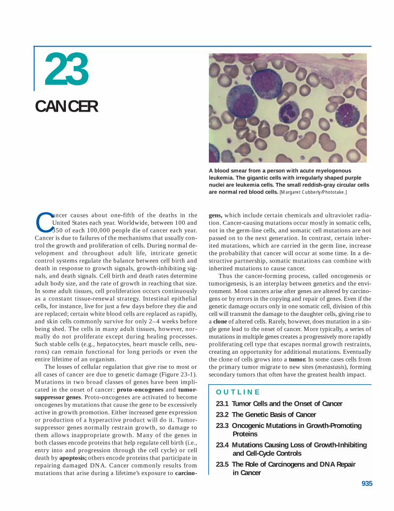
▲ FIGURE 23-1 Overview of changes in cells that cause
cancer. During carcinogenesis, six fundamental cellular propertiesare altered, as shown here, to give rise to the complete, mostdestructive cancer phenotype. Less dangerous tumors arise whenonly some of these changes occur. In this chapter we examinethe genetic changes that result in these altered cellular properties.[Adapted from D. Hanahan and R. A. Weinberg, 2000, Cell 100:57.]
harmful tumors of glial cells). Some malignant tumors, suchas those in the ovary or breast, remain localized and encap-sulated, at least for a time. When these tumors progress, thecells invade surrounding tissues, get into the body’s circula-tory system, and establish secondary areas of proliferation,a process called metastasis. Most malignant cells eventuallyacquire the ability to metastasize. Thus the major character-istics that differentiate metastatic (or malignant) tumorsfrom benign ones are their invasiveness and spread.
Cancer cells can often be distinguished from normal cellsby microscopic examination. They are usually less well differ-entiated than normal cells or benign tumor cells. In a specifictissue, malignant cells usually exhibit the characteristics of rap-idly growing cells, that is, a high nucleus-to-cytoplasm ratio,prominent nucleoli, and relatively little specialized structure.The presence of invading cells in an otherwise normal tissuesection is used to diagnose a malignancy (Figure 23-2).
Normal cells are restricted to their place in an organ or tis-sue by cell-cell adhesion and by physical barriers such as thebasal lamina, which underlies layers of epithelial cells and alsosurrounds the endothelial cells of blood vessels (Chapter 6).Cancer cells have a complex relation to the extracellular ma-trix and basal lamina. The cells must degrade the basal lam-ina to penetrate it and metastasize, but in some cases cells maymigrate along the lamina. Many tumor cells secrete a protein(plasminogen activator) that converts the serum protein plas-minogen to the active protease plasmin. Increased plasmin ac-tivity promotes metastasis by digesting the basal lamina, thusallowing its penetration by tumor cells. As the basal laminadisintegrates, some tumor cells will enter the blood, but fewerthan 1 in 10,000 cells that escape the primary tumor survive tocolonize another tissue and form a secondary, metastatictumor. In addition to escaping the original tumor and enter-ing the blood, cells that will seed new tumors must then adhereto an endothelial cell lining a capillary and migrate across orthrough it into the underlying tissue. The multiple crossings
of tissue layers that underlie malignancy often involve new orvariant surface proteins made by malignant cells.
In addition to important changes in cell-surface proteins,drastic changes occur in the cytoskeleton during tumor-cell for-mation and metastasis. These alterations can result fromchanges in the expression of genes encoding Rho and othersmall GTPases that regulate the actin cytoskeleton (Chapter 19).For instance, tumor cells have been found to over-express theRhoC gene, and this increased activity stimulates metastasis.
Cancers Usually Originate in Proliferating CellsIn order for most oncogenic mutations to induce cancer, theymust occur in dividing cells so that the mutation is passedon to many progeny cells. When such mutations occur innondividing cells (e.g., neurons and muscle cells), they gen-erally do not induce cancer, which is why tumors of muscleand nerve cells are rare in adults. Nonetheless, cancer canoccur in tissues composed mainly of nondividing differenti-ated cells such as erythrocytes and most white blood cells,absorptive cells that line the small intestine, and keratinizedcells that form the skin. The cells that initiate the tumors arenot the differentiated cells, but rather their precursor cells.Fully differentiated cells usually do not divide. As they dieor wear out, they are continually replaced by proliferationand differentiation of stem cells, and these cells are capableof transforming into tumor cells.
In Chapter 22, we learned that stem cells both perpetuatethemselves and give rise to differentiating cells that can re-generate a particular tissue for the life of an organism (seeFigure 22-2). For instance, many differentiated blood cellshave short life spans and are continually replenished fromhematopoietic (blood-forming) stem cells in the bone mar-row (see Figure 22-5). Populations of stem cells in the intes-tine, liver, skin, bone, and other tissues likewise give rise toall or many of the cell types in these tissues, replacing aged
23.1 • Tumor Cells and the Onset of Cancer 937
(b)(a) Tumor cells Normal cells
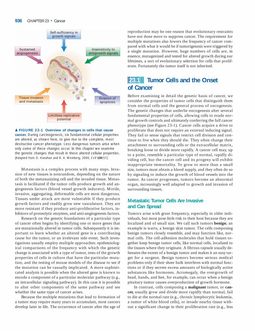
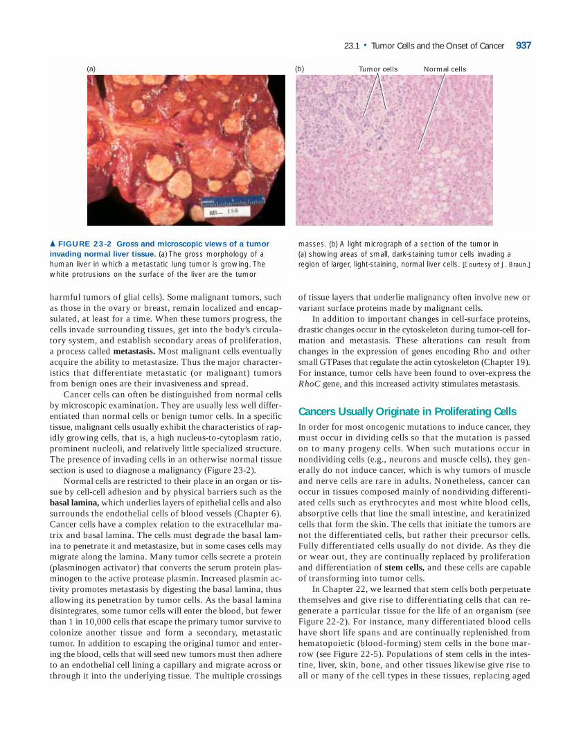
▲ FIGURE 23-2 Gross and microscopic views of a tumor
invading normal liver tissue. (a) The gross morphology of ahuman liver in which a metastatic lung tumor is growing. Thewhite protrusions on the surface of the liver are the tumor
masses. (b) A light micrograph of a section of the tumor in(a) showing areas of small, dark-staining tumor cells invading aregion of larger, light-staining, normal liver cells. [Courtesy of J. Braun.]
and dead cells, by pathways analogous to hematopoiesis inbone marrow. Similarly within a tumor there may be onlycertain cells with the ability to divide uncontrollably andgenerate new tumors; such cells are tumor stem cells.
Because stem cells can divide continually over the life ofan organism, oncogenic mutations in their DNA can accu-mulate, eventually transforming them into cancer cells. Cellsthat have acquired these mutations have an abnormal pro-liferative capacity and generally cannot undergo normalprocesses of differentiation. Many oncogenic mutations,such as ones that prevent apoptosis or generate an inappro-priate growth-promoting signal, also can occur in more dif-ferentiated, but still replicating, progenitor cells. Suchmutations in hematopoietic progenitor cells can lead to var-ious types of leukemia.
Normal animal cells are often classified according totheir embryonic tissue of origin, and the naming of tumorshas followed suit. Malignant tumors are classified as carci-nomas if they derive from endoderm (gut epithelium) or ec-toderm (skin and neural epithelia) and sarcomas if theyderive from mesoderm (muscle, blood, and connective tis-sue precursors). The leukemias, a class of sarcomas, grow asindividual cells in the blood, whereas most other tumors aresolid masses. (The name leukemia is derived from the Latinfor “white blood”: the massive proliferation of leukemic cellscan cause a patient’s blood to appear milky.)
Tumor Growth Requires Formation of New Blood VesselsTumors, whether primary or secondary, require recruitmentof new blood vessels in order to grow to a large mass. In the
absence of a blood supply, a tumor can grow into a mass ofabout 106 cells, roughly a sphere 2 mm in diameter. At thispoint, division of cells on the outside of the tumor mass isbalanced by death of those in the center due to an inadequatesupply of nutrients. Such tumors, unless they secrete hor-mones, cause few problems. However, most tumors inducethe formation of new blood vessels that invade the tumorand nourish it, a process called angiogenesis. This complexprocess requires several discrete steps: degradation of thebasal lamina that surrounds a nearby capillary, migration ofendothelial cells lining the capillary into the tumor, divisionof these endothelial cells, and formation of a new basementmembrane around the newly elongated capillary.
Many tumors produce growth factors that stimulate angiogenesis; other tumors somehow induce surroundingnormal cells to synthesize and secrete such factors. Basic fi-broblast growth factor (bFGF), transforming growth factor� (TGF�), and vascular endothelial growth factor (VEGF),which are secreted by many tumors, all have angiogenic prop-erties. New blood vessels nourish the growing tumor, allow-ing it to increase in size and thus increase the probability thatadditional harmful mutations will occur. The presence of anadjacent blood vessel also facilitates the process of metastasis.
Several natural proteins that inhibit angiogenesis(e.g., angiogenin and endostatin) or antagonists ofthe VEGF receptor have excited much interest as po-
tential therapeutic agents. Although new blood vessels are con-stantly forming during embryonic development, few formnormally in adults except after injury. Thus a specific inhibitorof angiogenesis not only might be effective against many kindsof tumors but also might have few adverse side effects. ❚
938 CHAPTER 23 • Cancer
(a)
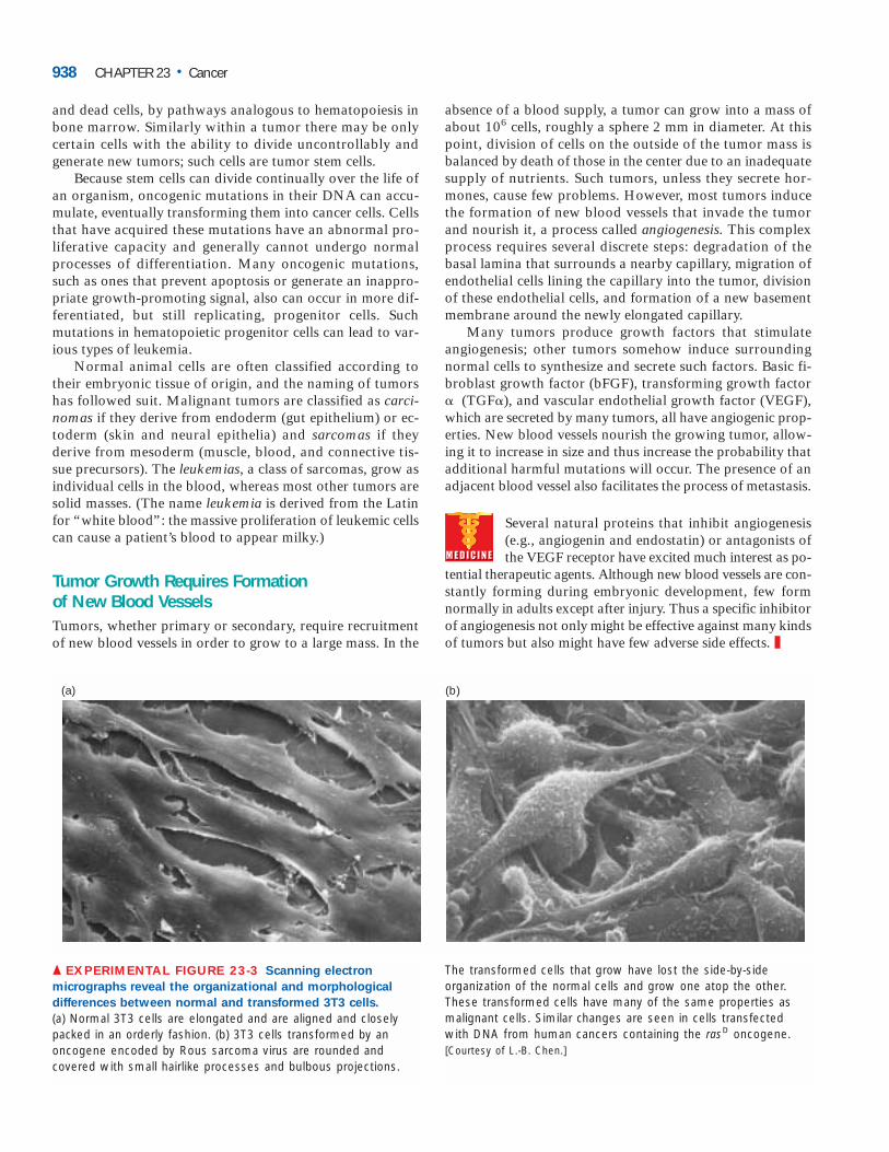
▲ EXPERIMENTAL FIGURE 23-3 Scanning electron
micrographs reveal the organizational and morphological
differences between normal and transformed 3T3 cells.
(a) Normal 3T3 cells are elongated and are aligned and closelypacked in an orderly fashion. (b) 3T3 cells transformed by anoncogene encoded by Rous sarcoma virus are rounded andcovered with small hairlike processes and bulbous projections.
The transformed cells that grow have lost the side-by-sideorganization of the normal cells and grow one atop the other.These transformed cells have many of the same properties asmalignant cells. Similar changes are seen in cells transfectedwith DNA from human cancers containing the rasD oncogene.[Courtesy of L.-B. Chen.]
(b)

Cultured Cells Can Be Transformed into Tumor CellsThe morphology and growth properties of tumor cells clearlydiffer from those of their normal counterparts; some of thesedifferences are also evident when cells are cultured. That mu-tations cause these differences was conclusively establishedby transfection experiments with a line of cultured mouse fi-broblasts called 3T3 cells. These cells normally grow onlywhen attached to the plastic surface of a culture dish and aremaintained at a low cell density. Because 3T3 cells stopgrowing when they contact other cells, they eventually forma monolayer of well-ordered cells that have stopped prolif-erating and are in the quiescent G0 phase of the cell cycle(Figure 23-3a).
When DNA from human bladder cancer cells is trans-fected into cultured 3T3 cells, about one cell in a millionincorporates a particular segment of the exogenous DNAthat causes a distinctive phenotypic change. The progenyof the affected cell are more rounded and less adherent toone another and to the dish than are the normal sur-rounding cells, forming a three-dimensional cluster of cells(a focus) that can be recognized under the microscope (Fig-ure 23-3b). Such cells, which continue to grow when thenormal cells have become quiescent, have undergone onco-genic transformation. The transformed cells have proper-ties similar to those of malignant tumor cells, includingchanges in cell morphology, ability to grow unattached toan extracellular matrix, reduced requirement for growthfactors, secretion of plasminogen activator, and loss ofactin microfilaments.
Figure 23-4 outlines the procedure for transforming 3T3cells with DNA from a human bladder cancer and cloningthe specific DNA segment that causes transformation. It wasremarkable to find a small piece of DNA with this capability;had more than one piece been needed, the experiment would
23.1 • Tumor Cells and the Onset of Cancer 939
Plate phageon E. coli
DNA from humantumor cells
Transfect mouse 3T3 cells
Culture for 2 weeks
Extract genomic DNA
Introduce intophage vector
Survivinghuman DNA
Alu probe
Oncogene
Replica onfilter paper
Phage library
Extract DNA,transform new mouse cells
Focus of transformedNIH/3T3 cells growingamong untransformedcells
Second cycle
� EXPERIMENTAL FIGURE 23-4 Transformation of mouse
cells with DNA from a human cancer cell permits
identification and molecular cloning of the rasD oncogene.
Addition of DNA from a human bladder cancer to a culture ofmouse 3T3 cells causes about one cell in a million to divideabnormally and form a focus, or clone, of transformed cells. Toclone the oncogene responsible for transformation, advantage istaken of the fact that most human genes have nearby repetitiveDNA sequences called Alu sequences. DNA from the initial focusof transformed mouse cells is isolated, and the oncogene isseparated from adventitious human DNA by secondary transferto mouse cells. The total DNA from a secondary transfectedmouse cell is then cloned into bacteriophage �; only the phagethat receives human DNA hybridizes with an Alu probe. Thehybridizing phage should contain part of or all the transformingoncogene. This expected result can be proved by showing eitherthat the phage DNA can transform cells (if the oncogene hasbeen completely cloned) or that the cloned piece of DNA isalways present in cells transformed by DNA transfer from theoriginal donor cell.
have failed. Subsequent studies showed that the cloned seg-ment included a mutant version of the cellular ras gene, des-ignated rasD. Normal Ras protein, which participates inmany intracellular signal-transduction pathways activated bygrowth factors, cycles between an inactive, “off” state withbound GDP and an active, “on” state with bound GTP. Themutated RasD protein hydrolyzes bound GTP very slowlyand therefore accumulates in the active state, sending agrowth-promoting signal to the nucleus even in the absenceof the hormones normally required to activate its signalingfunction.
The production and constitutive activation of RasD pro-tein are not sufficient to cause transformation of normal cellsin a primary (fresh) culture of human, rat, or mouse fibro-blasts. Unlike cells in a primary culture, however, cultured3T3 cells have undergone a loss-of-function mutation in thep16 gene, which encodes a cyclin-kinase inhibitor that re-stricts progression through the cell cycle. Such cells can growfor an unlimited time in culture if periodically diluted andsupplied with nutrients, which normal cells cannot (see Fig-ure 6-37b). These immortal 3T3 cells are transformed intofull-blown tumor cells only when they produce a constitu-tively active Ras protein. For this reason, transfection withthe rasD gene can transform 3T3 cells, but not normal cul-tured primary fibroblast cells, into tumor cells.
A mutant ras gene is found in most human colon, blad-der, and other cancers, but not in normal human DNA; thusit must arise as the result of a somatic mutation in one of thetumor progenitor cells. Any gene, such as rasD, that encodesa protein capable of transforming cells in culture or induc-ing cancer in animals is referred to as an oncogene. The nor-mal cellular gene from which it arises is called a proto-oncogene. The oncogenes carried by viruses that cause tu-mors in animals are often derived from proto-oncogenes thatwere hijacked from the host genome and altered to be onco-genic. When this was first discovered, it was startling to findthat these dangerous viruses were turning the animal’s owngenes against them.
A Multi-hit Model of Cancer Induction Is Supported by Several Lines of EvidenceAs noted earlier and illustrated by the oncogenic transfor-mation of 3T3 cells, multiple mutations usually are requiredto convert a normal body cell into a malignant one. Ac-cording to this “multi-hit” model, evolutionary (or “sur-vival of the fittest”) cancers arise by a process of clonalselection not unlike the selection of individual animals in alarge population. A mutation in one cell would give it aslight growth advantage. One of the progeny cells wouldthen undergo a second mutation that would allow its de-scendants to grow more uncontrollably and form a small be-nign tumor; a third mutation in a cell within this tumorwould allow it to outgrow the others and overcome con-straints imposed by the tumor microenvironment, and its
progeny would form a mass of cells, each of which wouldhave these three mutations. An additional mutation in oneof these cells would allow its progeny to escape into theblood and establish daughter colonies at other sites, the hall-mark of metastatic cancer. This model makes two easilytestable predictions.
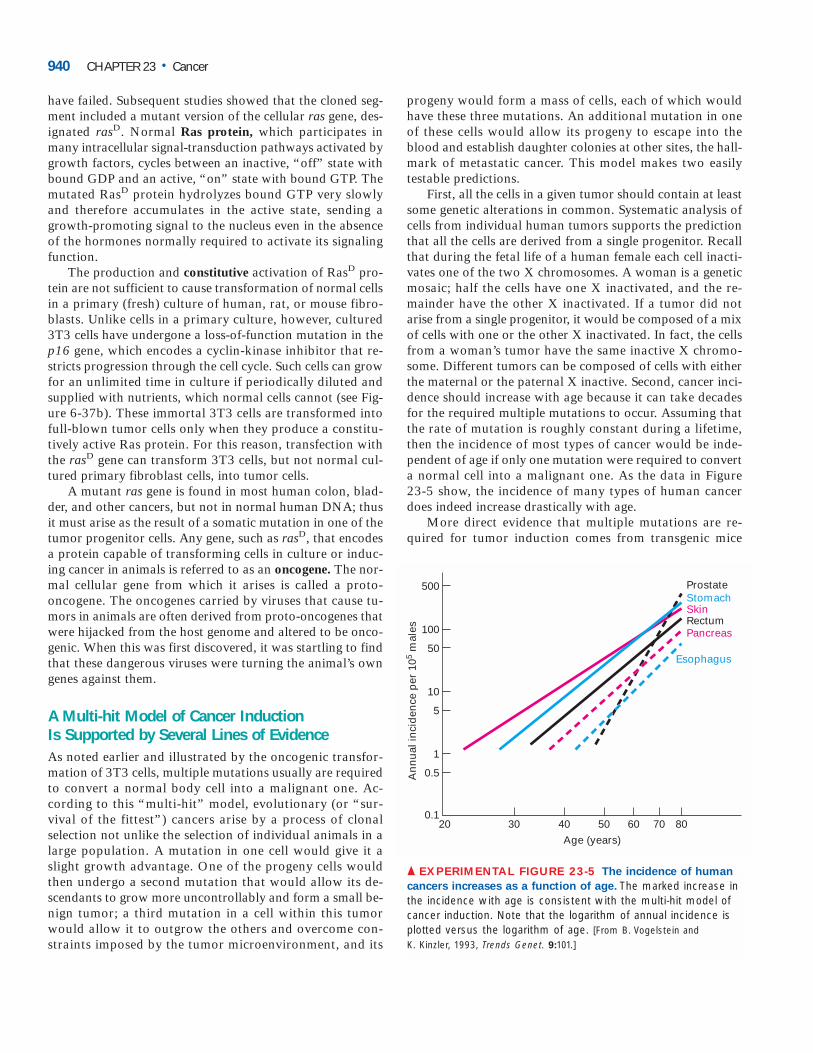
First, all the cells in a given tumor should contain at leastsome genetic alterations in common. Systematic analysis ofcells from individual human tumors supports the predictionthat all the cells are derived from a single progenitor. Recallthat during the fetal life of a human female each cell inacti-vates one of the two X chromosomes. A woman is a geneticmosaic; half the cells have one X inactivated, and the re-mainder have the other X inactivated. If a tumor did notarise from a single progenitor, it would be composed of a mixof cells with one or the other X inactivated. In fact, the cellsfrom a woman’s tumor have the same inactive X chromo-some. Different tumors can be composed of cells with eitherthe maternal or the paternal X inactive. Second, cancer inci-dence should increase with age because it can take decadesfor the required multiple mutations to occur. Assuming thatthe rate of mutation is roughly constant during a lifetime,then the incidence of most types of cancer would be inde-pendent of age if only one mutation were required to converta normal cell into a malignant one. As the data in Figure 23-5 show, the incidence of many types of human cancerdoes indeed increase drastically with age.
More direct evidence that multiple mutations are re-quired for tumor induction comes from transgenic mice
940 CHAPTER 23 • Cancer
10
50
100
500
0.5
1
5
0.1
An
nu
al in
cid
ence
per
105
mal
es
Age (years)
80706050403020
ProstateStomachSkinRectumPancreas
Esophagus
▲ EXPERIMENTAL FIGURE 23-5 The incidence of human
cancers increases as a function of age. The marked increase inthe incidence with age is consistent with the multi-hit model ofcancer induction. Note that the logarithm of annual incidence isplotted versus the logarithm of age. [From B. Vogelstein and K. Kinzler, 1993, Trends Genet. 9:101.]
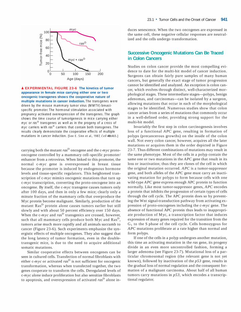
carrying both the mutant rasD oncogene and the c-myc proto-oncogene controlled by a mammary cell–specific promoter/enhancer from a retrovirus. When linked to this promoter, thenormal c-myc gene is overexpressed in breast tissue because the promoter is induced by endogenous hormonelevels and tissue-specific regulators. This heightened tran-scription of c-myc mimics oncogenic mutations that turn upc-myc transcription, converting the proto-oncogene into anoncogene. By itself, the c-myc transgene causes tumors onlyafter 100 days, and then in only a few mice; clearly only aminute fraction of the mammary cells that overproduce theMyc protein become malignant. Similarly, production of themutant RasD protein alone causes tumors earlier but stillslowly and with about 50 percent efficiency over 150 days.When the c-myc and rasD transgenics are crossed, however,such that all mammary cells produce both Myc and RasD,tumors arise much more rapidly and all animals succumb tocancer (Figure 23-6). Such experiments emphasize the syn-ergistic effects of multiple oncogenes. They also suggest thatthe long latency of tumor formation, even in the double-transgenic mice, is due to the need to acquire additional somatic mutations.
Similar cooperative effects between oncogenes can beseen in cultured cells. Transfection of normal fibroblasts witheither c-myc or activated rasD is not sufficient for oncogenictransformation, whereas when transfected together, the twogenes cooperate to transform the cells. Deregulated levels ofc-myc alone induce proliferation but also sensitize fibroblaststo apoptosis, and overexpression of activated rasD alone in-
duces senescence. When the two oncogenes are expressed inthe same cell, these negative cellular responses are neutral-ized and the cells undergo transformation.
Successive Oncogenic Mutations Can Be Tracedin Colon Cancers
Studies on colon cancer provide the most compelling evi-dence to date for the multi-hit model of cancer induction.Surgeons can obtain fairly pure samples of many humancancers, but generally the exact stage of tumor progressioncannot be identified and analyzed. An exception is colon can-cer, which evolves through distinct, well-characterized mor-phological stages. These intermediate stages—polyps, benignadenomas, and carcinomas—can be isolated by a surgeon,allowing mutations that occur in each of the morphologicalstages to be identified. Numerous studies show that coloncancer arises from a series of mutations that commonly occurin a well-defined order, providing strong support for themulti-hit model.
Invariably the first step in colon carcinogenesis involvesloss of a functional APC gene, resulting in formation ofpolyps (precancerous growths) on the inside of the colonwall. Not every colon cancer, however, acquires all the latermutations or acquires them in the order depicted in Figure23-7. Thus different combinations of mutations may result inthe same phenotype. Most of the cells in a polyp contain thesame one or two mutations in the APC gene that result in itsloss or inactivation; thus they are clones of the cell in whichthe original mutation occurred. APC is a tumor-suppressorgene, and both alleles of the APC gene must carry an inacti-vating mutation for polyps to form because cells with onewild-type APC gene express enough APC protein to functionnormally. Like most tumor-suppressor genes, APC encodesa protein that inhibits the progression of certain types of cellsthrough the cell cycle. The APC protein does so by prevent-ing the Wnt signal-transduction pathway from activating ex-pression of proto-oncogenes including the c-myc gene. Theabsence of functional APC protein thus leads to inappropri-ate production of Myc, a transcription factor that inducesexpression of many genes required for the transition from theG1 to the S phase of the cell cycle. Cells homozygous forAPC mutations proliferate at a rate higher than normal andform polyps.
If one of the cells in a polyp undergoes another mutation,this time an activating mutation in the ras gene, its progenydivide in an even more uncontrolled fashion, forming alarger adenoma (see Figure 23-7). Mutational loss of a par-ticular chromosomal region (the relevant gene is not yetknown), followed by inactivation of the p53 gene, results inthe gradual loss of normal regulation and the consequent for-mation of a malignant carcinoma. About half of all humantumors carry mutations in p53, which encodes a transcrip-tional regulator.
23.1 • Tumor Cells and the Onset of Cancer 941
0
20
40
60
80
100
Tu
mo
r-fr
ee m
ice
(%)
Age (days)
150100500 200
myc
rasD
myc + rasD
▲ EXPERIMENTAL FIGURE 23-6 The kinetics of tumor
appearance in female mice carrying either one or two
oncogenic transgenes shows the cooperative nature of
multiple mutations in cancer induction. The transgenes weredriven by the mouse mammary tumor virus (MMTV) breast-specific promoter. The hormonal stimulation associated withpregnancy activated overexpression of the transgenes. The graphshows the time course of tumorigenesis in mice carrying eithermyc or rasD transgenes as well as in the progeny of a cross ofmyc carriers with rasD carriers that contain both transgenes. Theresults clearly demonstrate the cooperative effects of multiplemutations in cancer induction. [See E. Sinn et al., 1987, Cell 49:465.]
Normal colon cells
Loss of APC tumor-suppressor gene (chromosome 5)
A polyp (smallgrowth) forms onthe colon wall
A benign,precanceroustumor grows Activation of K-ras
oncogene(chromosome 12)
Loss of tumor-suppressor gene inregion of DCC (chromosome 18)
Loss of p53 tumor-suppressorgene (chromosome 17)
Other changes
A class II adenoma(benign) grows
A class III adenoma(benign) grows
A malignant carcinomadevelops
Invasive tumor cells
Wallof colon
Lumenof colon
Bloodvessel
Normal colonepithelial cellsBasal lamina
Polyp
The cancer metastasizes(spreads to other tissues)
Tumor cells invadeblood vessels, allowingmetastasis to occur
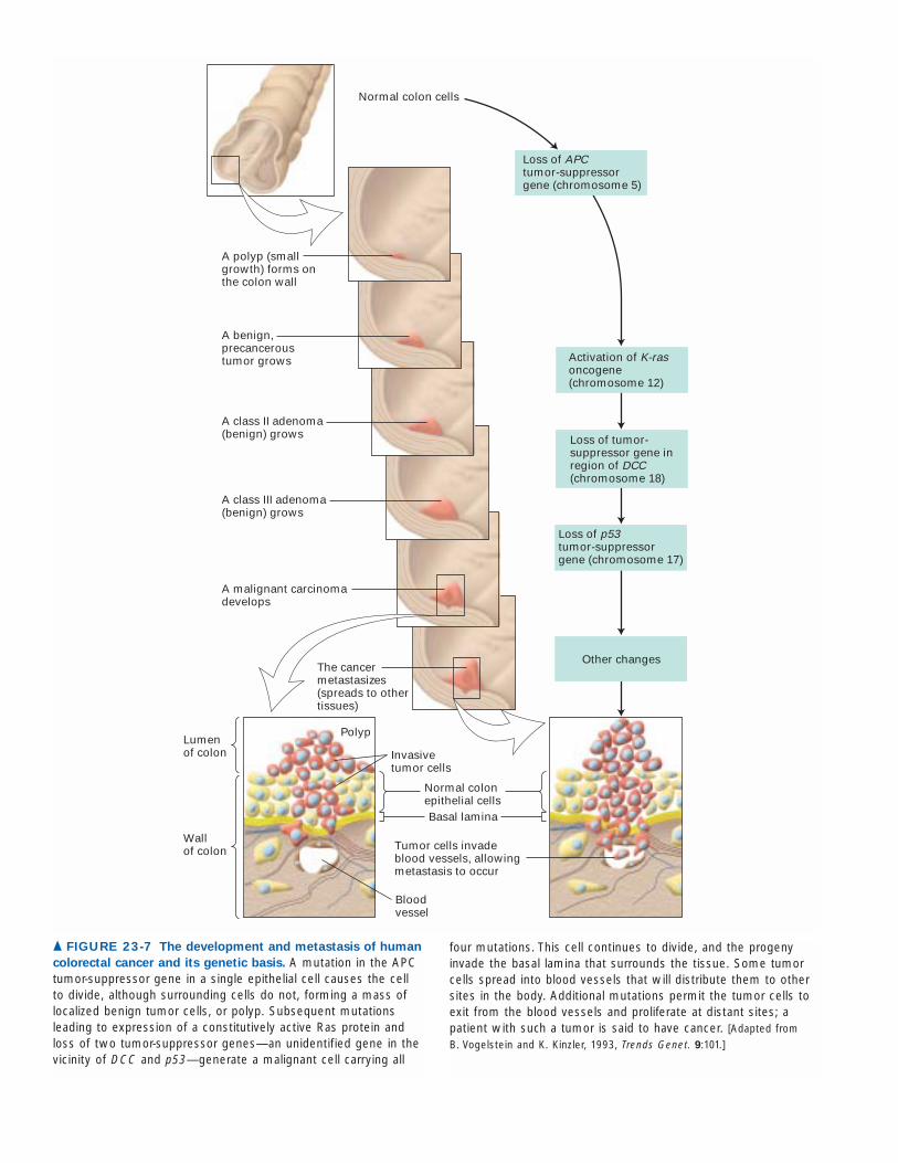
▲ FIGURE 23-7 The development and metastasis of human
colorectal cancer and its genetic basis. A mutation in the APCtumor-suppressor gene in a single epithelial cell causes the cellto divide, although surrounding cells do not, forming a mass oflocalized benign tumor cells, or polyp. Subsequent mutationsleading to expression of a constitutively active Ras protein andloss of two tumor-suppressor genes—an unidentified gene in thevicinity of DCC and p53—generate a malignant cell carrying all
four mutations. This cell continues to divide, and the progenyinvade the basal lamina that surrounds the tissue. Some tumorcells spread into blood vessels that will distribute them to othersites in the body. Additional mutations permit the tumor cells toexit from the blood vessels and proliferate at distant sites; apatient with such a tumor is said to have cancer. [Adapted from B. Vogelstein and K. Kinzler, 1993, Trends Genet. 9:101.]
DNA from different human colon carcinomas generallycontains mutations in all these genes—loss-of-function muta-tions in the tumor suppressors APC and p53, the as yet myste-rious gene, and an activating (gain-of-function) mutation in thedominant oncogene K-ras—establishing that multiple muta-tions in the same cell are needed for the cancer to form. Someof these mutations appear to confer growth advantages at anearly stage of tumor development, whereas other mutationspromote the later stages, including invasion and metastasis,which are required for the malignant phenotype. The numberof mutations needed for colon cancer progression may at firstseem surprising, seemingly an effective barrier to tumorigenesis.Our genomes, however, are under constant assault. Recent es-timates indicate that sporadically arising polyps have about11,000 genetic alterations in each cell, though very likely onlya few of these are relevant to oncogenesis.
Colon carcinoma provides an excellent example of themulti-hit mode of cancer. The degree to which this model ap-plies to cancer is only now being learned, but it is clear thatmultiple types of cancer involve multiple mutations. The adventof DNA microarray technology is allowing more detailed ex-amination of tumor properties by monitoring the spectrum ofmRNA molecules from tens of thousands of genes, and recentdata have provided some challenges to the multi-hit model. Notsurprisingly, primary tumors can often be distinguishable frommetastatic tumors by the pattern of gene expression. More in-terestingly, a subset of solid primary tumors has been found tohave characteristics more typical of metastatic tumors, suggest-ing that it may be possible to identify primary tumors that havea greater probability of becoming metastatic. This also raisesthe possibility that, at least for some types of cancer, the initi-ating events of the primary tumor may set a course towardmetastasis. This hypothesis can be distinguished from the emer-gence of a rare subset of cells within the primary tumor ac-quiring a necessary series of further mutations.
KEY CONCEPTS OF SECTION 23.1
Tumor Cells and the Onset of Cancer
■ Cancer is a fundamental aberration in cellular behavior,touching on many aspects of molecular cell biology. Mostcell types of the body can give rise to malignant tumor(cancer) cells.
■ Cancer cells usually arise from stem cells and other pro-liferating cells and bear more resemblance to these cellsthan to more mature differentiated cell types.
■ Cancer cells can multiply in the absence of at least someof the growth-promoting factors required for proliferationof normal cells and are resistant to signals that normallyprogram cell death (apoptosis).
■ Certain cultured cells transfected with tumor-cell DNAundergo transformation (see Figure 23-4). Such transformedcells share certain properties with tumor cells.
■ Cancer cells sometimes invade surrounding tissues, of-ten breaking through the basal laminae that define theboundaries of tissues and spreading through the body toestablish secondary areas of growth, a process called metas-tasis. Metastatic tumors often secrete proteases, which de-grade the surrounding extracellular matrix.
■ Both primary and secondary tumors require angiogene-sis, the formation of new blood vessels, in order to growto a large mass.
■ The multi-hit model, which proposes that multiple mu-tations are needed to cause cancer, is consistent with thegenetic homogeneity of cells from a given tumor, the ob-served increase in the incidence of human cancers with ad-vancing age, and the cooperative effect of oncogenic trans-genes on tumor formation in mice.
■ Most oncogenic mutations occur in somatic cells and arenot carried in the germ-line DNA.
■ Colon cancer develops through distinct morphologicalstages that commonly are associated with mutations inspecific tumor-suppressor genes and proto-oncogenes (seeFigure 23-7).
The Genetic Basis of CancerAs we have seen, mutations in two broad classes of genes—proto-oncogenes (e.g., ras) and tumor-suppressor genes(e.g., APC)—play key roles in cancer induction. These genesencode many kinds of proteins that help control cell growthand proliferation (Figure 23-8). Virtually all human tumorshave inactivating mutations in genes that normally act atvarious cell-cycle checkpoints to stop a cell’s progressthrough the cell cycle if a previous step has occurred incor-rectly or if DNA has been damaged. For example, most can-cers have inactivating mutations in the genes coding for oneor more proteins that normally restrict progression throughthe G1 stage of the cell cycle. Likewise, a constitutively ac-tive Ras or other activated signal-transduction protein isfound in several kinds of human tumor that have differ-ent origins. Thus malignancy and the intricate processes for controlling the cell cycle discussed in Chapter 21 are two faces of the same coin. In the series of events leading to growth of a tumor, oncogenes combine with tumor-suppressor mutations to give rise to the full spectrum oftumor cell properties described in the previous section (seeFigure 23-7).
In this section, we consider the general types of muta-tions that are oncogenic and see how certain viruses cancause cancer. We also explain why some inherited mutationsincrease the risk for particular cancers and consider the re-lation between cancer and developmentally importantgenes. We conclude this section with a brief discussion ofhow genomics methods are being used to characterize andclassify tumors.
23.2
23.2 • The Genetic Basis of Cancer 943
Gain-of-Function Mutations Convert Proto-oncogenes into OncogenesRecall that an oncogene is any gene that encodes a protein ableto transform cells in culture or to induce cancer in animals.Of the many known oncogenes, all but a few are derived fromnormal cellular genes (i.e., proto-oncogenes) whose productspromote cell proliferation. For example, the ras gene discussedpreviously is a proto-oncogene that encodes an intracellularsignal-transduction protein; the mutant rasD gene derivedfrom ras is an oncogene, whose encoded protein provides anexcessive or uncontrolled growth-promoting signal. Otherproto-oncogenes encode growth-promoting signal moleculesand their receptors, anti-apoptotic (cell-survival) proteins, andsome transcription factors.
Conversion, or activation, of a proto-oncogene into anoncogene generally involves a gain-of-function mutation. Atleast four mechanisms can produce oncogenes from the cor-responding proto-oncogenes:
• Point mutation (i.e., change in a single base pair) in aproto-oncogene that results in a constitutively active protein product
• Chromosomal translocation that fuses two genes together to produce a hybrid gene encoding a chimeric
protein whose activity, unlike that of the parent proteins,often is constitutive
• Chromosomal translocation that brings a growth-regulatory gene under the control of a different promoterthat causes inappropriate expression of the gene
• Amplification (i.e., abnormal DNA replication) of aDNA segment including a proto-oncogene, so that numerous copies exist, leading to overproduction of the encoded protein
An oncogene formed by either of the first two mecha-nisms encodes an “oncoprotein” that differs from the normalprotein encoded by the corresponding proto-oncogene. Incontrast, the other two mechanisms generate oncogeneswhose protein products are identical with the normal pro-teins; their oncogenic effect is due to production at higher-than-normal levels or in cells where they normally are notproduced.
The localized amplification of DNA to produce as manyas 100 copies of a given region (usually a region spanninghundreds of kilobases) is a common genetic change seen intumors. This anomaly may take either of two forms: the duplicated DNA may be tandemly organized at a single siteon a chromosome, or it may exist as small, independent
944 CHAPTER 23 • Cancer
Signaling molecule (I)
Signal receptor (II)
Intracellulartransducers(III)
Intracellularreceptors(II)
Transcription factors (IV)
DNA
RNA
mRNA
ProteinsApoptoticproteins (V)
Cell-cycle controlproteins (VI)
NUCLEUS
DNA-repairproteins (VII)
Virus-encodedactivators ofsignalreceptors (Ia)
Intracellulareffectorregion(often aprotein-tyrosinekinase)
Second messengers(phosphorylated proteins)
Transcription
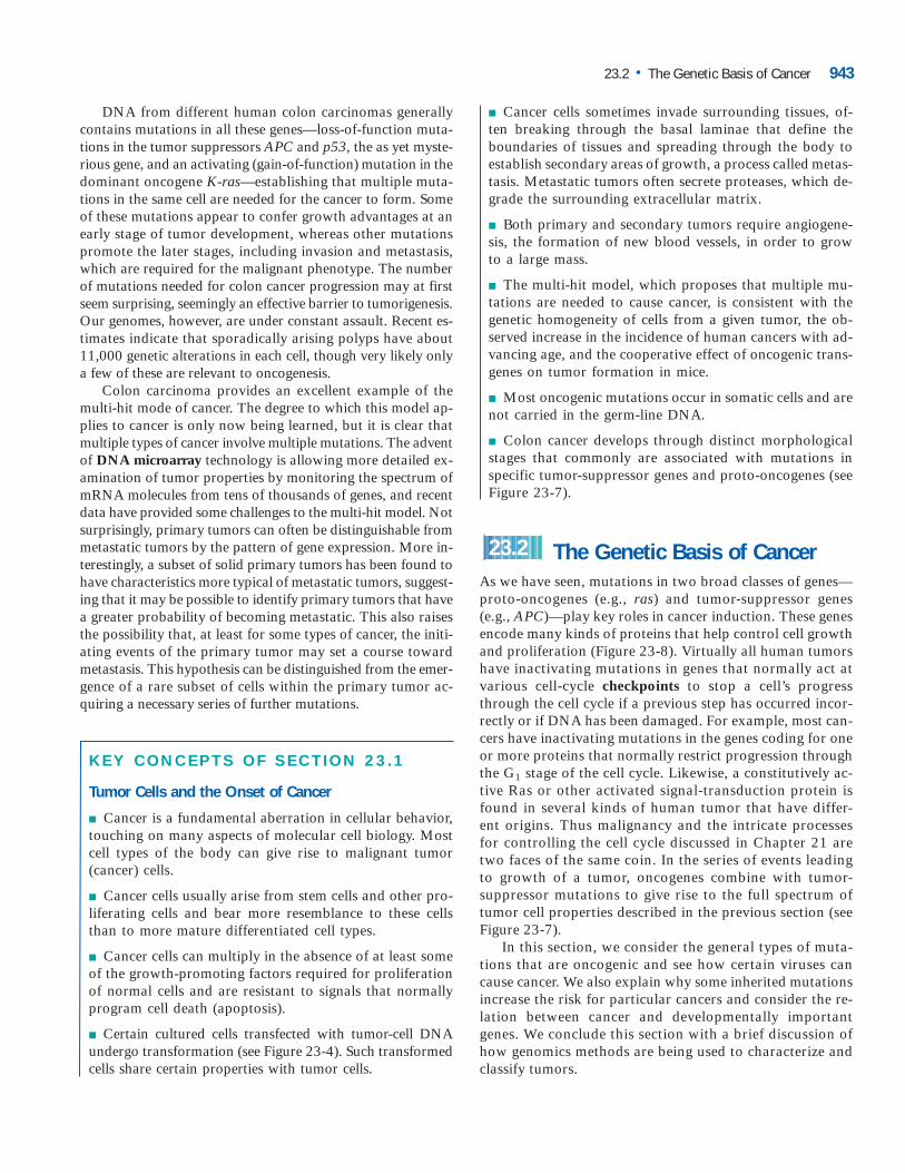
� FIGURE 23-8 Seven types of proteins that
participate in controlling cell growth and
proliferation. Cancer can result from expressionof mutant forms of these proteins. Mutationschanging the structure or expression of proteinsthat normally promote cell growth generally giverise to dominantly active oncogenes. Many, butnot all, extracellular signaling molecules (I), signalreceptors (II), signal-transduction proteins (III), andtranscription factors (IV) are in this category. Cell-cycle control proteins (VI) that function to restraincell proliferation and DNA-repair proteins (VII) areencoded by tumor-suppressor genes. Mutationsin these genes act recessively, greatly increasingthe probability that the mutant cells will becometumor cells or that mutations will occur in otherclasses. Apoptotic proteins (V) include tumorsuppressors that promote apoptosis andoncoproteins that promote cell survival. Virus-encoded proteins that activate signal receptors(Ia) also can induce cancer.
mini-chromosome-like structures. The former case leads toa homogeneously staining region (HSR) that is visible in thelight microscope at the site of the amplification; the lattercase causes extra “minute” chromosomes, separate from thenormal chromosomes that pepper a stained chromosomalpreparation (Figure 23-9).
Gene amplification may involve a small numberof genes, such as the N-myc gene and its neighborDDX1 that are amplified in neuroblastoma, or a
chromosome region containing many genes. It can be diffi-cult to determine which genes are amplified, a first step in de-termining which gene caused the tumor. DNA microarraysoffer a powerful approach for finding amplified regions ofchromosomes. Rather than look at gene expression, the ap-plication of microarrays we described earlier, these experi-ments involve looking for abnormally abundant DNAsequences. Genomic DNA from cancer cells is used to probearrays containing fragments of genomic DNA and spots withamplified DNA give stronger signals than control spots.Among the amplified genes, the strongest candidates for therelevant ones can be identified by also measuring gene ex-pression. A breast carcinoma cell line, with four known am-plified chromosome regions, was screened for amplifiedgenes, and the expression levels of those genes were alsostudied on microarrays. Fifty genes were found to be ampli-fied, but only five were also highly expressed. These five arenew candidates as oncogenes. ❚
However they arise, the gain-of-function mutations thatconvert proto-oncogenes to oncogenes are genetically domi-nant; that is, mutation in only one of the two alleles is suffi-cient for induction of cancer.
Cancer-Causing Viruses Contain Oncogenes or Activate Cellular Proto-oncogenesPioneering studies by Peyton Rous beginning in 1911 led tothe initial recognition that a virus could cause cancer wheninjected into a suitable host animal. Many years later mo-lecular biologists showed that Rous sarcoma virus (RSV) isa retrovirus whose RNA genome is reverse-transcribed intoDNA, which is incorporated into the host-cell genome (seeFigure 4-43). In addition to the “normal” genes present in allretroviruses, oncogenic transforming viruses like RSV con-tain the v-src gene. Subsequent studies with mutant formsof RSV demonstrated that only the v-src gene, not the otherviral genes, was required for cancer induction.
In the late 1970s, scientists were surprised to find thatnormal cells from chickens and other species contain a genethat is closely related to the RSV v-src gene. This normal cel-lular gene, a proto-oncogene, commonly is distinguishedfrom the viral gene by the prefix “c” (c-src). RSV and otheroncogene-carrying viruses are thought to have arisen by incorporating, or transducing, a normal cellular proto-oncogene into their genome. Subsequent mutation in thetransduced gene then converted it into a dominantly acting
23.2 • The Genetic Basis of Cancer 945
(a) (b)
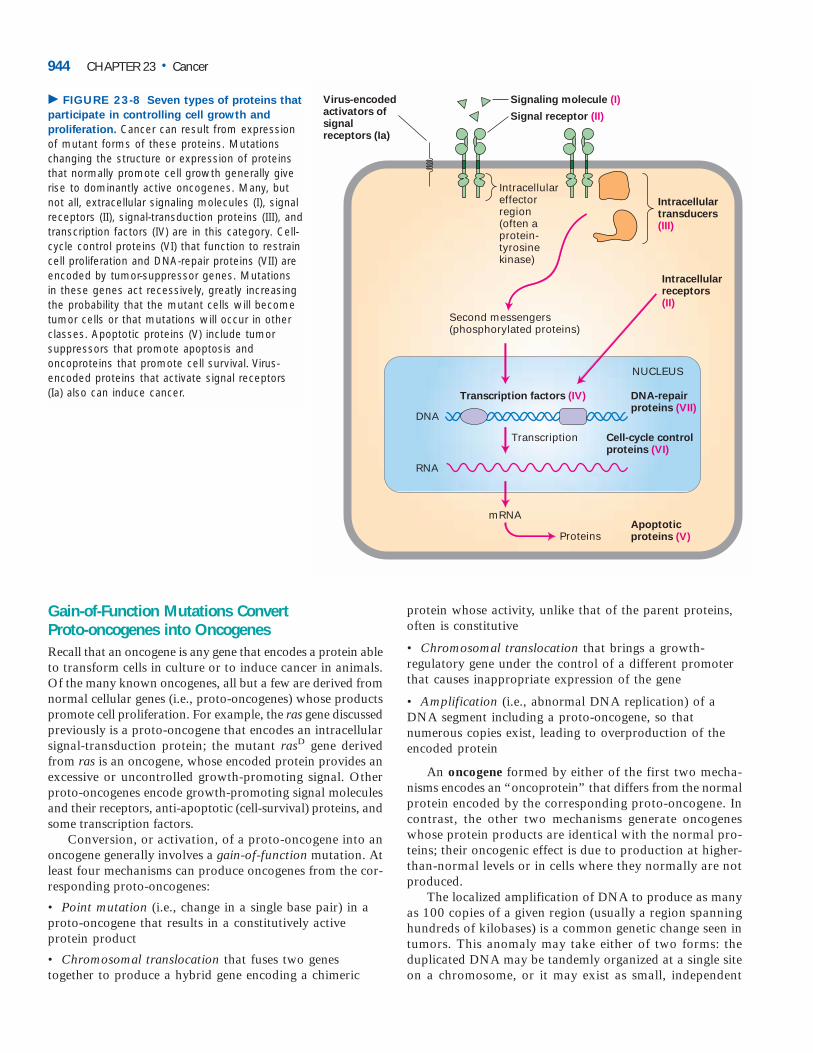
▲ EXPERIMENTAL FIGURE 23-9 DNA amplifications in
stained chromosomes take two forms, visible under the light
microscope. (a) Homogeneously staining regions (HSRs) in ahuman chromosome from a neuroblastoma cell. Thechromosomes are uniformly stained with a blue dye so that allcan be seen. Specific DNA sequences were detected usingfluorescent in situ hybridization (FISH) in which fluorescentlylabeled DNA clones are hybridized to denatured DNA in thechromosomes. The chromosome 4 pair is marked (red) by in situ hybridization with a large DNA cosmid clone containing
the N-myc oncogene. On one of the chromosome 4’s an HSR isvisible (green) after staining for a sequence enriched in the HSR.(b) Optical sections through nuclei from a human neuroblastomacell that contain double minute chromosomes. The normalchromosomes are the green and blue structures; the doubleminute chromosomes are the many small red dots. Arrowsindicate double minutes associated with the surface or interior of the normal chromosomes. [Parts (a) and (b) from I. Solovei et al.,2000, Genes Chromosomes Cancer 29:297–308, Figures 4 and 17.]
oncogene, which can induce cell transformation in the pres-ence of the normal c-src proto-oncogene. Such viruses arecalled transducing retroviruses because their genomes con-tain an oncogene derived from a transduced cellular proto-oncogene.
Because its genome carries the potent v-src oncogene, thetransducing RSV induces tumors within days. In contrast,most oncogenic retroviruses induce cancer only after a pe-riod of months or years. The genomes of these slow-actingretroviruses differ from those of transducing viruses in onecrucial respect: they lack an oncogene. All slow-acting, or“long latency,” retroviruses appear to cause cancer by integrating into the host-cell DNA near a cellular proto-oncogene and activating its expression. The long terminal repeat (LTR) sequences in integrated retroviral DNA can actas an enhancer or promoter of a nearby cellular gene,thereby stimulating its transcription. For example, in thecells from tumors caused by avian leukosis virus (ALV), theretroviral DNA is inserted near the c-myc gene. These cellsoverproduce c-Myc protein; as noted earlier, overproductionof c-Myc causes abnormally rapid proliferation of cells.Slow-acting viruses act slowly for two reasons: integrationnear a cellular proto-oncogene (e.g., c-myc) is a random, rareevent, and additional mutations have to occur before a full-fledged tumor becomes evident.
In natural bird and mouse populations, slow-acting retro-viruses are much more common than oncogene-containingretroviruses such as Rous sarcoma virus. Thus, insertionalproto-oncogene activation is probably the major mechanismby which retroviruses cause cancer. Although few human tu-mors have been associated with any retrovirus, the huge in-vestment in studying retroviruses as a model for human cancerpaid off both in the discovery of cellular oncogenes and in thesophisticated understanding of retroviruses, which later accelerated progress on the HIV virus that causes AIDS.
A few DNA viruses also are oncogenic. Unlike mostDNA viruses that infect animal cells, oncogenic DNA virusesintegrate their viral DNA into the host-cell genome. The viralDNA contains one or more oncogenes, which permanentlytransform infected cells. For example, many warts and otherbenign tumors of epithelial cells are caused by the DNA-containing papillomaviruses. Unlike retroviral oncogenes,which are derived from normal cellular genes and have nofunction for the virus except to allow their proliferation intumors, the known oncogenes of DNA viruses are integralparts of the viral genome and are required for viral replica-tion. As discussed later, the oncoproteins expressed from in-tegrated viral DNA in infected cells act in various ways tostimulate cell growth and proliferation.
Loss-of-Function Mutations in Tumor-SuppressorGenes Are OncogenicTumor-suppressor genes generally encode proteins thatin one way or another inhibit cell proliferation. Loss-of-
function mutations in one or more of these “brakes” con-tribute to the development of many cancers. Five broadclasses of proteins are generally recognized as being encodedby tumor-suppressor genes:
• Intracellular proteins that regulate or inhibit progressionthrough a specific stage of the cell cycle (e.g., p16 and Rb)
• Receptors or signal transducers for secreted hormonesor developmental signals that inhibit cell proliferation(e.g., TGF�, the hedgehog receptor patched)
• Checkpoint-control proteins that arrest the cell cycle ifDNA is damaged or chromosomes are abnormal (e.g., p53)
• Proteins that promote apoptosis
• Enzymes that participate in DNA repair
Although DNA-repair enzymes do not directly inhibitcell proliferation, cells that have lost the ability to repair er-rors, gaps, or broken ends in DNA accumulate mutations inmany genes, including those that are critical in controllingcell growth and proliferation. Thus loss-of-function muta-tions in the genes encoding DNA-repair enzymes preventcells from correcting mutations that inactivate tumor-suppressor genes or activate oncogenes.
Since generally one copy of a tumor-suppressor gene suf-fices to control cell proliferation, both alleles of a tumor-suppressor gene must be lost or inactivated in order to pro-mote tumor development. Thus oncogenic loss-of-functionmutations in tumor-suppressor genes are genetically reces-sive. In many cancers, tumor-suppressor genes have deletions or point mutations that prevent production of any protein orlead to production of a nonfunctional protein. Another mech-anism for inactivating tumor-suppressor genes is methyl-ation of cytosine residues in the promoter or other control elements. Such methylation is commonly found in nontran-scribed regions of DNA.
Inherited Mutations in Tumor-Suppressor GenesIncrease Cancer RiskIndividuals with inherited mutations in tumor-suppressorgenes have a hereditary predisposition for certain cancers.Such individuals generally inherit a germ-line mutation inone allele of the gene; somatic mutation of the second allelefacilitates tumor progression. A classic case is retinoblas-toma, which is caused by loss of function of RB, the firsttumor-suppressor gene to be identified. As we discuss later,the protein encoded by RB helps regulate progress throughthe cell cycle.
Hereditary versus Sporadic Retinoblastoma Childrenwith hereditary retinoblastoma inherit a single defectivecopy of the RB gene, sometimes seen as a small deletion onone of the copies of chromosome 13. The children developretinal tumors early in life and generally in both eyes. Oneessential event in tumor development is the deletion or
946 CHAPTER 23 • Cancer
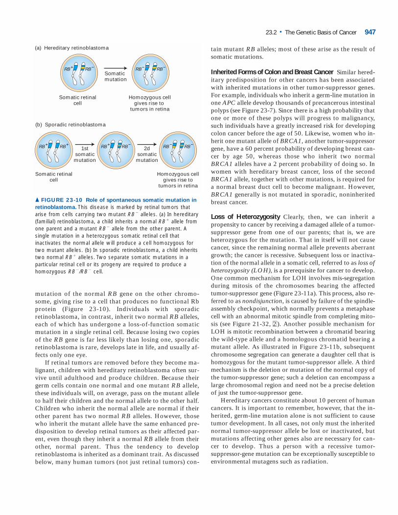
mutation of the normal RB gene on the other chromo-some, giving rise to a cell that produces no functional Rbprotein (Figure 23-10). Individuals with sporadicretinoblastoma, in contrast, inherit two normal RB alleles,each of which has undergone a loss-of-function somaticmutation in a single retinal cell. Because losing two copiesof the RB gene is far less likely than losing one, sporadicretinoblastoma is rare, develops late in life, and usually af-fects only one eye.
If retinal tumors are removed before they become ma-lignant, children with hereditary retinoblastoma often sur-vive until adulthood and produce children. Because theirgerm cells contain one normal and one mutant RB allele,these individuals will, on average, pass on the mutant alleleto half their children and the normal allele to the other half.Children who inherit the normal allele are normal if theirother parent has two normal RB alleles. However, thosewho inherit the mutant allele have the same enhanced pre-disposition to develop retinal tumors as their affected par-ent, even though they inherit a normal RB allele from theirother, normal parent. Thus the tendency to developretinoblastoma is inherited as a dominant trait. As discussedbelow, many human tumors (not just retinal tumors) con-
tain mutant RB alleles; most of these arise as the result ofsomatic mutations.
Inherited Forms of Colon and Breast Cancer Similar hered-itary predisposition for other cancers has been associatedwith inherited mutations in other tumor-suppressor genes.For example, individuals who inherit a germ-line mutation inone APC allele develop thousands of precancerous intestinalpolyps (see Figure 23-7). Since there is a high probability thatone or more of these polyps will progress to malignancy,such individuals have a greatly increased risk for developingcolon cancer before the age of 50. Likewise, women who in-herit one mutant allele of BRCA1, another tumor-suppressorgene, have a 60 percent probability of developing breast can-cer by age 50, whereas those who inherit two normalBRCA1 alleles have a 2 percent probability of doing so. Inwomen with hereditary breast cancer, loss of the secondBRCA1 allele, together with other mutations, is required fora normal breast duct cell to become malignant. However,BRCA1 generally is not mutated in sporadic, noninheritedbreast cancer.
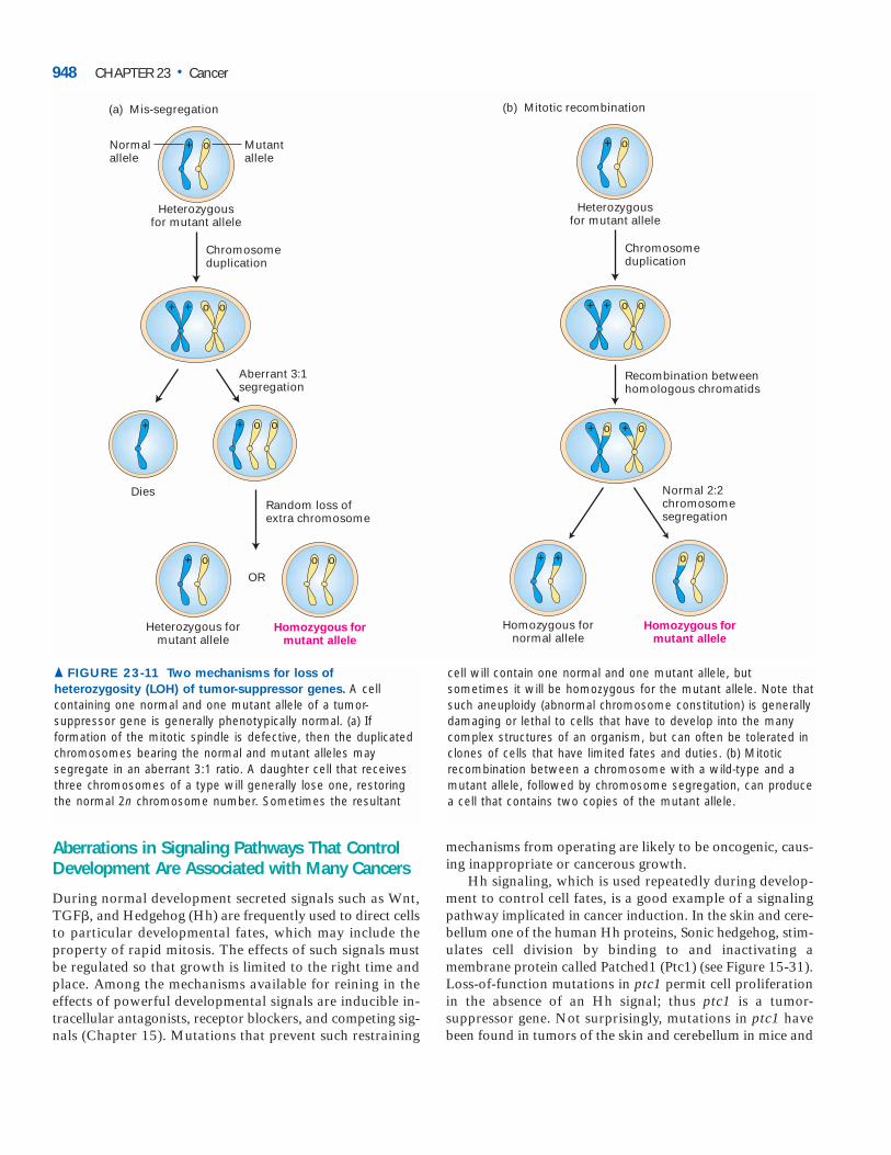
Loss of Heterozygosity Clearly, then, we can inherit apropensity to cancer by receiving a damaged allele of a tumor-suppressor gene from one of our parents; that is, we are heterozygous for the mutation. That in itself will not causecancer, since the remaining normal allele prevents aberrantgrowth; the cancer is recessive. Subsequent loss or inactiva-tion of the normal allele in a somatic cell, referred to as loss ofheterozygosity (LOH), is a prerequisite for cancer to develop.One common mechanism for LOH involves mis-segregationduring mitosis of the chromosomes bearing the affectedtumor-suppressor gene (Figure 23-11a). This process, also re-ferred to as nondisjunction, is caused by failure of the spindle-assembly checkpoint, which normally prevents a metaphasecell with an abnormal mitotic spindle from completing mito-sis (see Figure 21-32, 2 ). Another possible mechanism forLOH is mitotic recombination between a chromatid bearingthe wild-type allele and a homologous chromatid bearing amutant allele. As illustrated in Figure 23-11b, subsequentchromosome segregation can generate a daughter cell that ishomozygous for the mutant tumor-suppressor allele. A thirdmechanism is the deletion or mutation of the normal copy ofthe tumor-suppressor gene; such a deletion can encompass alarge chromosomal region and need not be a precise deletionof just the tumor-suppressor gene.
Hereditary cancers constitute about 10 percent of humancancers. It is important to remember, however, that the in-herited, germ-line mutation alone is not sufficient to causetumor development. In all cases, not only must the inheritednormal tumor-suppressor allele be lost or inactivated, butmutations affecting other genes also are necessary for can-cer to develop. Thus a person with a recessive tumor-suppressor-gene mutation can be exceptionally susceptible toenvironmental mutagens such as radiation.
23.2 • The Genetic Basis of Cancer 947
1stsomaticmutation
2dsomaticmutation
RB+ RB−
(a) Hereditary retinoblastoma
(b) Sporadic retinoblastoma
Somaticmutation
Somatic retinalcell
Somatic retinalcell
Homozygous cellgives rise to
tumors in retina
Homozygous cellgives rise to
tumors in retina
RB− RB−
RB+ RB+ RB+ RB− RB− RB−
▲ FIGURE 23-10 Role of spontaneous somatic mutation in
retinoblastoma. This disease is marked by retinal tumors thatarise from cells carrying two mutant RB� alleles. (a) In hereditary(familial) retinoblastoma, a child inherits a normal RB� allele fromone parent and a mutant RB� allele from the other parent. Asingle mutation in a heterozygous somatic retinal cell thatinactivates the normal allele will produce a cell homozygous fortwo mutant alleles. (b) In sporadic retinoblastoma, a child inheritstwo normal RB� alleles. Two separate somatic mutations in aparticular retinal cell or its progeny are required to produce ahomozygous RB�/RB� cell.
Aberrations in Signaling Pathways That ControlDevelopment Are Associated with Many Cancers
During normal development secreted signals such as Wnt,TGF�, and Hedgehog (Hh) are frequently used to direct cellsto particular developmental fates, which may include theproperty of rapid mitosis. The effects of such signals mustbe regulated so that growth is limited to the right time andplace. Among the mechanisms available for reining in the effects of powerful developmental signals are inducible in-tracellular antagonists, receptor blockers, and competing sig-nals (Chapter 15). Mutations that prevent such restraining
mechanisms from operating are likely to be oncogenic, caus-ing inappropriate or cancerous growth.
Hh signaling, which is used repeatedly during develop-ment to control cell fates, is a good example of a signalingpathway implicated in cancer induction. In the skin and cere-bellum one of the human Hh proteins, Sonic hedgehog, stim-ulates cell division by binding to and inactivating amembrane protein called Patched1 (Ptc1) (see Figure 15-31).Loss-of-function mutations in ptc1 permit cell proliferationin the absence of an Hh signal; thus ptc1 is a tumor-suppressor gene. Not surprisingly, mutations in ptc1 havebeen found in tumors of the skin and cerebellum in mice and
948 CHAPTER 23 • Cancer
(a) Mis-segregation
Normalallele
Dies
Heterozygous formutant allele
Homozygous formutant allele
Mutantallele
Aberrant 3:1segregation
+ ο
+ + ο
+ ο ο ο
ο
(b) Mitotic recombination
Heterozygousfor mutant allele
+ + ο ο
+ o + o
+ ο
+ + oo
Recombination betweenhomologous chromatids
Normal 2:2chromosomesegregation
Chromosomeduplication
Homozygous fornormal allele
Homozygous formutant allele
Heterozygousfor mutant allele
Chromosomeduplication
+ + ο ο
Random loss ofextra chromosome
OR
▲ FIGURE 23-11 Two mechanisms for loss of
heterozygosity (LOH) of tumor-suppressor genes. A cellcontaining one normal and one mutant allele of a tumor-suppressor gene is generally phenotypically normal. (a) Ifformation of the mitotic spindle is defective, then the duplicatedchromosomes bearing the normal and mutant alleles maysegregate in an aberrant 3:1 ratio. A daughter cell that receivesthree chromosomes of a type will generally lose one, restoringthe normal 2n chromosome number. Sometimes the resultant
cell will contain one normal and one mutant allele, butsometimes it will be homozygous for the mutant allele. Note thatsuch aneuploidy (abnormal chromosome constitution) is generallydamaging or lethal to cells that have to develop into the manycomplex structures of an organism, but can often be tolerated inclones of cells that have limited fates and duties. (b) Mitoticrecombination between a chromosome with a wild-type and amutant allele, followed by chromosome segregation, can producea cell that contains two copies of the mutant allele.
humans. Mutations in other genes in the Hh signaling path-way are also associated with cancer. Some such mutationscreate oncogenes that turn on Hh target genes inappropri-ately; others are recessive mutations that affect negative reg-ulators like Ptc1. As is the case for a number of othertumor-suppressor genes, complete loss of Ptc1 functionwould lead to early fetal death, since it is needed for devel-opment, so it is only the tumor cells that are homozygousptc1/ptc1.
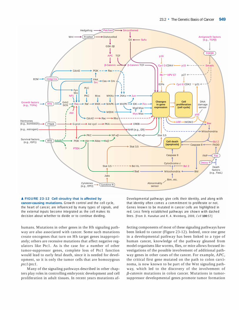
Many of the signaling pathways described in other chap-ters play roles in controlling embryonic development and cellproliferation in adult tissues. In recent years mutations af-
fecting components of most of these signaling pathways havebeen linked to cancer (Figure 23-12). Indeed, once one genein a developmental pathway has been linked to a type ofhuman cancer, knowledge of the pathway gleaned frommodel organisms like worms, flies, or mice allows focused in-vestigations of the possible involvement of additional path-way genes in other cases of the cancer. For example, APC,the critical first gene mutated on the path to colon carci-noma, is now known to be part of the Wnt signaling path-way, which led to the discovery of the involvement of�-catenin mutations in colon cancer. Mutations in tumor-suppressor developmental genes promote tumor formation
23.2 • The Genetic Basis of Cancer 949
Wnt Dishevelled
GSK-3β
APC
Rb
E2Fe
p16
p15 Smads
Antigrowth factors(e.g., TGFβ)
p53
Cycl D:CDK4
Cycl E:CDK2 p21
HPV E7 p27
DNAdamagesensor
Abnormalitysensor
TCF
β-Catenin β-Catenin:TCF
Frizzled
TGFβR
Growth factors(e.g., TGFα)
Hormones(e.g., bombesin)
(e.g., estrogen)
Grb2SOSRTK
Survival factors(e.g., IGF1)
Cytokines(e.g., EPO)
RTK
7TMR
Cytokine R
Fas
Decoy R
Integrins
NF1
Abl
Ras Raf
Rac Rho
PKC Mos MKKs
FynShc
FAKSrc
PI3K Rac
Cas Crk
Cdc42
ECM
PLC
MEK MAPK
Mitochondria
Caspase 8 FADDBcl 2
MEKK
Cdc42
Ad cycl
Akt Akkα
PKA
PKC Stat 3,5
Stat 3,5
Stat 3,5 Bcl XL
Bad
Bcl 2
Jaks
Caspase 9
Cytochrome cDeathfactors
(e.g., FasL)Mitochondria Bld
Bim, etc.
CREBARF
FAP
MDM2 Bax
NF-κβNF-κβ
Iκβ
NHR (e.g., ER)
G-prot
PI3K
? PTEN
Mad:Max
Myc:Max
MAPK
JNKs
Changesin gene
expression
Cellproliferation(cell cycle)
Cell death(apoptosis)
EIK
Jun
Fos
Patched SmoothenedHedgehog
Gli Sufu
▲ FIGURE 23-12 Cell circuitry that is affected by
cancer-causing mutations. Growth control and the cell cycle,the heart of cancer, are influenced by many types of signals, andthe external inputs become integrated as the cell makes itsdecision about whether to divide or to continue dividing.
Developmental pathways give cells their identity, and along withthat identity often comes a commitment to proliferate or not.Genes known to be mutated in cancer cells are highlighted inred. Less firmly established pathways are shown with dashedlines. [From D. Hanahan and R. A. Weinberg, 2000, Cell 100:57.]
in tissues where the affected gene normally helps restraingrowth, and not in cells where the primary role of the devel-opmental regulator is to control cell fate but not growth.Mutations in developmental proto-oncogenes may inducetumor formation in tissues where an affected gene normallypromotes growth or in another tissue where the gene has be-come aberrantly active.
DNA Microarray Analysis of Expression PatternsCan Reveal Subtle Differences Between Tumor CellsTraditionally the properties of tumor and normal cells havebeen assessed by staining and microscopy. The prognosis formany tumors could be determined, within certain limits,from their histology. However the appearance of cells alonehas limited information content, and better ways to discernthe properties of cells are desirable both to understand tumorigenesis and to arrive at meaningful and accurate de-cisions about prognosis and therapy.
As we’ve seen, genetic studies can identify the single initi-ating mutation or series of mutations that cause transforma-
tion of normal cells into tumor cells, as in the case of coloncancer. After these initial events, however, the cells of a tumorundergo a cascade of changes reflecting the interplay betweenthe initiating events and signals from outside. As a result,tumor cells can become quite different, even if they arise fromthe same initiating mutation or mutations. Although these dif-ferences may not be recognized from the appearance of cells,they can be detected from the cells’ patterns of gene expres-sion. DNA microarray analysis can determine the expressionof thousands of genes simultaneously, permitting complexphenotypes to be defined at the molecular genetic level. (SeeFigures 9-35 and 9-36 for an explanation of this technique.)
Microarray analysis recently has been applied todiffuse large B cell lymphoma, a disease marked bythe presence of abnormally large B lymphocytes
throughout lymph nodes. Affected patients have highly vari-able outcomes, so the disease has long been suspected to be,
950 CHAPTER 23 • Cancer
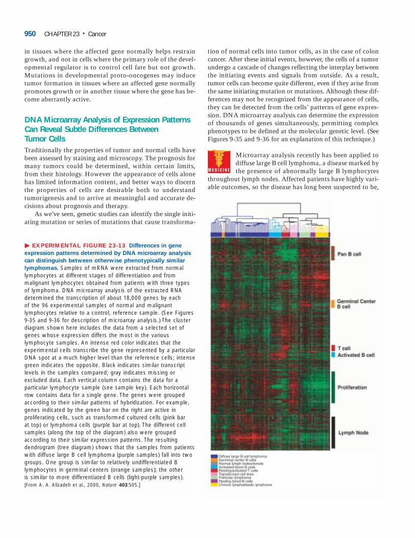
� EXPERIMENTAL FIGURE 23-13 Differences in gene
expression patterns determined by DNA microarray analysis
can distinguish between otherwise phenotypically similar
lymphomas. Samples of mRNA were extracted from normallymphocytes at different stages of differentiation and frommalignant lymphocytes obtained from patients with three typesof lymphoma. DNA microarray analysis of the extracted RNAdetermined the transcription of about 18,000 genes by each of the 96 experimental samples of normal and malignantlymphocytes relative to a control, reference sample. (See Figures9-35 and 9-36 for description of microarray analysis.) The clusterdiagram shown here includes the data from a selected set ofgenes whose expression differs the most in the variouslymphocyte samples. An intense red color indicates that theexperimental cells transcribe the gene represented by a particularDNA spot at a much higher level than the reference cells; intensegreen indicates the opposite. Black indicates similar transcriptlevels in the samples compared; gray indicates missing orexcluded data. Each vertical column contains the data for aparticular lymphocyte sample (see sample key). Each horizontalrow contains data for a single gene. The genes were groupedaccording to their similar patterns of hybridization. For example,genes indicated by the green bar on the right are active inproliferating cells, such as transformed cultured cells (pink bar at top) or lymphoma cells (purple bar at top). The different cellsamples (along the top of the diagram) also were groupedaccording to their similar expression patterns. The resultingdendrogram (tree diagram) shows that the samples from patientswith diffuse large B cell lymphoma (purple samples) fall into twogroups. One group is similar to relatively undifferentiated Blymphocytes in germinal centers (orange samples); the other is similar to more differentiated B cells (light-purple samples).[From A. A. Alizadeh et al., 2000, Nature 403:505.]
in fact, multiple diseases. Microarray analysis of lymphomasfrom different patients revealed two groups distinguished bytheir patterns of gene expression (Figure 23-13). No mor-phological or visible criteria were found that could distin-guish the two types of tumors. Patients with one tumor typedefined by the microarray data survived much longer thanthose with the other type. Lymphomas whose gene expres-sion is similar to that of B lymphocytes in the earliest stagesof differentiation have a better prognosis; lymphomas whosegene expression is closer to that of more differentiated Blymphocytes have a worse prognosis. Similar analyses of thegene expression patterns, or “signatures,” of other tumorsare likely to improve classification and diagnosis, allowinginformed decisions about treatments, and also provide in-sights into the properties of tumor cells. ❚
KEY CONCEPTS OF SECTION 23.2
The Genetic Basis of Cancer
■ Dominant gain-of-function mutations in proto-oncogenesand recessive loss-of-function mutations in tumor-suppressorgenes are oncogenic.
■ Among the proteins encoded by proto-oncogenes aregrowth-promoting signaling proteins and their receptors,signal-transduction proteins, transcription factors, andapoptotic proteins (see Figure 23-8).
■ An activating mutation of one of the two alleles of a proto-oncogene converts it to an oncogene. This can occur by pointmutation, gene amplification, and gene translocation.
■ The first human oncogene to be identified encodes a con-stitutively active form of Ras, a signal-transduction pro-tein. This oncogene was isolated from a human bladdercarcinoma (see Figure 23-4).
■ Slow-acting retroviruses can cause cancer by integratingnear a proto-oncogene in such a way that transcription of thecellular gene is activated continuously and inappropriately.
■ Tumor-suppressor genes encode proteins that directly orindirectly slow progression through the cell cycle, checkpoint-control proteins that arrest the cell cycle, components ofgrowth-inhibiting signaling pathways, pro-apoptotic proteins,and DNA-repair enzymes.
■ The first tumor-suppressor gene to be recognized, RB,is mutated in retinoblastoma and some other tumors.
■ Inheritance of a single mutant allele of RB greatly in-creases the probability that a specific kind of cancer willdevelop, as is the case for many other tumor-suppressorgenes (e.g., APC and BRCA1).
■ In individuals born heterozygous for a tumor-suppressorgene, a somatic cell can undergo loss of heterozygosity(LOH) by mitotic recombination, chromosome mis-segregation, mutation, or deletion (see Figure 23-11).
■ Many genes that regulate normal developmental processesencode proteins that function in various signaling pathways(see Figure 23-12). Their normal roles in regulating whereand when growth occurs are reflected in the character of thetumors that arise when the genes are mutated.
■ DNA microarray analysis can identify differences in geneexpression between types of tumor cells that are indistin-guishable by traditional criteria. Some tumor cells appearto be related to specific types of normal cells at certainstages of development based on similarities in their ex-pression patterns.
Oncogenic Mutations in Growth-Promoting ProteinsGenes encoding each class of cell regulatory protein depictedin Figure 23-8 have been identified as proto-oncogenes ortumor-suppressor genes. In this section we examine in moredetail how mutations that result in the unregulated, consti-tutive activity of certain proteins or in their overproductionpromote cell proliferation and transformation, thereby con-tributing to carcinogenesis. In each case we see how a rarecell that has undergone a very particular sort of mutation be-comes abundant owing to its uncontrolled proliferation.
Oncogenic Receptors Can Promote Proliferationin the Absence of External Growth FactorsAlthough oncogenes theoretically could arise from mutationsin genes encoding growth-promoting signaling molecules,this rarely occurs. In fact, only one such naturally occurringoncogene, sis, has been discovered. The sis oncogene, whichencodes a type of platelet-derived growth factor (PDGF), canaberrantly autostimulate proliferation of cells that normallyexpress the PDGF receptor.
In contrast, oncogenes encoding cell-surface receptorsthat transduce growth-promoting signals have been associ-ated with several types of cancer. The receptors for manysuch growth factors have intrinsic protein-tyrosine kinase ac-tivity in their cytosolic domains, an activity that is quiescentuntil activated. Ligand binding to the external domains ofthese receptor tyrosine kinases (RTKs) leads to their dimer-ization and activation of their kinase activity, initiating an in-tracellular signaling pathway that ultimately promotesproliferation.
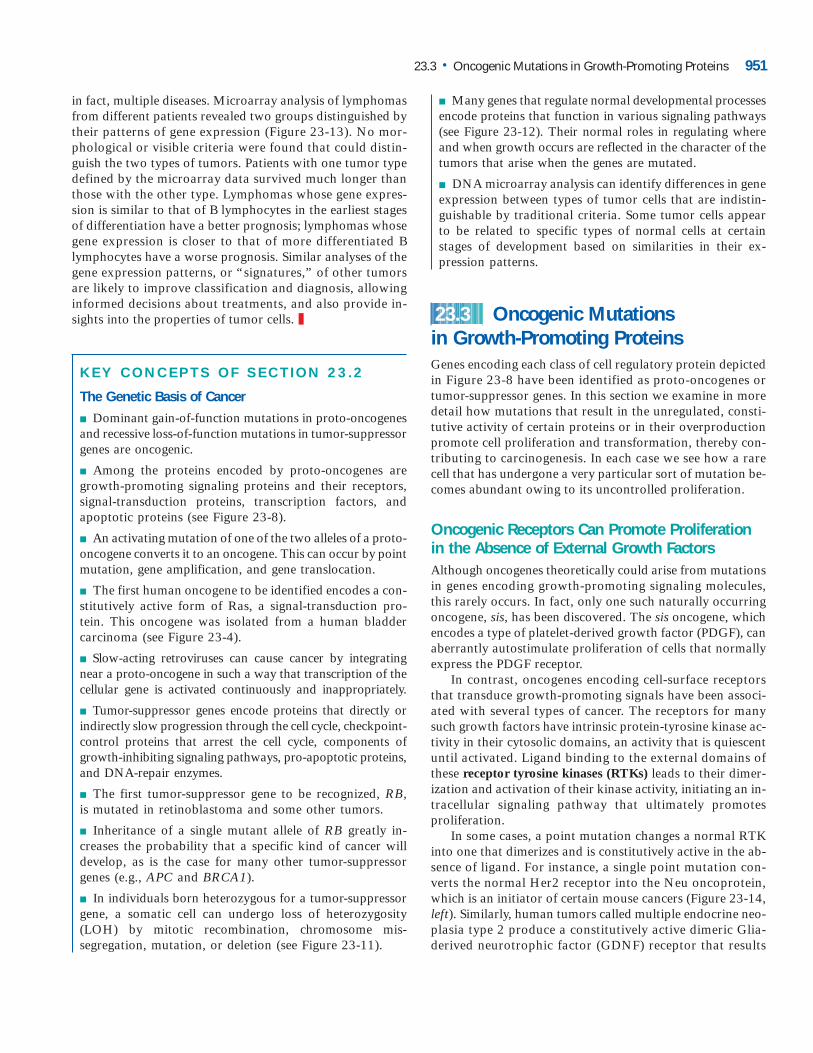
In some cases, a point mutation changes a normal RTKinto one that dimerizes and is constitutively active in the ab-sence of ligand. For instance, a single point mutation con-verts the normal Her2 receptor into the Neu oncoprotein,which is an initiator of certain mouse cancers (Figure 23-14,left). Similarly, human tumors called multiple endocrine neo-plasia type 2 produce a constitutively active dimeric Glia-derived neurotrophic factor (GDNF) receptor that results
23.3
23.3 • Oncogenic Mutations in Growth-Promoting Proteins 951
from a point mutation in the extracellular domain. In othercases, deletion of much of the extracellular ligand-bindingdomain produces a constitutively active oncogenic receptor.For example, deletion of the extracellular domain of the nor-mal EGF receptor converts it to the dimeric ErbB oncopro-tein (Figure 23-14, right).
Mutations leading to overproduction of a normal RTKalso can be oncogenic. For instance, many human breast can-cers overproduce a normal Her2 receptor. As a result, thecells are stimulated to proliferate in the presence of very lowconcentrations of EGF and related hormones, concentrationstoo low to stimulate proliferation of normal cells.
A monoclonal antibody specific for Her2 has beena strikingly successful new treatment for the subsetof breast cancers that overproduce Her2. Her2
antibody injected into the blood recognizes Her2 and causesit to be internalized, selectively killing the cancer cells with-out any apparent effect on normal breast (and other) cellsthat produce moderate amounts of Her2. ❚
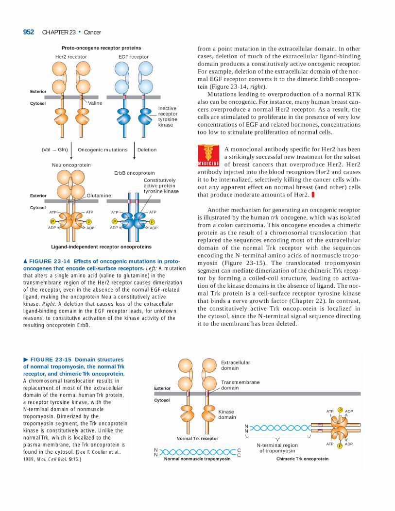
Another mechanism for generating an oncogenic receptoris illustrated by the human trk oncogene, which was isolatedfrom a colon carcinoma. This oncogene encodes a chimericprotein as the result of a chromosomal translocation that replaced the sequences encoding most of the extracellular domain of the normal Trk receptor with the sequences encoding the N-terminal amino acids of nonmuscle tropo-myosin (Figure 23-15). The translocated tropomyosin segment can mediate dimerization of the chimeric Trk recep-tor by forming a coiled-coil structure, leading to activa-tion of the kinase domains in the absence of ligand. The nor-mal Trk protein is a cell-surface receptor tyrosine kinase that binds a nerve growth factor (Chapter 22). In contrast,the constitutively active Trk oncoprotein is localized in the cytosol, since the N-terminal signal sequence directing it to the membrane has been deleted.
952 CHAPTER 23 • Cancer
Exterior
Cytosol
Neu oncoprotein
Exterior
Cytosol
Inactivereceptortyrosinekinase
Her2 receptor EGF receptor
Proto-oncogene receptor proteins
Oncogenic mutations(Val → Gln) Deletion
Valine
ErbB oncoprotein
Ligand-independent receptor oncoproteins
Constitutively active proteintyrosine kinase
PP
ATP
ADP
ATP
ADP
PP
ATP
ADP
ATP
ADP
Glutamine
▲ FIGURE 23-14 Effects of oncogenic mutations in proto-
oncogenes that encode cell-surface receptors. Left: A mutationthat alters a single amino acid (valine to glutamine) in thetransmembrane region of the Her2 receptor causes dimerizationof the receptor, even in the absence of the normal EGF-relatedligand, making the oncoprotein Neu a constitutively active kinase. Right: A deletion that causes loss of the extracellular ligand-binding domain in the EGF receptor leads, for unknownreasons, to constitutive activation of the kinase activity of theresulting oncoprotein ErbB.
NN
N-terminal regionof tropomyosin
PATP ADP
CC
NN
Normal nonmuscle tropomyosin
Normal Trk receptor
Extracellulardomain
Transmembranedomain
Kinasedomain
Chimeric Trk oncoprotein
PATP ADP
Exterior
Cytosol
� FIGURE 23-15 Domain structures
of normal tropomyosin, the normal Trk
receptor, and chimeric Trk oncoprotein.
A chromosomal translocation results inreplacement of most of the extracellulardomain of the normal human Trk protein, a receptor tyrosine kinase, with the N-terminal domain of nonmuscletropomyosin. Dimerized by thetropomyosin segment, the Trk oncoproteinkinase is constitutively active. Unlike thenormal Trk, which is localized to theplasma membrane, the Trk oncoprotein isfound in the cytosol. [See F. Coulier et al.,1989, Mol. Cell Biol. 9:15.]
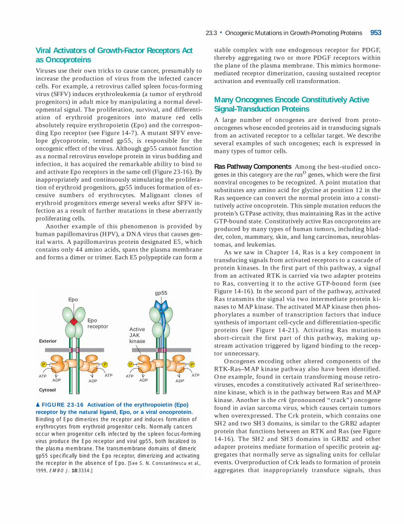
Viral Activators of Growth-Factor Receptors Actas OncoproteinsViruses use their own tricks to cause cancer, presumably toincrease the production of virus from the infected cancercells. For example, a retrovirus called spleen focus-formingvirus (SFFV) induces erythroleukemia (a tumor of erythroidprogenitors) in adult mice by manipulating a normal devel-opmental signal. The proliferation, survival, and differenti-ation of erythroid progenitors into mature red cellsabsolutely require erythropoietin (Epo) and the correspon-ding Epo receptor (see Figure 14-7). A mutant SFFV enve-lope glycoprotein, termed gp55, is responsible for theoncogenic effect of the virus. Although gp55 cannot functionas a normal retrovirus envelope protein in virus budding andinfection, it has acquired the remarkable ability to bind toand activate Epo receptors in the same cell (Figure 23-16). Byinappropriately and continuously stimulating the prolifera-tion of erythroid progenitors, gp55 induces formation of ex-cessive numbers of erythrocytes. Malignant clones oferythroid progenitors emerge several weeks after SFFV in-fection as a result of further mutations in these aberrantlyproliferating cells.
Another example of this phenomenon is provided byhuman papillomavirus (HPV), a DNA virus that causes gen-ital warts. A papillomavirus protein designated E5, whichcontains only 44 amino acids, spans the plasma membraneand forms a dimer or trimer. Each E5 polypeptide can form a
stable complex with one endogenous receptor for PDGF,thereby aggregating two or more PDGF receptors within the plane of the plasma membrane. This mimics hormone-mediated receptor dimerization, causing sustained receptoractivation and eventually cell transformation.
Many Oncogenes Encode Constitutively ActiveSignal-Transduction ProteinsA large number of oncogenes are derived from proto-oncogenes whose encoded proteins aid in transducing signalsfrom an activated receptor to a cellular target. We describeseveral examples of such oncogenes; each is expressed inmany types of tumor cells.
Ras Pathway Components Among the best-studied onco-genes in this category are the rasD genes, which were the firstnonviral oncogenes to be recognized. A point mutation thatsubstitutes any amino acid for glycine at position 12 in theRas sequence can convert the normal protein into a consti-tutively active oncoprotein. This simple mutation reduces theprotein’s GTPase activity, thus maintaining Ras in the activeGTP-bound state. Constitutively active Ras oncoproteins areproduced by many types of human tumors, including blad-der, colon, mammary, skin, and lung carcinomas, neuroblas-tomas, and leukemias.
As we saw in Chapter 14, Ras is a key component intransducing signals from activated receptors to a cascade ofprotein kinases. In the first part of this pathway, a signalfrom an activated RTK is carried via two adapter proteinsto Ras, converting it to the active GTP-bound form (seeFigure 14-16). In the second part of the pathway, activatedRas transmits the signal via two intermediate protein ki-nases to MAP kinase. The activated MAP kinase then phos-phorylates a number of transcription factors that inducesynthesis of important cell-cycle and differentiation-specificproteins (see Figure 14-21). Activating Ras mutationsshort-circuit the first part of this pathway, making up-stream activation triggered by ligand binding to the recep-tor unnecessary.
Oncogenes encoding other altered components of theRTK-Ras–MAP kinase pathway also have been identified.One example, found in certain transforming mouse retro-viruses, encodes a constitutively activated Raf serine/threo-nine kinase, which is in the pathway between Ras and MAPkinase. Another is the crk (pronounced “crack”) oncogenefound in avian sarcoma virus, which causes certain tumorswhen overexpressed. The Crk protein, which contains oneSH2 and two SH3 domains, is similar to the GRB2 adapterprotein that functions between an RTK and Ras (see Figure14-16). The SH2 and SH3 domains in GRB2 and otheradapter proteins mediate formation of specific protein ag-gregates that normally serve as signaling units for cellularevents. Overproduction of Crk leads to formation of proteinaggregates that inappropriately transduce signals, thus
23.3 • Oncogenic Mutations in Growth-Promoting Proteins 953
ATPADP
P
Exterior
Cytosol
Epo
ATPADP
ATPADP
PP
Eporeceptor Active
JAKkinase
ATPADP
P
gp55
▲ FIGURE 23-16 Activation of the erythropoietin (Epo)
receptor by the natural ligand, Epo, or a viral oncoprotein.
Binding of Epo dimerizes the receptor and induces formation oferythrocytes from erythroid progenitor cells. Normally cancersoccur when progenitor cells infected by the spleen focus-formingvirus produce the Epo receptor and viral gp55, both localized tothe plasma membrane. The transmembrane domains of dimericgp55 specifically bind the Epo receptor, dimerizing and activatingthe receptor in the absence of Epo. [See S. N. Constantinescu et al.,1999, EMBO J. 18:3334.]
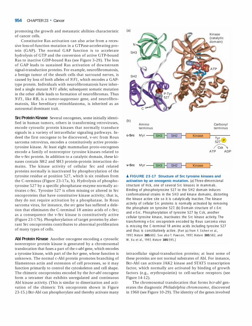
promoting the growth and metastatic abilities characteristicof cancer cells.
Constitutive Ras activation can also arise from a reces-sive loss-of-function mutation in a GTPase-accelerating pro-tein (GAP). The normal GAP function is to acceleratehydrolysis of GTP and the conversion of active GTP-boundRas to inactive GDP-bound Ras (see Figure 3-29). The lossof GAP leads to sustained Ras activation of downstream signal-transduction proteins. For example, neurofibromatosis,a benign tumor of the sheath cells that surround nerves, iscaused by loss of both alleles of NF1, which encodes a GAP-type protein. Individuals with neurofibromatosis have inher-ited a single mutant NF1 allele; subsequent somatic mutationin the other allele leads to formation of neurofibromas. ThusNF1, like RB, is a tumor-suppressor gene, and neurofibro-matosis, like hereditary retinoblastoma, is inherited as an autosomal dominant trait.
Src Protein Kinase Several oncogenes, some initially identi-fied in human tumors, others in transforming retroviruses,encode cytosolic protein kinases that normally transducesignals in a variety of intracellular signaling pathways. In-deed the first oncogene to be discovered, v-src from Roussarcoma retrovirus, encodes a constitutively active protein-tyrosine kinase. At least eight mammalian proto-oncogenesencode a family of nonreceptor tyrosine kinases related tothe v-Src protein. In addition to a catalytic domain, these ki-nases contain SH2 and SH3 protein-protein interaction do-mains. The kinase activity of cellular Src and relatedproteins normally is inactivated by phosphorylation of thetyrosine residue at position 527, which is six residues fromthe C-terminus (Figure 23-17a, b). Hydrolysis of phospho-tyrosine 527 by a specific phosphatase enzyme normally ac-tivates c-Src. Tyrosine 527 is often missing or altered in Srconcoproteins that have constitutive kinase activity; that is,they do not require activation by a phosphatase. In Roussarcoma virus, for instance, the src gene has suffered a dele-tion that eliminates the C-terminal 18 amino acids of c-Src;as a consequence the v-Src kinase is constitutively active(Figure 23-17b). Phosphorylation of target proteins by aber-rant Src oncoproteins contributes to abnormal proliferationof many types of cells.
Abl Protein Kinase Another oncogene encoding a cytosolicnonreceptor protein kinase is generated by a chromosomaltranslocation that fuses a part of the c-abl gene, which encodesa tyrosine kinase, with part of the bcr gene, whose function isunknown. The normal c-Abl protein promotes branching offilamentous actin and extension of cell processes, so it mayfunction primarily to control the cytoskeleton and cell shape.The chimeric oncoproteins encoded by the bcr-abl oncogeneform a tetramer that exhibits unregulated and continuous Abl kinase activity. (This is similar to dimerization and acti-vation of the chimeric Trk oncoprotein shown in Figure 23-15.) Bcr-Abl can phosphorylate and thereby activate many
intracellular signal-transduction proteins; at least some ofthese proteins are not normal substrates of Abl. For instance,Bcr-Abl can activate JAK2 kinase and STAT5 transcriptionfactor, which normally are activated by binding of growth factors (e.g., erythropoietin) to cell-surface receptors (see Figure 14-12).
The chromosomal translocation that forms bcr-abl gen-erates the diagnostic Philadelphia chromosome, discoveredin 1960 (see Figure 10-29). The identity of the genes involved
954 CHAPTER 23 • Cancer
Carboxylterminus
Aminoterminus
c-Src MyrTyr527
SH3 SH2 Kinase
v-Src Myr SH3 SH2 Kinase
Csk
P
(b)
ATP ADP
N
C
SH3
SH2
P-Tyr527
Kinase(catalyticdomain)
ATP
(a)
▲ FIGURE 23-17 Structure of Src tyrosine kinases and
activation by an oncogenic mutation. (a) Three-dimensionalstructure of Hck, one of several Src kinases in mammals. Binding of phosphotyrosine 527 to the SH2 domain inducesconformational strains in the SH3 and kinase domains, distortingthe kinase active site so it is catalytically inactive. The kinaseactivity of cellular Src proteins is normally activated by removingthe phosphate on tyrosine 527. (b) Domain structure of c-Src and v-Src. Phosphorylation of tyrosine 527 by Csk, anothercellular tyrosine kinase, inactivates the Src kinase activity. Thetransforming v-Src oncoprotein encoded by Rous sarcoma virus is missing the C-terminal 18 amino acids including tyrosine 527and thus is constitutively active. [Part (a) from F. Sicheri et al., 1997, Nature 385:602. See also T. Pawson, 1997, Nature 385:582, and W. Xu et al., 1997, Nature 385:595.]
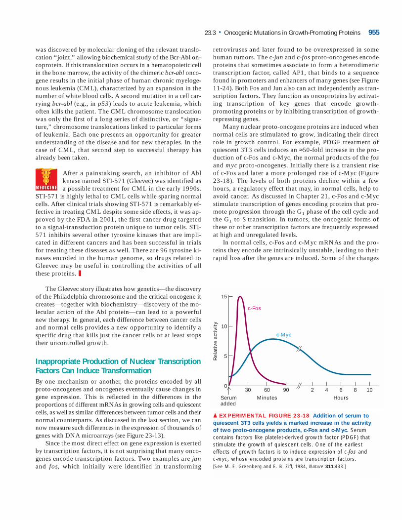
was discovered by molecular cloning of the relevant translo-cation “joint,” allowing biochemical study of the Bcr-Abl on-coprotein. If this translocation occurs in a hematopoietic cellin the bone marrow, the activity of the chimeric bcr-abl onco-gene results in the initial phase of human chronic myeloge-nous leukemia (CML), characterized by an expansion in thenumber of white blood cells. A second mutation in a cell car-rying bcr-abl (e.g., in p53) leads to acute leukemia, whichoften kills the patient. The CML chromosome translocationwas only the first of a long series of distinctive, or “signa-ture,” chromosome translocations linked to particular formsof leukemia. Each one presents an opportunity for greaterunderstanding of the disease and for new therapies. In thecase of CML, that second step to successful therapy has already been taken.
After a painstaking search, an inhibitor of Abl kinase named STI-571 (Gleevec) was identified asa possible treatment for CML in the early 1990s.
STI-571 is highly lethal to CML cells while sparing normalcells. After clinical trials showing STI-571 is remarkably ef-fective in treating CML despite some side effects, it was ap-proved by the FDA in 2001, the first cancer drug targetedto a signal-transduction protein unique to tumor cells. STI-571 inhibits several other tyrosine kinases that are impli-cated in different cancers and has been successful in trialsfor treating these diseases as well. There are 96 tyrosine ki-nases encoded in the human genome, so drugs related toGleevec may be useful in controlling the activities of allthese proteins. ❚
The Gleevec story illustrates how genetics—the discoveryof the Philadelphia chromosome and the critical oncogene itcreates—together with biochemistry—discovery of the mo-lecular action of the Abl protein—can lead to a powerfulnew therapy. In general, each difference between cancer cellsand normal cells provides a new opportunity to identify aspecific drug that kills just the cancer cells or at least stopstheir uncontrolled growth.
Inappropriate Production of Nuclear TranscriptionFactors Can Induce TransformationBy one mechanism or another, the proteins encoded by allproto-oncogenes and oncogenes eventually cause changes ingene expression. This is reflected in the differences in theproportions of different mRNAs in growing cells and quiescentcells, as well as similar differences between tumor cells and theirnormal counterparts. As discussed in the last section, we cannow measure such differences in the expression of thousands ofgenes with DNA microarrays (see Figure 23-13).
Since the most direct effect on gene expression is exertedby transcription factors, it is not surprising that many onco-genes encode transcription factors. Two examples are junand fos, which initially were identified in transforming
retroviruses and later found to be overexpressed in somehuman tumors. The c-jun and c-fos proto-oncogenes encodeproteins that sometimes associate to form a heterodimerictranscription factor, called AP1, that binds to a sequencefound in promoters and enhancers of many genes (see Figure11-24). Both Fos and Jun also can act independently as tran-scription factors. They function as oncoproteins by activat-ing transcription of key genes that encode growth-promoting proteins or by inhibiting transcription of growth-repressing genes.
Many nuclear proto-oncogene proteins are induced whennormal cells are stimulated to grow, indicating their directrole in growth control. For example, PDGF treatment ofquiescent 3T3 cells induces an ≈50-fold increase in the pro-duction of c-Fos and c-Myc, the normal products of the fosand myc proto-oncogenes. Initially there is a transient riseof c-Fos and later a more prolonged rise of c-Myc (Figure 23-18). The levels of both proteins decline within a fewhours, a regulatory effect that may, in normal cells, help toavoid cancer. As discussed in Chapter 21, c-Fos and c-Mycstimulate transcription of genes encoding proteins that pro-mote progression through the G1 phase of the cell cycle andthe G1 to S transition. In tumors, the oncogenic forms ofthese or other transcription factors are frequently expressedat high and unregulated levels.
In normal cells, c-Fos and c-Myc mRNAs and the pro-teins they encode are intrinsically unstable, leading to theirrapid loss after the genes are induced. Some of the changes
23.3 • Oncogenic Mutations in Growth-Promoting Proteins 955
5
10
15
0
Rel
ativ
e ac
tivi
ty
HoursMinutesSerumadded
6 8 10429060
c-Fos
c-Myc
30
▲ EXPERIMENTAL FIGURE 23-18 Addition of serum to
quiescent 3T3 cells yields a marked increase in the activity
of two proto-oncogene products, c-Fos and c-Myc. Serumcontains factors like platelet-derived growth factor (PDGF) thatstimulate the growth of quiescent cells. One of the earliesteffects of growth factors is to induce expression of c-fos and c-myc, whose encoded proteins are transcription factors. [See M. E. Greenberg and E. B. Ziff, 1984, Nature 311:433.]
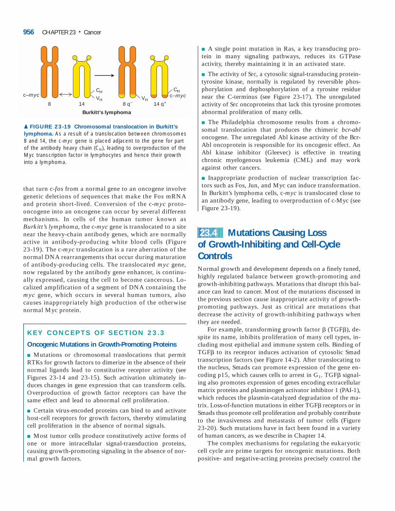
that turn c-fos from a normal gene to an oncogene involvegenetic deletions of sequences that make the Fos mRNAand protein short-lived. Conversion of the c-myc proto-oncogene into an oncogene can occur by several differentmechanisms. In cells of the human tumor known asBurkitt’s lymphoma, the c-myc gene is translocated to a sitenear the heavy-chain antibody genes, which are normallyactive in antibody-producing white blood cells (Figure 23-19). The c-myc translocation is a rare aberration of thenormal DNA rearrangements that occur during maturationof antibody-producing cells. The translocated myc gene,now regulated by the antibody gene enhancer, is continu-ally expressed, causing the cell to become cancerous. Lo-calized amplification of a segment of DNA containing themyc gene, which occurs in several human tumors, alsocauses inappropriately high production of the otherwisenormal Myc protein.
KEY CONCEPTS OF SECTION 23.3
Oncogenic Mutations in Growth-Promoting Proteins
■ Mutations or chromosomal translocations that permitRTKs for growth factors to dimerize in the absence of theirnormal ligands lead to constitutive receptor activity (seeFigures 23-14 and 23-15). Such activation ultimately in-duces changes in gene expression that can transform cells.Overproduction of growth factor receptors can have thesame effect and lead to abnormal cell proliferation.
■ Certain virus-encoded proteins can bind to and activatehost-cell receptors for growth factors, thereby stimulatingcell proliferation in the absence of normal signals.
■ Most tumor cells produce constitutively active forms ofone or more intracellular signal-transduction proteins,causing growth-promoting signaling in the absence of nor-mal growth factors.
■ A single point mutation in Ras, a key transducing pro-tein in many signaling pathways, reduces its GTPase activity, thereby maintaining it in an activated state.
■ The activity of Src, a cytosolic signal-transducing protein-tyrosine kinase, normally is regulated by reversible phos-phorylation and dephosphorylation of a tyrosine residuenear the C-terminus (see Figure 23-17). The unregulated activity of Src oncoproteins that lack this tyrosine promotesabnormal proliferation of many cells.
■ The Philadelphia chromosome results from a chromo-somal translocation that produces the chimeric bcr-abloncogene. The unregulated Abl kinase activity of the Bcr-Abl oncoprotein is responsible for its oncogenic effect. AnAbl kinase inhibitor (Gleevec) is effective in treatingchronic myelogenous leukemia (CML) and may workagainst other cancers.
■ Inappropriate production of nuclear transcription fac-tors such as Fos, Jun, and Myc can induce transformation.In Burkitt’s lymphoma cells, c-myc is translocated close toan antibody gene, leading to overproduction of c-Myc (seeFigure 23-19).
Mutations Causing Loss of Growth-Inhibiting and Cell-CycleControlsNormal growth and development depends on a finely tuned,highly regulated balance between growth-promoting andgrowth-inhibiting pathways. Mutations that disrupt this bal-ance can lead to cancer. Most of the mutations discussed inthe previous section cause inappropriate activity of growth-promoting pathways. Just as critical are mutations that decrease the activity of growth-inhibiting pathways whenthey are needed.
For example, transforming growth factor � (TGF�), de-spite its name, inhibits proliferation of many cell types, in-cluding most epithelial and immune system cells. Binding ofTGF� to its receptor induces activation of cytosolic Smadtranscription factors (see Figure 14-2). After translocating tothe nucleus, Smads can promote expression of the gene en-coding p15, which causes cells to arrest in G1. TGF� signal-ing also promotes expression of genes encoding extracellularmatrix proteins and plasminogen activator inhibitor 1 (PAI-1),which reduces the plasmin-catalyzed degradation of the ma-trix. Loss-of-function mutations in either TGF� receptors or inSmads thus promote cell proliferation and probably contributeto the invasiveness and metastasis of tumor cells (Figure 23-20). Such mutations have in fact been found in a varietyof human cancers, as we describe in Chapter 14.
The complex mechanisms for regulating the eukaryoticcell cycle are prime targets for oncogenic mutations. Bothpositive- and negative-acting proteins precisely control the
23.4
956 CHAPTER 23 • Cancer
c–myc c–myc
8 14 8 q− 14 q+VH
CH
VH
CH
Burkitt's lymphoma
▲ FIGURE 23-19 Chromosomal translocation in Burkitt’s
lymphoma. As a result of a translocation between chromosomes8 and 14, the c-myc gene is placed adjacent to the gene for partof the antibody heavy chain (CH), leading to overproduction of theMyc transcription factor in lymphocytes and hence their growthinto a lymphoma.

entry of cells into and their progression through the cellcycle, which consists of four main phases: G1, S, G2, and mi-tosis (see Figure 21-2). This regulatory system assures theproper coordination of cellular growth during G1 and G2,DNA synthesis during the S phase, and chromosome segre-gation and cell division during mitosis. In addition, cells thathave sustained damage to their DNA normally are arrestedbefore their DNA is replicated. This arrest allows time forthe DNA damage to be repaired; alternatively, the arrestedcells are directed to commit suicide via programmed celldeath. The whole cell-cycle control system functions to pre-vent cells from becoming cancerous. As might be expected,mutations in this system often lead to abnormal developmentor contribute to cancer.
Mutations That Promote Unregulated Passagefrom G1 to S Phase Are OncogenicOnce a cell progresses past a certain point in late G1, calledthe restriction point, it becomes irreversibly committed to en-tering the S phase and replicating its DNA (see Figure 21-28).D-type cyclins, cyclin-dependent kinases (CDKs), and the Rbprotein are all elements of the control system that regulatepassage through the restriction point.
The expression of D-type cyclin genes is induced by manyextracellular growth factors, or mitogens. These cyclins assemble with their partners CDK4 and CDK6 to generatecatalytically active cyclin-CDK complexes, whose kinase activity promotes progression past the restriction point. Mi-togen withdrawal prior to passage through the restrictionpoint leads to accumulation of p16. Like p15 mentionedabove, p16 binds specifically to CDK4 and CDK6, therebyinhibiting their kinase activity and causing G1 arrest. Undernormal circumstances, phosphorylation of Rb protein is ini-tiated midway through G1 by active cyclin D-CDK4 and cy-clin D-CDK6 complexes. Rb phosphorylation is completedby other cyclin-CDK complexes in late G1, allowing activa-tion of E2F transcription factors, which stimulate transcrip-tion of genes encoding proteins required for DNA synthesis.The complete phosphorylation of Rb irreversibly commitsthe cell to DNA synthesis. Most tumors contain an onco-genic mutation that causes overproduction or loss of one ofthe components of this pathway such that the cells are pro-pelled into the S phase in the absence of the proper extracel-lular growth signals (Figure 23-21).
Elevated levels of cyclin D1, for example, are found in many human cancers. In certain tumors of antibody-producing B lymphocytes, for instance, the cyclin D1 geneis translocated such that its transcription is under control ofan antibody-gene enhancer, causing elevated cyclin D1 pro-duction throughout the cell cycle, irrespective of extracellu-lar signals. (This phenomenon is analogous to the c-myctranslocation in Burkitt’s lymphoma cells discussed earlier.)That cyclin D1 can function as an oncoprotein was shown bystudies with transgenic mice in which the cyclin D1 gene was
placed under control of an enhancer specific for mammaryductal cells. Initially the ductal cells underwent hyperprolif-eration, and eventually breast tumors developed in thesetransgenic mice. Amplification of the cyclin D1 gene andconcomitant overproduction of the cyclin D1 protein is common in human breast cancer.
The proteins that function as cyclin-CDK inhibitors playan important role in regulating the cell cycle (Chapter 21). Inparticular, loss-of-function mutations that prevent p16 from
23.4 • Mutations Causing Loss of Growth-Inhibiting and Cell-Cycle Controls 957
P
−
−
Smad3
Smad4
Smad4
Smad3-P
TGFβ
PPPP
P
Cytosol
Nucleus
Type IIreceptors
Type Ireceptors
Transcription of gene
encoding cell-cycle
inhibitor
p15 promoter p15
Transcription of gene
encoding inhibitor of
protease that degrades
extracellular matrix
proteins
PAI-1 promoter PAI-1
PP
Loss-of-function
mutations in
Smads block
TGFβ signal
Decreased production of
p15 increases proliferation
Decreased production
of PAI-1 allows increased
extracellular matrix
degradation and, hence,
metastasis
−
−Loss-of-function
mutations in
receptors block
TGFβ signal
−
▲ FIGURE 23-20 Effect of loss of TGF� signaling.
Binding of TGF�, an antigrowth factor, causes activation of Smad transcription factors. In the absence of TGF�
signaling due to either a receptor mutation or a SMADmutation, cell proliferation and invasion of the surroundingextracellular matrix (ECM) increase. [See X. Hua et al., 1998,Genes & Develop. 12:3084.]
ME
DIA
C
ON
NE
CT
IO
NS
Focus Anim
ation: TGF
�Signaling Pathw
ay
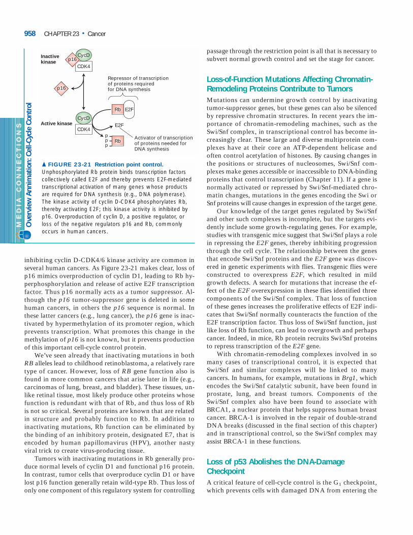
inhibiting cyclin D-CDK4/6 kinase activity are common inseveral human cancers. As Figure 23-21 makes clear, loss ofp16 mimics overproduction of cyclin D1, leading to Rb hy-perphosphorylation and release of active E2F transcriptionfactor. Thus p16 normally acts as a tumor suppressor. Al-though the p16 tumor-suppressor gene is deleted in somehuman cancers, in others the p16 sequence is normal. Inthese latter cancers (e.g., lung cancer), the p16 gene is inac-tivated by hypermethylation of its promoter region, whichprevents transcription. What promotes this change in themethylation of p16 is not known, but it prevents productionof this important cell-cycle control protein.
We’ve seen already that inactivating mutations in bothRB alleles lead to childhood retinoblastoma, a relatively raretype of cancer. However, loss of RB gene function also isfound in more common cancers that arise later in life (e.g.,carcinomas of lung, breast, and bladder). These tissues, un-like retinal tissue, most likely produce other proteins whosefunction is redundant with that of Rb, and thus loss of Rbis not so critical. Several proteins are known that are relatedin structure and probably function to Rb. In addition to inactivating mutations, Rb function can be eliminated by the binding of an inhibitory protein, designated E7, that isencoded by human papillomavirus (HPV), another nastyviral trick to create virus-producing tissue.
Tumors with inactivating mutations in Rb generally pro-duce normal levels of cyclin D1 and functional p16 protein.In contrast, tumor cells that overproduce cyclin D1 or havelost p16 function generally retain wild-type Rb. Thus loss ofonly one component of this regulatory system for controlling
passage through the restriction point is all that is necessary tosubvert normal growth control and set the stage for cancer.
Loss-of-Function Mutations Affecting Chromatin-Remodeling Proteins Contribute to TumorsMutations can undermine growth control by inactivatingtumor-suppressor genes, but these genes can also be silencedby repressive chromatin structures. In recent years the im-portance of chromatin-remodeling machines, such as theSwi/Snf complex, in transcriptional control has become in-creasingly clear. These large and diverse multiprotein com-plexes have at their core an ATP-dependent helicase andoften control acetylation of histones. By causing changes inthe positions or structures of nucleosomes, Swi/Snf com-plexes make genes accessible or inaccessible to DNA-bindingproteins that control transcription (Chapter 11). If a gene isnormally activated or repressed by Swi/Snf-mediated chro-matin changes, mutations in the genes encoding the Swi orSnf proteins will cause changes in expression of the target gene.
Our knowledge of the target genes regulated by Swi/Snfand other such complexes is incomplete, but the targets evi-dently include some growth-regulating genes. For example,studies with transgenic mice suggest that Swi/Snf plays a rolein repressing the E2F genes, thereby inhibiting progressionthrough the cell cycle. The relationship between the genesthat encode Swi/Snf proteins and the E2F gene was discov-ered in genetic experiments with flies. Transgenic flies wereconstructed to overexpress E2F, which resulted in mildgrowth defects. A search for mutations that increase the ef-fect of the E2F overexpression in these flies identified threecomponents of the Swi/Snf complex. That loss of functionof these genes increases the proliferative effects of E2F indi-cates that Swi/Snf normally counteracts the function of theE2F transcription factor. Thus loss of Swi/Snf function, justlike loss of Rb function, can lead to overgrowth and perhapscancer. Indeed, in mice, Rb protein recruits Swi/Snf proteinsto repress transcription of the E2F gene.
With chromatin-remodeling complexes involved in somany cases of transcriptional control, it is expected thatSwi/Snf and similar complexes will be linked to many cancers. In humans, for example, mutations in Brg1, whichencodes the Swi/Snf catalytic subunit, have been found inprostate, lung, and breast tumors. Components of theSwi/Snf complex also have been found to associate withBRCA1, a nuclear protein that helps suppress human breastcancer. BRCA-1 is involved in the repair of double-strandDNA breaks (discussed in the final section of this chapter)and in transcriptional control, so the Swi/Snf complex mayassist BRCA-1 in these functions.
Loss of p53 Abolishes the DNA-DamageCheckpointA critical feature of cell-cycle control is the G1 checkpoint,which prevents cells with damaged DNA from entering the
958 CHAPTER 23 • Cancer
CDK4
Rb
Rbppp
E2F
E2F
Repressor of transcriptionof proteins required for DNA synthesis
Activator of transcriptionof proteins needed forDNA synthesis
CycD
CDK4
Inactivekinase
p16
p16CycD
Active kinase
▲ FIGURE 23-21 Restriction point control.
Unphosphorylated Rb protein binds transcription factorscollectively called E2F and thereby prevents E2F-mediatedtranscriptional activation of many genes whose productsare required for DNA synthesis (e.g., DNA polymerase).The kinase activity of cyclin D-CDK4 phosphorylates Rb,thereby activating E2F; this kinase activity is inhibited byp16. Overproduction of cyclin D, a positive regulator, orloss of the negative regulators p16 and Rb, commonlyoccurs in human cancers.
ME
DIA
C
ON
NE
CT
IO
NS
Ove
rvie
w A
nnim
atio
n: C
ell-C
ycle
Con
trol
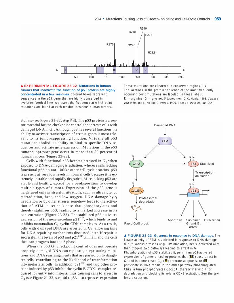
S phase (see Figure 21-32, step 4a). The p53 protein is a sen-sor essential for the checkpoint control that arrests cells withdamaged DNA in G1. Although p53 has several functions, itsability to activate transcription of certain genes is most rele-vant to its tumor-suppressing function. Virtually all p53 mutations abolish its ability to bind to specific DNA se-quences and activate gene expression. Mutations in the p53tumor-suppressor gene occur in more than 50 percent ofhuman cancers (Figure 23-22).
Cells with functional p53 become arrested in G1 whenexposed to DNA-damaging irradiation, whereas cells lackingfunctional p53 do not. Unlike other cell-cycle proteins, p53is present at very low levels in normal cells because it is ex-tremely unstable and rapidly degraded. Mice lacking p53 areviable and healthy, except for a predisposition to developmultiple types of tumors. Expression of the p53 gene isheightened only in stressful situations, such as ultraviolet or� irradiation, heat, and low oxygen. DNA damage by �irradiation or by other stresses somehow leads to the activa-tion of ATM, a serine kinase that phosphorylates andthereby stabilizes p53, leading to a marked increase in itsconcentration (Figure 23-23). The stabilized p53 activatesexpression of the gene encoding p21CIP, which binds to andinhibits mammalian G1 cyclin-CDK complexes. As a result,cells with damaged DNA are arrested in G1, allowing timefor DNA repair by mechanisms discussed later. If repair issuccessful, the levels of p53 and p21CIP will fall, and the cellsthen can progress into the S phase.
When the p53 G1 checkpoint control does not operateproperly, damaged DNA can replicate, perpetuating muta-tions and DNA rearrangements that are passed on to daugh-ter cells, contributing to the likelihood of transformationinto metastatic cells. In addition, p21CIP and two other pro-teins induced by p53 inhibit the cyclin B-CDK1 complex re-quired for entry into mitosis, thus causing cells to arrest inG2 (see Figure 21-32, step 4d). p53 also represses expression
23.4 • Mutations Causing Loss of Growth-Inhibiting and Cell-Cycle Controls 959
I II III IV V
200 250 300 350 39350 100 150
N C
R248
R175 G245
R282
R249
R273
▲ EXPERIMENTAL FIGURE 23-22 Mutations in human
tumors that inactivate the function of p53 protein are highly
concentrated in a few residues. Colored boxes representsequences in the p53 gene that are highly conserved inevolution. Vertical lines represent the frequency at which pointmutations are found at each residue in various human tumors.
These mutations are clustered in conserved regions II–V. The locations in the protein sequence of the most frequentlyoccurring point mutations are labeled. In these labels, R � arginine; G � glycine. [Adapted from C. C. Harris, 1993, Science262:1980, and L. Ko and C. Prives, 1996, Genes & Develop. 10:1054.]
ppp
Chk2
p
Cdc25A
Rapid G1/S block
Proteasomaldegradation
p53 Stabilized
Transcriptionactivation
ATM
Damaged DNA
Apoptosis SustainedG1 and G2
arrest
DNA repair
pp
p53
▲ FIGURE 23-23 G1 arrest in response to DNA damage. Thekinase activity of ATM is activated in response to DNA damagedue to various stresses (e.g., UV irradiation, heat). Activated ATMthen triggers two pathways leading to arrest in G1.Phosphorylation of p53 stabilizes it, permitting p53-activatedexpression of genes encoding proteins that ( ) cause arrest inG1 and in some cases G2, ( ) promote apoptosis, or ( )participate in DNA repair. In the other pathway phosphorylatedChk2 in turn phosphorylates Cdc25A, thereby marking it fordegradation and blocking its role in CDK2 activation. See the textfor a discussion.
321
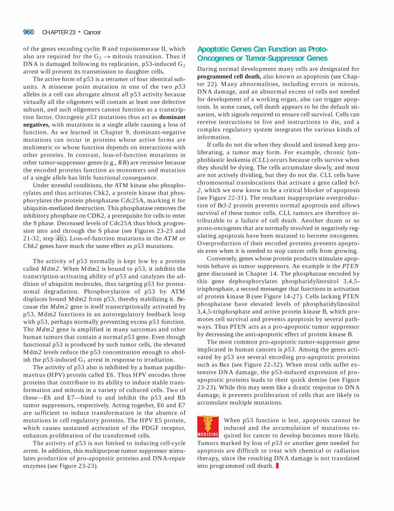
of the genes encoding cyclin B and topoisomerase II, whichalso are required for the G2 n mitosis transition. Thus ifDNA is damaged following its replication, p53-induced G2
arrest will prevent its transmission to daughter cells.The active form of p53 is a tetramer of four identical sub-
units. A missense point mutation in one of the two p53alleles in a cell can abrogate almost all p53 activity becausevirtually all the oligomers will contain at least one defectivesubunit, and such oligomers cannot function as a transcrip-tion factor. Oncogenic p53 mutations thus act as dominantnegatives, with mutations in a single allele causing a loss offunction. As we learned in Chapter 9, dominant-negativemutations can occur in proteins whose active forms are multimeric or whose function depends on interactions withother proteins. In contrast, loss-of-function mutations inother tumor-suppressor genes (e.g., RB) are recessive becausethe encoded proteins function as monomers and mutationof a single allele has little functional consequence.
Under stressful conditions, the ATM kinase also phospho-rylates and thus activates Chk2, a protein kinase that phos-phorylates the protein phosphatase Cdc25A, marking it forubiquitin-mediated destruction. This phosphatase removes theinhibitory phosphate on CDK2, a prerequisite for cells to enterthe S phase. Decreased levels of Cdc25A thus block progres-sion into and through the S phase (see Figures 23-23 and 21-32, step 4b). Loss-of-function mutations in the ATM orChk2 genes have much the same effect as p53 mutations.
The activity of p53 normally is kept low by a proteincalled Mdm2. When Mdm2 is bound to p53, it inhibits thetranscription-activating ability of p53 and catalyzes the ad-dition of ubiquitin molecules, thus targeting p53 for protea-somal degradation. Phosphorylation of p53 by ATMdisplaces bound Mdm2 from p53, thereby stabilizing it. Be-cause the Mdm2 gene is itself transcriptionally activated byp53, Mdm2 functions in an autoregulatory feedback loopwith p53, perhaps normally preventing excess p53 function.The Mdm2 gene is amplified in many sarcomas and otherhuman tumors that contain a normal p53 gene. Even thoughfunctional p53 is produced by such tumor cells, the elevatedMdm2 levels reduce the p53 concentration enough to abol-ish the p53-induced G1 arrest in response to irradiation.
The activity of p53 also is inhibited by a human papillo-mavirus (HPV) protein called E6. Thus HPV encodes threeproteins that contribute to its ability to induce stable trans-formation and mitosis in a variety of cultured cells. Two ofthese—E6 and E7—bind to and inhibit the p53 and Rbtumor suppressors, respectively. Acting together, E6 and E7are sufficient to induce transformation in the absence of mutations in cell regulatory proteins. The HPV E5 protein,which causes sustained activation of the PDGF receptor, enhances proliferation of the transformed cells.
The activity of p53 is not limited to inducing cell-cycle arrest. In addition, this multipurpose tumor suppressor stimu-lates production of pro-apoptotic proteins and DNA-repairenzymes (see Figure 23-23).
Apoptotic Genes Can Function as Proto-Oncogenes or Tumor-Suppressor GenesDuring normal development many cells are designated forprogrammed cell death, also known as apoptosis (see Chap-ter 22). Many abnormalities, including errors in mitosis,DNA damage, and an abnormal excess of cells not neededfor development of a working organ, also can trigger apop-tosis. In some cases, cell death appears to be the default sit-uation, with signals required to ensure cell survival. Cells canreceive instructions to live and instructions to die, and a complex regulatory system integrates the various kinds of information.
If cells do not die when they should and instead keep pro-liferating, a tumor may form. For example, chronic lym-phoblastic leukemia (CLL) occurs because cells survive whenthey should be dying. The cells accumulate slowly, and mostare not actively dividing, but they do not die. CLL cells havechromosomal translocations that activate a gene called bcl-2, which we now know to be a critical blocker of apoptosis(see Figure 22-31). The resultant inappropriate overproduc-tion of Bcl-2 protein prevents normal apoptosis and allowssurvival of these tumor cells. CLL tumors are therefore at-tributable to a failure of cell death. Another dozen or soproto-oncogenes that are normally involved in negatively reg-ulating apoptosis have been mutated to become oncogenes.Overproduction of their encoded proteins prevents apopto-sis even when it is needed to stop cancer cells from growing.
Conversely, genes whose protein products stimulate apop-tosis behave as tumor suppressors. An example is the PTENgene discussed in Chapter 14. The phosphatase encoded bythis gene dephosphorylates phosphatidylinositol 3,4,5-trisphosphate, a second messenger that functions in activationof protein kinase B (see Figure 14-27). Cells lacking PTENphosphatase have elevated levels of phosphatidylinositol3,4,5-trisphosphate and active protein kinase B, which pro-motes cell survival and prevents apoptosis by several path-ways. Thus PTEN acts as a pro-apoptotic tumor suppressorby decreasing the anti-apoptotic effect of protein kinase B.
The most common pro-apoptotic tumor-suppressor geneimplicated in human cancers is p53. Among the genes acti-vated by p53 are several encoding pro-apoptotic proteinssuch as Bax (see Figure 22-32). When most cells suffer ex-tensive DNA damage, the p53-induced expression of pro-apoptotic proteins leads to their quick demise (see Figure23-23). While this may seem like a drastic response to DNAdamage, it prevents proliferation of cells that are likely to accumulate multiple mutations.
When p53 function is lost, apoptosis cannot be induced and the accumulation of mutations re-quired for cancer to develop becomes more likely.
Tumors marked by loss of p53 or another gene needed forapoptosis are difficult to treat with chemical or radiationtherapy, since the resulting DNA damage is not translatedinto programmed cell death. ❚
960 CHAPTER 23 • Cancer
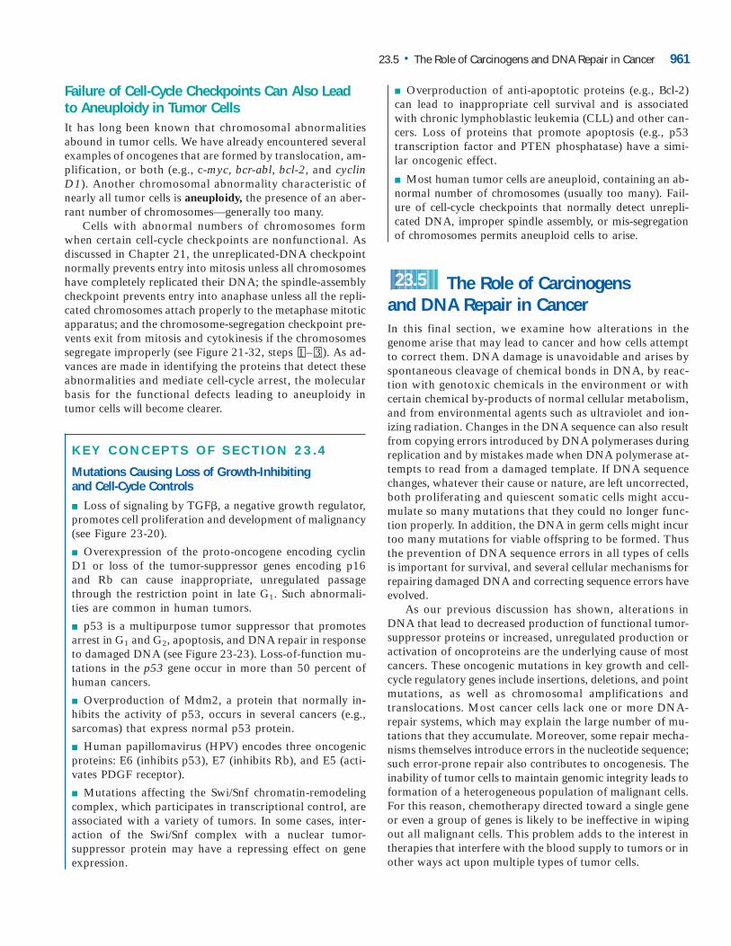
Failure of Cell-Cycle Checkpoints Can Also Leadto Aneuploidy in Tumor CellsIt has long been known that chromosomal abnormalitiesabound in tumor cells. We have already encountered severalexamples of oncogenes that are formed by translocation, am-plification, or both (e.g., c-myc, bcr-abl, bcl-2, and cyclinD1). Another chromosomal abnormality characteristic ofnearly all tumor cells is aneuploidy, the presence of an aber-rant number of chromosomes—generally too many.
Cells with abnormal numbers of chromosomes formwhen certain cell-cycle checkpoints are nonfunctional. Asdiscussed in Chapter 21, the unreplicated-DNA checkpointnormally prevents entry into mitosis unless all chromosomeshave completely replicated their DNA; the spindle-assemblycheckpoint prevents entry into anaphase unless all the repli-cated chromosomes attach properly to the metaphase mitoticapparatus; and the chromosome-segregation checkpoint pre-vents exit from mitosis and cytokinesis if the chromosomessegregate improperly (see Figure 21-32, steps – ). As ad-vances are made in identifying the proteins that detect theseabnormalities and mediate cell-cycle arrest, the molecularbasis for the functional defects leading to aneuploidy intumor cells will become clearer.
KEY CONCEPTS OF SECTION 23.4
Mutations Causing Loss of Growth-Inhibiting and Cell-Cycle Controls
■ Loss of signaling by TGF�, a negative growth regulator,promotes cell proliferation and development of malignancy(see Figure 23-20).
■ Overexpression of the proto-oncogene encoding cyclinD1 or loss of the tumor-suppressor genes encoding p16and Rb can cause inappropriate, unregulated passagethrough the restriction point in late G1. Such abnormali-ties are common in human tumors.
■ p53 is a multipurpose tumor suppressor that promotesarrest in G1 and G2, apoptosis, and DNA repair in responseto damaged DNA (see Figure 23-23). Loss-of-function mu-tations in the p53 gene occur in more than 50 percent ofhuman cancers.
■ Overproduction of Mdm2, a protein that normally in-hibits the activity of p53, occurs in several cancers (e.g.,sarcomas) that express normal p53 protein.
■ Human papillomavirus (HPV) encodes three oncogenicproteins: E6 (inhibits p53), E7 (inhibits Rb), and E5 (acti-vates PDGF receptor).
■ Mutations affecting the Swi/Snf chromatin-remodelingcomplex, which participates in transcriptional control, areassociated with a variety of tumors. In some cases, inter-action of the Swi/Snf complex with a nuclear tumor-suppressor protein may have a repressing effect on geneexpression.
31
■ Overproduction of anti-apoptotic proteins (e.g., Bcl-2)can lead to inappropriate cell survival and is associatedwith chronic lymphoblastic leukemia (CLL) and other can-cers. Loss of proteins that promote apoptosis (e.g., p53transcription factor and PTEN phosphatase) have a simi-lar oncogenic effect.
■ Most human tumor cells are aneuploid, containing an ab-normal number of chromosomes (usually too many). Fail-ure of cell-cycle checkpoints that normally detect unrepli-cated DNA, improper spindle assembly, or mis-segregationof chromosomes permits aneuploid cells to arise.
The Role of Carcinogens and DNA Repair in CancerIn this final section, we examine how alterations in thegenome arise that may lead to cancer and how cells attemptto correct them. DNA damage is unavoidable and arises byspontaneous cleavage of chemical bonds in DNA, by reac-tion with genotoxic chemicals in the environment or withcertain chemical by-products of normal cellular metabolism,and from environmental agents such as ultraviolet and ion-izing radiation. Changes in the DNA sequence can also resultfrom copying errors introduced by DNA polymerases duringreplication and by mistakes made when DNA polymerase at-tempts to read from a damaged template. If DNA sequencechanges, whatever their cause or nature, are left uncorrected,both proliferating and quiescent somatic cells might accu-mulate so many mutations that they could no longer func-tion properly. In addition, the DNA in germ cells might incurtoo many mutations for viable offspring to be formed. Thusthe prevention of DNA sequence errors in all types of cellsis important for survival, and several cellular mechanisms forrepairing damaged DNA and correcting sequence errors haveevolved.
As our previous discussion has shown, alterations inDNA that lead to decreased production of functional tumor-suppressor proteins or increased, unregulated production oractivation of oncoproteins are the underlying cause of mostcancers. These oncogenic mutations in key growth and cell-cycle regulatory genes include insertions, deletions, and pointmutations, as well as chromosomal amplifications andtranslocations. Most cancer cells lack one or more DNA-repair systems, which may explain the large number of mu-tations that they accumulate. Moreover, some repair mecha-nisms themselves introduce errors in the nucleotide sequence;such error-prone repair also contributes to oncogenesis. Theinability of tumor cells to maintain genomic integrity leads toformation of a heterogeneous population of malignant cells.For this reason, chemotherapy directed toward a single geneor even a group of genes is likely to be ineffective in wipingout all malignant cells. This problem adds to the interest intherapies that interfere with the blood supply to tumors or inother ways act upon multiple types of tumor cells.
23.5
23.5 • The Role of Carcinogens and DNA Repair in Cancer 961
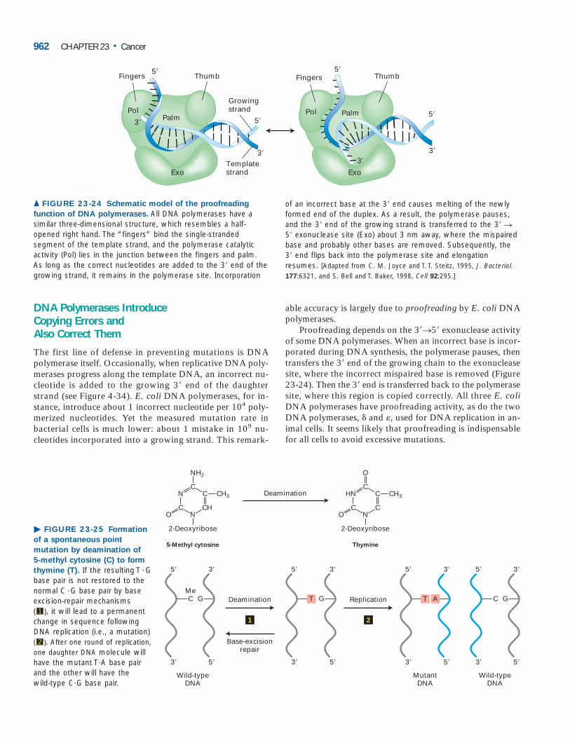
DNA Polymerases Introduce Copying Errors and Also Correct Them
The first line of defense in preventing mutations is DNApolymerase itself. Occasionally, when replicative DNA poly-merases progress along the template DNA, an incorrect nu-cleotide is added to the growing 3� end of the daughterstrand (see Figure 4-34). E. coli DNA polymerases, for in-stance, introduce about 1 incorrect nucleotide per 104 poly-merized nucleotides. Yet the measured mutation rate inbacterial cells is much lower: about 1 mistake in 109 nu-cleotides incorporated into a growing strand. This remark-
able accuracy is largely due to proofreading by E. coli DNApolymerases.
Proofreading depends on the 3�n5� exonuclease activityof some DNA polymerases. When an incorrect base is incor-porated during DNA synthesis, the polymerase pauses, thentransfers the 3� end of the growing chain to the exonucleasesite, where the incorrect mispaired base is removed (Figure23-24). Then the 3� end is transferred back to the polymerasesite, where this region is copied correctly. All three E. coliDNA polymerases have proofreading activity, as do the twoDNA polymerases, � and �, used for DNA replication in an-imal cells. It seems likely that proofreading is indispensablefor all cells to avoid excessive mutations.
962 CHAPTER 23 • Cancer
Fingers Thumb
Templatestrand
Growingstrand
3�3�
5�
3� 5�
3�
5�
Exo
PalmPol
Fingers Thumb
Exo
Palm
5�
Pol
▲ FIGURE 23-24 Schematic model of the proofreading
function of DNA polymerases. All DNA polymerases have asimilar three-dimensional structure, which resembles a half-opened right hand. The “fingers” bind the single-strandedsegment of the template strand, and the polymerase catalyticactivity (Pol) lies in the junction between the fingers and palm.As long as the correct nucleotides are added to the 3� end of thegrowing strand, it remains in the polymerase site. Incorporation
of an incorrect base at the 3� end causes melting of the newlyformed end of the duplex. As a result, the polymerase pauses,and the 3� end of the growing strand is transferred to the 3� n5� exonuclease site (Exo) about 3 nm away, where the mispairedbase and probably other bases are removed. Subsequently, the3� end flips back into the polymerase site and elongationresumes. [Adapted from C. M. Joyce and T. T. Steitz, 1995, J. Bacteriol.177:6321, and S. Bell and T. Baker, 1998, Cell 92:295.]
5�
3�
G
3�
5�
T
O
2-Deoxyribose
5-Methyl cytosine Thymine
Deamination
Deamination Replication
Base-excisionrepair
Wild-typeDNA
MutantDNA
N
C CH3
NH2
C
C
N
CHO
2-Deoxyribose
N
C CH3
MeC
5�
3�
G
3�
5�
1 2
C
O
C
HN
C
Wild-typeDNA
5�
3�
A
3�
5�
T
5�
3�
G
3�
5�
C
� FIGURE 23-25 Formation
of a spontaneous point
mutation by deamination of
5-methyl cytosine (C) to form
thymine (T). If the resulting T Gbase pair is not restored to thenormal C G base pair by baseexcision-repair mechanisms ( ), it will lead to a permanentchange in sequence followingDNA replication (i.e., a mutation)( ). After one round of replication,one daughter DNA molecule willhave the mutant TA base pairand the other will have the wild-type CG base pair.
2
1
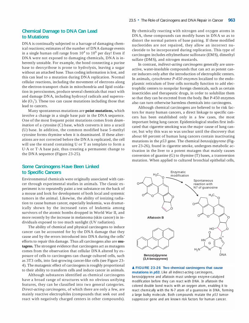
Chemical Damage to DNA Can Lead to MutationsDNA is continually subjected to a barrage of damaging chem-ical reactions; estimates of the number of DNA damage eventsin a single human cell range from 104 to 106 per day! Even ifDNA were not exposed to damaging chemicals, DNA is in-herently unstable. For example, the bond connecting a purinebase to deoxyribose is prone to hydrolysis, leaving a sugarwithout an attached base. Thus coding information is lost, andthis can lead to a mutation during DNA replication. Normalcellular reactions, including the movement of electrons alongthe electron-transport chain in mitochondria and lipid oxida-tion in peroxisomes, produce several chemicals that react withand damage DNA, including hydroxyl radicals and superox-ide (O2
.-). These too can cause mutations including those thatlead to cancers.
Many spontaneous mutations are point mutations, whichinvolve a change in a single base pair in the DNA sequence.One of the most frequent point mutations comes from deam-ination of a cytosine (C) base, which converts it into a uracil(U) base. In addition, the common modified base 5-methylcytosine forms thymine when it is deaminated. If these alter-ations are not corrected before the DNA is replicated, the cellwill use the strand containing U or T as template to form aUA or TA base pair, thus creating a permanent change tothe DNA sequence (Figure 23-25).
Some Carcinogens Have Been Linked to Specific CancersEnvironmental chemicals were originally associated with can-cer through experimental studies in animals. The classic ex-periment is to repeatedly paint a test substance on the back ofa mouse and look for development of both local and systemictumors in the animal. Likewise, the ability of ionizing radia-tion to cause human cancer, especially leukemia, was dramat-ically shown by the increased rates of leukemia amongsurvivors of the atomic bombs dropped in World War II, andmore recently by the increase in melanoma (skin cancer) in in-dividuals exposed to too much sunlight (UV radiation).
The ability of chemical and physical carcinogens to inducecancer can be accounted for by the DNA damage that theycause and by the errors introduced into DNA during the cells’efforts to repair this damage. Thus all carcinogens also are mu-tagens. The strongest evidence that carcinogens act as mutagenscomes from the observation that cellular DNA altered by ex-posure of cells to carcinogens can change cultured cells, suchas 3T3 cells, into fast-growing cancer-like cells (see Figure 23-4). The mutagenic effect of carcinogens is roughly proportionalto their ability to transform cells and induce cancer in animals.
Although substances identified as chemical carcinogenshave a broad range of structures with no obvious unifyingfeatures, they can be classified into two general categories.Direct-acting carcinogens, of which there are only a few, aremainly reactive electrophiles (compounds that seek out andreact with negatively charged centers in other compounds).
By chemically reacting with nitrogen and oxygen atoms inDNA, these compounds can modify bases in DNA so as todistort the normal pattern of base pairing. If these modifiednucleotides are not repaired, they allow an incorrect nu-cleotide to be incorporated during replication. This type ofcarcinogen includes ethylmethane sulfonate (EMS), dimethylsulfate (DMS), and nitrogen mustards.
In contrast, indirect-acting carcinogens generally are unre-active, water-insoluble compounds that can act as potent can-cer inducers only after the introduction of electrophilic centers.In animals, cytochrome P-450 enzymes localized to the endo-plasmic reticulum of liver cells normally function to add elec-trophilic centers to nonpolar foreign chemicals, such as certaininsecticides and therapeutic drugs, in order to solubilize themso that they can be excreted from the body. But P-450 enzymesalso can turn otherwise harmless chemicals into carcinogens.
Although chemical carcinogens are believed to be risk fac-tors for many human cancers, a direct linkage to specific can-cers has been established only in a few cases, the mostimportant being lung cancer. Epidemiological studies first indi-cated that cigarette smoking was the major cause of lung can-cer, but why this was so was unclear until the discovery thatabout 60 percent of human lung cancers contain inactivatingmutations in the p53 gene. The chemical benzo(a)pyrene (Fig-ure 23-26), found in cigarette smoke, undergoes metabolic ac-tivation in the liver to a potent mutagen that mainly causesconversion of guanine (G) to thymine (T) bases, a transversionmutation. When applied to cultured bronchial epithelial cells,
23.5 • The Role of Carcinogens and DNA Repair in Cancer 963
Enzymaticmodification
Spontaneousreaction withN7 of guanosine
Aflatoxin B
Benzo(a)pyrene(3,4-benzpyrene)
DNA
H2N
H3CO
O
O
O O
O
H3CO
O
O
O
N
O
OHOO
G
+N
N
HN
▲ FIGURE 23-26 Two chemical carcinogens that cause
mutations in p53. Like all indirect-acting carcinogens,benzo(a)pyrene and aflatoxin must undergo enzyme-catalyzedmodification before they can react with DNA. In aflatoxin thecolored double bond reacts with an oxygen atom, enabling it toreact chemically with the N-7 atom of a guanosine in DNA, forminga large bulky molecule. Both compounds mutate the p53 tumor-suppressor gene and are known risk factors for human cancer.
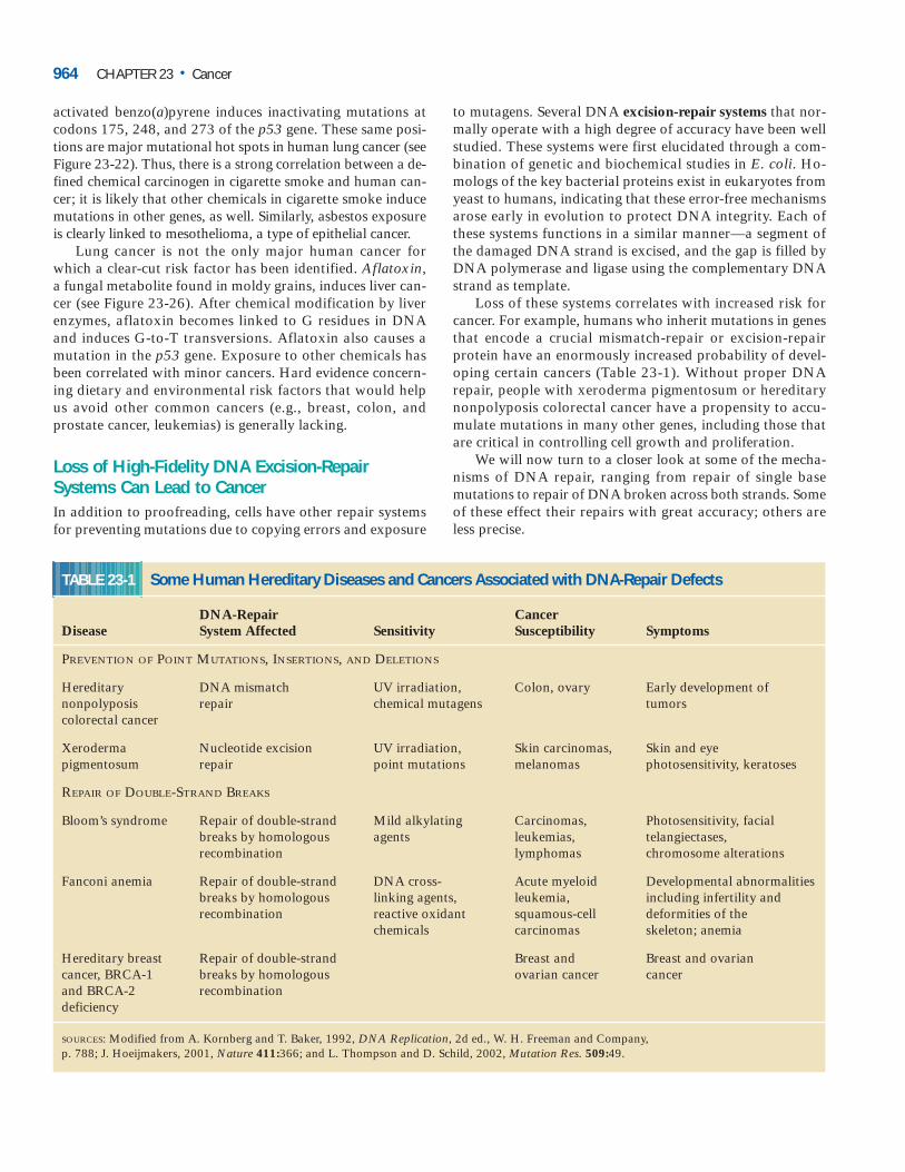
activated benzo(a)pyrene induces inactivating mutations atcodons 175, 248, and 273 of the p53 gene. These same posi-tions are major mutational hot spots in human lung cancer (seeFigure 23-22). Thus, there is a strong correlation between a de-fined chemical carcinogen in cigarette smoke and human can-cer; it is likely that other chemicals in cigarette smoke inducemutations in other genes, as well. Similarly, asbestos exposureis clearly linked to mesothelioma, a type of epithelial cancer.
Lung cancer is not the only major human cancer forwhich a clear-cut risk factor has been identified. Aflatoxin,a fungal metabolite found in moldy grains, induces liver can-cer (see Figure 23-26). After chemical modification by liverenzymes, aflatoxin becomes linked to G residues in DNAand induces G-to-T transversions. Aflatoxin also causes amutation in the p53 gene. Exposure to other chemicals hasbeen correlated with minor cancers. Hard evidence concern-ing dietary and environmental risk factors that would help us avoid other common cancers (e.g., breast, colon, andprostate cancer, leukemias) is generally lacking.
Loss of High-Fidelity DNA Excision-RepairSystems Can Lead to Cancer In addition to proofreading, cells have other repair systemsfor preventing mutations due to copying errors and exposure
to mutagens. Several DNA excision-repair systems that nor-mally operate with a high degree of accuracy have been wellstudied. These systems were first elucidated through a com-bination of genetic and biochemical studies in E. coli. Ho-mologs of the key bacterial proteins exist in eukaryotes fromyeast to humans, indicating that these error-free mechanismsarose early in evolution to protect DNA integrity. Each ofthese systems functions in a similar manner—a segment ofthe damaged DNA strand is excised, and the gap is filled byDNA polymerase and ligase using the complementary DNAstrand as template.
Loss of these systems correlates with increased risk forcancer. For example, humans who inherit mutations in genesthat encode a crucial mismatch-repair or excision-repair protein have an enormously increased probability of devel-oping certain cancers (Table 23-1). Without proper DNA repair, people with xeroderma pigmentosum or hereditarynonpolyposis colorectal cancer have a propensity to accu-mulate mutations in many other genes, including those thatare critical in controlling cell growth and proliferation.
We will now turn to a closer look at some of the mecha-nisms of DNA repair, ranging from repair of single base mutations to repair of DNA broken across both strands. Someof these effect their repairs with great accuracy; others areless precise.
964 CHAPTER 23 • Cancer
TABLE 23-1 Some Human Hereditary Diseases and Cancers Associated with DNA-Repair Defects
DNA-Repair Cancer Disease System Affected Sensitivity Susceptibility Symptoms
PREVENTION OF POINT MUTATIONS, INSERTIONS, AND DELETIONS
Hereditary DNA mismatch UV irradiation, Colon, ovary Early development of nonpolyposis repair chemical mutagens tumorscolorectal cancer
Xeroderma Nucleotide excision UV irradiation, Skin carcinomas, Skin and eye pigmentosum repair point mutations melanomas photosensitivity, keratoses
REPAIR OF DOUBLE-STRAND BREAKS
Bloom’s syndrome Repair of double-strand Mild alkylating Carcinomas, Photosensitivity, facial breaks by homologous agents leukemias, telangiectases, recombination lymphomas chromosome alterations
Fanconi anemia Repair of double-strand DNA cross- Acute myeloid Developmental abnormalitiesbreaks by homologous linking agents, leukemia, including infertility and recombination reactive oxidant squamous-cell deformities of the
chemicals carcinomas skeleton; anemia
Hereditary breast Repair of double-strand Breast and Breast and ovarian cancer, BRCA-1 breaks by homologous ovarian cancer cancerand BRCA-2 recombinationdeficiency
SOURCES: Modified from A. Kornberg and T. Baker, 1992, DNA Replication, 2d ed., W. H. Freeman and Company, p. 788; J. Hoeijmakers, 2001, Nature 411:366; and L. Thompson and D. Schild, 2002, Mutation Res. 509:49.
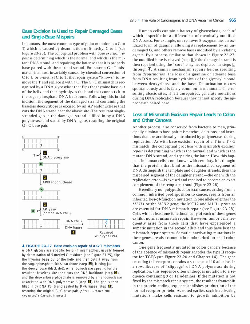
Base Excision Is Used to Repair Damaged Basesand Single-Base MispairsIn humans, the most common type of point mutation is a C toT, which is caused by deamination of 5-methyl C to T (seeFigure 23-25). The conceptual problem with base excision re-pair is determining which is the normal and which is the mu-tant DNA strand, and repairing the latter so that it is properlybase-paired with the normal strand. But since a G · T mis-match is almost invariably caused by chemical conversion ofC to U or 5-methyl C to T, the repair system “knows” to re-move the T and replace it with a C. The G · T mismatch is rec-ognized by a DNA glycosylase that flips the thymine base outof the helix and then hydrolyzes the bond that connects it tothe sugar-phosphate DNA backbone. Following this initialincision, the segment of the damaged strand containing thebaseless deoxyribose is excised by an AP endonuclease thatcuts the DNA strand near the abasic site. The resultant single-stranded gap in the damaged strand is filled in by a DNApolymerase and sealed by DNA ligase, restoring the originalG · C base pair.
Human cells contain a battery of glycosylases, each ofwhich is specific for a different set of chemically modifiedDNA bases. For example, one removes 8-oxyguanine, an ox-idized form of guanine, allowing its replacement by an un-damaged G, and others remove bases modified by alkylatingagents. By a process similar to that shown in Figure 23-27,the modified base is cleaved (step ); the damaged strand isthen repaired using the “core” enzymes depicted in stepsthrough . A similar mechanism repairs lesions resultingfrom depurination, the loss of a guanine or adenine basefrom DNA resulting from hydrolysis of the glycosylic bondbetween deoxyribose and the base. Depurination occursspontaneously and is fairly common in mammals. The re-sulting abasic sites, if left unrepaired, generate mutationsduring DNA replication because they cannot specify the ap-propriate paired base.
Loss of Mismatch Excision Repair Leads to Colonand Other CancersAnother process, also conserved from bacteria to man, prin-cipally eliminates base-pair mismatches, deletions, and inser-tions that are accidentally introduced by polymerases duringreplication. As with base excision repair of a T in a T · Gmismatch, the conceptual problem with mismatch excisionrepair is determining which is the normal and which is themutant DNA strand, and repairing the latter. How this hap-pens in human cells is not known with certainty. It is thoughtthat the proteins that bind to the mismatched segment ofDNA distinguish the template and daughter strands; then themispaired segment of the daughter strand—the one with thereplication error—is excised and repaired to become an exactcomplement of the template strand (Figure 23-28).
Hereditary nonpolyposis colorectal cancer, arising from acommon inherited predisposition to cancer, results from aninherited loss-of-function mutation in one allele of either theMLH1 or the MSH2 gene; the MSH2 and MLH1 proteinsare essential for DNA mismatch repair (see Figure 23-28).Cells with at least one functional copy of each of these genesexhibit normal mismatch repair. However, tumor cells fre-quently arise from those cells that have experienced a somatic mutation in the second allele and thus have lost themismatch repair system. Somatic inactivating mutations inthese genes are also common in noninherited forms of coloncancer.
One gene frequently mutated in colon cancers becauseof the absence of mismatch repair encodes the type II recep-tor for TGF� (see Figure 23-20 and Chapter 14). The geneencoding this receptor contains a sequence of 10 adenines ina row. Because of “slippage” of DNA polymerase duringreplication, this sequence often undergoes mutation to a se-quence containing 9 or 11 adenines. If the mutation is notfixed by the mismatch repair system, the resultant frameshiftin the protein-coding sequence abolishes production of thenormal receptor protein. As noted earlier, such inactivatingmutations make cells resistant to growth inhibition by
42
1
23.5 • The Role of Carcinogens and DNA Repair in Cancer 965
DNA glycosylase
GT
APEI endonuclease
Repairedwild-type DNA
AP lyase(part of DNA Pol β)
DNA Pol β
DNA ligase
G
G
T
C
1
2
3
4
G G
5� 3�
3� 5�
▲ FIGURE 23-27 Base excision repair of a G�T mismatch
A DNA glycosylase specific for G T mismatches, usually formedby deamination of 5-methyl C residues (see Figure 23-25), flipsthe thymine base out of the helix and then cuts it away from the sugar-phosphate DNA backbone (step ), leaving just the deoxyribose (black dot). An endonuclease specific for theresultant baseless site then cuts the DNA backbone (step ),and the deoxyribose phosphate is removed by an endonucleaseassociated with DNA polymerase � (step ). The gap is thenfilled in by DNA Pol � and sealed by DNA ligase (step ),restoring the original G C base pair. [After O. Schärer, 2003,Angewandte Chemie, in press.]
43
2
1
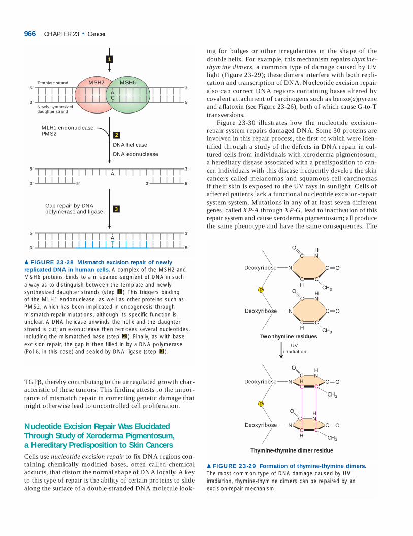
TGF�, thereby contributing to the unregulated growth char-acteristic of these tumors. This finding attests to the impor-tance of mismatch repair in correcting genetic damage thatmight otherwise lead to uncontrolled cell proliferation.
Nucleotide Excision Repair Was ElucidatedThrough Study of Xeroderma Pigmentosum, a Hereditary Predisposition to Skin CancersCells use nucleotide excision repair to fix DNA regions con-taining chemically modified bases, often called chemicaladducts, that distort the normal shape of DNA locally. A keyto this type of repair is the ability of certain proteins to slidealong the surface of a double-stranded DNA molecule look-
ing for bulges or other irregularities in the shape of the double helix. For example, this mechanism repairs thymine-thymine dimers, a common type of damage caused by UVlight (Figure 23-29); these dimers interfere with both repli-cation and transcription of DNA. Nucleotide excision repairalso can correct DNA regions containing bases altered by covalent attachment of carcinogens such as benzo(a)pyreneand aflatoxin (see Figure 23-26), both of which cause G-to-Ttransversions.
Figure 23-30 illustrates how the nucleotide excision-repair system repairs damaged DNA. Some 30 proteins areinvolved in this repair process, the first of which were iden-tified through a study of the defects in DNA repair in cul-tured cells from individuals with xeroderma pigmentosum,a hereditary disease associated with a predisposition to can-cer. Individuals with this disease frequently develop the skincancers called melanomas and squamous cell carcinomas if their skin is exposed to the UV rays in sunlight. Cells ofaffected patients lack a functional nucleotide excision-repairsystem system. Mutations in any of at least seven differentgenes, called XP-A through XP-G, lead to inactivation of thisrepair system and cause xeroderma pigmentosum; all producethe same phenotype and have the same consequences. The
966 CHAPTER 23 • Cancer
MSH2 MSH6Template strand
MLH1 endonuclease,PMS2
DNA helicase
DNA exonuclease
Newly synthesizeddaughter strand
Gap repair by DNApolymerase and ligase
A5�
3�
3�
5�5� 3�
1
2
3
AC
5�
3�
3�
5�
5�
3�
3�
5�
AT
▲ FIGURE 23-28 Mismatch excision repair of newly
replicated DNA in human cells. A complex of the MSH2 andMSH6 proteins binds to a mispaired segment of DNA in such a way as to distinguish between the template and newlysynthesized daughter strands (step ). This triggers binding of the MLH1 endonuclease, as well as other proteins such asPMS2, which has been implicated in oncogenesis throughmismatch-repair mutations, although its specific function isunclear. A DNA helicase unwinds the helix and the daughterstrand is cut; an exonuclease then removes several nucleotides,including the mismatched base (step ). Finally, as with baseexcision repair, the gap is then filled in by a DNA polymerase (Pol �, in this case) and sealed by DNA ligase (step ).3
2
1
Deoxyribose
Deoxyribose
P
Two thymine residues
Thymine-thymine dimer residue
N
N
CHN
CH
C
C
O
O
Deoxyribose
Deoxyribose
P
CHN
CH
C
C
UVirradiation
CH3
CH3
O
O
NC
HN
HC C
C
O
O
CH3
NC
HN
CC
O
O
CH3
CH
▲ FIGURE 23-29 Formation of thymine-thymine dimers.
The most common type of DNA damage caused by UVirradiation, thymine-thymine dimers can be repaired by anexcision-repair mechanism.
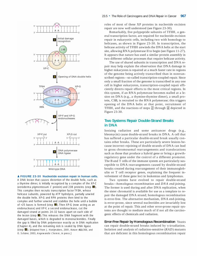
roles of most of these XP proteins in nucleotide excision repair are now well understood (see Figure 23-30).
Remarkably, five polypeptide subunits of TFIIH, a gen-eral transcription factor, are required for nucleotide excisionrepair in eukaryotic cells, including two with homology tohelicases, as shown in Figure 23-30. In transcription, the helicase activity of TFIIH unwinds the DNA helix at the startsite, allowing RNA polymerase II to begin (see Figure 11-27).It appears that nature has used a similar protein assembly intwo different cellular processes that require helicase activity.
The use of shared subunits in transcription and DNA re-pair may help explain the observation that DNA damage inhigher eukaryotes is repaired at a much faster rate in regionsof the genome being actively transcribed than in nontran-scribed regions—so-called transcription-coupled repair. Sinceonly a small fraction of the genome is transcribed in any onecell in higher eukaryotes, transcription-coupled repair effi-ciently directs repair efforts to the most critical regions. Inthis system, if an RNA polymerase becomes stalled at a le-sion on DNA (e.g., a thymine-thymine dimer), a small pro-tein, CSB, is recruited to the RNA polymerase; this triggersopening of the DNA helix at that point, recruitment ofTFIIH, and the reactions of steps through depicted inFigure 23-30.
Two Systems Repair Double-Strand Breaks in DNAIonizing radiation and some anticancer drugs (e.g.,bleomycin) cause double-strand breaks in DNA. A cell thathas suffered a particular double-strand break usually con-tains other breaks. These are particularly severe lesions be-cause incorrect rejoining of double strands of DNA can leadto gross chromosomal rearrangements and translocationssuch as those that produce a hybrid gene or bring a growth-regulatory gene under the control of a different promoter.The B and T cells of the immune system are particularly sus-ceptible to DNA rearrangements caused by double-strandbreaks created during rearrangement of their immunoglob-ulin or T cell receptor genes, explaining the frequent in-volvement of these gene loci in leukemias and lymphomas.
Two systems have evolved to repair double-strandbreaks—homologous recombination and DNA end-joining.The former is used during and after DNA replication, whenthe sister chromatid is available for use as a template to re-pair the damaged DNA strand; homologous recombinationis error-free. The alternative mechanism, DNA end-joining,is error-prone, since several nucleotides are invariably lostat the point of repair. This and other error-prone repair sys-tems are thought to mediate much of if not all the carcino-genic effects of chemicals and radiation.
Error-Free Repair by Homologous Recombination Yeastscan repair double-strand breaks induced by �-irradiation.Isolation and analysis of radiation-sensitive (RAD) mutantsthat are deficient in this homologous recombination repair
42
23.5 • The Role of Carcinogens and DNA Repair in Cancer 967
Initial damagerecognition
5�
3�
3�
A
T
A
3� 5�A A
T
Opening of DNA double helix
DNA polymeraseDNA ligase
Wild-type DNA
5�
3�
3�
5�
5�
T T
A A
XP-C
23B
XP-F and XP-Gendonucleases
5�
3�
3�
5�
T T
A A
RPA
5� 5� 3�3�
TFIIH XP-G
XP-F cut XP-G cut
3� 5�A AT T
5� 3�
T T
1
2
4
3
▲ FIGURE 23-30 Nucleotide excision repair in human cells.
A DNA lesion that causes distortion of the double helix, such asa thymine dimer, is initially recognized by a complex of the XP-C(xeroderma pigmentosum C protein) and 23B proteins (step ).This complex then recruits transcription factor TFIIH, whosehelicase subunits, powered by ATP hydrolysis, partially unwindthe double helix. XP-G and RPA proteins then bind to thecomplex and further unwind and stabilize the helix until a bubbleof ≈25 bases is formed (step ). Then XP-G (now acting as anendonuclease) and XP-F, a second endonuclease, cut thedamaged strand at points 24–32 bases apart on each side of the lesion (step ). This releases the DNA fragment with thedamaged bases, which is degraded to mononucleotides. Finallythe gap is filled by DNA polymerase exactly as in DNA replication(Chapter 4), and the remaining nick is sealed by DNA ligase (step ). [Adapted from J. Hoeijmakers, 2001, Nature 411:366, and O. Schärer, 2003, Angewandte Chemie, in press.]
4
3
2
1
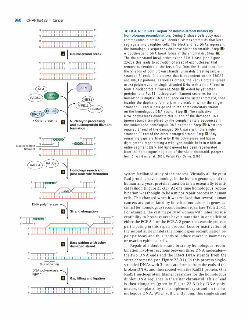
system facilitated study of the process. Virtually all the yeastRad proteins have homologs in the human genome, and thehuman and yeast proteins function in an essentially identi-cal fashion (Figure 23-31). At one time homologous recom-bination was thought to be a minor repair process in humancells. This changed when it was realized that several humancancers are potentiated by inherited mutations in genes es-sential for homologous recombination repair (see Table 23-1).For example, the vast majority of women with inherited sus-ceptibility to breast cancer have a mutation in one allele ofeither the BCRA-1 or the BCRA-2 genes that encode proteinsparticipating in this repair process. Loss or inactivation ofthe second allele inhibits the homologous recombination re-pair pathway and thus tends to induce cancer in mammaryor ovarian epithelial cells.
Repair of a double-strand break by homologous recom-bination involves reactions between three DNA molecules—the two DNA ends and the intact DNA strands from thesister chromatid (see Figure 23-31). In this process single-stranded DNAs with 3� ends are formed from the ends of thebroken DNAs and then coated with the Rad51 protein. OneRad51 nucleoprotein filament searches for the homologousduplex DNA sequence in the sister chromatid. This 3� endis then elongated (green in Figure 23-31) by DNA poly-merase, templated by the complementary strand on the ho-mologous DNA. When sufficiently long, this single strand
968 CHAPTER 23 • Cancer
� FIGURE 23-31 Repair of double-strand breaks by
homologous recombination. During S phase cells copy eachchromosome to create two identical sister chromatids that latersegregate into daughter cells. The black and red DNAs representthe homologous sequences on these sister chromatids. Step :A double-strand DNA break forms in the chromatids. Step :The double-strand break activates the ATM kinase (see Figure 23-23); this leads to activation of a set of exonucleases thatremove nucleotides at the break first from the 3� and then fromthe 5� ends of both broken strands, ultimately creating single-stranded 3� ends. In a process that is dependent on the BRCA1and BRCA2 proteins, as well as others, the Rad51 protein (greenovals) polymerizes on single-stranded DNA with a free 3� end toform a nucleoprotein filament. Step : Aided by yet otherproteins, one Rad51 nucleoprotein filament searches for thehomologous duplex DNA sequence on the sister chromatid, theninvades the duplex to form a joint molecule in which the single-stranded 3� end is base-paired to the complementary strand on the homologous DNA strand. Step : The replicative DNA polymerases elongate this 3� end of the damaged DNA(green strand), templated by the complementary sequences inthe undamaged homologous DNA segment. Step : Next thisrepaired 3� end of the damaged DNA pairs with the single-stranded 3� end of the other damaged strand. Step : Anyremaining gaps are filled in by DNA polymerase and ligase (light green), regenerating a wild-type double helix in which anentire segment (dark and light green) has been regenerated from the homologous segment of the sister chromatid. [Adaptedfrom D. van Gant et al., 2001, Nature Rev. Genet. 2:196.]
6
5
4
3
21
Double-strand break
Nucleolytic processingand nucleoprotein-filamentformation
Homology search andjoint molecule formation
Strand elongation
Base pairing with otherdamaged strand
Gap filling and ligation
1
2
3
4
5
6
NBS1
RAD50MRE11
ATM
RAD52
RAD52RAD54
DNA polymerase
DNA polymerase,ligase
RAD51
RPA
5� 3�
3�
5�
5�
3�
3� 5�
5� 5� 3�3�
5� 3�
3� 5�
3�5�Nucleoproteinfilament
+ Other proteins
5�5�
5� 3�
3�
5� 3�
5� 3�
3� 3� 5�
5� 3�
3� 5�
5�
5' 3�
5� 3�
3� 5�
5�3�
5� 3�
3� 5�
5� 3�
5� 3�
3� 5�
3� 5�
5�
3�
5� 3�
5�
3�
5� 3�
3� 5�
BRCA1BRCA2
Site of pairing
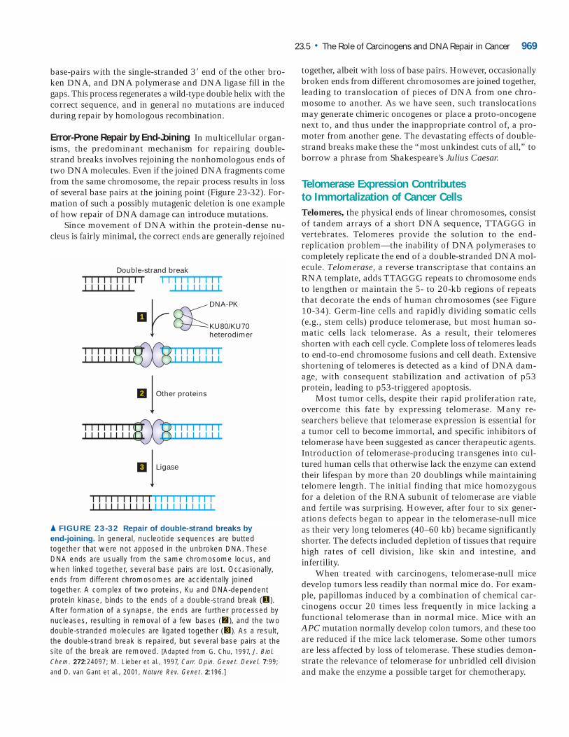
base-pairs with the single-stranded 3� end of the other bro-ken DNA, and DNA polymerase and DNA ligase fill in thegaps. This process regenerates a wild-type double helix with thecorrect sequence, and in general no mutations are inducedduring repair by homologous recombination.
Error-Prone Repair by End-Joining In multicellular organ-isms, the predominant mechanism for repairing double-strand breaks involves rejoining the nonhomologous ends oftwo DNA molecules. Even if the joined DNA fragments comefrom the same chromosome, the repair process results in lossof several base pairs at the joining point (Figure 23-32). For-mation of such a possibly mutagenic deletion is one exampleof how repair of DNA damage can introduce mutations.
Since movement of DNA within the protein-dense nu-cleus is fairly minimal, the correct ends are generally rejoined
together, albeit with loss of base pairs. However, occasionallybroken ends from different chromosomes are joined together,leading to translocation of pieces of DNA from one chro-mosome to another. As we have seen, such translocationsmay generate chimeric oncogenes or place a proto-oncogenenext to, and thus under the inappropriate control of, a pro-moter from another gene. The devastating effects of double-strand breaks make these the “most unkindest cuts of all,” toborrow a phrase from Shakespeare’s Julius Caesar.
Telomerase Expression Contributes to Immortalization of Cancer CellsTelomeres, the physical ends of linear chromosomes, consistof tandem arrays of a short DNA sequence, TTAGGG in vertebrates. Telomeres provide the solution to the end-replication problem—the inability of DNA polymerases tocompletely replicate the end of a double-stranded DNA mol-ecule. Telomerase, a reverse transcriptase that contains anRNA template, adds TTAGGG repeats to chromosome endsto lengthen or maintain the 5- to 20-kb regions of repeatsthat decorate the ends of human chromosomes (see Figure10-34). Germ-line cells and rapidly dividing somatic cells(e.g., stem cells) produce telomerase, but most human so-matic cells lack telomerase. As a result, their telomeresshorten with each cell cycle. Complete loss of telomeres leadsto end-to-end chromosome fusions and cell death. Extensiveshortening of telomeres is detected as a kind of DNA dam-age, with consequent stabilization and activation of p53 protein, leading to p53-triggered apoptosis.
Most tumor cells, despite their rapid proliferation rate,overcome this fate by expressing telomerase. Many re-searchers believe that telomerase expression is essential fora tumor cell to become immortal, and specific inhibitors oftelomerase have been suggested as cancer therapeutic agents.Introduction of telomerase-producing transgenes into cul-tured human cells that otherwise lack the enzyme can extendtheir lifespan by more than 20 doublings while maintainingtelomere length. The initial finding that mice homozygousfor a deletion of the RNA subunit of telomerase are viableand fertile was surprising. However, after four to six gener-ations defects began to appear in the telomerase-null miceas their very long telomeres (40–60 kb) became significantlyshorter. The defects included depletion of tissues that requirehigh rates of cell division, like skin and intestine, and infertility.
When treated with carcinogens, telomerase-null mice develop tumors less readily than normal mice do. For exam-ple, papillomas induced by a combination of chemical car-cinogens occur 20 times less frequently in mice lacking afunctional telomerase than in normal mice. Mice with anAPC mutation normally develop colon tumors, and these tooare reduced if the mice lack telomerase. Some other tumorsare less affected by loss of telomerase. These studies demon-strate the relevance of telomerase for unbridled cell divisionand make the enzyme a possible target for chemotherapy.
23.5 • The Role of Carcinogens and DNA Repair in Cancer 969
Double-strand break
DNA-PK
KU80/KU70heterodimer
Other proteins
Ligase
1
2
3
▲ FIGURE 23-32 Repair of double-strand breaks by
end-joining. In general, nucleotide sequences are buttedtogether that were not apposed in the unbroken DNA. TheseDNA ends are usually from the same chromosome locus, andwhen linked together, several base pairs are lost. Occasionally,ends from different chromosomes are accidentally joinedtogether. A complex of two proteins, Ku and DNA-dependentprotein kinase, binds to the ends of a double-strand break ( ).After formation of a synapse, the ends are further processed bynucleases, resulting in removal of a few bases ( ), and the twodouble-stranded molecules are ligated together ( ). As a result,the double-strand break is repaired, but several base pairs at thesite of the break are removed. [Adapted from G. Chu, 1997, J. Biol.Chem. 272:24097; M. Lieber et al., 1997, Curr. Opin. Genet. Devel. 7:99;and D. van Gant et al., 2001, Nature Rev. Genet. 2:196.]
32
1
KEY CONCEPTS OF SECTION 23.5
The Role of Carcinogens and DNA Repair in Cancer
■ Changes in the DNA sequence result from copying er-rors and the effects of various physical and chemical agents,or carcinogens. All carcinogens are mutagens; that is, theyalter one or more nucleotides in DNA.
■ Many copying errors that occur during DNA replicationare corrected by the proofreading function of DNA poly-merases that can recognize incorrect (mispaired) bases atthe 3� end of the growing strand and then remove themby an inherent 3�n5� exonuclease activity (see Figure 23-24).
■ Indirect carcinogens, the most common type, must beactivated before they can damage DNA. In animals, meta-bolic activation occurs via the cytochrome P-450 system,a pathway generally used by cells to rid themselves of nox-ious foreign chemicals.
■ Benzo(a)pyrene, a component of cigarette smoke, causesinactivating mutations in the p53 gene at the same posi-tions that are mutated in many human lung tumors.
■ Eukaryotic cells have three excision-repair systems forcorrecting mispaired bases and for removing UV-inducedthymine-thymine dimers or large chemical adducts fromDNA. Base excision repair, mismatch repair, and nucleotideexcision repair operate with high accuracy and generallydo not introduce errors.
■ Inherited defects in the nucleotide excision-repair path-way, as in individuals with xeroderma pigmentosum, pre-dispose them to skin cancer. Inherited colon cancer fre-quently is associated with mutant forms of proteinsessential for the mismatch repair pathway.
■ Error-free repair of double-strand breaks in DNA is ac-complished by homologous recombination using the un-damaged sister chromatid as template.
■ Defects in repair by homologous recombination are as-sociated with inheritance of one mutant allele of the BRCA-1 or BRCA-2 gene and result in predisposition to breastcancer.
■ Repair of double-strand breaks by the end-joining path-way can link segments of DNA from different chromo-somes, possibly forming an oncogenic translocation. Therepair mechanism also produces a small deletion, evenwhen segments from the same chromosome are joined.
■ Inherited defects in other cellular DNA-repair processesfound in certain human diseases are associated with an in-creased susceptibility for certain cancers (see Table 23-1).
■ Cancer cells, like germ cells and stem cells but unlikemost differentiated cells, produce telomerase, which pre-vents shortening of chromosomes during DNA replicationand may contribute to their immortalization. The absenceof telomerase is associated with resistance to generation ofcertain tumors.
P E R S P E C T I V E S F O R T H E F U T U R E
The recognition that cancer is fundamentally a genetic dis-ease has opened enormous new opportunities for preventingand treating the disease. Carcinogens can now be assessedfor their effects on known steps in cell-cycle control. Geneticdefects in the checkpoint controls for detecting damagedDNA and in the systems for repairing it can be readily rec-ognized and used to explore the mechanisms of cancer. Themultiple changes that must occur for a cell to grow into adangerous tumor present multiple opportunities for inter-vention. Identifying mutated genes associated with cancerpoints directly to proteins at which drugs can be targeted.
Diagnostic medicine is being transformed by our new-found ability to monitor large numbers of cell characteristics.The traditional methods of assessing possible tumor cells,mainly microscopy of stained cells, will be augmented or re-placed by techniques for measuring the expression of tens ofthousands of genes, focusing particularly on genes whose ac-tivities are identified as powerful indicators of the cell’sgrowth properties and the patient’s prognosis. Currently,DNA microarray analysis permits measurement of gene tran-scription. In the future, techniques for systematically meas-uring protein production, modification, and localization, allimportant measures of cell states, will give us even more re-fined portraits of cells. Tumors now viewed as identical orvery similar will instead be recognized as distinctly differentand given appropriately different treatments. Earlier detec-tion of tumors, based on better monitoring of cell properties,should allow more successful treatment. A focus on that par-ticularly destructive process, metastasis, should be successfulin identifying more of the mechanisms used by cells to migrate, attach, and invade. Manipulation of angiogenesiscontinues to look hopeful as a means of suffocating tumors.
The molecular cell biology of cancer provides avenues fornew therapies, but prevention remains crucial and preferableto therapy. Avoidance of obvious carcinogens, in particularcigarette smoke, can significantly reduce the incidence oflung cancer and perhaps other kinds as well. Beyond mini-mizing exposure to carcinogens such as smoke or sunlight,certain specific approaches are now feasible. New knowledgeof the involvement of human papillomavirus 16 in mostcases of cervical cancer holds promise for developing a can-cer vaccine that will prevent viral action. Antibodies againstcell surface markers that distinguish cancer cells are a sourceof great hope, especially after successes with the clinical useof monoclonal antibodies against human EGF receptor 2(Her2), a protein involved in some cases of human breastcancer. Further steps must involve medicine and science. Un-derstanding the cell biology of cancer is a critical first step to-ward prevention and cure, but the next steps are hard. Thesuccess with Gleevec (STI-571) against leukemia is excep-tional; many cancers remain difficult to treat and cause enor-mous suffering. Since cancer is a term for a group of highlydiverse diseases, interventions that are successful for one typemay not be useful for others. Despite these daunting realities,
970 CHAPTER 23 • Cancer
we are beginning to reap the benefits of decades of researchexploring the molecular biology of the cell. We hope thatmany of the readers of this book will help to overcome theobstacles that remain.
KEY TERMS
aflatoxin 964aneuploidy 961benign 936Burkitt’s lymphoma 956carcinogens 935carcinomas 938deamination 963DNA end-joining 967excision-repair
systems 964leukemias 938loss of heterozygosity
(LOH) 947malignant 936metastasis 937mutagens 963
REVIEW THE CONCEPTS
1. What characteristics distinguish benign from malignanttumors? With respect to gene mutations, what distinguishesbenign colon polyps from malignant colon carcinoma?
2. Ninety percent of cancer deaths are caused by metastaticrather than primary tumors. Define metastasis. Explain the rationale for the following new cancer treatments: (a) batima-stat, an inhibitor of matrix metalloproteinases and of the plas-minogen activator receptor, (b) antibodies that block thefunction of integrins, integral membrane proteins that mediateattachment of cells to the basal laminae and extracellular ma-trices of various tissues, and (c) bisphosphonate, which in-hibits the function of bone-digesting osteoclasts.
3. Because of oxygen and nutrient requirements, cells in atissue must reside within 100 m of a blood vessel. Basedon this information, explain why many malignant tumorsoften possess gain-of-function mutations in one of the fol-lowing genes: bFGF, TGF�, and VEGF.
4. What hypothesis explains the observations that inci-dence of human cancers increases exponentially with age?Give an example of data that confirm the hypothesis.
5. Distinguish between proto-oncogenes and tumor-suppressor genes. To become cancer promoting, do proto-oncogenes and tumor-suppressor genes undergo gain-of-function or loss-of-function mutations? Classify the follow-
ing genes as proto-oncogenes or tumor-suppressor genes:p53, ras, Bcl-2, telomerase, jun, and p16.
6. Hereditary retinoblastoma generally affects children inboth eyes, while spontaneous retinoblastoma usually occursduring adulthood only in one eye. Explain the genetic basis forthe epidemiologic distinction between these two forms ofretinoblastoma. Explain the apparent paradox: loss-of-functionmutations in tumor-suppressor genes act recessively, yet hered-itary retinoblastoma is inherited as an autosomal dominant.
7. Explain the concept of loss of heterozygosity (LOH).Why do most cancer cells exhibit LOH of one or moregenes? How does failure of the spindle assembly checkpointlead to loss of heterozygosity?
8. Many malignant tumors are characterized by the activa-tion of one or more growth factor receptors. What is the cat-alytic activity associated with transmembrane growth factorreceptors such as the EGF receptor? Describe how the fol-lowing events lead to activation of the relevant growth factorreceptor: (a) expression of the viral protein gp55, (b) translo-cation that replaces the extracellular domain of the Trk re-ceptor with the N-terminal region of tropomyosin, (c) pointmutation that converts a valine to glutamine within thetransmembrane region of the Her2 receptor.
9. Describe the common signal-transduction event that isperturbed by cancer-promoting mutations in the genes en-coding Ras and NF-1. Why are mutations in Ras more com-monly found in cancers than mutations in NF-1?
10. What is the structural distinction between the proteinsencoded by c-src and v-src? How does this difference renderv-src oncogenic?
11. Describe the mutational event that produces the myconcogene in Burkitt’s lymphoma. Why does the particularmechanism for generating oncogenic myc result in a lym-phoma rather than another type of cancer? Describe anothermechanism for generating oncogenic myc.
12. Pancreatic cancers often possess loss-of-function muta-tions in the gene that encodes the SMAD4 protein. How doesthis mutation promote the loss of growth inhibition andhighly metastatic phenotype of pancreatic tumors?
13. Loss of p53 function occurs in the majority of humantumors. Name two ways in which loss of p53 functioncontributes to a malignant phenotype. Explain the mecha-nism by which the following agents cause loss of p53function: (a) human papillomavirus and (b) benzo(a)pyrene.
14. DNA-repair systems are responsible for maintaining ge-nomic fidelity in normal cells despite the high frequency withwhich mutational events occur. What type of DNA mutationis generated by (a) UV irradiation and (b) ionizing radiation?Describe the system responsible for repairing each of thesetypes of mutations in mammalian cells. Postulate why a lossof function in one or more DNA-repair systems typifiesmany cancers.
Review the Concepts 971
nondisjunction 947oncogene 940p53 protein 959Philadelphia
chromosome 954proto-oncogene 935sarcomas 938slow-acting
retroviruses 946thymine-thymine
dimers 966transducing
retroviruses 946transformation 939tumor-suppressor
gene 935
15. Which human cell types possess telomerase activity?What characteristic of cancer is promoted by expression oftelomerase? What concerns does this pose for medical ther-apies involving stem cells?
ANALYZE THE DATA
In a recent study by Elizabeth Snyderwine and colleagues,cDNA microarray profiling was used to compare the expres-sion profiles for 6900 genes in normal and malignant breast tis-sues from rats. RNA was extracted from the following tissues:
a. Breast tissue from virgin rats
b. Breast tissue from pregnant rats
c. Breast tissue from lactating rats
d. Breast carcinoma induced by the meat-derived carcinogen PhIP
e. Breast carcinoma induced by the experimental carcinogen DMBA
After the microarray slides were hybridized with labeledcDNAs derived from these 5 populations of RNAs, the datawere analyzed by several different comparisons.
In comparison 1, tissues a, b, and c were grouped to-gether as “normal” tissue samples, and d and e were groupedas “carcinoma” samples. Genes that were either induced(more than twofold increase in expression) or repressed(more than twofold decrease in expression) in both carci-noma samples relative to all three normal samples were de-termined. A partial listing of differentially expressed genesis shown in the accompanying table.
2. What characteristic of cancer is promoted by the overex-pression of PDGF-A, Cdk4, and cyclin D?
3. What characteristic of cancer might be promoted by therepression of extracellular matrix genes?
4. STAT5a is a transcription factor that regulates the ex-pression of cyclin D, Bcl-XL, and other genes. Why is it pos-sible for these carcinomas that no mutation occurs in thecyclin D gene despite its overexpression? Why are mutationsin transcription-factor genes like STAT5a commonly foundin cancer cells?
In comparison 2, expression profiles of carcinomas in-duced by PhIP and DMBA were compared with each other.In this analysis, some distinctions were found, but the num-ber of differentially expressed genes was far less than thenumber of genes identified when comparing the grouped“normal” samples (a, b, and c) to the grouped “carcinoma”samples (d and e).
5. On the basis of this information, what can be generalizedabout the molecular profile of breast carcinomas?
6. Would you anticipate greater or fewer differences in geneexpression if two distinct types of cancer (e.g., breast carci-noma vs. B-cell lymphoma) induced by the same carcinogenwere compared by microarray analysis?
REFERENCES
Tumor Cells and the Onset of CancerFidler, I. J. 2002. The pathogenesis of cancer metastasis: the “seed
and soil” hypothesis revisited. Nature Rev. Cancer 3:1–6.Folkman, J. 2002. Role of angiogenesis in tumor growth and
metastasis. Semin. Oncol. 29:15–18.Hanahan, D., and R. A. Weinberg. 2000. The hallmarks of can-
cer. Cell 100:57–70.Jain, M., et al. 2002. Sustained loss of a neoplastic phenotype
by brief inactivation of MYC. Science 297:102–104.Kinzler, K. W., and B. Vogelstein. 1996. Lessons from hereditary
colo-rectal cancer. Cell 87:159–170.Klein, G. 1998. Foulds’ dangerous idea revisited: the multistep
development of tumors 40 years later. Adv. Cancer Res. 72:1–23.Rafii, S., et al. 2002. Vascular and haematopoietic stem cells:
novel targets for anti-angiogenesis therapy? Nature Rev. Cancer2:826–835.
Ramaswamy, S., et al. 2003. A molecular signature of metasta-sis in primary solid tumors. Nature Genet. 33:49–54.
Trusolino, L., and P. M. Comoglio. 2002. Scatter-factor and sem-aphorin receptors: cell signalling for invasive growth. Nature Rev.Cancer 2:289–300.
Yancopoulos, G., M. Klagsburn, and J. Folkman. 1998. Vascu-logenesis, angiogenesis, and growth factors: ephrins enter the fray atthe border. Cell 93:661–664.
The Genetic Basis of CancerBienz, M., and H. Clevers. 2000. Linking colorectal cancer to
Wnt signaling. Cell 103:311–320.Clark, J., et al. 2002. Identification of amplified and expressed
genes in breast cancer by comparative hybridization onto microar-
972 CHAPTER 23 • Cancer
Induced Genes
Cell-Growth and Cell-Cycle-related Genes
Platelet-derived growth factor A chain (PDGF-A)
Cyclin-dependent kinase 4 (Cdk4)
Cyclin D
Signal-Transduction and Transcription-Related Genes
STAT5a
Repressed Genes
Extracellular Matrix Genes
Alpha 1 type V collagen
Fibronectin 1
Desmin
1. What was the purpose of analyzing breast tissue frompregnant and lactating rats?
rays of randomly selected cDNA clones. Genes Chrom. Cancer34:104–114.
Classon, M., and E. Harlow. 2002. The retinoblastoma tumoursuppressor in development and cancer. Nature Rev. Cancer 2:910–917.
Hesketh, R., ed. 1997. The Oncogene and Tumor SuppressorGene Facts Book, 2d ed. Academic Press.
Hunter, T. 1997. Oncoprotein networks. Cell 88:333–346.Moon, R. T., et al. 2002. The promise and perils of Wnt sig-
naling through beta-catenin. Science 296:1644–1646.Nevins, J. R. 2001. The Rb/E2F pathway and cancer. Hum. Mol.
Genet. 10:699–703.Polakis, P. 2000. Wnt signaling and cancer. Genes Devel.
14:1837–1851.Sasaki, T., et al. 2000. Colorectal carcinomas in mice lacking the
catalytic subunit of PI(3)Kgamma. Nature 406:897–902.Sherr, C. J., and F. McCormick. 2002. The RB and p53 path-
ways in cancer. Cancer Cell 2:103–112.Taipale, J., and P. A. Beachy. 2001. The Hedgehog and Wnt sig-
nalling pathways in cancer. Nature 411:349–354.van ’t Veer, L. J., et al. 2002. Gene expression profiling predicts
clinical outcome of breast cancer. Nature 415:530–536.West, M., et al. 2001. Predicting the clinical status of human
breast cancer by using gene expression profiles. Proc. Nat’l. Acad.Sci. USA 98:11462–11467.
White, R. 1998. Tumor suppressor pathways. Cell 92:591–592.
Oncogenic Mutations in Growth-Promoting ProteinsCapdeville, R., et al. 2002. Glivec (STI571, imatinib), a ration-
ally developed, targeted anticancer drug. Nature Rev. Drug Discov.1:493–502.
Downward, J. 2003. Targeting RAS signalling pathways in can-cer therapy. Nature Rev. Cancer 3:11–22.
Rowley, J. D. 2001. Chromosome translocations: dangerous liaisons revisited. Nature Rev. Cancer 1:245–250.
Sahai, E., and C. J. Marshall. 2002. RHO-GTPases and cancer.Nature Rev. Cancer 2:133–142.
Shaulian, E., and M. Karin. 2002. AP-1 as a regulator of cell lifeand death. Nature Cell Biol. 4:E131–E136.
Shawver, L. K., D. Slamon, and A. Ullrich. 2002. Smart drugs:tyrosine kinase inhibitors in cancer therapy. Cancer Cell 1:117–123.
Mutations Causing Loss of Growth-Inhibiting and Cell-Cycle Controls
Chau, B. N., and J. Y. Wang. 2003. Coordinated regulation oflife and death by RB. Nature Rev. Cancer 3:130–138.
Malumbres, M., and M. Barbacid. 2001. To cycle or not to cy-cle: a critical decision in cancer. Nature Rev. Cancer 1:222–231.
Mathon, N. F., and A. C. Lloyd. 2001. Cell senescence and can-cer. Nature Rev. Cancer 1:203–213.
Paulovich, A. G., D. P. Toczyski, and L. H. Hartwell. 1997. Whencheckpoints fail. Cell 88:315–321.
Planas-Silva, M. D., and R. A. Weinberg. 1997. The restrictionpoint and control of cell proliferation. Curr. Opin. Cell Biol.9:768–772.
Sherr, C. J. 2000. Cell cycle control and cancer. Harvey Lect.96:73–92.
Westphal, C. H. 1997. Cell-cycle signaling: Atm displays its manytalents. Curr. Biol. 7:R789–R792.
Zhang, L., et al. 2000. Role of BAX in the apoptotic responseto anticancer agents. Science 290:989–992.
The Role of Carcinogens and DNA Repair in CancerBatty, D., and R. Wood. 2000. Damage recognition in nucleotide
excision repair of DNA. Gene 241:193–204.D’Andrea, A., and M. Grompe. 2003. The Fanconi
anemia/BRCA pathway. Nature Rev. Cancer 3:23–34.Fishel, R. 1998. Mismatch repair, molecular switches, and sig-
nal transduction. Genes Devel. 12:2096–2101.Flores-Rozas, H., and R. Kolodner. 2000. Links between repli-
cation, recombination and genome instability in eukaryotes. TrendsBiochem. Sci. 25:196–200.
Friedberg, E. 2003. DNA damage and repair. Nature 421:436–440.Friedberg, E., G. Walker, and W. Siede. 1995. DNA Repair and
Mutagenesis. ASM Press.Hoeijmakers, J. 2001. Genome maintenance mechanisms for pre-
venting cancer. Nature 411:366–374.Lindahl, T. 2001. Keynote: past, present, and future aspects of
base excision repair. Prog. Nucl. Acid Res. Mol. Biol. 68:xvii–xxx.Muller, A., and R. Fishel. 2002. Mismatch repair and the hered-
itary non-polyposis colorectal cancer syndrome (HNPCC). CancerInvest. 20:102–109.
Schärer, O. 2003. Chemistry and biology of DNA repair. Ange-wandte Chemie, in press.
Schärer, O., and J. Jiricny. 2001. Recent progress in the biology,chemistry and structural biology of DNA glycosylases. Bioessays23:270–281.
Somasundaram, K. 2002. Breast cancer gene 1 (BRCA1): role incell cycle regulation and DNA repair—perhaps through transcrip-tion. J. Cell Biochem. 88:1084–1091.
Thompson, L., and D. Schild. 2002. Recombinational DNA re-pair and human disease. Mut. Res. 509:49–78.
van Gant, D., J. Hoeijmakers, and R. Kanaar. 2001. Chromo-somal stability and the DNA double-stranded break connection. Nature Rev. Genet. 2:196–205.
References 973