Embed Size (px)
Citation preview
8/26/2020
1
1
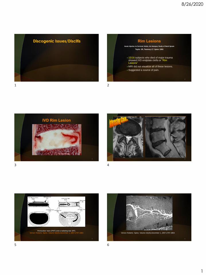
15/16 subjects who died of major trauma showed IVD endplate clefts or “Rim Lesions”
MRI did not visualize all of these lesions.
Suggested a source of pain.
Acute Injuries to Cervical Joints. An Autopsy Study of Neck Sprain
Taylor JR, Twomey LT. Spine 1993
2
3 4
Perinuclear tears (PNT) and a radiating tear (RT).
Vernon Roberts: Spine, Volume 32(25).December 1, 2007.2797-2804
5
Vernon Roberts: Spine, Volume 32(25).December 1, 2007.2797-2804
6
1 2
3 4
5 6
8/26/2020
2
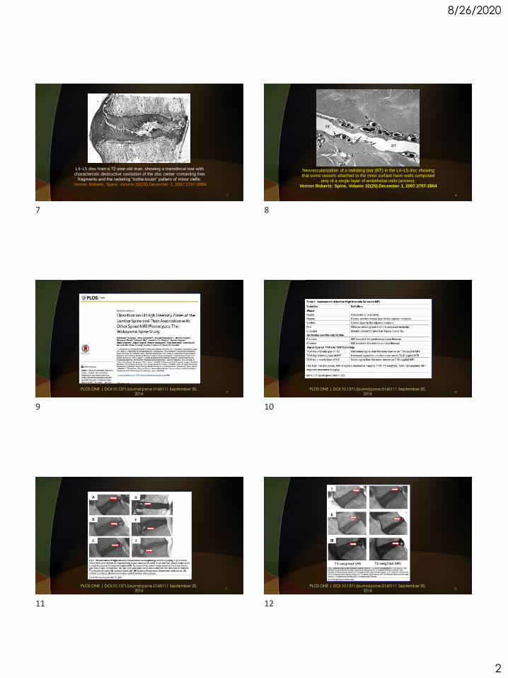
L4–L5 disc from a 72-year-old man, showing a transdiscal tear with
characteristic destructive cavitation of the disc center containing free
fragments and the radiating “bottle-brush” pattern of minor clefts.
Vernon Roberts: Spine, Volume 32(25).December 1, 2007.2797-2804
7
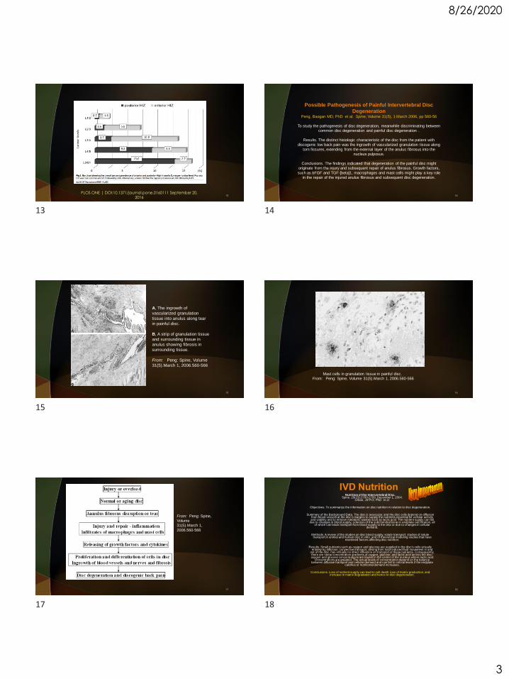
Neovascularization of a radiating tear (RT) in the L4–L5 disc showing
that some vessels attached to the inner surface have walls composed
only of a single layer of endothelial cells (arrows).
Vernon Roberts: Spine, Volume 32(25).December 1, 2007.2797-2804
8
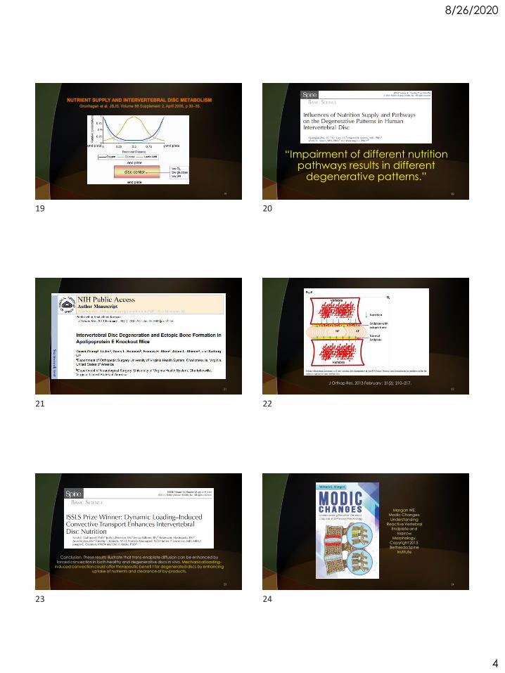
PLOS ONE | DOI:10.1371/journal.pone.0160111 September 20, 2016
9PLOS ONE | DOI:10.1371/journal.pone.0160111 September 20,
201610
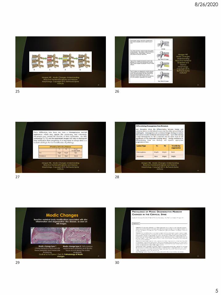
PLOS ONE | DOI:10.1371/journal.pone.0160111 September 20, 2016
11PLOS ONE | DOI:10.1371/journal.pone.0160111 September 20,
201612
7 8
9 10
11 12
8/26/2020
3
PLOS ONE | DOI:10.1371/journal.pone.0160111 September 20, 2016
13
Possible Pathogenesis of Painful Intervertebral Disc
DegenerationPeng, Baogan MD, PhD et al. Spine, Volume 31(5), 1 March 2006, pp 560-56
To study the pathogenesis of disc degeneration, meanwhile discriminating between common disc degeneration and painful disc degeneration .
Results. The distinct histologic characteristic of the disc from the patient with discogenic low back pain was the ingrowth of vascularized granulation tissue along
torn fissures, extending from the external layer of the anulus fibrosus into the nucleus pulposus.
Conclusions. The findings indicated that degeneration of the painful disc might originate from the injury and subsequent repair of anulus fibrosus. Growth factors,
such as bFGF and TGF-[beta]1, macrophages and mast cells might play a key role in the repair of the injured anulus fibrosus and subsequent disc degeneration.
14
A, The ingrowth of
vascularized granulation
tissue into anulus along tear
in painful disc.
B, A strip of granulation tissue
and surrounding tissue in
anulus showing fibrosis in
surrounding tissue.
From: Peng: Spine, Volume
31(5).March 1, 2006.560-566
15
Mast cells in granulation tissue in painful disc.From: Peng: Spine, Volume 31(5).March 1, 2006.560-566
16
From: Peng: Spine, Volume 31(5).March 1,
2006.560-566
17
Nutrition of the Intervertebral Disc.Spine. 29(23):2700-2709, December 1, 2004.
Urban, Jill P.G. PhD et al.
Objectives. To summarize the information on disc nutrition in relation to disc degeneration.
Summary of the Background Data. The disc is avascular, and the disc cells depend on diffusion from blood vessels at the disc's margins to supply the nutrients essential for cellular activity and viability and to remove metabolic wastes such as lactic acid. The nutrient supply can fail
due to changes in blood supply, sclerosis of the subchondral bone or endplate calcification, all of which can block transport from blood supply to the disc or due to changes in cellular
demand.
Methods. A review of the studies on disc blood supply, solute transport, studies of solute transport in animal and human disc in vitro, and of theoretical modeling studies that have
examined factors affecting disc nutrition.
Results. Small nutrients such as oxygen and glucose are supplied to the disc's cells virtually entirely by diffusion; convective transport, arising from load-induced fluid movement in and
out of the disc, has virtually no direct influence on transport of these nutrients. Consequently, there are steep concentration gradients of oxygen, glucose, and lactic acid across the disc; oxygen and glucose concentrations are lowest in the center of the nucleus where lactic acid
concentrations are greatest. The actual levels of concentration depend on the balance between diffusive transport and cellular demand and can fall to critical levels if the endplate
calcifies or nutritional demand increases.
Conclusions. Loss of nutrient supply can lead to cell death, loss of matrix production, and increase in matrix degradation and hence to disc degeneration.
18
13 14
15 16
17 18
8/26/2020
4
19
“Impairment of different nutrition pathways results in different
degenerative patterns.”
20
21
J Orthop Res. 2013 February ; 31(2): 210–217.
22
Conclusion. These results illustrate that trans-endplate diffusion can be enhanced by forced convection in both healthy and degenerative discs in vivo. Mechanical loading–
induced convection could offer therapeutic benefi t for degenerated discs by enhancing uptake of nutrients and clearance of by-products.
23
Morgan WE. Modic Changes:
Understanding Reactive Vertebral
Endplate and
Marrow Morphology.
Copyright 2015. Bethesda Spine
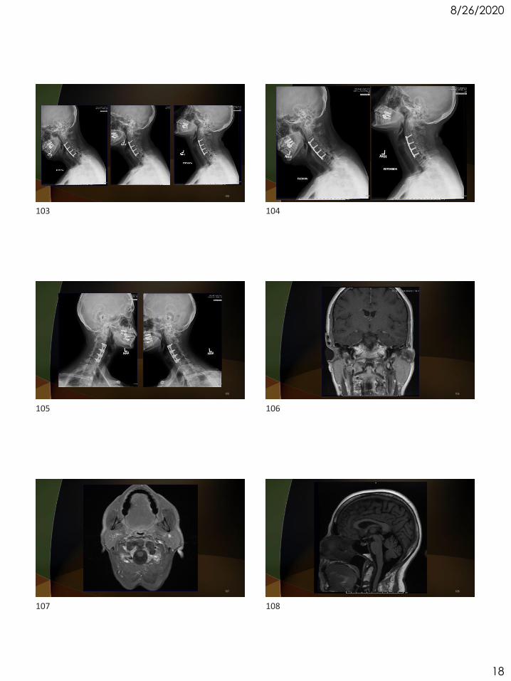
Institute
24
19 20
21 22
23 24
8/26/2020
5
Morgan WE. Modic Changes: Understanding Reactive Vertebral Endplate and Marrow
Morphology. Copyright 2015. Bethesda Spine Institute
25
Morgan WE. Modic Changes:
Understanding Reactive Vertebral
Endplate and
Marrow Morphology.
Copyright 2015. Bethesda Spine
Institute7
26
Morgan WE. Modic Changes: Understanding Reactive Vertebral Endplate and Marrow
Morphology. Copyright 2015. Bethesda Spine Institute
27
Morgan WE. Modic Changes: Understanding Reactive Vertebral Endplate and Marrow
Morphology. Copyright 2015. Bethesda Spine Institute
28
Reactive vertebral body modifications associated with disc inflammation and degenerative disc disease, as seen on
MR images.
Dudli et al. Eur Spine J. Feb 25. Pathobiology of Modic changes.
Modic change type 1 characterized by fibrovascular
tissue (asterisks) and trabecular thickening.
Modic change type 2. Fatty marrow replacement (asterisks) and fibrotic
tissue occurs along the entire endplates.
29 30
25 26
27 28
29 30
8/26/2020
6
31 32
33 34
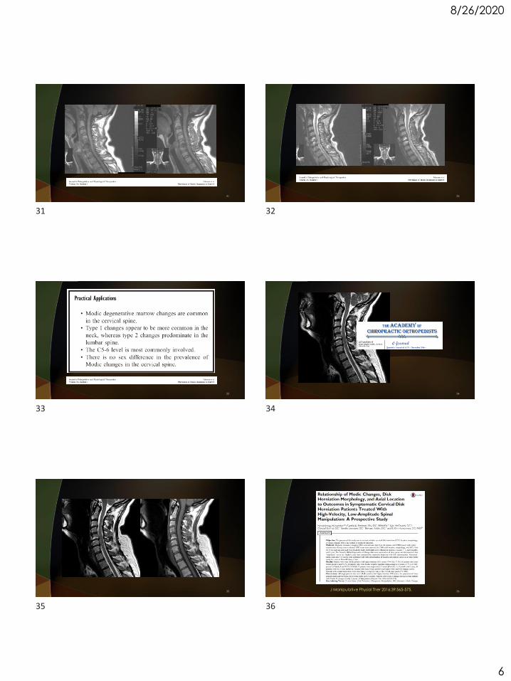
35J Manipulative Physiol Ther 2016;39:565-575.
36
31 32
33 34
35 36
8/26/2020
7
J Manipulative Physiol Ther 2016;39:565-575.37
J Manipulative Physiol Ther 2016;39:565-575.38
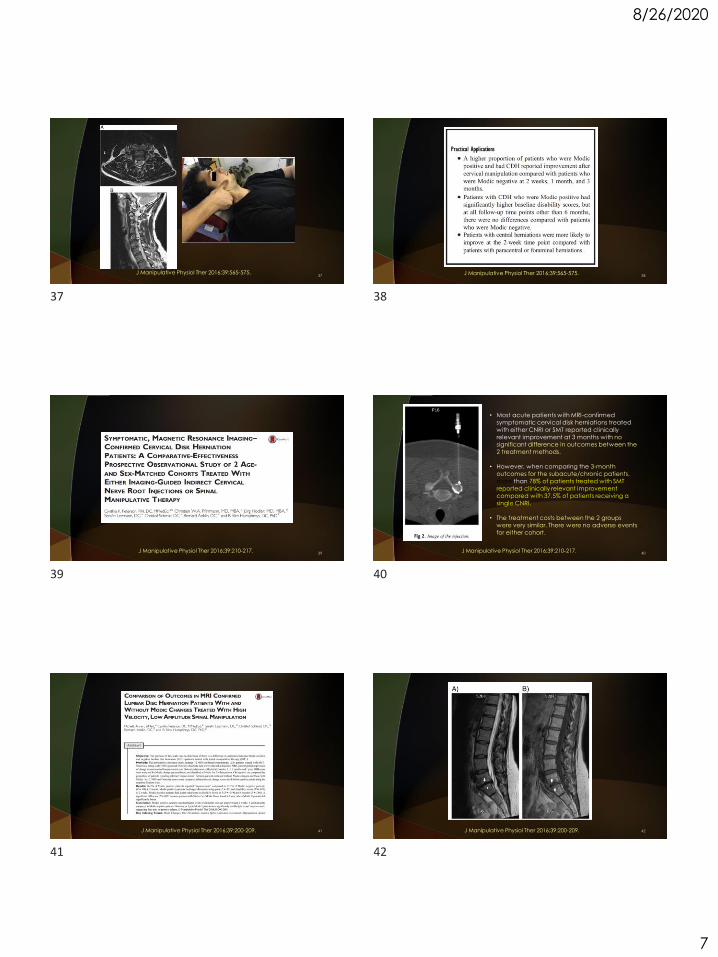
J Manipulative Physiol Ther 2016;39:210-217.39
• Most acute patients with MRI-confirmed
symptomatic cervical disk herniations treated
with either CNRI or SMT reported clinically
relevant improvement at 3 months with no
significant difference in outcomes between the
2 treatment methods.
• However, when comparing the 3-month
outcomes for the subacute/chronic patients,
more than 78% of patients treated with SMT
reported clinically relevant improvement
compared with 37.5% of patients receiving a
single CNRI.
• The treatment costs between the 2 groups
were very similar. There were no adverse events
for either cohort.
J Manipulative Physiol Ther 2016;39:210-217.40
J Manipulative Physiol Ther 2016;39:200-209. 41 J Manipulative Physiol Ther 2016;39:200-209. 42
37 38
39 40
41 42
8/26/2020
8
J Manipulative Physiol Ther 2016;39:200-209. 43 J Manipulative Physiol Ther 2016;39:200-209. 44
• Patients with sequestered herniations treated with SMT to the level of herniation reported significantly higher levels of leg pain reduction at 1 month and a higher proportion reported
improvement at all data collection time points compared to patients with extruded disc herniations but this did not reach statistical significance.
• Further investigation is needed to determine mechanisms for this finding. This also calls into question the seriousness of disc sequestration in determining appropriate treatment.
J Manipulative Physiol Ther 2016;39:192-19945
J Manipulative Physiol Ther 2016;39:192-19946
J Manipulative Physiol Ther 2016;39:192-19947
J Manipulative Physiol Ther 2016;39:192-19948
43 44
45 46
47 48
8/26/2020
9
J Manipulative Physiol Ther 2016;39:192-19949 50
Neurosurgery 79:315–335, 2016. 51
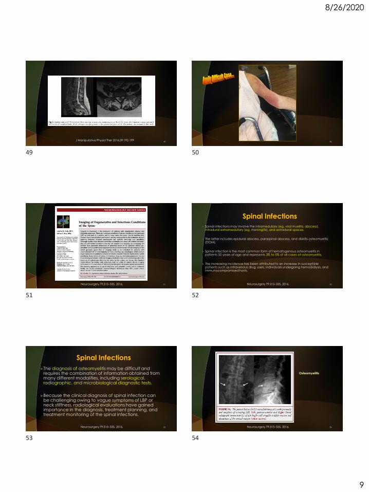
Spinal infections may involve the intramedullary (eg, viral myelitis, abscess), intradural extramedullary (eg, meningitis), and extradural spaces.
The latter includes epidural abscess, paraspinal abscess, and diskitis-osteomyelitis (DOM).
Spinal infection is the most common form of hematogenous osteomyelitis in patients 50 years of age and represents 3% to 5% of all cases of osteomyelitis.
The increasing incidence has been attributed to an increase in susceptible patients such as intravenous drug users, individuals undergoing hemodialysis, and immunocompromised hosts.
Neurosurgery 79:315–335, 2016. 52
The diagnosis of osteomyelitis may be difficult and requires the combination of information obtained from many different modalities, including serological, radiographic, and microbiological diagnostic tests.
Because the clinical diagnosis of spinal infection can be challenging owing to vague symptoms of LBP or neck stiffness, radiological evaluations have gained importance in the diagnosis, treatment planning, and treatment monitoring of the spinal infections.
Neurosurgery 79:315–335, 2016. 53
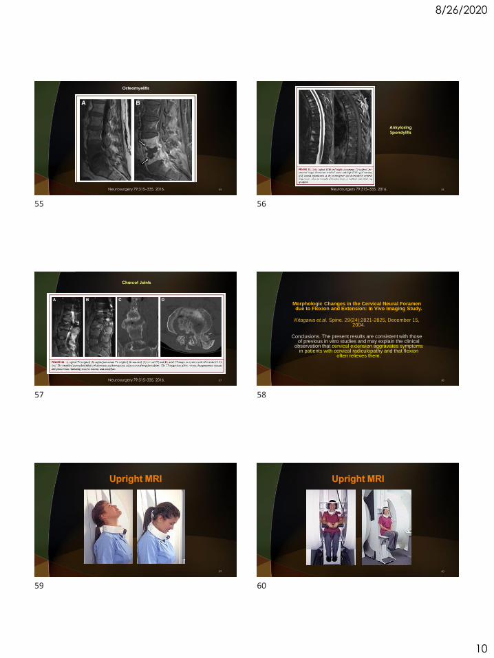
Osteomyelitis
Neurosurgery 79:315–335, 2016. 54
49 50
51 52
53 54
8/26/2020
10
Osteomyelitis
Neurosurgery 79:315–335, 2016. 55
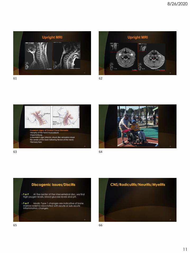
Ankylosing
Spondylitis
Neurosurgery 79:315–335, 2016. 56
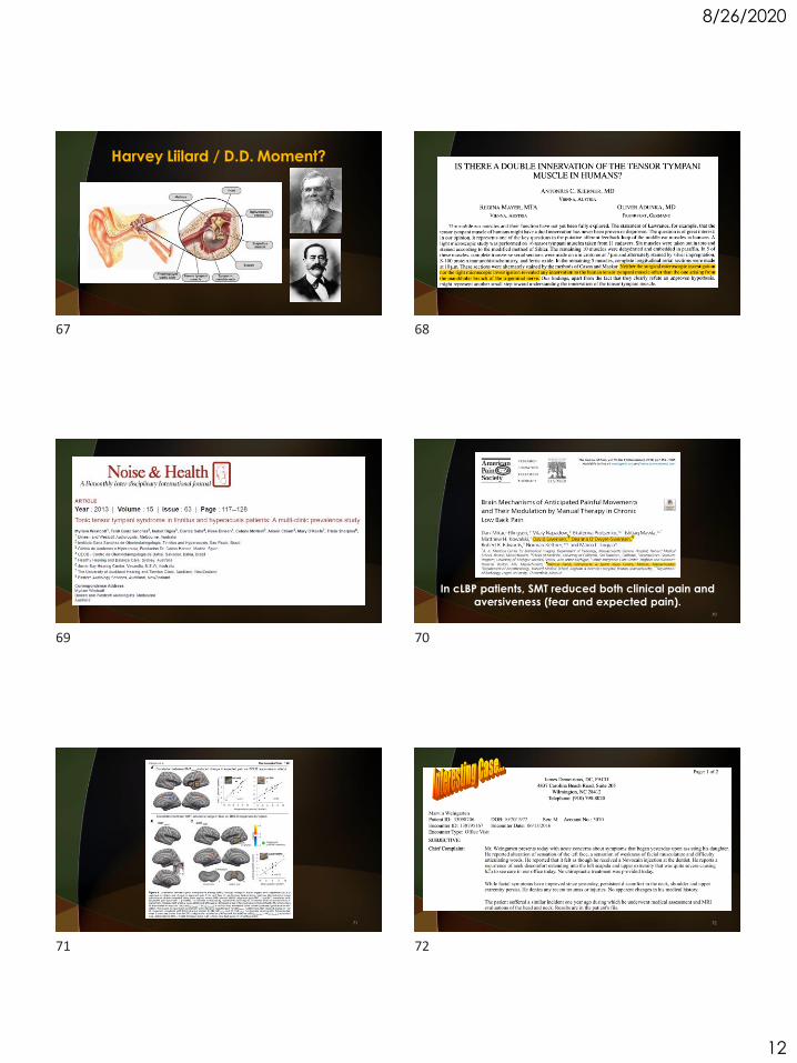
Charcot Joints
Neurosurgery 79:315–335, 2016. 57
Morphologic Changes in the Cervical Neural Foramen due to Flexion and Extension: In Vivo Imaging Study.
Kitagawa et.al. Spine. 29(24):2821-2825, December 15, 2004.
Conclusions. The present results are consistent with those of previous in vitro studies and may explain the clinical
observation that cervical extension aggravates symptoms in patients with cervical radiculopathy and that flexion
often relieves them.
58
59 60
55 56
57 58
59 60
8/26/2020
11
61 62
Common signs of Central Canal Stenosis
•Atrophy of the hand musculature
•Hyperreflexia
•Lhermitte's sign (electric shock-like sensation down
the center of the back following flexion of the neck)
•Sensory loss
63 64
T or F At the center of the intervertebral disc, we find high oxygen levels, blood glucose levels and pH.
T or F Modic Type 1 changes are indicative of bone marrow edema associated with acute or sub-acute inflammatory changes.
65 66
61 62
63 64
65 66
8/26/2020
12
70
In cLBP patients, SMT reduced both clinical pain and
aversiveness (fear and expected pain).
71 72
67 68
69 70
71 72
8/26/2020
13
73 74
6/23/16: MRI Brain with and without contrast: solitary small, 4 mm cortical
abnormality involving the right precentral gyrus of uncertain etiology and significance. No acute ischemia on diffusion imaging. Absence of edema would weigh against a small metastatic lesion. Likely represents and enhancing small subacute cortical ischemic lesion.
MRA of the intracranial arteries: negative intracranial MRA.
MRA of the carotid and vertebral arteries with contrast: negative cervical MRA.
8/19/16: MRI Brain with and without contrast: previous area of abnormal
enhancement in the right lateral posterior frontal will has resolved with mild residual underlying T2 signal change, compatible with a chronic infarct. No new infarct or new enhancing intracranial lesion is observed, nor hematoma or mass effect.
75
Unknown etiology until approved discussion with the patient’s mother:
Discussion with his mother revealed that she was had a recessive Factor V history.
Factor V Leiden is a variant (mutated form) of human Factor V (one of several substances that helps blood clot), which causes an increase in blood clotting (hypercoagulability). With this mutation, the anticoagulant protein secreted (which normally inhibits the pro-clotting activity of factor V) is not able to bind normally to Factor V, leading to a hypercoagulable state, i.e., an increased tendency for the patient to form abnormal and potentially harmful blood clots.
The patient was placed on a daily aspirin regimen.
76
A 64-year-old male presents your office with the clinical triad of:
Acute onset dementia
Urinary incontinence
Ataxic gait
What is your immediate diagnostic consideration?
Follow-up assessments?
77 78
73 74
75 76
77 78
8/26/2020
14
79 80
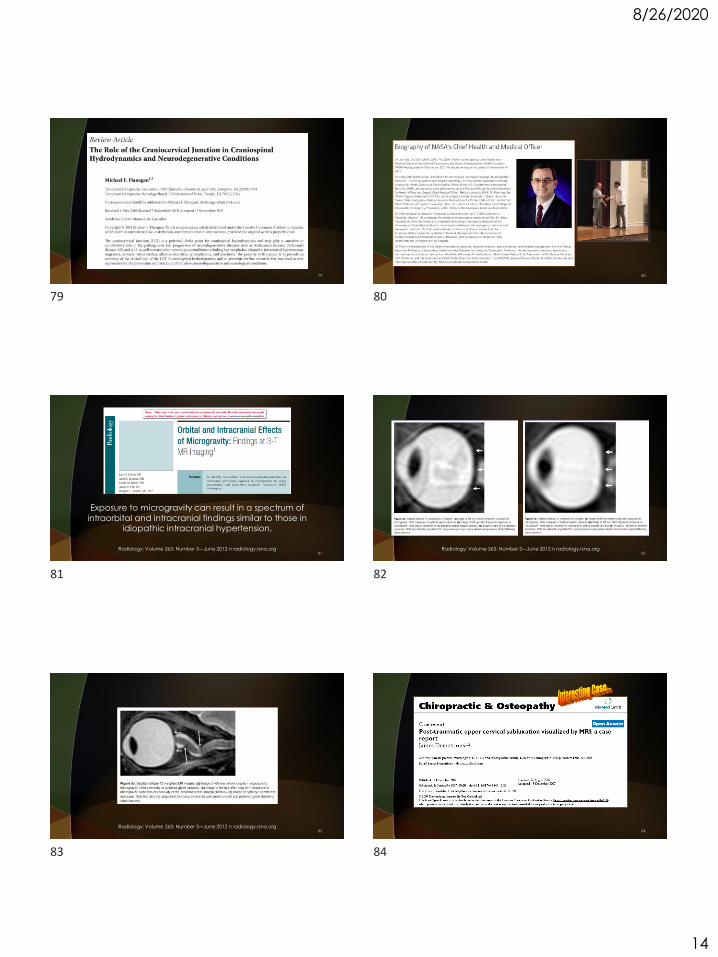
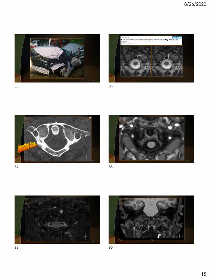
81Radiology: Volume 263: Number 3—June 2012 n radiology.rsna.org
Exposure to microgravity can result in a spectrum of
intraorbital and intracranial findings similar to those in idiopathic intracranial hypertension.
82Radiology: Volume 263: Number 3—June 2012 n radiology.rsna.org
83Radiology: Volume 263: Number 3—June 2012 n radiology.rsna.org
84
79 80
81 82
83 84
8/26/2020
15
85 86
87 88
89 90
85 86
87 88
89 90
8/26/2020
16
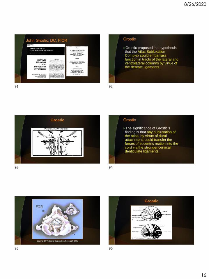
Grostic proposed the hypothesis that the Atlas Subluxation Complex could embarrass function in tracts of the lateral and ventrolateral columns by virtue of the dentate ligaments.
Grostic
The significance of Grostic'sfinding is that any subluxation of the atlas, by virtue of duralattachment, could transfer the forces of eccentric motion into the cord via the stronger cervical denticulate ligaments.
Journal Of Vertebral Subluxation Research 2001
Grostic
91 92
93 94
95 96
8/26/2020
17
Conclusions
The results strongly favour the theory that CSM is caused by tensile stresses transmitted to the spinal cord from the dura via the dentate ligaments.
A spondylotic bar can increase dentate tension by displacing the spinal cord dorsally, while the dural attachments of the dentate, anchored by the dural root sleeves and dural ligaments, are displaced less.
The spondylotic bar may also increase dentate tension by interfering locally with dural stretch during neck flexion, the resultant increase in dural stress being transmitted to the spinal cord via the dentate ligaments.
Flexion of the neck increases dural tension and should be avoided in the conservative treatment of CSM.
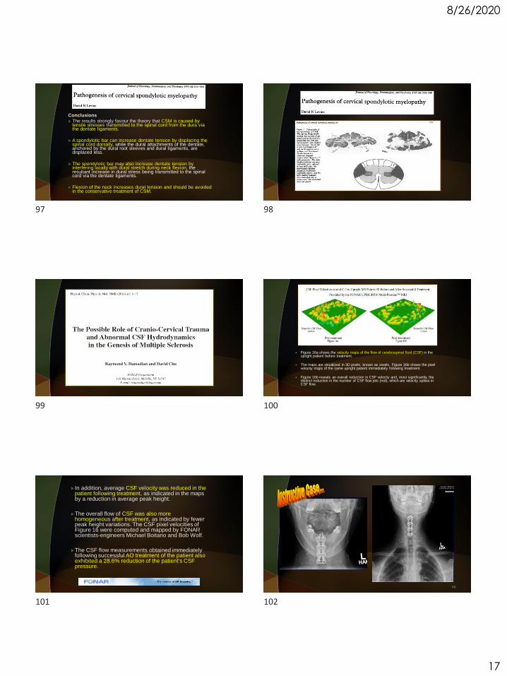
Figure 16a shows the velocity maps of the flow of cerebrospinal fluid (CSF) in the upright patient before treatment.
The maps are visualized in 3D pixels, known as voxels. Figure 16b shows the pixel velocity maps of the same upright patient immediately following treatment.
Figure 16b reveals an overall reduction in CSF velocity and, most significantly, the distinct reduction in the number of CSF flow jets (red), which are velocity spikes in CSF flow.
In addition, average CSF velocity was reduced in the patient following treatment, as indicated in the maps by a reduction in average peak height.
The overall flow of CSF was also more homogeneous after treatment, as indicated by fewer peak height variations. The CSF pixel velocities of Figure 16 were computed and mapped by FONAR scientists-engineers Michael Boitano and Bob Wolf.
The CSF flow measurements obtained immediately following successful AO treatment of the patient also exhibited a 28.6% reduction of the patient's CSF pressure.
102
97 98
99 100
101 102
8/26/2020
18
103 104
105 106
107 108
103 104
105 106
107 108
8/26/2020
19
109 110
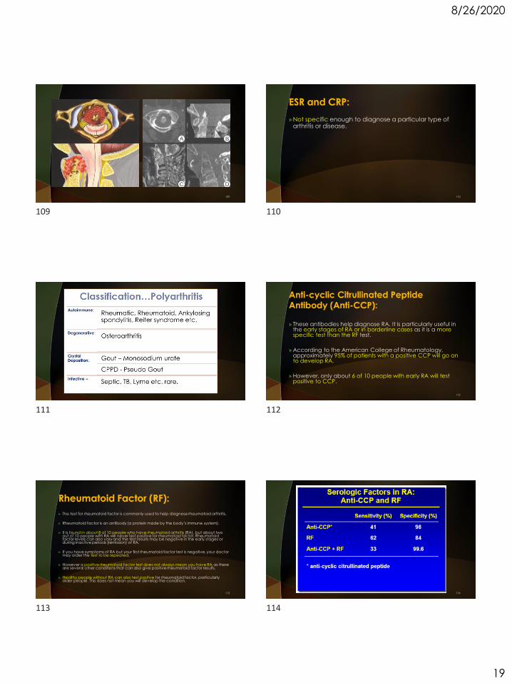
Not specific enough to diagnose a particular type of arthritis or disease.
112
These antibodies help diagnose RA. It is particularly useful in the early stages of RA or in borderline cases as it is a more specific test than the RF test.
According to the American College of Rheumatology, approximately 95% of patients with a positive CCP will go on to develop RA.
However, only about 6 of 10 people with early RA will test positive to CCP.
113
The test for rheumatoid factor is commonly used to help diagnose rheumatoid arthritis.
Rheumatoid factor is an antibody (a protein made by the body’s immune system).
It is found in about 8 of 10 people who have rheumatoid arthritis (RA), but about two out of 10 people with RA will never test positive for rheumatoid factor. Rheumatoid factor levels can also vary and the test results may be negative in the early stages or during inactive periods (remission) of RA.
If you have symptoms of RA but your first rheumatoid factor test is negative, your doctor may order the test to be repeated.
However a positive rheumatoid factor test does not always mean you have RA as there are several other conditions that can also give positive rheumatoid factor results.
Healthy people without RA can also test positive for rheumatoid factor, particularly older people. This does not mean you will develop the condition.
114
109 110
111 112
113 114
8/26/2020
20
115
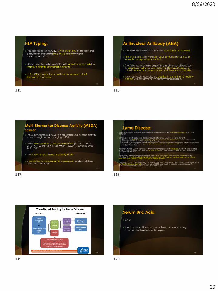
This test looks for HLA-B27. Present in 8% of the general population including healthy people without spondyloarthritis.
Commonly found in people with ankylosing spondylitis, reactive arthritis or psoriatic arthritis.
HLA – DR4 is associated with an increased risk of rheumatoid arthritis.
116
The ANA test is used to screen for autoimmune disorders.
95% of people with systemic lupus erythematosus (SLE or lupus) have a positive ANA test.
The ANA test may also be positive in other conditions, such as Sjogrens syndrome, scleroderma, Raynaud’s disease, mixed connective tissue disease and rheumatoid arthritis.
ANA test results can also be positive in up to 1 in 10 healthy people without any known autoimmune disease.
117
The MBDA score is a novel blood-test based disease activity score of single integer ranging 1-100.
Score derived from 12 serum biomarkers (VCAM-1, EGF, VEGF-A, IL-6, TNF-RI, YKL-40, MMP-1, MMP-3, leptin, resistin, SAA, CRP).
The MBDA reflects disease activity in RA.
Is predictive for radiographic progression and risk of flare after drug reduction.
118
Lyme disease (LD) is caused by infection with a member of the Borrelia burgdorferi sensu latocomplex.
Transmission of LD-associated Borrelia requires at least 36 hours of tick attachment. Approximately 80% of infected individuals will develop a unique expanding skin lesion with a central zone of
clearing, referred to as erythema migrans (EM; stage 1).
In the absence of treatment, patients may progress to early disseminated disease (stage 2), which is characterized by neurologic manifestations (eg, meningitis, cranial neuropathy, radiculoneuropathy) and is often associated with B garinii infection.
Patients with late LD often present with intermittent or persistent arthralgia, most often associated with B burgdorferi infection, or with acrodermatitis chronica atrophicans (ACA), typically due to infection with B afzelii.
Importantly, while serologic assessment for LD may be negative in the early weeks following infection, over 90% of patients with later stages of infection are seropositive by serology, which remains the diagnostic method of choice for this disease.
Diagnosis of LD is currently based on a 2-tiered serologic testing algorithm, as recommended by the Centers for Disease Control and Prevention (CDC), and involves an initial screening assay for detection of antibodies to LD-causing Borrelia species.
119 120
Gout
Monitor elevations due to cellular turnover during chemo- and radiation therapies
115 116
117 118
119 120
8/26/2020
21
121 122
123 124
125 126
121 122
123 124
125 126
8/26/2020
22
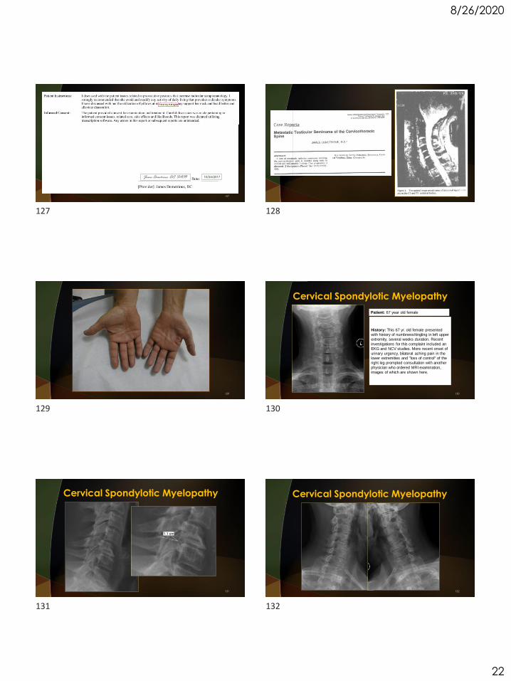
James Demetrious, DC, FACO 10/24/2017
127 128
129
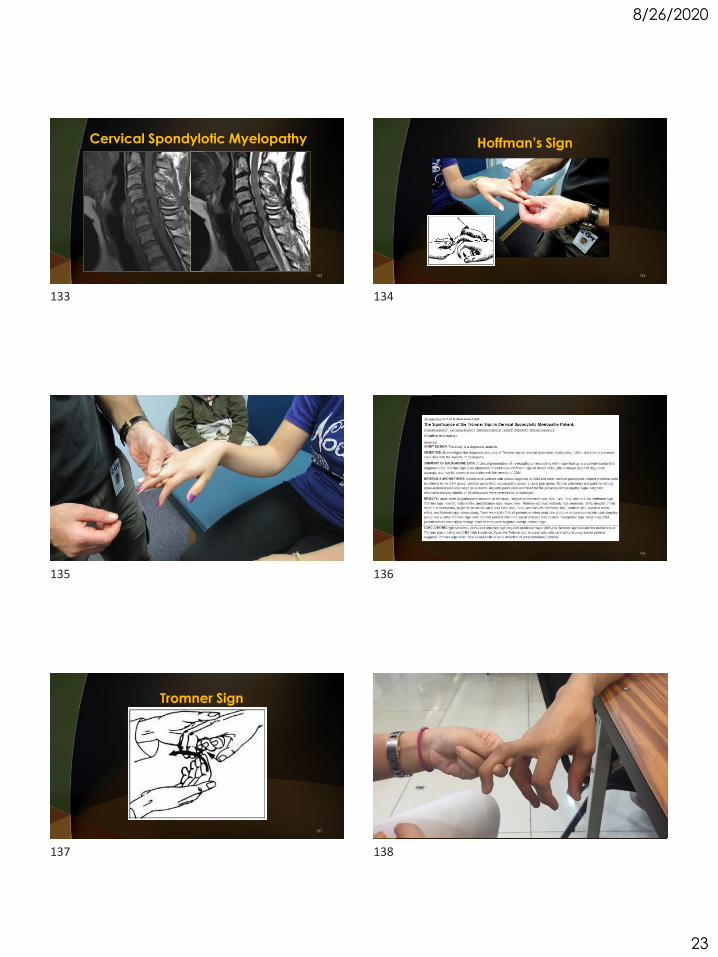
Patient: 67 year old female
History: This 67 yr. old female presented with history of numbness/tingling in left upper extremity, several weeks duration. Recent
investigations for this complaint included an EKG and NCV studies. More recent onset of
urinary urgency, bilateral aching pain in the lower extremities and "loss of control" of the right leg prompted consultation with another
physician who ordered MRI examination, images of which are shown here.
Cervical Spondylotic Myelopathy
130
Cervical Spondylotic Myelopathy
131
Cervical Spondylotic Myelopathy
132
127 128
129 130
131 132
8/26/2020
23
Cervical Spondylotic Myelopathy
133 134
136
137
133 134
135 136
137 138
8/26/2020
24
139 140
141 142
143 144
139 140
141 142
143 144
8/26/2020
25
145 146
147 148
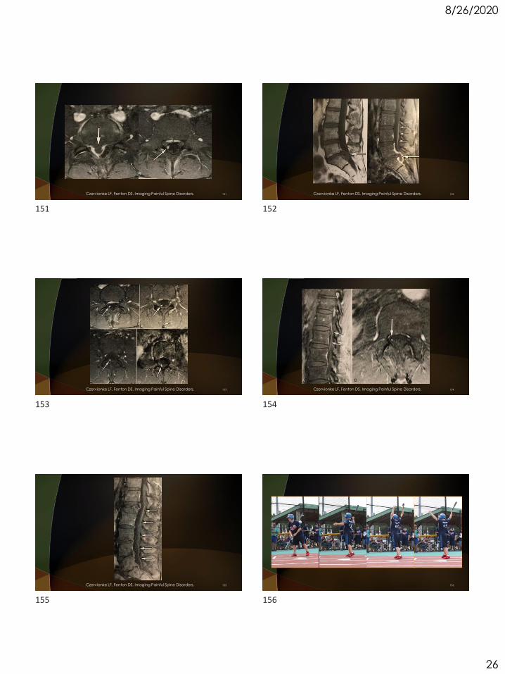
Czervionke LF, Fenton DS. Imaging Painful Spine Disorders. 149 Czervionke LF, Fenton DS. Imaging Painful Spine Disorders. 150
145 146
147 148
149 150
8/26/2020
26
Czervionke LF, Fenton DS. Imaging Painful Spine Disorders. 151 Czervionke LF, Fenton DS. Imaging Painful Spine Disorders. 152
Czervionke LF, Fenton DS. Imaging Painful Spine Disorders. 153 Czervionke LF, Fenton DS. Imaging Painful Spine Disorders. 154
Czervionke LF, Fenton DS. Imaging Painful Spine Disorders. 155 156
151 152
153 154
155 156
8/26/2020
27
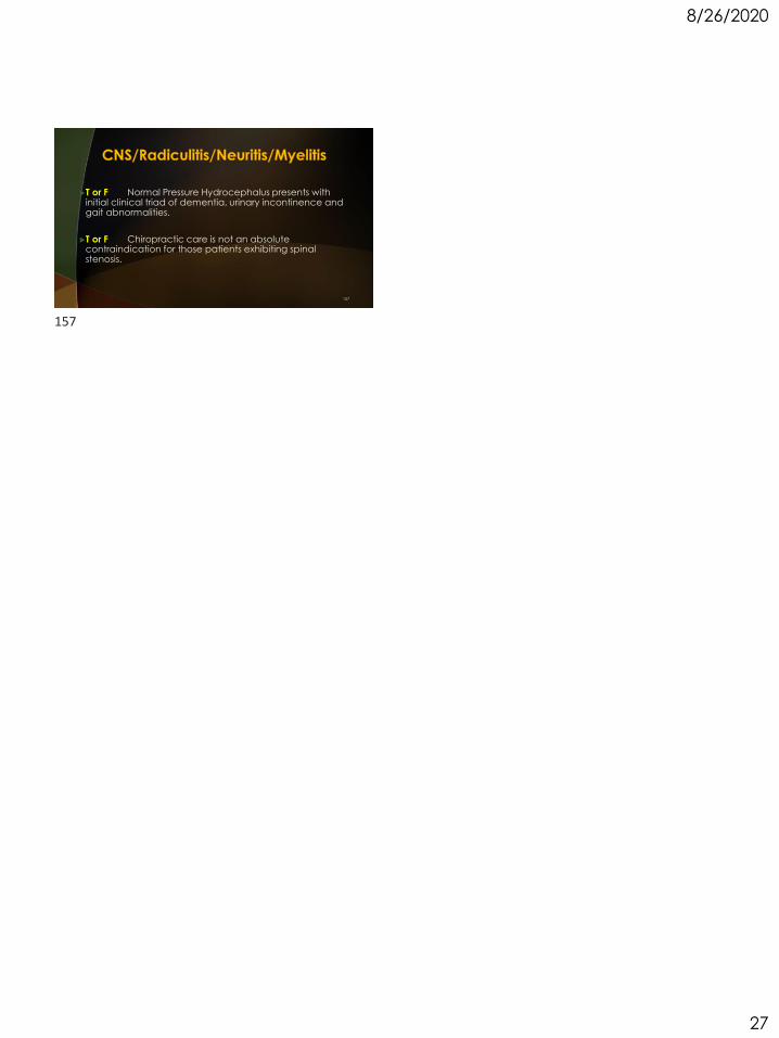
T or F Normal Pressure Hydrocephalus presents with initial clinical triad of dementia, urinary incontinence and gait abnormalities.
T or F Chiropractic care is not an absolute contraindication for those patients exhibiting spinal stenosis.
157
157