Embed Size (px)
Citation preview

7th Annual UC Davis Lung Research Day
June 17, 2016 UCD Genome and Biomedical Sciences Facility (GBSF) Auditorium 1005
https://www.surveymonkey.com/r/LungDay2016

7th Annual Lung Research Symposium ARDS: From Bioinformatics to Precision
Medicine
1

PROGRAM:
7:45‐8:30 Registration and continental breakfast 8:30‐8:35 Welcome and overview: Angela Haczku, MD, PhD, UC Davis 8:35 Session 1: ARDS Clinical Science Moderators: Marc Avdalovic, MD & Brooks Kuhn, MD, UC Davis
8:35‐9:10 Kelly LaMarco, PhD (Science Translational Medicine) How to publish the science of translational medicine?
9:10‐9:45 Timothy Albertson, MD (UC Davis) Clinical trials in the treatment of ARDS: Past, Present & Future
9:45‐10:20 Vincent Liu, MD, MS (Kaiser Permanente) Data that Drives: Ventilator management in the learning hospital system
10:20‐10:55 Jason Adams, MD, MS (UC Davis) Bedside to Cloud: Novel Informatics for ARDS
Coffee Break 10:55‐11:05 11:05 Session 2: Keynote Lecture & Panel Discussion: ARDS ‐ State of the Art Moderator: Christian Sandrock, MD, UC Davis
11:10‐12:00 Michael Matthay, MD, (UC San Francisco) Mesenchymal Stromal Cells: Biology and Potential Treatment for ARDS
12:‐12:30 Drs. Albertson, Adams, LaMarco, Liu,Looney, Matthay, Rogers, Simon
Panel Discussion
Lunch 12:30 – 1:00 Poster Presentations 1:00‐2:00 Facilitators: Samuel Chung, PhD, Richart Harper, MD, Nicholas Kenyon, MD, Jason Lee, MD, Angela
Linderholm, PhD, DV Nguyen, MD, Kent Pinkerton, PhD, Laura Van Winkle, PhD
2:00‐2:15 Presentation of the Phil Thai Memorial Award ‐Amir Zeki, MD & Awardee 2:15‐2:30 Presentation of the Benjamin Davis Memorial Award ‐Kent Pinkerton, PhD & Awardee
2:35 Session 3: Biology of ARDS Moderators: Angela Rogers, MD, Stanford University & Moyar Ge, PhD, UC Davis
2:35‐3:10 Mark Looney, MD (UC San Francisco)Neutrophil and platelet biology in the lungs as illuminated by intravital microscopy
3:10‐3:45 Scott Simon, PhD (UC Davis) Innate immune response: Inflammatory lung injury
3:45‐4:20 Angela Rogers, MD, (Stanford University) From genetics to metabolomics: what has genomics taught us about ARDS
4:20‐4:30 Closing Remarks and Adjourn: Jason Adams, MD, MS 4:30‐6:30 Beer, Wine & Cheese Networking Social
2

PLANNING COMMITTEE Angela Haczku
Jason Adams
Angela Linderholm
Chue Xiong
Gina McDuff
Nicholas Kenyon
Moyar Ge
Amir Zeki
PRESENTERS Dr. Michael Matthay -- Keynote Speaker
Dr. Kelly LaMarco
Dr. Timothy Albertson
Dr. Vincent Liu
Dr. Jason Adams
Dr. Mark Looney
Dr. Scott Simon
Dr. Angela Rogers
3

Abstract #1 Effects of ozone exposure on innate immunity
Leif S. Anderson*, Cameron H Flayer, Angela Haczku and Scott I Simon* *UC Davis, Biomedical Engineering
Rationale: Exposure to high levels of air pollution is a major problem in our industrialized society. One such pollutant, ozone, can increase mortality and hospital visits due to exacerbation of respiratory diseases brought on by infection. Polymorphonuclear leukocytes (PMN), commonly called neutrophils, are the first immune cells to arrive at a site of tissue insult and are endowed with the capacity to produce proinflammatory cytokines and reactive oxygen species, which are critical components for regulation of pathogens. However, under conditions of particulate and ozone insult, PMN exhibit diminished phagocytic and intracellular killing capacity. The impaired PMN contribute to a pathophysiologic environment in the lung, where chronic inflammation and infection cause airway hyperresponsiveness (AHR) and respiratory disease. Little is known regarding the mechanisms underlying the altered PMN function in response to ozone that leads to impairment of the host’s defense against infection. The hypothesis of this study is that ozone has a systemic effect on PMN, characterized by an increase in inflammatory markers and decreased anti-microbial capacity across PMN in the bone marrow, circulation, and bronchoalveolar lavage fluid (BAL), and these changes promote lung neutrophilia as well as reduce a host’s capacity to resolve bacterial infection (Figure 1).
Methods: C57BL/6 mice were exposed to 3 ppm ozone for 2 hours. 12 hours later, lung function was assessed using the flexiVent and mouseOX systems. AHR was determined by measuring total lung resistance as a function of methacholine concentration. Following in vivo lung functional studies, bone marrow, peripheral blood, and BAL were collected and stained with fluorescently labeled primary antibodies, fixed with 4% paraformaldehyde and assessed on the Attune NxT flow cytometer. Ozone exposed samples were normalized to the filtered air (FA) control.
Results: Ozone exposed mice had increased numbers of PMN in circulation and BAL and a decreased number of PMN in Bone Marrow compared to FA controls. Ozone exposed mice had lower oxygen saturation and increased AHR compared to FA controls. PMN from ozone exposed mice expressed more of the adhesion molecule, CD11b, across all compartments, and the PMN expressed less CXCR4, indicating an increase in PMN maturity (Figure 2).
Conclusions: Supporting previously published studies, we observed that ozone exposure causes neutrophilia and AHR. Interestingly, ozone has a systemic effect on PMN. An increase of CD11b, the alpha subunit of the MAC-1 integrin, and a decrease of CXCR4, the SDF-1 receptor that homes cells to the bone marrow, on the surface of PMN from ozone exposed mice provides initial insight into the mechanisms that drives neutrophilia. Future studies will look into possible signaling molecules (G-CSF, GM-CSF, IL-1β) and the source of these inflammatory signals (lung-resident macrophages, ILC-2).
Acknowledgements: NIH T32 GM099608 to Department of Pharmacology & NIH R01 AI47294 to Scott Simon
Figure 1: Ozone stimulates airway epithelium to secrete inflammatory mediators that systemically activate PMN. These cells undergo changes to promote migratory efficiency and reduce anti-
Airway Epithelium Lung injury
Airway Neutrophilia
O3 Systemic Activation
Figure 2: The effect of ozone on the activation and maturation of PMN in bone marrow. Bone marrow was collected from mice 12 hours after ozone exposure. The MFI of CD11b and CXCR4 was assessed on PMN gated events (CD15+, CD16+, CD49d-).
4

Abstract #2
IL13-Induces Eotaxin-3 (CCL26) Production by a Geranylgeranylpyrophosphate-Dependent Mechanism in Human Bronchial Epithelial Cells: Implications for Airway Eosinophilia
Authors: Muhammad Arif, Sean Ott, Amir A. Zeki.
Rational: The influx of eosinophils into the airways, a hallmark of allergic asthma, is mediated in part by the IL13-dependent chemokine eotaxin-3 (CCL26), which is secreted from airway epithelial cells. IL13 induces eotaxin-3 gene expression via JAK1/STAT6 signaling. Eotaxin-3 is the most clinically significant of the three eotaxins, indeed of all the eosinophil chemokines, and has direct correlation with blood and lung eosinophilia, and serum IL5 in asthmatics. We previously demonstrated that simvastatin (Sim), a HMG-CoA reductase (HMGCR) inhibitor, can inhibit IL13-stimulated eotaxin-2 (CCL24) and eotaxin-3 gene expression and peptide secretion. Because statins inhibit both the isoprenoids (IPP, FPP, GGPP) and downstream sterols (squalene, cholesterol) in the mevalonate (MA) pathway, we investigated which arm of this pathway is primarily responsible for the observed beneficial statin effect. We hypothesized that IL13-induced eotaxin-3 production depends on FPP and GGPP, rather than cholesterol.
Methods: Human Bronchial Epithelial (HBE-1) cells were grown to 95% confluence in submerged culture with serum-free D-Media and 6 growth factors. Cells were then pre-treated with 5 uM Simvastatin (Sim) with or without the following: 2 mM Mevalonate (MA), 10 uM isopentenylpyrophosphate (IPP), 10 uM farnesyl pyrophosphate (FPP), 10 uM geranylgeranyl pyrophosphate (GGPP), 50 uM squalene (SQ), and 2 or 20 ug/mL of soluble cholesterol for 72 hours total. In the last 12 hours, cells were stimulated with IL13 (20 ng/mL) in different experiments to activate IL13/STAT6 signaling and induce eotaxin-3 production. ELISA was used to measure extracellular secreted eotaxin-3 protein (pg/mL) in cell-free media, and mRNA expression was measured using RT-qPCR normalized to beta-actin. Protein levels were assessed via Western blot using GAPDH as a loading control. Cell viability assays to assess cytotoxicity were done using alamar blue or MTT assays, and cell death was assessed by trypan blue exclusion.
Results: Simvastatin inhibited IL13-induced JAK1 and STAT6 phosphorylation, eotaxin-3 mRNA levels, and eotaxin-3 peptide secretion (without significantly affecting total JAK1 or total STAT6). This Sim effect was completely abrogated with MA co-treatment, confirming that the MA pathway mediates IL13 signaling. Of the isoprenoids (IPP, FPP, GGPP), only GGPP completely abrogated the inhibitory effect of Sim on STAT6 phosphorylation, eotaxin-3 mRNA, and eotaxin-3 peptide secretion while FPP had a partial and insignificant effect. For the sterol arm, neither SQ nor cholesterol abrogated the inhibitory effect of Sim on STAT6 and eotaxin-3 secretion. These data suggest that the GGPP arm of the MA pathway mediates IL13/STAT6 signaling, and therefore, controls eotaxin-3 production. Unexpectedly, we also observed that independent of any statin treatment, exposing cells to FPP, GGPP, or cholesterol augmented IL13-induced STAT6 phosphoprylation and eotaxin-3 peptide secretion. The most powerful of these was GGPP where we observed an 18-fold increase in eotaxin-3 mRNA expression and 3-fold increase in eotaxin-3 extracellular secretion, along with a significant increase in STAT6 phosphorylation in IL13+GGPP treated cells as compared to IL13 alone. These data suggest that an excess in the pool of available isoprenoids and cholesterol, in particular GGPP, significantly promotes IL13-induced STAT6 activation, eotaxin-3 production, and eotaxin-3 secretion. There was no evidence of statin cytotoxic effect at the Sim doses used (1 to 5 uM).
Conclusion: The GGPP arm of the MA pathway mediates IL13/STAT6-induced eotaxin-3 production and extracellular secretion. These results highlight not only the mechanism of the statin drugs, but also elucidates the novel role of GGPP in mucosal immune responses and further adds to our basic understanding of IL13 signaling and airway eosinophilia. Beyond the statins, pharmacologic agents that deplete GGPP such as bisphosphonates may be potential therapies for asthma.
5

Abstract #3 Targeting tumor-associated exosomes with novel integrin binding peptides Randy P. Carney, Sidhartha Hazari, Alisha Knudson, and Kit S. Lam Department of Biochemistry and Molecular Medicine, UC Davis
Rationale: There is an urgent need in clinical sciences for the development of new non-invasive technologies capable of rapidly diagnosing cancers in their early-stage and according to subtype. Fortuitously, all cells dynamically excrete into circulation nano-sized packages called exosomes for normal cell-to-cell communication. It was recently discovered that cancer cells hijack exosomal pathways for means of immune system suppression and metastasis. Yet distinguishing tumor- associated exosomes from healthy ones is not currently possible. We hypothesize that there exist ligands capable of binding to specific subpopulations of circulating extracellular vesicles (e.g. ones derived from tumor cells) and furthermore that these ligands could be used to target those vesicles for further use.
Methods: Using a combinatorial library-based screening methodology, our lab has recently discovered several unique peptide ligands capable of binding specific tumor
cells through their overexpressed integrins (e.g. LXY30 peptide binding to a3B1 integrin). To investigate whether these ligands are capable of specific binding to the exosomes derived from those tumor cells, we have employed a combination of characterization schemes for both bulk exosomes, like flow cytometry and proteomic profiling, and also for single exosomes, including Laser tweezers Raman spectroscopy (LTRS) and nanoparticle tracking analysis (NTA).
Results: We have measured strong binding for two unique peptide ligands, LXY30 and LLP2A, to specific integrins present on single exosomes derived from ovarian, brain, and lung tumor cells (Fig. 2). Moreover, these ligands show little affinity to other types of normal cell-derived exosomes or to tumor exosomes with varying integrin profiles. Furthermore, we show that these ligands, when reversibly bound to magnetic beads, can be used to fish out tumor-associated exosomes from blood in
human cancer patients. Conclusion: Given our single exosome analysis techniques and the ability to enrich tumor-associated exosomes from isolated biofluids, we believe that our novel ligands could be used to sense very small numbers of tumor-associated exosomes amongst the normal secretome background. Such a ligand-based targeting approach has the potential to transform both the understanding of compositional differences amongst circulating exosomes and also the ease in which cancer could be diagnosed.
6
Abstract #4 Naphthalene DNA Adduct Formation in Ex-Vivo Rodent Tissues Sarah A. Carratt (1), Bruce A. Buchholz (2), Xinxin Ding (3), Laura S. Van Winkle (1)
(1) UC Davis (2) Lawrence Livermore National Lab (3) SUNY Polytechnic Institute
RATIONALE: Naphthalene (NA) is a respiratory toxicant and possible human carcinogen. Human epidemiology data are unclear on the long-term effects of NA exposure and risk assessment relies heavily on animal data. Recurrent cycles of cytotoxicity and proliferation are thought to be the driving force behind formation of mouse lung tumors and rat nasal tumors; however, the formation of DNA adducts from NA metabolites has not been ruled out and could impact the decision to evaluate NA as a genotoxic agent. METHODS: This study evaluated DNA adduct formation in target tissues for carcinogenesis in the National Toxicology Program’s chronic bioassays: female mouse airway epithelium and male rat nasal epithelium (respiratory, olfactory). Metabolically active epithelial tissue was isolated and incubated in explant culture with 14C-labeled NA (0, 2.5, 25, 250 µM) for 1 h. The tissue was then rinsed 12-15 times to remove unbound NA; DNA was isolated and prepared for 14C-AMS (accelerator mass spectrometry) analyses. Male rat airway, which is not susceptible to tumor formation by NA, was used as a negative control. Benzo(a)pyrene (25 µM), a known carcinogen and DNA adductor, was the positive control.
RESULTS: We found the highest level of ex vivo formed NA-DNA adducts in mouse airway, followed by rat airway and nasal olfactory tissue, in a manner corresponding with the previously reported, differing rates of in vitro NA bioactivation at these sites. CONCLUSION: We conclude that NA is capable of forming DNA adducts in target tissues of NA carcinogenesis in mice and rats. Future experiments will determine adduct structure and examine adduct stability, both properties may impact the carcinogenic potential of the detected DNA adducts. Funded by T32 ES007059, R01 ES020867, ES020867S1 and DOD LC130820.) This work performed in part under the auspices of the U.S. Department of Energy by Lawrence Livermore National Laboratory under Contract DE- AC52-07NA27344.
7

Abstract #5
Repressing MARCKS Signaling Pathway to Suppress Lung Cancer Stemness
Chun-Lung Chiu, Ching-Hsien Chen, Yelin Hu, Meng-Chen Lee, Suzanne Miyamoto, Ken Yoneda, and Reen Wu.
University of California Davis
RATIONALE: Lung cancer, especially non-small cell lung carcinoma, NSCLC, is the leading cause of cancer death worldwide. There is no remedy for the treatment, except targeted therapy. However, cancer cells have the ability to evade these inhibitors, and have become resistance to these treatments. The difficulty may be related to cancer heterogeneity and the protective nature of cancer stemness. We have recently uncovered that an elevated phosphorylated MARCKS expression is a biomarker for the poor outcome of overall lung cancer survival. To further elaborate this finding, we propose to test the hypothesis that MARCKS phosphorylation serves as a non-genetic mean in the control of lung cancer NSCLC stemness and drug resistance.
METHODS: Lung cancer stem/tumor initiating cells (CSICs) were enriched and isolated from NSCLC cell lines through serum-free non-adhering culture system and flow cytometer approaches. Stemness nature of these CSICs were further characterized in vivo by xenograft approach, and in vitro by Q-RT-PCR, flow cytometer, immunohistochemical staining, and western blot analyses. Sensitivities of CSICs and parent/adhered cells to various chemo-therapeutic treatment were carried out with MTS assays as described before. Molecular/genetic silencing approach was used to establish the interdependent nature of MARCKS expression with stemness gene expression. MPS peptides that are targeting MARCKS phosphorylation sequence were used to characterize the effectiveness of inhibition of MARCKS phosphorylation step in the regulation of cancer growth and drug sensitivity.
RESULTS: NSCLC CSICs were enriched from cultures of a tyrosine-kinase inhibitors (TKIs) sensitive PC9 line and TKI-resistant primary pleural effusion cell line, LG704. Flow cytometer and in vivo tumor xenograft approaches confirmed the nature of these CSICs in the expression of stemness cell surface markers, CD44 and CD 133, and the potency in inducing tumor xenograft with less than 10,000 cells. Q-RT-PCR analyses had confirmed a coordinated expression of various stemness genes and MARCKS. Immunohistochemical staining and western blot analyses had confirmed the elevation of phospho-MARCKS and MARCKS expression in isolated/enriched CSICs. Molecular siRNA MARCKS silencing approach had shown the suppression of the expression of various stemness genes and surface markers, in addition to MARCKS itself. Similar results were seen if MARCKS phosphorylation was suppressed in vitro by MPS peptides. These results suggest that MARCKS, and perhaps, the MARCKS’ phosphorylation step, is serving as a non-genetic mean in coordinating stemness gene expression. Additionally, isolated/enriched CSICs of PC9 have elevated TKI resistance, while there is no change in TKI sensitivity with LG704 CSICs. Interestingly, both PC9 and LG704 cells, regardless the stemness were all sensitive to MPS peptide treatment. CONCLUSIONS: These results suggest that MARCKS, especially the phosphorylated MARCKS is participated in coordinating regulation of CSIC stemness expression and targeting MARCKS phosphorylation step is an effective way in the suppression of lung cancer stemness and growth.
8

Abstract #6 Neutral Sphingomyelinase 2 promotes cell proliferation of human airway epithelial cells during chronic cigarette smoke exposure via Sphingosine-1-Phosphate Samuel Chung@, Simon Vu, Simone Filosto and Tzipora Goldkorn#. Center for Comparative Respiratory Biology and Medicine (CCRBM); Genome and Biomedical Sciences Facility (GBSF); UCD School of Medicine, Davis, CA. E-mails: @ [email protected]; #[email protected] RATIONALE: Smoking-related lung diseases, such as Chronic Obstructive Pulmonary Disease (COPD) and lung cancer, continue to remain significant sources of morbidity and mortality worldwide. Interestingly, past epidemiological studies concluded that smokers with COPD-like symptoms have elevated risk of developing lung cancer. Therefore, the emergence of proliferation-dominant lung cancer from apoptosis-dominant lung injury suggests that processes at the molecular- and cell-level are inexplicably linked between the two diseases. We propose that the neutral sphingomyelinase 2 (nSMase2), which hydrolyzes sphingomyelin to the pro-apoptotic ceramide, may serves as a common molecular link.
We previously showed that nSMase2 expression is highly-elevated in the airway of emphysema patients and in rodents exposed to whole-body cigarette smoke (CS). Importantly, nSMase2 knockdown via siRNA prevented CS-induced ceramide generation and apoptosis of airway epithelial cells in vitro and in vivo. Recently, however, we demonstrated that nSMase2 and ceramide generation are up-regulated by Src during CS exposure via p38 MAPK. Given that a proto-oncogene (Src) paradoxically regulates nSMase2 which is linked to apoptosis and smoking-induced lung injury, we now hypothesize that nSMase2 is elevated in smoking-related lung cancer and has a functional role in tumorigenesis. METHODS: Lung tumor biopsy samples were requested from the UC Davis Biorepository Shared Resource. In situ nSMase2 and ceramide were measured by immunofluorescence using confocal microscopy. Standard tissue culture practices were used to maintain A549 lung adenocarcinoma cells and derived cell lines. CS extract (CSE) was prepared by puffing 60 cc of smoke (1 cigarette) for 30 min. Cell proliferation was assessed by MTT assay and 3H-Thymidine Incorporation assay. Sphingolipids were quantified by LC-MS/MS by the Lipidomics Core of Medical University of South Carolina. RESULTS: First, we observed in situ that nSMase2 and ceramide are elevated in lung tumors of smokers compared to tumors of non-smokers and normal parenchyma of smokers. Second, A549 chronically exposed to 10% CSE (“A549+CSE”) have altered morphology and increased proliferation. Importantly, A549+CSE cells have elevated nSMase2 expression and nSMase2 knockdown via shRNA suppressed their proliferation. Furthermore, A549 adapted to overexpress nSMase2 (“A549+nSMase2”) show enhanced proliferation. Lastly, the sphingolipid profiles of A549+CSE and A549+nSMase2 cells have elevated sphingosine-1-phosphate (S1P), a sphingolipid associated with proliferative signaling. CONCLUSIONS: Our data shows that adaptation to elevated nSMase2/ceramide promotes S1P production, suggesting that S1P-mediated proliferative signaling may be a novel mechanism of lung-specific tumor promotion unique to chronic smokers. Future studies will investigate the molecular mechanisms of nSMase2-dependent S1P production/secretion during chronic CS exposure.
9

Abstract #7 HEPATIC STELLATE CELLS REGULATE CCR2 MRNA STABILITY AND RECRUITMENT OF INFLAMMATORY CELLS VIA NOX4 IN ALCOHOLIC LIVER INJURY Dehnad A1,2, Sasaki Y1,2, Jiang JX1, Sato A1, Chao TI1, Tian J1 ,Török NJ1,2 1Division of Gastroenterology and Hepatology, University of California Davis, Sacramento, CA, USA 2VA Medical Center, VA Northern California Health Care System, Mather, CA, USA Rationale: Inflammatory cell recruitment is a major feature of alcoholic liver injury however; the signals and cellular sources regulating the migration of these cells to the liver are still not well defined. C-C chemokine receptor type 2 (CCR2) is mainly expressed by active hepatic stellate cells (HSC) and is a key recruitment signal. Active HSC are also important sources of hydrogen peroxide resulting from the activation of NADPH oxidase 4 (NOX4). As the role of this NOX in early alcoholic liver injury has not been well studied, we hypothesized that NOX4 via hydrogen peroxide modulates the mRNA stability of CCR2 thereby inducing recruitment of inflammatory cells. Methods: NOX4 was studied in healthy human livers and in patients with alcoholic liver disease. Fl/fl and HSC specific [GFAP-cre NOX4 knockout mice (KO)] were pair-fed with the Lieber deCarli or control isocaloric diets, and CCR2, MCP-1, TNFα, IL-1β, IL-6, and Ly6C expression in the livers were assessed by real time qPCR. In vitro, NOX4 expression was studied in primary HSC exposed to acetaldehyde (Ac). CCR2 mRNA stability was assessed 1) in primary wt or NOX4KO HSC, and 2) in LX2 human HSC line transfected by Ad-NOX4 or control vector. Immunohistochemistry and western blots were done to detect the mRNA binding protein HuR subcellular localization and phosphorylation. Results: NOX4 was co-localized with αSMA-expressing activated HSC in liver biopsies of patients with alcoholic hepatitis. NOX4 mRNA was significantly induced in patients, as well as in mice on the Lieber deCarli diet. In the fl/fl mice on Lieber deCarli diet, TG content, lipid peroxidation and the expression of CCR2, TNFα, IL-6, MCP-1, Ly6C were significantly increased compared to the HSCNOX4Komice (p <0.05). NOX4 was induced in primary HSC by Ac treatment (p<0.05). NOX4 has significantly increased transcript stability of CCR2 (p<0.05) coinciding with cytoplasmic shuttling and phosphorylation of HuR. In conclusion: NOX4 is induced in early alcoholic liver injury in HSC and regulates CCR2 mRNA stability thereby promotes recruitment of inflammatory cells and production of proinflammatory cytokines.
10

Abstract #8 Helicobacter Pylori Elicits a Heightened Innate Immune Response in Pediatric Airway Epithelium: Evidence of NOD1 Developmental Regulation Authors: M.A. Dela Pena1, M.T. Jimenez1, J.V. Solnick1, L.A. Miller2M 1California National Primate Research Center O Davis, CA/US, 2UC Davis School of Veterinary Medicine O Davis, CA/US
RATIONALE: Epidemiologic studies suggest that childhood infection with the gramOnegative stomach bacteria Helicobacter pylori (H. pylori) confers protection from development of asthma. Because oral aspirates are known to contribute to the airway microbiome, we hypothesized that H. pylori can directly elicit innate immune responses in airway epithelium. Based upon our published findings that show attenuation of pattern recognition receptor (PRR) ligand induced cytokine signaling in pediatric airway epithelium (Maniar Hew et. al. 2013, Clay, et. al. 2014), we further proposed that mechanisms for epithelial innate immune responses to H. pylori are age dependent.
METHODS: To test our hypothesis, we established an in vitro H. pylori infection model of airway epithelium using primary tracheobronchial epithelial cell cultures derived from infant or adult rhesus macaque monkeys. Cultures were inoculated with wildOtype or cag pathogenicity island mutant H. pylori strains for 24 hours, followed by evaluation of IL-8 and IL-6 protein synthesis. Because the H. pylori type IV secretion system is known to mediate cellular activation through recognition of "injected" peptidoglycan by nucleotideObinding oligomerization domainOcontaining protein 1 (NOD1), we also assessed this signaling pathway via selective agonists and inhibitors.
RESULTS: Inoculation of primary tracheobronchial epithelial cell cultures with H. pylori increased secretion of ILO8 and ILO 6 in a MOIOdependent fashion. Induction of IL-8 synthesis by H. pylori was ageOdependent (p<0.01), with a greater than 4 fold increased secretion by infant cultures. Comparatively, there were no significant differences in IL-6 synthesis between infant and adult cultures following H. pylori inoculation. H. pylori mediated synthesis of IL-8 in infant cultures was significantly reduced by cag pathogenecity island mutants, indicating a requirement for the type IV secretion system and NOD1. There was a modest, but significant (p<0.01) increase in NOD1 mRNA for infant versus adult cultures. Treatment of cultures with the selective NOD1 inhibitor ML130 (Nodinitib-1) resulted in complete suppression of IL-8 synthesis in infant cultures, with minimal effects in adult cultures.
CONCLUSIONS: Our data show that H. pylori can directly immunomodulate airway epithelium in an MOI and age dependent manner. Induction of IL-8 synthesis in infant cultures by H. pylori was dramatically increased relative to adult cultures, whereas induction of IL-6 was not significantly age dependent, suggesting that pathways upstream of NF Kappa B are selective with chronological age. Infant cultures expressed higher levels of NOD1 mRNA and were uniquely sensitive to NOD1 inhibition, indicating that signaling via this cytosolic PRR is developmentally regulated in pediatric airway epithelium.
11
Abstract #9
Age-Related Modulation of Pulmonary Innate Immunity against Influenza A/PR/8/34 by Environmental Tobacco Smoke in Mice
J.K. Dhillon, L. Wang, M. Kaur, K.E. Pinkerton University of California at Davis - Davis, CA/US
Abstract Body
Rationale: There is minimal information on the susceptibility to influenza virus infections in children or adults who are exposed to environmental stressors such as environmental tobacco smoke (ETS) in life.
Objective: To define and compare the influence of ETS on pulmonary and systemic innate immunity in response to primary influenza virus infection between neonatal mice and adult mice.
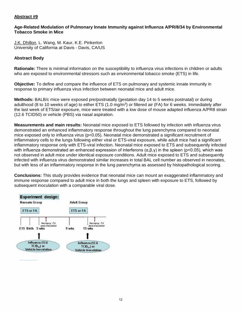
Methods: BALB/c mice were exposed pre/postnatally (gestation day 14 to 5 weeks postnatal) or during adulthood (8 to 10 weeks of age) to either ETS (1.0 mg/m3) or filtered air (FA) for 6 weeks. Immediately after the last week of ETS/air exposure, mice were treated with a low dose of mouse adapted influenza A/PR8 strain (12.6 TCID50) or vehicle (PBS) via nasal aspiration.
Measurements and main results: Neonatal mice exposed to ETS followed by infection with influenza virus demonstrated an enhanced inflammatory response throughout the lung parenchyma compared to neonatal mice exposed only to influenza virus (p<0.05). Neonatal mice demonstrated a significant recruitment of inflammatory cells to the lungs following either viral or ETS-viral exposure, while adult mice had a significant inflammatory response only with ETS-viral infection. Neonatal mice exposed to ETS and subsequently infected with influenza demonstrated an enhanced expression of interferons (α,β,γ) in the spleen (p<0.05), which was not observed in adult mice under identical exposure conditions. Adult mice exposed to ETS and subsequently infected with influenza virus demonstrated similar increases in total BAL cell number as observed in neonates, but with less of an inflammatory response in the lung parenchyma as assessed by histopathological scoring.
Conclusions: This study provides evidence that neonatal mice can mount an exaggerated inflammatory and immune response compared to adult mice in both the lungs and spleen with exposure to ETS, followed by subsequent inoculation with a comparable viral dose.
12

Abstract #10
Developmental Regulation of IL-22R1 in Infant Airway Epithelium is Dependent Upon Interferon-λ and Enhanced By TLR3 Signaling
Daniel T. Dugger1,2 Lisa A. Miller2,3 1Department of Pathology, Microbiology, and Immunology, School of Veterinary Medicine, University of California, Davis, California 2California National Primate Research Center, University of California, Davis, California 3Department of Anatomy, Physiology, and Cell Biology, School of Veterinary Medicine, University of California, Davis, California Rationale: Respiratory tract infections are a leading cause of morbidity and mortality in pediatric populations. Using a nonhuman primate model of childhood lung development, we have reported that interleukin-22 receptor (IL-22R1) expression in airway epithelium is attenuated in early life. Reduced IL-22R1 expression was linked to diminished innate responses to IL-22 in infant airway epithelial cell cultures relative to adults, suggesting this deficiency may lead to disparate responses to lung infections. In this study we hypothesized that maturation of the IL-22R1 response in airway epithelium is mediated by Toll-like receptor (TLR) ligands that are encountered during infancy. Because the IL-22R1 gene is immediately downstream of the IL-28(IFN-λ) receptor gene on chromosome 1, we further speculated that IFN-λ has a regulatory role in IL-22R1 expression. Methods: Tracheal specimens from infant and adult rhesus monkeys were acquired from the California National Primate Research Center Pathology unit. Primary tracheobronchial epithelial (TBE) cell cultures were established from tissue isolates and cultured under air-liquid interface conditions. Cultures were treated with TLR ligands or recombinant human IFN for 24hr prior to evaluation. IL-22R1 and IFN mRNA were measured by qRT-PCR. Results: Treatment of infant and adult TBE cell cultures with ligands for TLR2, TLR4, TLR5, and TLR7 did not significantly alter IL-22R1 expression in either age group. In contrast, poly(I:C), a ligand for TLR3, induced a 2-fold expression of IL-22R1 mRNA in adult TBE cell cultures, while a 3-fold increase was observed in infant TBE cell cultures. From these findings, we subsequently determined if downstream products of TLR3 ligation could induce IL-22R1 expression. Constitutive expression of IFN-α mRNA was detected in both infant and adult cultures at comparable levels. However, after treatment with poly(I:C), IFN-α mRNA increased 6-fold in adult cultures but did not change in infant cultures. IFN-λ mRNA was detectable in the majority of adult cultures at baseline but not found in infant cultures. Unlike IFN-α, poly(I:C) treatment of dramatically increased IFN-λ mRNA in both adults (100-fold) and infants (2000-fold). To determine whether IFN-λ may directly mediate IL-22R1 expression, treatment of either infant or adult cultures with recombinant human IFN-λ resulted in significantly increased IL-22R1 mRNA. Conclusions: We conclude that maturation of IL-22R1 expression in airway epithelium is dependent upon IFN-λ and can be enhanced by TLR3 signaling. These data suggest that innate immune function in the infant lung may be augmented by supplementation with mucosal IFN-λ. Funded by: R01HL097087, R21AI116129, T32 AI060555, P51 OD011107
13

Abstract # 11 Cellular and molecular Imaging Core at The Center for Health and the Environment Patricia Edwards & Laura Van Winkle, University of California, Davis The goal of this facility is to provide research quality standard light and fluorescent microscopy facilities for histology, as well as sample preparation capability, in one location. Use of the facility, and consultation on techniques, is aided by technical personnel expert in the use of the equipment, Ms. Patricia Edwards and the presence of a faculty member, Dr. Laura Van Winkle, who has expertise in microscopes, imaging and immunohistochemistry.
The Cellular and Molecular Imaging Core (CAMI) specializes in histologic imaging, primarily of tissues on sections. Imaging capabilities in
this core are coupled with on-site histologic sample preparation equipment for users who need to control every aspect of their sample prior to imaging. This enables custom and sequential embedment and processing for specialized applications and provides onsite trouble shooting and analysis. The core contains research quality upright microscopes with color cameras for standard light microscopy and fluorescence microscopy. Also available: a state of the art Leica LMD6000 laser capture microdissection instrument equipped with high quality cameras and image capture and manipulation software and a new Leica TCS LSI confocal microscope. Equipment is available for low magnification image capture and near real-time movies of dissection techniques, an ultrawidefield stereomicroscope is included in the core. Embedding and sample processing capabilities include frozen, paraffin and resin embedding equipment as well as 2 microtomes and a cryostat for cutting tissue sections. Support equipment for sample analysis also includes nanodrop and spectramax
spectrophotometers and a cellometer for cell counting. A fully equipped histology lab is also available for special stains and immunohistochemistry (IHC). We are available for consultation on imaging and histology projects.
14

Abstract #12 Metabolomic and Lipidomic Analyses Reveal that Inhalation of Statins Affects Pulmonary and Systemic Metabolism in the Rhesus Macaque Non-Human Primate Model Mona Elbadawi-Sidhu1, Sean Ott2, Michael La Frano3, John Newman3, Lisa A. Miller4, Angela Haczku2, Oliver Fiehn1, Amir A. Zeki2
1. Pharmacology & Toxicology Graduate Group, U.C. Davis, Davis, CA, USA; West Coast Metabolomics Center, Genome Center, U.C. Davis, CA, USA 2. Dept. of Internal Medicine. Division of Pulmonary, Critical Care, and Sleep Medicine. U.C. Davis, CA, USA. 3. Western Human Nutrition Research Center; U.S. Department of Agriculture, Agricultural Research Service; Department of Nutrition, U.C. Davis 4. CA National Primate Research Center, U. C. Davis, CA, USA. RATIONALE: Novel inhalers for the treatment of airway diseases such as asthma are limited and difficult to develop. Statins are known to have systemic and pulmonary anti-inflammatory effects in rodent models of asthma when administered systemically. However, we do not know whether the inhalation of statins is a viable approach. We therefore hypothesized that 1) statins can be safely nebulized into the airway and 2) statin inhalation will significantly alter pulmonary and systemic metabolic signatures in naïve non-human primates. METHODS: Hydrophilic pravastatin and lipophilic simvastatin were independently nebulized into healthy monkeys (n=6) for 7 consecutive days; control animals were administered the drug vehicle (PBS or 10% ethanol) as placebo and underwent the same procedures as the treatment group. Plasma, airway epithelial cells, and bronchoalveolar lavage fluid (BALF) were sampled on day 0, 8, and 12. Tracheal and left and right lung parenchymal tissues were obtained post-sacrifice. Statins and their metabolites were quantified using ultra performance liquid chromatography mass spectrometry. Metabolomics platforms were combined in a targeted and untargeted manner (TargetPLUS) to enhance metabolic coverage. Complex lipids analysis was performed by reversed-phase liquid chromatography-quadrupole-time-of-flight mass spectrometry (LC-QTOF MS) using an Agilent 1290 LC coupled to an Agilent 6550 qTOF MS. Lipid mediators were analyzed by reversed-phase quadrupole-ion trap mass spectrometry (LC-QTRAP) using a Shimadzu LC with a SCIEX 6500 QTRAP MS . Biogenic amines and other small polar metabolites were profiled using hydrophilic-interaction chromatography (HILIC) QTOF MS on Agilent instrumentation. Primary metabolism, including amino acids and their keto-acids, were profiled using gas chromatography-time-of-flight mass spectrometry (GC-MS) using an Agilent 6890 GC coupled to a Leco Pegasus IV TOF. RESULTS: Pulmonary and systemic metabolomic differences were detected between control and drug-treated subjects 24 hours after 7 days of consecutive statin treatment. Hydrophilic pravastatin and lipophilic simvastatin both induced changes in plasma amino acid and lipid profiles. Triacylglycerides significantly decreased in the plasma of drug-treated subjects while ceramides and acyl-carnitines generally increased. Differences between the left cranial lobe and right lower accessory lobe were detected in drug-treated animals. Metabolic pravastatin-induced changes in the left bronchial epithelial cells were primarily related to protein biosynthesis and the urea cycle. Changes in the right bronchial epithelial cells were primarily related to insulin signaling and galactose metabolism. Alterations in lipid mediators were detected in plasma and homogenized lung and tracheal tissue following simvastatin treatment. There was no evidence of harm or injury to any of the animals studied with nebulized statin treatment. CONCLUSION: Hydrophilic pravastatin and lipophilic simvastatin can effectively be nebulized into the airways and lungs of non-human primates. Using multi-platform metabolomics and lipidomics analyses, we identified metabolic signatures related to drug treatment and demonstrated that both statins altered airway metabolism in a potentially beneficial manner for lung health. Although further research is needed into the effect and metabolism of inhaled statins in the airways and systemic system, the changes detected in our study indicate that statins may potentially be repurposed for treatment of airway disorders via this route of administration.
15

Abstract #13
Group-2 Innate Lymphoid Cells Mediate Ozone Induced Neutrophilia and Airway Hyperresponsiveness
C.H. Flayer1, M.Q. Ge1, C.E. Jereb 1,S. Ott1,Q. Yang2 , A. Haczku1 1University of California at Davis - Davis, CA/US, 2 University of Pennsylvania - Philadelphia, PA/US
RATIONALE: Ozone is a toxic air pollutant that activates the innate immune system causing an influx of neutrophilic granulocytes to the airways (neutrophilia) and airway hyperresponsiveness (AHR). The mechanisms of ozone- induced AHR are unclear. Type two innate lymphoid cells (ILC2) are recently described innate immune cells that reside in close proximity to the airway epithelium. We have previously shown that ozone exposure activates ILC2. Here we hypothesized that ILC2 plays a significant role in mediating the ozone effects on airway neutrophilia and AHR. METHODS: Balb/c mice were exposed to 3 ppm ozone for 2 hours. 12 hours later bronchoalveolar lavage (BAL) cell count was assessed to determine infiltration of inflammatory cells into the airways. IL-33 mRNA and protein expression was studied in the mouse lung and in tert-Butyl hydroperoxide-treated human A549 airway epithelial cells. Mice were treated with anti-Thy1 .2 mAb to deplete innate lymphoid and T cells. BAL cell count and AHR was determined. Thy1 .1+ILC2 were added back by intratracheal transfer and the same outcomes were assessed. RESULTS: Ozone exposure of mice resulted in an influx of neutrophils to the airways associated with an induction of IL-33 mRNA and protein expression in the lung, in a time dependent manner. In an in vitro model, A549 cells responded to tert-Butyl hydroperoxide treatment by a dose-dependent increase of IL-33 mRNA indicating de novo gene activation of this protein. IL-33 is a potent activator of ILC2. To investigate the significance of ILC2 activation we depleted these cells prior to ozone exposure. Depletion of ILC2 but not of CD4 T cells abolished ozone-induced neutrophil influx to the airways. In contrast, replacement of ILC2 restored airway neutrophilia. Similarly, AHR was inhibited by elimination of ILC2 while replacement of these cells significantly increased AHR after ozone inhalation in Balb/c mice. CONCLUSIONS: These data indicate that ILC2 are required and sufficient to induce neutrophilic airway inflammation following ozone exposure in mice. Ozone induces IL-33 mRNA and protein release in the airways thereby activating ILC2. We speculate that lung residential ILC2 are at least in part, responsible for mediating the effects of air pollution in the lung.
16

Abstract #14
Love Is In The Air: A Case of Inhaled Cosmetic Talc-Related Pulmonary Granulomatosis
Sonia Jasuja, M.D., Brooks Kuhn, M.D., Jason Y. Adams, M.D.
Department of Internal Medicine
UC Davis Medical Center
Introduction:
Cosmetic talc, also known as crystallized magnesium silicate, is found in common household items such as baby powder, cosmetics and pharmaceutical tablets. Cosmetic talc is a known cause of pulmonary foreign-body granulomatosis in intravenous drug users. Less commonly, inhalation of cosmetic talc can also lead to pulmonary foreign-body granulomatosis; fewer than ten cases have been reported in adults.
Case Report.
We report a case of a 64-year-old Caucasian man who presented to pulmonology clinic with indolent, worsening shortness of breath and productive cough. Physical exam showed fine rales at the bases of his lungs. CT scan showed diffuse, bilateral pulmonary nodules and hilar and mediastinal lymphadenopathy. Trans-bronchial biopsy showed foreign body giant cell reaction with intracellular round to oval polarizable material.
Upon obtaining further history, the patient admitted to applying copious amounts of baby powder to his obese, bed-bound wife twice daily for the last year, which corresponded to symptom onset. A diagnosis of inhaled cosmetic talc-related pulmonary granulomatosis (ICTRPG) was made. After cessation of talc exposure, the patient demonstrated clinical and radiographic improvement without the use of corticosteroids.
Discussion:
ICTRPG is a clinical entity that is rarely seen in clinical practice. Multiple cases of ICTRPG have demonstrated either stagnant or worsening fibrosis and granulomatosis, eventually culminating in interstitial lung disease, despite cessation of exposure to cosmetic talc. The use of corticosteroids has shown some positive results based on select case studies.
In contrast, we chose to take a conservative approach by eliminating the offending agent. There have been no documented cases of ICTRPG that have shown clinical and radiographic improvement with cessation of exposure alone, thus demonstrating the clinical significance of our case. This significance likely arises from the fact that our patient had chronic, low-dose re-exposure to cosmetic talc.
Conclusion:
Our case underlines the fact that there are no standard approaches to treating this rare disease, due to a paucity of objective studies. Furthermore, our case demonstrates that a conservative approach with cessation of exposure alone, without the use of corticosteroids, can be an effective therapy.
17
Abstract #15 Hepatic Contribution to Naphthalene-Induced Lung Epithelial Toxicity. J.S Kelty, UC Davis, Davis, CA, P.C. Edwards, UC Davis, Davis, CA, X. Ding, SUNY Polytechnic Institute, Albany, NY, L.S. Van Winkle, UC Davis, Davis, CA. Rationale: Naphthalene toxicity in the lung requires metabolic activation by cytochrome P450 monooxygenases. We are developing an ex vivo approach to determine whether hepatic naphthalene bioactivation contributes to lung toxicity. A source of viable hepatic bioactivation enzymes is provided to replicate metabolism found in whole animal exposures. We expect that hepatocytes will be capable of increasing lung epithelial damage caused by naphthalene in this system. Methods: Cytotoxicity was tested with a human bronchiolar epithelial cell line, HBE1, essentially incapable of bioactivating naphthalene. Naphthalene metabolite formation was catalyzed by mouse liver microsomes, serving as a precursor for future studies using microsomes from humans and genetically modified mice. HBE1 cells were exposed to naphthalene or 1,4-naphthoquinone in a Transwell permeable support system with a basal compartment for liver microsomes. Cell viability was determined by measuring HBE1 membrane permeability. Results: Naphthalene alone did not cause toxicity to HBE1 cells at concentrations ranging from 38 ng/mL to 38 μg/mL. In contrast, significant HBE1 cell death was produced by 1,4-naphthoquinone in the same concentration range. Co-incubation of the cells with mouse liver microsomes doubled the extent of HBE1 cell death resulting from 38 μg/mL naphthalene exposure. Conclusion: Liver microsome-generated naphthalene metabolites can cause toxicity to human lung epithelial cells in the ex vivo system. Supported by NIEHS grant ES020867 and Schwall Fellowship.
18

Abstract #16 Characterization of airway immune and physiological changes in response to inhalational exposure in non-human primates S.S. Killingbeck, S. Ott, M.Q. Ge, C.H. Flayer, L. Olsen, C. Jereb, K. Chun, LA Miller, AA Zeki, A. Haczku; University of California at Davis - Davis, CA/US
Abstract Body Rationale: Rhesus macaque models are invaluable for preclinical drug testing because non-human primates spontaneously develop asthma, similarly to humans. The effects of inhalational exposure on the airways of these animals are not well documented. We assessed lung physiology and immune cell changes in response to repeated exposures to aerosolized vehicles (PBS +/- 10% ethanol) commonly used to deliver inhaled drugs.
Methods: The response to nebulized aerosol inhalation for seven consecutive days was measured on days 0, S (one day post final exposure, dpt) and 12 (5 dpt) followed by necropsy. Respiratory physiology measurements, bronchoalveolar lavage (BAL) and differential blood cell assessment were performed. Cells derived from peripheral blood, BAL, lung parenchyma, and lung-draining lymph nodes (LNs) were characterized by flow cytometry. Upon necropsy, lung and lung-draining LNs were collected for flow cytometry.
Results: Treatment with neither vehicle had any significant effect on lung physiology, measured by weight- normalized peak inspiratory and expiratory volumes (-20 ml), minute volume (-150 ml/min), or total ventilation (-4000 ml). Neutrophil, lymphocyte and basophil percentages in peripheral blood were unaffected by both treatments. PBS nebulization reduced monocytes, while eosinophil percentages remained unchanged. In contrast both monocyte and eosinophil percentages increased 1 dpt with 10% ethanol. Within the lung, pro-inflammatory CD16+ myeloid dendritic cell (mDCs) proportions increased five-fold 1 dpt with PBS, but returned to baseline 5 dpt. Intracellular IL-12p40 expression doubled concurrently while CD40 expression was reduced by one-third. Both returned to baseline 5 dpt. Chemotactic CD1c+ DCs were unaffected by PBS treatment, while plasmacytoid DCs (pDCs) decreased three-fold 1 dpt but returned to baseline 5 dpt. 10% ethanol nebulization had no effect on CD16+ mDC or pDCs, while CD1c+ mDCs decreased slightly. IL-12p40 expression in CD16+ mDCs was unchanged 1 dpt but increased four-fold 5 dpt, while CD40 expression doubled 1 dpt decreasing slightly by 5 dpt. No trend was observed in CD4+ or CDS+ T cell percentage or functionality, measured by IL-4 and interferon-y (IFN- y), post PBS treatment. IL-4 and IFN-y expression tripled 1 dpt with 10% ethanol in both CD4+ and CDS+ T cells, returning to baseline by 5 dpt.
Conclusions: lnhalational exposure induces pro-inflammatory changes in pulmonary innate and adaptive immune cells in rhesus macaques. Inhalation of 10% ethanol activated T cells and increased monocyte and eosinophil counts in circulation. This study paves the way for evaluation of novel in vivo inhalation therapies in non-human primates.
19

Abstract #17 Impact of High Sugar Diet on L-Arginine Metabolism in the Lung Angela L. Linderholm Ph.D., Megan R. Showalter B.S., Eric Nonnecke B.S., Bo Lonnerdahl Ph.D., Oliver Fiehn Ph.D. and Nicholas J. Kenyon MD University of California, Davis, Davis, CA Rationale: Obesity is common in asthma and it is correlated with poor asthma control. The relationship between the two is complex and the exact causal effect of obesity on asthma is unknown. In this study, we investigated two different diet induced obesity models (one with high sugar and one without) on L-arginine metabolism in the lung. Methods: C57BL/6 mice, 6-7 weeks of age upon diet assignment were fed for 150 days and weighed daily. Mice were euthanized with an overdose of pentobarbital IP and lungs were flash frozen. Six milligrams of lung tissue was extracted for HILIC-QTOFMS analysis. HILIC-QTOFMS samples were analyzed using Agilent 6530 QTOFMS in ESI (+) mode. Raw data was processed using MSDial for peak finding and identifications. MSMS matching, in addition to mz/RT in house library were used for identifications. Statistical analysis was performed using Metaboanalyst. The remaining lung tissue was homogenized in RIPA buffer for western blot analysis of Arg1, Arg2, NOS2, NOS3 and phosphorylated NOS3 (pNOS3) expression. Results: Briefly, MS analysis yielded decreases in arginine, citrulline and ornithine but not ADMA in the lung of animals fed the high fat sugar diet when compared to animals fed the control diet. Arginine levels were decreased in animals fed very high fat compared to control animals. Interestingly, arginase 1, 2 and NOS3 protein levels were not different between the lean, very high fat or high fat sugar groups. However, NOS2 and pNOS3 protein levels were increased and decreased respectively, in both obese diets compared to the control diet. Interestingly, the high fat sugar diet showed the most dramatic changes in phosphorylated NOS3 and NOS2 compared to the control diet. Conclusion: Our results suggest that both high fat sugar and very high fat diets impact arginine metabolism in the lung. The protein data suggests a more dramatic difference in the NOS arm of the arginine metabolic pathway and not in the arginase arm. However, the metabolomics data demonstrate changes in substrate availability in both arms of the arginine metabolic pathway suggesting both are involved in a mechanism by which obesity impacts asthma control.
20
Abstract #18 Ozone suppresses pulmonary dendritic cell migration to regional lymph nodes via SP-D and NKp46 dependent pathways M.Q. Ge1, B. Kokalari2 , C.H. Flayer1, S.S. Killingbeck1, I. Redai2 , A. Macfarlane3, J.W. Hwang2,A. Kolupoti2, D.M. Kemeny", K.S. Campbell3, A. Haczku1; 1University of California at Davis - Davis, CA 2University of Pennsylvania - Philadelphia, PA 3Fox Chase Cancer Center - Philadelphia, PA 4 National University of Singapore - Singapore
Rationale: Inhalation of ozone, a common air pollutant induces airway inflammation. The effects of ozone exposure on pulmonary dendritic cell (DC) migration are unknown. We previously showed that natural killer (NK) cells and surfactant protein D (SP-D) are both involved in DC lymph node homing, but whether SP-D and NK cells interact is not known. NKp46 is a surface activating receptor that induces IFN-y production in NK cells. We aimed to study the regulatory role of SP-D and NKp46 on DC migration during ozone-induced pulmonary inflammation. Methods: Bronchoalveolar (BAL) cellularity, pulmonary dendritic cell migration, cytokine and chemokine profile and NK cell functions were assessed using in vivo models of ozone exposure (3ppm for 2hours) in C57BL/6, NKp46"'- and SP-D"'- mice. Naive splenic cells and pulmonary NK cells were cultured in vitro with recombinant SP- D to assess IFN-y production and SP-D surface binding via ELISA and FACS analysis. Results: Ozone exposure induced neutrophil recruitment into the lung in C57BL/6 mice. Concurrently, a reduction in pulmonary DC migration to mediastinal lymph nodes, accompanied by dampened pulmonary IFN-y and CCL21 expression was observed. Deficient IFN-y production was particularly pronounced in lung NK cells. Compared to C57BL/6 mice, SP-D-/- mice showed an enhanced neutrophil influx into the lung with severely impaired DC lymph node homing, IFN-y and CCL21 expression post ozone exposure. NKp46-/- mice also had a significant reduction in DC homing and IFN-y expression post ozone exposure. In vitro treatment with recombinant SP-D enhanced IFN-y release from naive splenocytes. SP-D-/- (but not NKp46-/-) lung NK cells dose-dependently bound recombinant SP-D. Conclusions: Ozone inhalation inhibits pulmonary DC migration to the draining mediastinal lymph nodes by dampening IFN-y mediated pathways. SP-D promotes IFN-y production from lung resident NK cells at least partly through engaging NKp46. The impairment of SP-D and NKp46 mediated pathways leads to retention of pulmonary DC and contributes to lung injury.
21
Abstract #19 L-arginine and Glucagon-like Peptide 1, a Novel Pathway to Asthma Control Dan-Vinh Nguyen MD, Angela Linderholm PhD, Lisa Franzi, Nicholas Kenyon MD, MAS Rationale: L-arginine supplementation has been found to improve various outcomes in mouse models of asthma. These effects may result from arginine-stimulated production of the incretin, glucagon-like peptide 1 (GLP-1). GLP-1 receptors are found abundantly in the lung. Activation of these receptors promotes further receptor mRNA expression, stimulates surfactant production, and decreases inflammation. We propose that L-arginine’s effects may be partly mediated by the GLP-1 pathway. We hypothesized that arginine supplementation would decrease bronchoalveolar lavage fluid cell counts and increase GLP-1 receptor expression in an ovalbumin mouse model of allergic asthma. Further, we hypothesized that with GLP-1 receptor antagonism, allergic responses would be potentiated while attenuating L-arginine’s anti-inflammatory properties in a house dust mite antigen model. Methods: Twenty-eight C57 mice with and without ovalbumin-exposure were examined. Mice were sensitized to ovalbumin via intraperitoneal injection and exposed to ovalbumin aerosol for thirty minutes, three times per week for two weeks. Mice were given intraperitoneal injections of 20 mg/kg of L-arginine,
4 mg/kg of N(omega)-hydroxy-nor-l-arginine (norNOHA, an arginase inhibitor and L-arginine potentiator), or combined L- arginine and norNOHA, thirty minutes prior to ovalbumin exposures. Bronchoalveolar lavage for cell count analysis was performed and lung sections were examined via semi-quantitative analysis of immunohistochemical staining with a GLP-1 receptor antibody (Santa Cruz Biotechnology, sc-66911) to assess for changes in receptor expression. Sixty C57Bl/6 mice with and without house dust mite antigen exposure were then examined. Eight groups of 5 mice underwent intratracheal administration of house dust mite antigens at 2 mg/kg weekly for 4 weeks, while four control groups underwent PBS administration intratracheally. Thirty minutes before exposure, mice were given intraperitoneal injections of PBS, 40 mg/kg L- arginine, 25 nmol/kg exendin 9-39 (GLP-1 receptor antagonist), or co- administration of L-arginine and
exendin 9-39. The mice were then sacrificed and bronchoalveolar lavage fluid was analyzed. Results: Mice with ovalbumin-induced asthma had higher total cell counts (4502 ± 2932 versus 234 ± 153, p 0.015) and eosinophilia (3264 ± 2241 versus 0) in lavage, as compared to control mice. L-arginine supplementation decreased total cell counts (753 ± 483 versus 4502 ± 2932, p 0.022) and eosinophilia (545 ± 528 versus 3264 ± 2241, p 0.027). Nor-NOHA did not have significant effects upon cell counts with or without L-arginine. L-arginine supplementation correlated with greater GLP-1 staining intensity. In the GLP-1 receptor antagonist experiment, house dust mite antigen exposure was correlated with higher total cell counts (1417 ± 1270 versus 341 ± 101, p 0.034) and eosinophilia (918 ± 1124 versus 3 ± 5, p 0.04) in lavage, as compared to control mice. However, there were no significant changes with L-arginine or exendin 9-39 administration. Conclusions: L-arginine and norNOHA administration appeared to attenuate inflammatory responses in an ovalbumin mouse model of asthma, while the combination of norNOHA and L-arginine had no additive effect. L-arginine administration was further correlated with increased GLP-1 receptor staining in both control and ovalbumin- exposed mice. This suggests that with L-arginine supplementation, subsequent increases in GLP-1 production result in receptor up-regulation and probable downstream activity. However, testing L-arginine’s effects in a house dust mite mouse model in the presence of a GLP-1 receptor antagonist has been inconclusive. Evaluation of GLP-1 receptor knockout mice may better elucidate GLP-1’s role in asthma.
22

Abstract #20 A Diagnostic Challenge: A Rare Case of Cavitary Pulmonary Sarcoidosis Lam-Phuong Nguyen1,2 Amir Zeki 1,2 Richart Harper1,2 1- University of California, Davis, Division of Pulmonary, Critical Care, and Sleep Medicine, Sacramento, USA 2- VA Northern California Health Care System, Mather, USA The pulmonary manifestations of sarcoidosis are highly variable and can mimic many other conditions such as interstitial lung disease. Classic radiographic findings of pulmonary sarcoidosis include bilateral hilar and mediastinal lymphadenopathy, micronodules with perilymphatic distribution, and upper and middle lung zones parenchymal abnormalities. However, sarcoidosis can manifest with isolated ground glass opacities without micronodules, mosaic attenuation, macronodules, unilateral lymphadenopathy, or isolated cavities, which creates significant diagnostic challenges. Pulmonary cavitary lesions in the absence of concomitant comorbidities is an uncommon manifestation of sarcoidosis, but is associated with active, severe sarcoidosis with an unpredictable course. Here we present a rare case of cavitary pulmonary sarcoidosis diagnosed by VATS biopsy.
A 53-year-old Caucasian woman with a previous diagnosis of sarcoidosis, made by biopsy of an ulcerative skin lesion, presented with microscopic hematuria, acute kidney injury, and hemoptysis after several months of progressive cough, hoarse voice, weight loss, fatigue, and night sweats. Chest x-ray and chest computed tomography scan showed evidence of a large thick-wall cavitary process of the LUL, bullous emphysema with near-complete destruction of the RUL and ill-defined multifocal areas of alveolar consolidation. Multiple prior sputum cultures grew Aspergillus and she was started on voriconazole for presumed concurrent semi-invasive aspergillus infection in the background of an abnormal lung process. Bronchoscopy with bronchial wash revealed highly vascularized and friable mucosa but negative cultures. The combination of clinical findings prompted a kidney biopsy to evaluate for a pulmonary-renal syndrome. This was unremarkable for vasculitis or granulomatous inflammation. She subsequently underwent left VATS with biopsy to evaluate for invasive aspergillosis or vasculitis. Surgical pathology revealed non-necrotizing granuloma consistent with sarcoidosis and no evidence of vasculitis or fungal elements.
Pulmonary cavitary sarcoidosis is an uncommon radiologic manifestation of sarcoidosis. Prevalence of cavitary lesions solely related to sarcoidosis is estimated at 2.2% to 3.9%. In a study conducted by Hours et. al., those with cavitary lesions tend be thin-walled (69.6%), have active disease with high serum ACE level, are more likely to present with stage IV radiographic findings (52.2%) and less likely to recover despite treatment (15%) However, those who presents with thick-walled lesions are typically associated with active infection in 83% of the cases. The natural course of the disease is highly unpredictable with frequent and often severe complications. Given the prognostic and treatment implications, it is imperative to differentiate cavitary sarcoidosis from other cavitary lung diseases, including pulmonary angiitis and granulomatosis.
References:
1. Veltkamp M, Gutters JC, The pulmonary manifestations of sarcoidosis. Respiratory Medicine 17. 2014: 19-39
2. Hours S, Nunes H, Kambouchner M, Uzunhan Y, Brauner MW, Valeyre D, Brillet PY, Pulmonary cavitray sarcoidosis – Clinico-radiologic characteristics and natural history of a rare form of sarcoidosis. Medicine 2008. 87(3): 142-152
Figure 1: LUL VATS biopsy with non-necrotizing granuloma consisted with sarcoidosis
23

Abstract #21 Response to Treatment in Interstitial Pneumonia with Autoimmune Features Justin M. Oldham, MD; Ayodeji Adegunsoye, MD; Leah Witt, MD; Eleanor Valenzi, MD; Cathryn Lee, MD; Lena Chen, BA; Scully Hsu, BA; Jonathan H. Chung, MD; Steven Montner, MD; Imre Noth, MD; Rekha Vij, MD; Mary E. Strek, MD Introduction: Patients with interstitial lung disease (ILD) may have features suggestive of connective tissue disease (CTD), but lack findings diagnostic of a specific CTD. A recent European Respiratory Society/American Thoracic Society joint research statement proposed interstitial pneumonia with autoimmune features (IPAF) criteria. While these patients have features of autoimmune disease, it remains unknown whether immunosuppressive therapy alters the course of the disease. In this investigation, we perform a longitudinal survival and pulmonary function analysis to determine whether patients meeting IPAF criteria have differential survival or pulmonary function trajectory when using immunosuppressive therapy. Methods: The University of Chicago ILD registry was used to retrospectively identify patients meeting IPAF criteria. Patients were drawn from a cohort of individuals previously characterized as idiopathic interstitial pneumonia or an undifferentiated CTD. Patients with less than 2 clinic visits were excluded. Cox proportional hazards regression was used to determine mortality risk associated with prednisone and immunosuppressive therapy, including azathioprine, mycophenolate mofetil and cyclophosphamide. Transplant-free survival was defined as time from diagnostic test (surgical lung biopsy or HRCT) to death, transplant, loss-to-follow-up or end of study period. Mixed effects linear regression was performed to conduct longitudinal pulmonary function analysis using forced vital capacity (FVC) as the response variable. Results: Of 144 patients meeting IPAF criteria, 102 had longitudinal follow-up data and were included. Overall, 50 (49%) patients received chronic prednisone therapy and 35 (34.3%) patients received an immunosuppressive agent, including azathioprine (n=28), mycophenolate mofetil (n=11) and cyclophosphamide (n=1). Neither prednisone, nor individual or aggregated immunosuppressive agents were associated with differential mortality risk in unadjusted analysis (Table 1), but azathioprine and aggregated immunosuppressive use were associated with significantly decreased mortality risk in multivariable analysis after adjusting for age, gender, race, usual interstitial pneumonia pattern on imaging or biopsy and concurrent prednisone use. Mycophenolate mofetil was associated with a trend towards survival benefit in the multivariable model. In longitudinal PFT analysis, aggregated immunosuppressive use was associated with stabilization in forced vital capacity over the first year of therapy. Conclusion: While prednisone use is not associated with differential mortality risk among individuals meeting IPAF criteria, azathioprine and mycophenolate mofetil therapy appear to improve transplant-free survival. Furthermore, the use of an immunosuppressive agent appeared to stabilize pulmonary function over the first year of therapy. Further research is needed to test these therapies in a controlled fashion.
Table 1. Mortality Risk Associated with Immunosuppressive Therapy in IPAF
Unadjusted (n=102) Adjusted* (n=101)
Therapy HR p-value 95% CI HR p-value 95% CI
Prednisone 0.88 0.65 0.51-1.52
0.91 0.74 0.51-1.62
Azathioprine 0.58 0.10 0.31-1.12
0.42 0.03 0.18-0.93
Mycophenolate mofetil 0.50 0.18 0.18-1.39
0.35 0.08 0.11-1.14
Aggregated agents** 0.55 0.06 0.29-1.01
0.39 0.03 0.17-0.93
* Adjusted for age, gender, race, usual interstitial pneumonia pattern on imaging or lung biopsy, forced vital capacity, diffusion capacity and prednisone use
** Azathioprine, mycophenolate mofetil and/or cyclophosphamide
24

Abstract #22
Effects of particle surface charge for inhaled aerosols of mesoporous silica nanoparticles
A. Parker, R. Wang, X. Sun, J. Zhang, O. G. Raabe, J.I. Zink, K.E. Pinkerton, S. H. Risbud
University of California, Davis
Inhalation is one method of therapeutic drug delivery that is both effective and simple for patients. Depending on drug particle size, the inhaled drug can deposit in the deep lung either for localized treatment or for transport into the central nervous system. Mesoporous silica nanoparticles (MSN) have shown great potential in drug delivery due to their architecture, functionality, and biocompatibility. We wish to investigate the influence of nanoparticle charge and its effects on deposition, uptake and translocation in the respiratory tract. Two sets of fluorescently-labeled 50 nm mesoporous silica nanoparticles, one with a positive surface charge and one with a negative surface charge, were aerosolized into aqueous droplets to model particulate matter. CD-1 male mice were exposed to either the positive or negative MSN aerosol for 5.5 hours. The mice were sacrificed immediately, 1 day, 7 days, or 21 days after the exposure. These timepoints allow for both acute and long-term effects of exposure to be analyzed. BALF (bronchoalveolar lavage fluid) will be analyzed for inflammatory responses and fluorescent observation of nanoparticle uptake. TEM will also be used to visualize the effects of charged particle uptake by macrophages in the BALF. Our most current results from this work will be presented.
This study is funded by the Integrated Health Sciences Facility Core at UC Davis.
25

Abstract #23
Indices of Pulmonary Function in a Nonhuman Primate Model of Nonatopic Asthma: Contribution of Lung Growth to Airway Hyperresponsiveness
C.M. Royer1, K. Chun2, E.S. Schelegle1, L.A. Miller1, J.P. Capitanio1
1California National Primate Research Center - Davis, CA/US,
2University of California, Los Angeles - Los Angeles, CA/US
RATIONALE: Asthma has been associated with behavioral stress and an inhibited temperament in humans. In both prospective and retrospective studies, we identified rhesus monkeys that recapitulate this relationship, demonstrating an intrinsic airway hyperresponsiveness (AHR) endotype unrelated to a Th2 profile. To determine the mechanistic link between behaviorally inhibited temperament and airways physiology, we conducted extensive evaluation of pulmonary function measures in the rhesus monkey model of nonatopic asthma. We hypothesized that a behaviorally inhibited temperament may be related to abnormalities in lung growth, resulting in AHR.
METHODS: Monkeys (n=25) identified as behaviorally inhibited (BI) at 93 to 124 days of age were age-matched to a group of non-BI monkeys. At 1.1 to 1.4yrs of age monkeys underwent pulmonary function tests to determine functional residual capacity, lung compliance, vital capacity (VC), forced expiratory flow, and AHR. A ratio of mid-expiratory flow to (elastic pressure*VC) was used as an index of disproportionate airways to parenchyma growth (dysanapsis). AHR was defined by the effective concentration of methacholine causing a 200% increase in pulmonary resistance (EC200) which was less than 8mg/ml. Monkeys were grouped into one of four groups based on the presence or absence of BI and AHR. The effect of sex on these indices relative to BI and AHR status was also determined.
RESULTS: There were no significant differences in body weight, lung size (VC), or lung compliance amongst animal groups, regardless of sex. The dysanapsis index was significantly related to VC(-4/3), indicating that airway diameter is unrelated to lung size in three of four groups, with the nonBI/AHR+ group trending toward significance (p=0.087). Within this model the individual groups did not differ from each other. For animals with AHR, the dysanapsis index did not correlate with the EC200.
CONCLUSIONS: Dysanapsis correlates well to AHR in asthmatics and arises from a disproportionate growth of airways relative to lung parenchyma. The rhesus monkey model of nonatopic asthma exhibited a range of values in the index of dysanapsis, however the disproportionate airway to lung size did not correlate with AHR. Lung volumes, lung compliance, and expiratory flows were also not associated with BI, regardless of sex. Thus, the association of BI with AHR appears unrelated to abnormalities of lung growth which could otherwise contribute to lung function decrements, implicating alternative neurophysiologic mechanisms for elicitation of intrinsic airways reactivity independent of allergy.
26
Abstract #24
Dry Powder Insufflator for Mice Intratracheal Particle Delivery
Carlos Ruvalcaba, * Jean-Pierre Delplanque*, Jerold Last+ *Mechanical and Aerospace Engineering
+Pulmonary, Critical Care, and Sleep Medicine
University of California Davis
RATIONALE – Studying particle deposition in the lungs is used to determine the effects of toxicity of certain particulate matter (PM) or efficacy of targeted drug therapeutics. However, current animal models use liquid solvents to dissolve PM and then deliver these solutions directly into the trachea. This research study aims at developing a dry powder insufflator (DPI) to remove the dependency of liquid solvents and use air as the carrier fluid. Hypothesis: Using a dry powder insufflator (DPI) versus intratracheal liquid instillation (IT) to deliver particulate matter (PM) to a mouse lung, a comparable pro-inflammatory response will be seen.
METHODS – We use carbon black powder initially to troubleshoot the device efficiency and qualitatively inspect particle deposition in the lung by use of dissection microscope. We will use 6 BALB-c male mice per treatment group and instill coarse dairy barn PM (10.2 𝜇𝜇m -2.1 𝜇𝜇m) using liquid saline via IT and using air via DPI. Histological analysis of lung tissue and bronchial alveolar lavage are performed to evaluate inflammation in the lung.
RESULTS – Qualitative inspection of lung physiology did not show comparable deposition of carbon black. Other preliminary data of IT coarse diary barn PM elicited a predominantly neutrophilic inflammatory response. We anticipate that using the DPI versus IT to deliver PM to a mouse lung will not affect the observed pro-inflammatory response.
CONCLUSIONS – The preliminary testing of this device has led to the further mitigation of issues of powder loading and dosage control. In particular, qualitative inspection of lung lobes under dissecting microscope has shown the difficulties in dry powder delivery. Either a diluent powder will be used to evaluate further qualitative proof-of-concept testing or more accurate powder loading mechanisms will be necessary.
This work is funded under the Pre-Doctoral Training Program Grant: T32 HL007013, NIH/NHLBI
27

Abstract #25
Chlorpyrifos Induces Airway Hyperreactivity in Rats
Frances Shaffo1, Ana Cristina Grodzki1, Edward Schelegle2 and Pamela Lein1 1Molecular Bioscience and 2Anatomy, Physiology & Cell Biology, University of California-Davis Davis, CA 95616 USA
Rationale: Organophosphorus pesticides have been associated with increased incidence of human asthma. The canonical mechanism of OP neurotoxicity is inhibition of acetylcholinesterase (AChE), but we previously demonstrated that the OP chlorpyrifos (CPF) causes airway hyperreactivity in non-sensitized guinea pigs at levels that do not inhibit AChE in the lung or brain. Rather the mechanism appears to involve inflammatory mediators. While these findings are significant, they cannot be readily generalized to other species, including humans, because the guinea pig is a highly reactive species with a more robust inflammatory response to asthmatic stimuli than humans or other rodents. Therefore, in this study, we determined whether CPF similarly influences the airway physiology of rats, a far less reactive species, to provide cross-species validation of these findings.
Methods: Pulmonary function was measured in 8 week old male Sprague Dawley rats during electrical stimulation of the vagus nerves on a Flexivent mechanical ventilator 24 h after subcutaneous injection of 10 or 30 mg/kg CPF or vehicle control. Vagus nerves were electrically stimulated at 30 Hz every 2 minutes, 30 seconds after the animal received a total lung inflation maneuver. Stimulation voltage (V) was from 0-30 V, at increments of 2 V. Real-time breath-by-breath measurements were recorded for airway resistance (R) and elastance (E) for the duration of the experiment. Immediately following physiological measurements, blood was collected via cardiac puncture, and animals were perfused with phosphate buffered saline to clear organs of blood. Lungs and cerebellum were collected for measurement of AChE specific activity using Ellman’s assay.
Results: 30 mg/kg CPF increased airway resistance (Figure 1) and elastance, and decreased lung compliance relative to control while 10 mg/kg CPF had no effect. CPF did not significantly decreased AChE activity in peripheral blood or extracts of lung or cerebellar tissues (Figure 2).
Conclusions: These data indicate that CPF induces airway hyperreactivity in rats at levels that do not inhibit AChE activity in the blood, lung or brain, suggesting that OP-induced airway hyperreactivity is a cross-species phenomenon. This work was supported by the NIH (grants R01 ES017592 and T32 HL07013).
*
Fig 1: Voltage-response of airway resistance (R). 30 mg/kg CPF increased R (*curve significantly different from all other groups, p<0.001 as determined by one-way repeated measures ANOVA). Data presented as the mean ± S.E.
Fig 2: Specific AChE activity as determined by one-way ANOVA (α set at 0.05). Data presented as the mean ± S.E.
28

Abstract #26 Comparison of the Biological Effects of Wintertime California and China PM in Healthy Young Mice X.L. Sun1, H.Y. Wei2, K.J. Bein3, D.E. Young3, Q. Zhang3, A.R. Castañeda3, K.E. Pinkerton3 1Shandong University - Jinan/CN, 2Shanxi University - Taiyuan/CN, 3University of California at Davis - Davis, CA/US Abstract Body RATIONALE: Air pollution has become a major environmental concern worldwide with growing public health issues. Particulate matter (PM) is associated with adverse health effects to the cardiorespiratory system, in which fine PM with a diameter < 2.5 μm (PM2.5) are observed to be potentially most toxic. In this study, we collected PM2.5 during the winter season from Sacramento, California and Taiyuan, China to compare the differential response of acute exposure to PM2.5 via oropharyngeal aspiration (OPA) in mice. METHODS: PM2.5 samples were collected during the winter of 2013-2014 and suspended in Hank’s Balanced Salt Solution (HBSS) at the concentration of 1μg/μl. 18 mice were divided into 3 groups (n=6/group), 1) sham control, 2) China (CH) PM, and 3) California (CA) PM. A single 50μl suspension of HBSS, California PM or China PM was administered by OPA. Necropsy was performed 24 hours later. Bronchoalveolar lavage (BAL) was performed to determine cell counts and differentials. Frozen caudal right lung lobes were homogenized and analyzed by ELISA for TNFα. One-way ANOVA and Tukey’s Multiple post-test were performed for data analysis. PM2.5 samples were suspended in Milli-Q H2O at the concentration of 1µg/µl, atomized, dried, and analyzed using a High-Resolution Time-of-Flight Aerosol Mass Spectrometer (HR-AMS) to characterize the chemical composition of each sample. RESULTS: No statistically significant differences were found for total BAL cell number among the 3 groups. However, significant differences were noted in the percent of neutrophils between all 3 groups: (control (2.4±0.9%), CH (19.0±2.4%) and CA (73.8±1.7%)). The level of TNFα in the CA Group was also significantly different from the other two groups (p<0.05); control (1.8±0.2), CH (1.9±0.4), CA (4.2±0.9) (pg/mg protein). HR-AMS analysis demonstrated significantly different chemical compositions for the two samples; China PM was dominated by ammonium sulfate followed by organic compounds, whereas CA PM was composed predominantly of organic species and ammonium nitrate. Another striking difference between the two samples was noted in the composition of the organic matter; a higher abundance of coal combustion compounds was found in the China PM, whereas wood burning compounds were more abundant in the CA sample. CONCLUSION: Oropharyngeal aspiration of CA and CH PM2.5 in mice induces acute lung inflammation. The toxicity of California PM on an equal mass basis is significantly greater than that found for China PM, for which a possible reason could be the difference in the composition of organic matter between samples.
29

Abstract #27 Parsimony or Poor Luck: Concurrent Birt-Hogg- Dubé Syndrome and Sarcoidosis Priyanka Teckchandani, MD, Brooks Kuhn, MD, and Richart Harper, MD INTRODUCTION While sarcoidosis is notorious for myriad manifestations including cystic lung changes, Birt-Hogg-Dubé syndrome (BHD) is a rare, autosomal dominant genetic disorder characterized by numerous thin-walled, irregular cysts, with typically normal lung function typically normal. We present a case of BHD syndrome and concurrent granulomatous lung disease consistent with sarcoidosis. CASE STUDY A Caucasian man in his 50s initially presented with a pneumothorax and multiple cystic lesions on CT chest. He was subsequently diagnosed with cystic sarcoidosis after surgical lung biopsy revealed non-caseating granulomas in the mediastinal lymph nodes and lung. He was seen in our clinic several years later due to worsening dyspnea off immunosuppressive therapy. Of note, he had two siblings with cryptogenic cystic lung disease. Significant physical exam findings included multiple, cutaneous nodules and fine rales at the bases of his lungs. Pulmonary function tests demonstrated severe restriction and a severe decrease in diffusion capacity. CT scan of the chest showed thin-walled, irregularly shaped cysts predominantly in the lower lobes, peribronchial thickening, and bulky hilar adenopathy (Figure 1). Genetic testing demonstrated a mutation in the folliculin gene consistent with BHD. Surgical lung biopsy demonstrated diffuse non-caseating granulomas without evidence of infectious etiology. DISCUSSION The patient was diagnosed with BHD and sarcoidosis. BHD is characterized by a clinical triad of fibrofolliculomas, premature renal cell carcinoma, and lung cysts. The incidence of sarcoidosis in white men in the US is 10.9 per 100,000. BHD is a very rare disease, with prevalence estimated at 1 per 200,000. Assuming independence, the probability of concurrently having BHD and sarcoidosis would be 1 in 9.5 billion. We will discuss the postulated biomechanical etiologies of lung disease in BHD, including upregulation of mTOR and disrupted cell-cell adhesion, and their potential overlaps with sarcoidosis. Figure 1: CT of the chest. Note the dense peribronchiolar consolidations and irregularly shaped, peripheral cysts.
30

Abstract #28
Investigating the links between chemical composition of atmospheric particulates and adverse health effects
Dominique E. Young, Xiaolin Sun, Haiying Wei, Kent E. Pinkerton, Keith J. Bein, and Qi Zhang
RATIONALE: Atmospheric particulate matter (PM) is an important air pollutant associated with cardiovascular disease and other detrimental health outcomes. As PM can be composed of complex chemical compounds that vary by region and time of year, the connection between the chemical properties of PM and negative health outcomes remains poorly understood. In this study, fine PM (PM2.5) collected during the winter from two different urban locations were compared to investigate these connections. It is hypothesized that the degree of toxicity of the particles is influenced by the chemical composition of the PM. The goal of this project is to identify both the sources and key chemical components of PM that are potentially responsible for adverse health effects through detailed analysis of the PM samples.
METHODS: PM2.5 samples were collected on filters during the winter from Sacramento in the Central Valley of California (CV) and Taiyuan, China. To investigate chemical composition, the PM was analyzed using an aerosol mass spectrometer (AMS) to determine their chemical composition. The PM was analyzed for bulk chemical composition, tracer species for source identification, and metal content. To investigate the toxicological effects of the particles, mice were exposed to the PM from each of the two locations. The China PM and CV PM samples were delivered to the mice via oropharyngeal aspiration and the bronchoalveolar lavage fluid (BALF) was assessed 24-hours after the exposure for various inflammatory and toxicological responses.
RESULTS: Notable differences in the composition between the California and China samples were detected including a greater contribution of organics to total PM mass that were also more oxidized in the CV PM than the China PM. Different sources of the PM were indicated by the abundance of different compounds such as wood burning tracers for residential heating in California and coal combustion tracers in China. In addition, the China PM also contained a greater abundance of high molecular weight organics compared to the CV PM. Oropharyngeal aspiration of both the China and CV PM in mice induced acute lung inflammation. However, there were significantly different inflammatory responses between the CV and China PM, with a greater response in the mice exposed to the CV PM.
CONCLUSIONS: Differences were observed in the chemical composition and toxicological responses between the two PM2.5 samples. The CV PM contained more organics and more oxidized compounds than the China PM. PM2.5 from both China and CV induced lung inflammation in the mice exposed to the samples. However, on an equal mass basis, the toxicity of California PM appears to be much greater than that observed for China PM. The greater degree of oxygenation of CV PM than China PM may be responsible for the greater toxicological responses observed.
31

Abstract #29
AN OPTIMIZED NON-INVASIVE RESPIRATORY SAMPLER FOR HUMAN BREATH METABOLOMICS
Konstantin Zamuruyev
RATIONALE: Chemical analysis of exhaled breath metabolites can be an alternative to traditional clinical testing for many physiological conditions. The advantage of breath analysis is its non-invasive nature and ease of sample collection. The interest to develop this health diagnostic method drives creation of multiple commercially available as well as researcher-built devices. However to date, there has been very limited information available to determine an optimized breath sampling approach, especially regarding the dependence of metabolomic content on the hardware design and collection methodologies. The lack of an optimized standard procedure results in a range of results that are sometimes contradictory in the literature. It is hypothesized that the choice of condenser surface material, condensation temperature and its stability over breath sampling period, as well as sampling methodology may have an effect on the metabolomic content of the sample. Here, we present a design of a portable human breath sampler that allows to investigate effects of these hardware parameters and develop an optimal methodology for breath collection. METHODS: The device design is developed and optimized for collection and preservation of rich metabolomic content of the biological sample with material choice, sufficient heat transfer, and fluid flow modeling. The apparatus design allows efficient collection of exhaled breath condensate at a range of cold temperatures and connection of a standard commercial mouthpiece or mask accessories. The device is fabricated of chemically inert materials, can be sanitized, and reused. We describe the analysis methodology developed to profile the metabolomic content of the breath samples using multiple types of mass spectrometry analysis. The performance of the device is compared to two commercially available breath collection instruments; Rtube® and Turbo DeccsTM. The three devices are compared in terms of the condenser surface temperature and its
stability over 10 minutes sampling period, collected exhaled breath condensate volume and its metabolomic content. Three sample groups, collected from six healthy volunteers with each device, are compared in terms of the metabolomic content using established mass spectrometry methods: gas chromatography, hydrophilic interaction liquid chromatography and reverse phase liquid chromatography. RESULTS: Analysis of the metabolomic content shows that the optimized human breath sampler (HBS) captures both volatile and non-volatile fractions of exhaled compounds with higher efficiency than available commercial devices. Study and comparison of the
detected peaks show that HBS collects a number of unique compounds in addition to all common peaks. Lower number of common peaks for Turbo DeccsTM device is explained by the fact that it operates at much higher temperature than the other two devices. The difference in condenser surface materials, plastics vs. glass in HBS, also has effect on the nature and number of collected compounds. CONCLUSIONS: HBS demonstrates higher efficiency for collection and preservation of metabolomic content of EBC than commercial devices. An optimal methodology for analysis of volatile and non-volatile fraction of metabolites is developed. Multiple design parameters are investigated and adjustable (temperature and its stability, material choice, connection of accessories) that allows comparative studies and may lead to development of standardized EBC sampling procedures.
Detected peaks in EBC sample collected with each device. The number of common peaks includes common peaks for all three devices and number of common peaks between compared device and the other two counter devices. All numbers are average and based on multiple experimental runs.
32

Abstract #30
A Sub-Phenotype of Severe Asthma Patients Meet Criteria for Asthma-COPD Overlap Syndrome (ACOS): A Comparative Analysis of Clinical Criteria and Biomarkers.
A.A. Zeki, Ali Al-Athari, S. Louie.
RATIONALE: The Asthma-COPD Overlap Syndrome (ACOS) is a real clinical phenotype not yet fully defined. Up to 20% of patients with airflow obstruction manifest ACOS. We hypothesized that a sub-phenotype of patients with severe asthma may in fact be consistent with the ACOS phenotype. Further, we hypothesized that based on the GINA 2015 Guidelines, ACOS patients can be clearly distinguished from severe asthmatics based on clinical and biomarker criteria.
METHODS: Cross sectional, observational study, single center, academic severe asthma clinic (UC Davis UCANTM Clinic). A total of >300 patients with moderate-to-severe asthma were evaluated using ATS and GINA criteria, respectively. We used the GINA 2015 Guidelines to determine which patients had ACOS. Clinical, physiological, demographic, and biomarker data were collected and analyzed using summary statistics, univariate and multivariate analyses. A p-value of <0.05 (2 tailed alpha) was considered statistically significant.
RESULTS: We identified 165 patients with severe asthma and 84 patients with ACOS. Patients with ACOS had a mean age of 64.9±12.7 years, were almost half men and women (45.2% male/54.8% female), had a mean BMI of 30.7±7.9, and the majority had a current or former history of smoking (65.5%). The majority (77.5%) of ACOS patients had an initial diagnosis of asthma before COPD. Mean FEV1 %predicted was 62.9±22.3 and a majority had a negative or equivocal post-bronchodilator response. RV/TLC ratio was elevated at 47% and DLCO mildly reduced at 68.2±16.5 %predicted. The asthma control test (ACT) score was 17.2±5.5 indicating inadequate symptom control. Mean fraction of exhaled nitric oxide (FeNO) was 28.9±25.5 with absolute eosinophils of 0.29±0.3. Mean IgE was 421.4±1031.9 with elevated C-reactive protein (CRP) of 1.8±3.3 and normal fasting lipid panel. The majority of patients (>80%) were on ICS or ICS/LABA combination, 52% on LAMA inhalers, 36% systemic corticosteroids, 33% LTRA, 33.3% statins, and 23.8% PDE4 inhibitor. Compared to severe asthmatics, those with ACOS were older, more likely to be male and smokers, have lower lung function and lower CRP, but have higher ACT score (15.6±5.04 (severe asthma) vs. 17.2±5.5 ACOS) as compared to severe asthmatics. ACOS statin-users had significantly higher ACT scores than nonusers.
CONCLUSION: In a population of moderate-to-severe asthmatics, we were able to identify a unique sub-phenotype of asthmatics that is consistent with ACOS. This indicates that ACOS is prevalent and may subsume subgroups of severe asthmatics needing further study. Furthermore, ACOS patients may represent a sicker group needing more aggressive therapy.
33

Abstract #31 The Organic Anion Transporter Polypeptides (OATP) in the Mouse Lung and Human Airway Epithelium: Important Molecular Targets in the Development of Statin Inhalers Authors: Amir A. Zeki, Sean Ott, Kishorchandra Gohil. Rationale: The physiochemical properties of the statin drugs range on a spectrum from lipophilic to hydrophilic. The lipophilic statins such as simvastatin enter cells via passive diffusion. Whereas the hydrophilic statins such as pravastatin require cell transporters in order to enter the cell. These organic anion transporter polypeptides (OATP) are involved in transporting a variety of molecules across cell membranes of which the statins is one group. We hypothesized that the SLCO genes encoding OATPs are expressed in both mouse and human airways, specifically in the epithelium. To test this hypothesis, we searched for the expression of OATPs in mouse lung tissues and human airway epithelial cells. Methods: Human study participants (n=10) with moderate-to-severe asthma underwent bronchoscopic cytobrush biopsies of their distal tracheal epithelium, and epithelial cells were processed for RNA. Then using Affymetrix HuGene 2.0 ST Arrays, we determined the relative gene expression of >53,000 probe sets corresponding to unique genes. In C57BL naive mice (n=10), whole lung homogenate was similarly processed for RNA. Gene expression was determined using Affymetrix MOE403A Arrays. Data were tested for normality then analyzed for the expression of SLCO genes using 1-way ANOVA with Tukey’s post-test correction (p-value < 0.05 being significant). Statistical analysis was done compared to SLCO2A1 given its ubiquitous expression. Results: In human airway epithelial cells, there is robust gene expression of SLCO genes where SLCO4C1 and SLCO2A1 have the highest expression (Fig. 1). Similarly, in mouse lungs the SLCO genes are also expressed in the lung where SLCO2A1 and SLCO3A1 have the highest expression (Fig. 2). It is interesting to see that SLCO2A1 had very high expression in both human and mouse tissues (the highest in mouse, 2nd highest in human) suggesting an important and conserved physiological role for these OATPs in the airways. Conclusions: This is the first report of a comprehensive list of OATP genes in mouse lungs and human airway epithelial cells that we are aware of. These data indicate that the human airway epithelium expresses molecular signatures of potential statin transport into the cell. This becomes especially important for experimental design in mice, and for the selection of the right statin to deliver via inhalation in both mouse pre-clinical studies and future human clinical trials. Further research is needed to characterize which OATPs can readily transport statins across epithelial cell membranes.
Fig.1
Fig.2
34

Abstract #32
Macrophage and airway epithelial cell enhanced M1 expression in response to silver nanoparticle exposure
J.J. Zhang1, O.K. Kurt2, T.N. Kadir3, A.K. Pham3, J.K. Dhillon3, X.L. Sun4, D.A. Anderson3, L.S. Van Winkle3, K.E. Pinkerton3
1Sichuan University - Chengdu/CN, 2Hacettepe University - Ankara/TR, , 3University of California at Davis - Davis, CA/US, 4Shandong University - Jinan/CN
RATIONALE: Advances in nanotechnology has allowed silver nanoparticles (AgNP) to be widely used in numerous consumer products including clothing, soaps and sprays that take advantage of their antimicrobial properties. Due to the widespread applications of AgNP, a major target of exposure continues to be the lungs. The objective of this study is to better understand how AgNP affects the activation of the inflammatory response in the lungs. Specifically, our goal is to characterize the mechanisms of persistent inflammation produced by citrate-coated 20 and 110 nm AgNP (C20 and C110) in the lung.
MATERIALS AND METHODS: Male Sprague Dawley rats were exposed to 1.0 mg/kg of C20 or C110 by intratracheal instillation and necropsied 21 days post-exposure. Immunohistochemical staining for the classically activated (M1) pro-inflammatory macrophage phenotype, and the alternatively activated (M2) regulatory anti-inflammatory macrophage phenotype, was performed on paraffin-embedded lung tissue sections. M1 and M2 are specific to identify the activation of antigen presenting cells, including macrophages, dendritic cells, and airway epithelial cells. In addition, histopathology assessment was performed to evaluate the overall inflammatory response.
RESULTS: Macrophages were found to be the primary inflammatory cell type in the lung following 21 days post-exposure to both C20 and C110. Lung tissues from animals exposed to C20 and C110 AgNP showed strong M1 staining and minimal M2 staining, while sham controls showed no significant staining of either the M1 or M2 phenotype. M1 staining was found to be in single macrophages as well as large clusters of macrophages. M1 staining within macrophage clusters followed a reticular pattern of cytoplasmic staining associated with a high degree of vacuolization within these cells (please see figure below), along with strong expression in cell plasma membranes. Airway epithelial cells (predominantly Clara or Club cells) were also found to be stained with M1 in animals exposed to C20 and C110, while little M2 staining was detected in these same cells.
CONCLUSIONS: Even at 21 days post-exposure, AgNP continues to induce a M1 phenotype expression in macrophages as well as airway epithelial cells. This suggests a persistent inflammatory injury response in the lungs of both C20 and C110-exposed rats.
35

Abstract #33
Silver nanomaterials: A comparative study of pulmonary response.
T.N. Kadir1, J.J. Zhang2, R.M. Silva1, D.S. Anderson1, L.S. Van Winkle1, K.E. Pinkerton1 1University of California at Davis - Davis, CA/US, 2Sichuan University - Chengdu/CN
RATIONALE: Silver engineered nanomaterials (AgNM) are used in many different technologies due to their desirable antibacterial, thermal, and electrical properties. Given their widespread usage, and the potential for human exposure via inhalation, there has been a rising interest in understanding the pulmonary effects of AgNM and whether certain types of AgNM are safer than others. Previous findings from our laboratory indicate AgNM can produce size- and dose-dependent pulmonary toxicity. This study provides a novel comparative insight into the in vivo pulmonary effects of different AgNM shapes and sizes.
METHODS: Male Sprague Dawley rats were exposed via intratracheal instillation to 1.0 mg/kg of PVP-coated silver nanoparticles 20 nm (20AgNP) or 110 nm (110AgNP) in diameter, or PVP-coated nanowires 2 µm (S-AgNW) or 20 µm (L-AgNW) in length. Total cell numbers in bronchoalveolar lavage fluid (BALF) were analyzed at 1, 7, and 21 days post exposure by 3-way ANOVA. Qualitative histopathology was assessed at 21 days post exposure using severity and morphometric measures of inflammation in lung tissue to assess inflammatory persistence following 3 weeks of recovery from instillation.
RESULTS: At post exposure days 1 and 7, 20AgNP in rats induced significantly greater numbers of total BALF cells than all other AgNM, suggesting 20AgNP have the highest inflammatory effects at early time points following exposure. By post exposure day 21, total BALF cell numbers for all treatments had returned to control levels. In contrast, histopathological scoring showed a persistence of macrophages within the centriacinar region for all AgNM types to varying degrees. S-AgNW were associated with distinct clusters of alveolar macrophages, multi-nucleated foreign body giant cells and pulmonary macules, while L-AgNW demonstrated focal areas of septal thickening, occasional granulomas, and pulmonary macules. Eosinophils persisting in the interstitial space surrounding terminal airways and blood vessels were also noted, but most prominently following exposure to both S- and L-NWs. Overall, the histopathological and morphometic scoring of lung tissues suggested the following ranking for persistent AgNM inflammatory potential:
S-AgNW>L-AgNW>110AgNP>20AgNP>control.
CONCLUSION: The results from this study suggest that shape, size, and time post-exposure influence the inflammatory potential of AgNM.
36

Abstract #34
Metabolic Pertubations of Postnatal Growth Restriction and Hyperoxia-induced Neonatal Pulmonary Hypertension in a Rat Model of Bronchopulmonary Dysplasia
Michael R. La Frano*1, Johannes F. Fahrmann*1,Dmitry Grapov3, John W. Newman1,2, Oliver Fiehn1,4, Karen Mestan5, Robin H. Steinhorn6, Stephen Wedgwood6#
1 NIH West Coast Metabolomics Center, Davis, CA
2 USDA-ARS Western Human Nutrition Research Center, Davis, California, United States of America; Department of Nutrition, University of California Davis, Davis, California, United States of America.
3 CDS Creative Data Solutions, Ballwin, MO
4 Department of Biochemistry, Faculty of Sciences, King Abdulaziz University, Jeddah 21589, Saudi-Arabia
5 Northwestern University Feinberg School of Medicine, Chicago, Illinois
6 Department of Pediatrics, University of California Davis Medical Center, Sacramento, California
Pulmonary hypertension (PH) is a common manifestation of bronchopulmonary dysplasia (BPD) and contributes to increased morbidity and mortality of preterm birth. Postnatal growth restriction has emerged as an independent contributor to the development of PH. Utilizing a rat model combining hyperoxia and post-natal growth restriction, our lab recently investigated the lung histological and protein expression changes caused by BPD-associated vasculature remodeling and consequent PH. The study presented herein utilized a multi-platform approach consisting of primary metabolism, complex lipids, and lipid mediators to characterize the metabolome of lung tissue and plasma from this rodent model. Specifically, Sprague-Dawley neonates underwent three modes of PH (growth-restriction, hyperoxia or combined) relative to control. Univariate analyses, multivariate modeling and biochemical networks were utilized to identify and visualize metabolic perturbations. Metabolic alterations were characterized by lung-specific 1) changes in pathway intermediates indicative of decreased glycolysis, increased TCA cycle activity and beta-oxidation , 2) altered polyol pathway and xanthine oxidase pathways, reduced glutathione, and increased total CYP450-derived oxylipins, together with increases in ß-oxidation and mitochondrial respiration, were indicative of increased reactive oxygen species production, and 3) reduction of plasmalogens may be linked to surfactant degradation. Although circulating metabolite changes generally showed similar trends as the lung, especially in regards to fatty acids and amino acids, there were subtle differences. Most notably, plasma and lung 3-hydroxybutyric acid were strongly positively correlated (R=0.953; p-value <0.001). Plasma phosphatidylcholines changes were growth restriction-specific while increases in plasma 5-LOX and sEH-derived non-esterified oxylipins were indicative of systemic inflammation and altered vascular tone. The present study identifies unique and conserved metabolic perturbations which accompany growth-restriction or hyperoxia-induced PH, providing unique insight into disease pathophysiology.
37
Abstract #35
Circadian Changes in Glucocorticoid Receptor Expression and Function in Airway Epithelial cells E.D. Larson, Z. Jiang, S. Ott, K. Chmiel, A.A. Zeki, A. Haczku UC Davis - Davis, CA/US
Rationale: The pathogenesis and treatment of airway inflammation is strongly influenced by the circadian clock, but the underlying mechanisms are unknown. Corticosteroids were shown to selectively induce the expression of certain clock genes and clock proteins have also been shown to chemically modify the glucocorticoid receptor (GR) thereby affecting specific gene targeting. The relationship between expression of the clock gene Bmal1 and the GR and their significance in airway epithelial cell function remain unclear. We hypothesized that the GR in these cells is under circadian regulation. Methods: Primary human bronchial epithelial cells (HBE) and lung type II alveolar epithelial cells (A549) were cultured under standard conditions and were shocked with 50% horse serum for 2 hours to synchronize their circadian rhythm. A549 cells were maintained in DMEM with 1% FBS serum and HBE cells were maintained in serum-free MEM supplied with growth factors. Cells were harvested for every 2 hours starting at the 6 hour time point after serum shock, for qRT-PCR, GR DNA binding activity and Western blot analysis. To assess the function of the GR rhythm, we studied mRNA expression of the immunoprotective surfactant protein D (SP-D, a glucocorticoid dependent gene) and the proinflammatory chemokine, CCL26 (eotaxin 3, which is inhibited by GR activation). Results: Expression of both the clock gene BMAL1 and GR gene were increased in the afternoon with a peak at around 8 pm in A549 cells. BMAL1 and GR mRNA levels were positively correlated. In contrast to mRNA, GR protein levels and GR DNA binding activity to the c/EBP binding site (one of the SP-D gene promoter binding sites) were the lowest between noon and 8 pm in A549 cells. Similar results were observed in HBE cells. In addition, SP- D mRNA expression also oscillated over 24 hours with levels positively correlating to the BMAL 1 and GR mRNA expression levels. We also found that CCL26 mRNA levels negatively correlated with the GR DNA binding activity in HBE cells implying a potential role of GR activity in controlling the proinflammatory CCL26 expression. Conclusion: We conclude that GR and BMAL 1 mRNA expression oscillates in proximal airway (HBE) and distal alveolar (A549) epithelial cells in a similar fashion. GR gene expression positively correlated with SP-D mRNA while GR DNA binding activity negatively correlated with CCL26 mRNA suggesting an important role of the circadian rhythm in immunoprotective and proinflammatory gene regulation in airway epithelial cells.
38

The mission of the CNPRC is to improve human health and quality of life through support of exceptional
nonhuman primate research programs.
RESPIRATORY BIOLOGY AND DISEASE MODELS
The CNPRC has been a leader in understanding nonhuman primate lung biology and respiratory disease for more than 30 years. It has the distinction of being the only NIH supported National Primate Research Center with a Respiratory Diseases research unit and scientific expertise in airway immunity, environmental air pollutants, lung physiology, and asthma.
The research program includes faculty from the UC Davis School of Veterinary Medicine and School of Medicine, with capabilities in rodent and nonhuman primate models. The CNPRC developed the first rhesus monkey model of adult and childhood asthma using a human allergen, which has given researchers the ability to test numerous biological mechanisms and new therapies.
In 2014 the CNPRC opened the Respiratory Disease Center (RDC). The state-of-the-art 19,000 square-foot Center, funded by the American Recovery and Reinvestment Act of 2009 and UC Davis, has wet laboratories and an inhalation exposure facility that is setting the standards for research in respiratory health. Research using nonhuman primate models of lung disease may be conducted by independent investigators with the assistance of CNPRC Research Cores. Scientists and staff associated with each of the Cores provide consultation in experimental design, sample collection, and date analysis, and offer assays that utilize species-specific reagents wherever possible. Core scientists can also work with users to develop new assays to meet research needs.
For more information on conducting research at the CNPRC, contact:Director of Research; [email protected]
Unique Strength of the CNPRC
The scientific staff at the CNPRC utilize nonhuman primate models ranging from infants to geriatric animals to study the respiratory system across the entire life span. This includes close collaboration with other areas of research interest including infectious disease, behavior, and vision sciences.
Lifespan Health
Scientists in the Respiratory Disease Center program have three areas of focus:
• Inhalation Toxicology• Pulmonary Immunology• Respiratory Physiology
Scientists at the CNPRC have developed several animal models including:
• Allergic asthma models (rodent, primate)• Unique model of intrinsic non-atopic asthma• Pediatric influenza model• Tobacco smoke-induced model of COPD
CNPRC Respiratory Disease Center
39

SCIENTIFIC EXPERTISE
For more information on conducting research at the CNPRC, contact:Director of Research; [email protected]
Engaging in corporate partnerships to support the mission of UC Davis
The University of California has a long history of cooperation with industry in the support of research that is consonant with the university’s mission of teaching, research and public service. One of the primary purposes of the university is to carry out research to advance the frontiers of science and technology and further the university’s educational programs.
LISA MILLER, PhDCNPRC Core ScientistLeader, Respiratory Diseases Unit
Pulmonary Immunology
KENT PINKERTON, PhDCNPRC Core Scientist CNPRC Pathogen Detection Laboratory
Inhalation Toxicology
EDWARD SCHELEGLE, PhDCNPRC Core Scientist
Pulmonary Neurophysiology
CHRIS ROYER, DVM, PhDManager, CNPRC Inhalation Exposure Core
IL-22R expression in developing lung
Enhanced viral replication and modulated innate immune responses in infant airway epithelium following H1N1 infection
Clay CC, Reader JR, Gerriets JE, Wang TT,Harrod KS, Miller LA
J. Virol., 88:7412-7425, 2014
Global Climate Change and Public Health
Pinkerton KE and Rom WNKent E. Pinkerton and William N. Rom, (ed)
Global Climate Change and Public Health, 2014, Springer Humana Press, New York
Women and Lung Disease Sex Differences and Global Health Disparities
Pinkerton KE, Harbaugh M, Han MK, Jourdan Le Saux C, Van Winkle LS, Martin II WJ, Kosgei RJ, Carter EJ, Sitkinn N, Smiley-Jewell SM, George M
American Journal of Respiratory and Critical Care Medicine, 2015 192(1): 11-16.
Attenuated airway epithelial cell interleukin-22R1 expression in the infant nonhuman primate lung
Dugger DT, Gerriets JE, and Miller LA
Am J Respir Cell Mol Biol 53:761-8, 2015 PMID: 26309027
40