Embed Size (px)
Citation preview

3,350+OPEN ACCESS BOOKS
108,000+INTERNATIONAL
AUTHORS AND EDITORS115+ MILLION
DOWNLOADS
BOOKSDELIVERED TO
151 COUNTRIES
AUTHORS AMONG
TOP 1%MOST CITED SCIENTIST
12.2%AUTHORS AND EDITORS
FROM TOP 500 UNIVERSITIES
Selection of our books indexed in theBook Citation Index in Web of Science™
Core Collection (BKCI)
Chapter from the book Neck Dissection - Clinical Application and Recent AdvancesDownloaded from: http://www.intechopen.com/books/neck-dissection-clinical-application-and-recent-advances
PUBLISHED BY
World's largest Science,Technology & Medicine
Open Access book publisher
Interested in publishing with IntechOpen?Contact us at [email protected]
6
Advanced Developments in Neck Dissection Technique:
Perspectives in Minimally Invasive Surgery
Jandee Lee and Woong Youn Chung Department of Surgery, Yonsei Univeristy College of Medicine
South Korea
1. Introduction
Over the last decade, surgeons have experienced dramatic changes in operative procedures as a result of the development of remarkable new technological tools that have enabled significant advances in minimally invasive surgical techniques and instruments. These advances have led to the increased application of minimally invasive techniques for “non-conventional” procedures. The potential benefits of minimally invasive surgery have included reduced levels of trauma to the tissues, decreased postoperative pain, reduced length of hospital stay, and better cosmetic outcomes. Various types of minimally invasive operative techniques have been introduced, including mini-incision, video-assisted, endoscopic, robotic, laparoendoscopic single-site surgery (LESS), and natural orifice trans-luminal endoscopic surgery (NOTES™). In head and neck surgery, where vital structures are in close proximity to each other, and the operative field is a deep and narrow space, these minimally invasive approaches can be especially challenging. Minimally invasive surgery is not minimum surgery, and the principle of complete tumor resection must still be followed. Therefore, head and neck surgeons have often avoided minimally invasive techniques due to concerns about visualization, damage to vital structures, and limited availability of instruments specific to the delicate tasks required of the head and neck surgeon. Minimally invasive neck surgery through totally endoscopic or video-assisted techniques, which are currently being used around the globe for thyroid and parathyroid surgeries, enables a smaller wound size or allows for the positioning of the wound in areas of cosmetic benefit. Since Michel Gagner first described endoscopic neck surgery in 1996, endoscopic procedures based on various approaches have been widely applied. In addition to minimized scarring and improved cosmetic results, the adoption of endoscopic procedures has offered several extra advantages, such as diminished postoperative hyperesthesia or paresthesia of the anterior neck and less patient discomfort during swallowing, which may sometimes result from the conventional transverse cervical incision. However even with these potential advantages, the technical limitations of endoscopic neck surgery, which are shared by many other types of minimally invasive surgery, have remained a significant consideration. The skills required in using straight, rigid endoscopic instruments without articulations and a two-dimensional (2D) view are radically different from those applied in
www.intechopen.com
Neck Dissection – Clinical Application and Recent Advances
88
the 3D milieu of conventional surgery performed directly by the surgeon’s expert hands. Furthermore, particularly in the head and neck area, the workspace during endoscopic surgery is narrow and restrictive. Although several minimally invasive techniques have been adopted in the attempt to avoid long cervical scars, purely endoscopic methods have been technically limited when procedures are complex. In many fields, surgeons have introduced robotic techniques to minimally invasive procedures and have gradually overcome such limitations. The new da Vinci S surgical robot system (Intuitive, Inc., Sunnyvale, CA) is increasingly available, and because of the complexities of certain laparoscopic procedures, the extended capabilities offered by this robotic technology are gaining wide acceptance. The da Vinci S system allows operations to be performed more safely and meticulously than conventional endoscopic procedures by providing a 3D, magnified, and stable operative view. Head and neck surgeons have begun to incorporate surgical robotics in minimally invasive neck surgery to overcome the constraints observed during endoscopic surgery. In head and neck surgery, robotic techniques permit better visualization and a wider range of manipulations that can fit in a deep and narrow space. The authors have recently reported our initial experience with 33 patients who underwent modified radical neck dissection using robotic techniques. The results seem promising, with greater surgical scope and no serious complications. In this chapter, we introduce the newly developed technologies in neck surgery and evaluate how some of these developments might improve surgical outcomes. These advanced technologies include the development of various endoscopic techniques, and the da Vinci robot surgical system.
2. Minimally invasive neck dissection
2.1 History of neck dissection The first documented neck dissection was performed in 1888 by Franciszek Jawdynski, but the first description of neck dissection technique was presented by George Crile in 1906. Since then, neck dissection has evolved into a more refined set of procedures that allow for a greater degree of conservation and reduced morbidity. This modern technique, radical block dissection of all the deep lymphatic structures in the neck, has been described in detail (Rinaldo et al, 2008). Radical neck dissection in a series of 132 patients was found to have a mortality rate of 8% and a 3-year survival rate of 38% (Kazi, 2003); however 86 of these 132 patients underwent types of dissection that likely corresponded to modern selective neck dissections rather than en bloc radical neck dissection. Following a report showing the results of 665 operations in 599 patients by Martin et al. (1951), the technique of Martin, similar in most respects to that of Crile, became the standard “radical neck dissection”, and for many years was considered the only truly curative procedure for regional lymph node disease in patients with head and neck cancer. This operation involved the removal of all lymphatic and non-lymphatic structures from the mandible to the clavicle and between the platysma and the prevertebral fascia, except for the carotid arteries; hypoglossal, lingual, vagus and phrenic nerves; and brachial plexus. The lateral boundary of the dissection was the anterior border of the trapezius muscle, and the medial border was the midline of the neck, superficial to the infrahyoid muscles, and the opposite digastric muscle superficial to the suprahyoid (mylohyoid) muscle (Ferlito et al, 2009). A standard selective neck dissection that spares the spinal accessory nerve was also described (Ward & Robben, 1951). At that time, the technique of neck dissection included the en-bloc resection of the spinal accessory
www.intechopen.com
Advanced Developments in Neck Dissection Technique: Perspectives in Minimally Invasive Surgery
89
nerve, the jugular vein and the sternocleidomastoid muscle, and in some cases, the resection of the vagus nerve. This method, however, was not widely accepted until the 1980s, when studies comparing radical and modified radical neck dissections revealed similar oncologic results but more compromised function and greater shoulder pain for patients who underwent radical neck procedures. This change in extent of surgery had an important impact on elective neck dissection, maximizing the use of a preventive treatment that is less invasive but that does not diminish oncologic results (Kowalski & Sanabria, 2007).
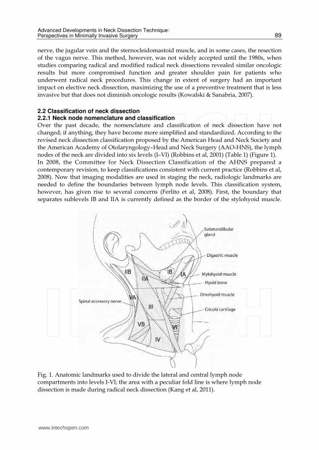
2.2 Classification of neck dissection 2.2.1 Neck node nomenclature and classification Over the past decade, the nomenclature and classification of neck dissection have not changed; if anything, they have become more simplified and standardized. According to the revised neck dissection classification proposed by the American Head and Neck Society and the American Academy of Otolaryngology–Head and Neck Surgery (AAO-HNS), the lymph nodes of the neck are divided into six levels (I–VI) (Robbins et al, 2001) (Table 1) (Figure 1). In 2008, the Committee for Neck Dissection Classification of the AHNS prepared a contemporary revision, to keep classifications consistent with current practice (Robbins et al, 2008). Now that imaging modalities are used in staging the neck, radiologic landmarks are needed to define the boundaries between lymph node levels. This classification system, however, has given rise to several concerns (Ferlito et al, 2008). First, the boundary that separates sublevels IB and IIA is currently defined as the border of the stylohyoid muscle.
Fig. 1. Anatomic landmarks used to divide the lateral and central lymph node compartments into levels I-VI; the area with a peculiar fold line is where lymph node dissection is made during radical neck dissection (Kang et al, 2011).
www.intechopen.com
Neck Dissection – Clinical Application and Recent Advances
90
Neck node level
Superior boundary
Inferior boundary
Anteromedial boundary
Posterolateral boundary
IA (submental)
Symphysis of mandible
Body of hyoid Anterior belly of
contralateral digastric muscle
Anterior belly of ipsilateral digastric
muscle
IB (submandib
ular)
Body of mandible
Posterior belly of digastric muscle
Anterior belly of digastric muscle
Stylohyoid muscle
IIA (upper jugular)
Skull base
Horizontal plane defined by the
inferior body of the hyoid bone
Stylohyoid muscle
Vertical plane defined by the
spinal accessory nerve
IIB (upper jugular)
Skull base
Horizontal plane defined by the
inferior body of the hyoid bone
Vertical plane defined by the
spinal accessory nerve
Lateral border of the
sternocleidomastoid muscle
III (middle jugular)
Horizontal plane defined by the inferior
body of the hyoid bone
Horizontal plane defined by the
inferior border of the cricoid cartilage
Lateral border of the sternohyoid
muscle
Lateral border of the
sternocleidomastoid muscle
IV (lower jugular)
Horizontal plane defined by the inferior border of the
cricoid cartilage
Clavicle Lateral border of the sternohyoid
muscle
Lateral border of the
sternocleidomastoid muscle
VA (posterior triangle)
Apex of convergence of
the sternocleidomas
toid and trapezius muscle
Horizontal plane defined by the
inferior border of the cricoid cartilage
Posterior border of the
sternocleidomastoid muscle
Anterior border of the trapezius
muscle
VB (posterior triangle)
Horizontal plane defined by the inferior border of the
cricoid cartilage
Clavicle
Posterior border of the
sternocleidomastoid muscle
Anterior border of the trapezius
muscle
VI (anterior compartme
nt) Hyoid bone Suprasternal
Common carotid artery
Common carotid artery
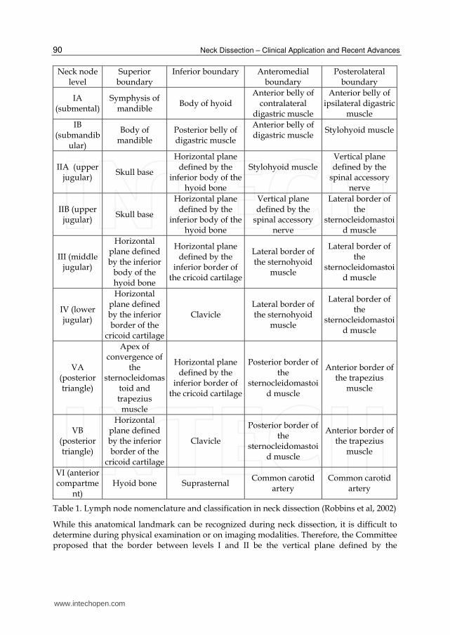
Table 1. Lymph node nomenclature and classification in neck dissection (Robbins et al, 2002)
While this anatomical landmark can be recognized during neck dissection, it is difficult to determine during physical examination or on imaging modalities. Therefore, the Committee proposed that the border between levels I and II be the vertical plane defined by the
www.intechopen.com
Advanced Developments in Neck Dissection Technique: Perspectives in Minimally Invasive Surgery
91
posterior edge of the submandibular gland, that the boundary between levels II and III be the hyoid bone, and the boundary between levels III and IV be the cricoid cartilage. Level VI is currently defined as lying below the body of the hyoid bone, above the top of the manubrium, and between the lateral borders of the sternohyoid muscles, which separate level VI from levels II and III. However, the sternohyoid muscles are not easy to define radiologically. Thus, the Committee proposed that the lateral borders of level VI be defined as the inner margins of the carotid arteries, which in most patients can be easily palpated as well as viewed radiologically (Ferlito et al, 2008). According to the neck node classification of the AHNS, both lymph nodes of the superior mediastinum (often referred to as level VII) and lymph nodes outside the neck groupings (i.e. the retropharyngeal, periparotid, and buccinator nodes) are not included in this classification, but are designated by their specific group. Although the superior mediastinal lymph nodes have been referred to as ろlevel VII,わ the Neck Dissection Classification
Committee of the AHNS does not recommend use of this term, as it defines a region outside the typical boundaries of the neck. The Committee has sought to prevent the establishment of new levels defining other lymph node groups, thus avoiding a more complex numbering system (Ferlito et al, 2008). However, the term level VII continues to be employed in many publications to represent the lymph nodes in the superior mediastinal group. Thus, the new Committee recommends that level VII refer to the extension of the chain of paratracheal nodes below the suprasternal notch (the dividing line between levels VI and VII) to the level of the innominate artery only. As an alternative to naming this group level VII, these nodes may be designated as ろthe superior mediastinal lymph nodes, above the level of the
innominate artery.わ This level is defined by the sternal notch superiorly and the
innominate artery inferiorly, landmarks that are readily identifiable on imaging modalities. The Committee noted that nodes in level VII are usually accessible through the cervical incision. Mediastinal lymph nodes inferior to the innominate artery require sternotomy for access, and are not included in level VII (Robbins et al, 2008).
2.2.2 Neck dissection classification The updated American Joint Committee on Cancer (AJCC) staging system has highlighted
the significance of the biology of lymph node metastases and has refined selective neck
dissection procedures by correlating surgical with radiologic landmarks, thus facilitating
multidisciplinary cooperation among surgeons, radiologists and oncologists. The currently
employed definitions of neck dissection terminology and definitions and indications for
types of selective node dissection are shown in Table 2 (Ferlito et al, 2009).
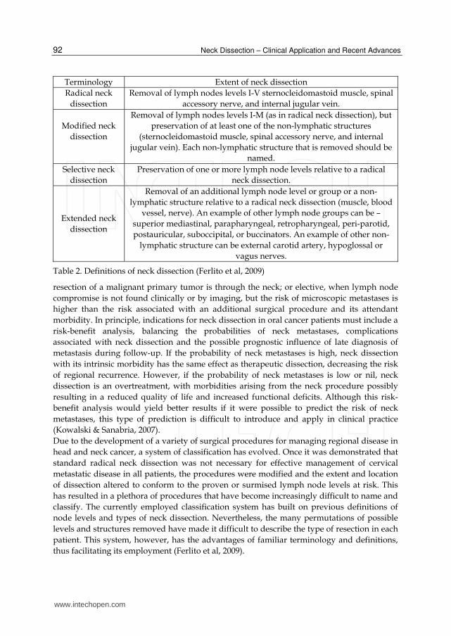
Several types of neck dissection have been described. Radical neck dissection consists of
levels I–V with the associated sternocleidomastoid muscle, jugular vein and spinal accessory
nerve. Modified radical neck dissection consists of levels I–V without any of the
aforementioned non-lymphatic structures. Selective neck dissection consists of any
dissection that excludes one or more lymph node levels included in a radical neck dissection
(i.e. levels II–IV). Extended neck dissection includes one or more additional lymph node
groups or nonlymphatic structures in addition to those of a radical neck dissection,
including periparotid lymph nodes and parotidectomy or superior mediastinal nodes and
level VI.
The purpose of neck dissection may be therapeutic, to treat lymph node metastases found during a physical or imaging examination; opportune, when the approach for exposure and
www.intechopen.com
Neck Dissection – Clinical Application and Recent Advances
92
Terminology Extent of neck dissection
Radical neck
dissection
Removal of lymph nodes levels I-V sternocleidomastoid muscle, spinal
accessory nerve, and internal jugular vein.
Modified neck
dissection
Removal of lymph nodes levels I-M (as in radical neck dissection), but
preservation of at least one of the non-lymphatic structures
(sternocleidomastoid muscle, spinal accessory nerve, and internal
jugular vein). Each non-lymphatic structure that is removed should be
named.
Selective neck
dissection
Preservation of one or more lymph node levels relative to a radical
neck dissection.
Extended neck
dissection
Removal of an additional lymph node level or group or a non-
lymphatic structure relative to a radical neck dissection (muscle, blood
vessel, nerve). An example of other lymph node groups can be –
superior mediastinal, parapharyngeal, retropharyngeal, peri-parotid,
postauricular, suboccipital, or buccinators. An example of other non-
lymphatic structure can be external carotid artery, hypoglossal or
vagus nerves.
Table 2. Definitions of neck dissection (Ferlito et al, 2009)
resection of a malignant primary tumor is through the neck; or elective, when lymph node
compromise is not found clinically or by imaging, but the risk of microscopic metastases is
higher than the risk associated with an additional surgical procedure and its attendant
morbidity. In principle, indications for neck dissection in oral cancer patients must include a
risk-benefit analysis, balancing the probabilities of neck metastases, complications
associated with neck dissection and the possible prognostic influence of late diagnosis of
metastasis during follow-up. If the probability of neck metastases is high, neck dissection
with its intrinsic morbidity has the same effect as therapeutic dissection, decreasing the risk
of regional recurrence. However, if the probability of neck metastases is low or nil, neck
dissection is an overtreatment, with morbidities arising from the neck procedure possibly
resulting in a reduced quality of life and increased functional deficits. Although this risk-
benefit analysis would yield better results if it were possible to predict the risk of neck
metastases, this type of prediction is difficult to introduce and apply in clinical practice
(Kowalski & Sanabria, 2007).
Due to the development of a variety of surgical procedures for managing regional disease in
head and neck cancer, a system of classification has evolved. Once it was demonstrated that
standard radical neck dissection was not necessary for effective management of cervical
metastatic disease in all patients, the procedures were modified and the extent and location
of dissection altered to conform to the proven or surmised lymph node levels at risk. This
has resulted in a plethora of procedures that have become increasingly difficult to name and
classify. The currently employed classification system has built on previous definitions of
node levels and types of neck dissection. Nevertheless, the many permutations of possible
levels and structures removed have made it difficult to describe the type of resection in each
patient. This system, however, has the advantages of familiar terminology and definitions,
thus facilitating its employment (Ferlito et al, 2009).
www.intechopen.com
Advanced Developments in Neck Dissection Technique: Perspectives in Minimally Invasive Surgery
93
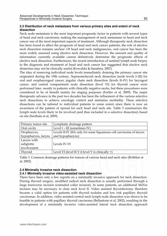
2.3 Distribution of neck metastasis from various primary sites and extent of neck dissection Neck node metastasis is the most important prognostic factor in patients with several types of head and neck carcinoma, making the management of neck metastases in head and neck cancer one of the most important aspects of treatment. Although therapeutic neck dissection has been found to affect the prognosis of head and neck cancer patients, the role of elective neck dissection remains unclear. Of head and neck malignancies, oral cancer has been the most widely assessed using elective neck dissection. However, the amount and quality of information currently available cannot definitively determine the prognostic effects of elective neck dissection. Furthermore, the recent introduction of sentinel lymph node biopsy in the diagnosis and treatment of head and neck cancer has suggested that elective neck dissection may not be clinically useful (Kowalski & Sanabria, 2007). The idea of removing individual node levels immediately draining the primary cancer site
originated during the 19th century. Supraomohyoid neck dissection (node levels I–III) for
oral and oropharyngeal cancer, jugular chain neck dissection (levels II–IV) for laryngeal
cancer, and central compartment node dissection (level VI) for thyroid cancer were
performed later, mostly in patients with clinically negative necks, but these procedures were
considered to be of benefit mainly for staging purposes (Ferlito et al, 2009). The major
therapeutic advance in the past two decades has been the refinement of the various selective
neck dissections to achieve oncologic control and minimize morbidity. These selective
dissections can be tailored to individual patients to some extent since there is now an
awareness of the pattern of spread for each head and neck site. Table 3 summarizes the
lymph node levels likely to be involved (and thus included in a selective dissection) based
on site (Seethala et al, 2009).
Primary tumor site Lymphatic drainage pattern
Oral cavity Level I – III (sometimes IV)
Oropharynx, hypopharynx, larynx
Levels II-IV (IIA only for some Squamous cell carcinoma of larynx and hypopharynx)
Larynx with subglottic involvement
Levels IV-VI
Thyroid Level VI (level II-V if level V is clinically +)
Table 3. Common drainage patterns for tumors of various head and neck sites (Robbins et al, 2002)
2.4 Minimally invasive neck dissection 2.4.1 Minimally invasive video-assisted neck dissection There have been only a few reports on a minimally invasive approach for neck dissection. During thyroid surgery, modified radical neck dissection is usually performed through a large transverse incision (extended collar incision). In some patients, an additional McFee incision may be necessary to clear neck level II. Video assisted thyroidectomy therefore became a valid option for patients with thyroid nodules and low risk papillary thyroid carcinomas. In addition, video assisted central neck lymph node dissection was shown to be feasible in patients with papillary thyroid carcinoma (Bellantone et al, 2002), resulting in the development of a minimally invasive video-assisted lateral neck dissection approach
www.intechopen.com
Neck Dissection – Clinical Application and Recent Advances
94
(VALNED) (Lombardi et al, 2007). This type of surgery begins by making a 4 cm cervical incision between the cricoid cartilage and sternal notch. A 30° endoscope (5 mm) is used for better vision and the operating field is exposed by retractors. Under visual control the neck dissection is performed with conventional instruments, although use of a harmonic scalpel is preferred. The mean number of nodes removed per side was 25. The cosmetic results of the 4 cm horizontal incision were superior to those of conventional approaches. Although VALNED is a safe and feasible technique, additional studies are needed to show that the completeness of resection is similar to that of conventional open approaches.
2.4.2 Endoscopic neck dissection The outcomes of minimally invasive video assisted thyroidectomies have suggested that endoscopic techniques have advantages for other types of head and neck surgery. The relatively longer operation time using this approach is likely due to the narrower operative field and the presence of many vital structures in the neck. Although endoscopic operations were initially limited to regions with natural cavities such as the peritoneum and pleura, the use of endoscopic approaches for head and neck surgery has extended their indications to regions without a natural cavity. All validated methods try to reduce the extent of surgical trauma and its associated morbidity (Muenscher et al, 2011). The main reasons for the development of endoscopic neck surgery are the unpredictable risks of unsatisfactory cosmetic results. For patients with benign neck lesions, this would mean replacing one deformity with another. Further, use of endoscopic methods results in faster wound healing and reduced morbidity due to complications. Ten endoscopic neck dissections on five human cadavers showed that the majority of neck
lymph nodes could be removed by this approach (Dulguerov et al, 2001). Endoscopic
selective neck dissection has been utilized in a porcine model (Terris et al, 2003), and
endoscopic neck surgery with lymph node dissection has been performed on patients with
thyroid neoplasms (Kitagawa et al, 2003; Miccoli & Materazzi, 2004). Gasless skin lifting
techniques, approaching lateral neck levels during thyroidectomy, have also been
performed (Kitagawa et al, 2003). The results of endoscopic lymph node excisions in
patients with squamous cell carcinomas of the upper aerodigestive tract located at different
sites (uvula, epiglottis and glottis), as well as those of endoscopic sentinel
lymphadenectomy for diagnosis of the N0 neck, were presented in 2004 (Werner et al, 2004).
It is unclear whether the N0 neck in surgically treated head and neck carcinomas should be
accessed by neck dissection or regular clinical follow up, although an endoscopic approach
may be an alternative to tracer uptake by sentinel lymph nodes. A small skin incision chosen
for endoscopy may be extended for standard neck dissection. In this method, a rigid
endoscope is introduced through a specially designed tube, allowing the labeled lymph
node to be dissected after removing subcutaneous adipose tissue. The sentinel node concept
combines endoscopic lymph node dissection with frozen section analysis to explore the N0
neck. Alternatively, an approach called stealth surgery can be used for transaxillary
subcutaneous endoscopic excision of benign neck lesions (Dutta et al, 2008). This endoscopic
method may reduce the degree of invasiveness frequently associated with sentinel
lymphadenectomy. A recent editorial concluded that ‘‘It will take a lot of work before we
know if endoscopic neck dissection is a good, oncologic operation, but the trip to learn such
a truth should be interesting’’ (Richtsmeier, 2003). At present, however, this procedure has
not achieved widespread acceptance in clinical practice (Ferlito et al, 2006).
www.intechopen.com
Advanced Developments in Neck Dissection Technique: Perspectives in Minimally Invasive Surgery
95
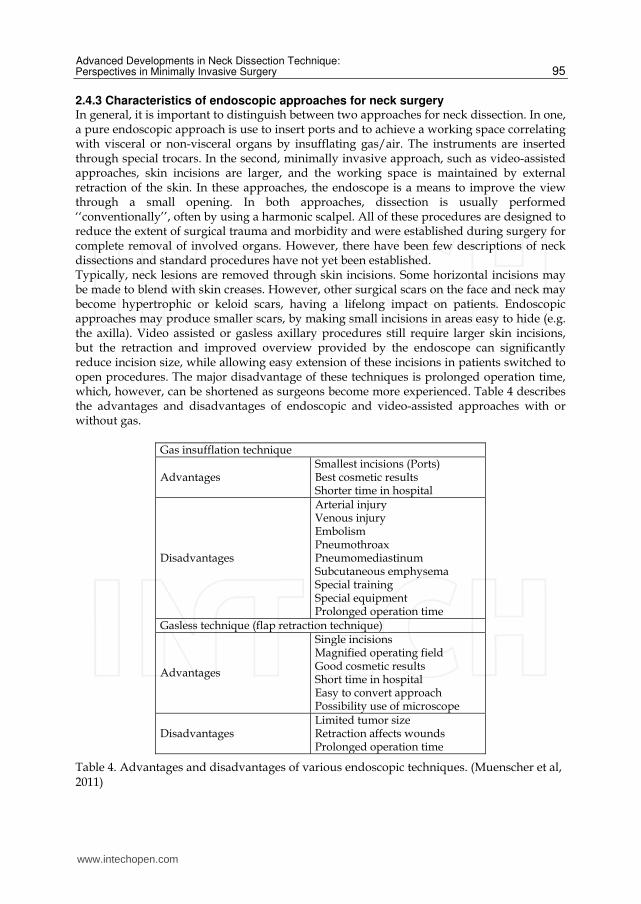
2.4.3 Characteristics of endoscopic approaches for neck surgery In general, it is important to distinguish between two approaches for neck dissection. In one, a pure endoscopic approach is use to insert ports and to achieve a working space correlating with visceral or non-visceral organs by insufflating gas/air. The instruments are inserted through special trocars. In the second, minimally invasive approach, such as video-assisted approaches, skin incisions are larger, and the working space is maintained by external retraction of the skin. In these approaches, the endoscope is a means to improve the view through a small opening. In both approaches, dissection is usually performed ‘‘conventionally’’, often by using a harmonic scalpel. All of these procedures are designed to reduce the extent of surgical trauma and morbidity and were established during surgery for complete removal of involved organs. However, there have been few descriptions of neck dissections and standard procedures have not yet been established. Typically, neck lesions are removed through skin incisions. Some horizontal incisions may be made to blend with skin creases. However, other surgical scars on the face and neck may become hypertrophic or keloid scars, having a lifelong impact on patients. Endoscopic approaches may produce smaller scars, by making small incisions in areas easy to hide (e.g. the axilla). Video assisted or gasless axillary procedures still require larger skin incisions, but the retraction and improved overview provided by the endoscope can significantly reduce incision size, while allowing easy extension of these incisions in patients switched to open procedures. The major disadvantage of these techniques is prolonged operation time, which, however, can be shortened as surgeons become more experienced. Table 4 describes the advantages and disadvantages of endoscopic and video-assisted approaches with or without gas.
Gas insufflation technique
Advantages Smallest incisions (Ports)Best cosmetic results Shorter time in hospital
Disadvantages
Arterial injuryVenous injury Embolism Pneumothroax Pneumomediastinum Subcutaneous emphysema Special training Special equipment Prolonged operation time
Gasless technique (flap retraction technique)
Advantages
Single incisionsMagnified operating field Good cosmetic results Short time in hospital Easy to convert approach Possibility use of microscope
Disadvantages Limited tumor sizeRetraction affects wounds Prolonged operation time
Table 4. Advantages and disadvantages of various endoscopic techniques. (Muenscher et al, 2011)
www.intechopen.com
Neck Dissection – Clinical Application and Recent Advances
96
Furthermore, endoscopic and minimally invasive/video assisted dissections require special instruments and are more costly and time consuming. Although complication rates are low after endoscopic neck surgery, several morbidities, such as injuries to arteries and veins, embolism, subcutaneous emphysema, pneumothorax and pneumomediastinum, were described in early reports on the use of these methods in thyroid surgery. Many of these complications, however, may have been due to gas insufflation to enhance working space. Moreover, as in any other type of endoscopic surgery, common surgical complications, such as nerve injury and wound infection, can occur. These complications depend on patient selection, especially since indications for minimally invasive approaches have not been determined. Conversion to open procedures is common in oncologic settings such as proven N+ status in patients with head and neck carcinomas. To date, there have been no prospective randomized clinical trials comparing open with endoscopic or video assisted surgery, especially regarding the extent of resection. Minimally invasive approaches are advantageous for patients with benign neck lesions, thyroid disease, and selective/sentinel lymph node dissections, due to better cosmetic results and shorter wound healing times. Surgeons tend to favor video assisted minimally invasive techniques or endoscopic surgery using a gasless transaxillary approach, creating the working space by retraction, because the gas filling procedures, especially at level IV, bear some risks (Muenscher et al, 2011).
2.5 Robot technique for head and neck cancer The endoscopic technique represents a considerable technologic advance and has recently
been applied to head and neck surgery. Several trials of endoscopic neck surgery plus
radical node dissection in patients with head and neck as well as thyroid cancer have shown
that the endoscopic approach to neck dissection eliminates the long cervical scar.
Furthermore, to overcome displeasing cosmetic outcomes, several endoscopic approaches to
neck dissection have been conducted using remote skin incision. However, endoscopic
surgery is more demanding and requires more time than open surgery, primarily because of
instrumental and anatomical limitations. The instruments used to perform these minimally
invasive endoscopic surgeries have definite limitations such as a 2-dimensional flat monitor,
rigid and straight endoscopic instruments, and no tactile sense. Endoscopic surgery is
particularly problematic for complex and difficult procedures such as radical neck
dissection for head and neck cancer, in keeping with the principles of oncologic safety. The
da Vinci surgical robot system (Intuitive Surgical, Sunnyvale, CA, USA) was developed to
overcome these limitations (Chung et al, 2011). This surgical robot system promises more
precise, improved endoscopic techniques and enables compartment-oriented anatomical
neck dissection. Moreover, the robotic technique for minimally invasive surgery has other
advantages, including the increased dexterity of the instrumentation used. Use of the robot
system in head and neck surgery eliminates some of the technical pitfalls and limitations of
endoscopic surgery. Furthermore, advances in robotic techniques, such as a steady camera
platform, a 3-dimensional magnified operative view, 7 degrees of freedom, scaled and
tremor-filtered movements, and a multi-articulated endo-wrist, allow precise and complex
endoscopic procedures to be performed. Accordingly, the meticulous and precise motions of
modern robotic instruments have introduced new levels of technical safety and feasibility to
robotic thyroidectomy.
We recently described 33 patients who underwent robotic modified radical neck dissection using a gasless transaxillary approach, and provided details of operative techniques and
www.intechopen.com
Advanced Developments in Neck Dissection Technique: Perspectives in Minimally Invasive Surgery
97
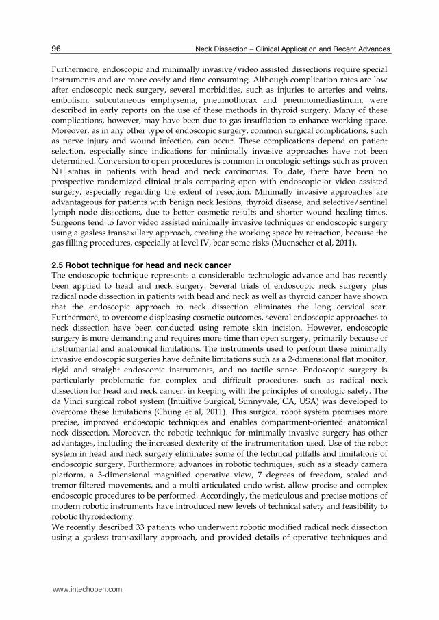
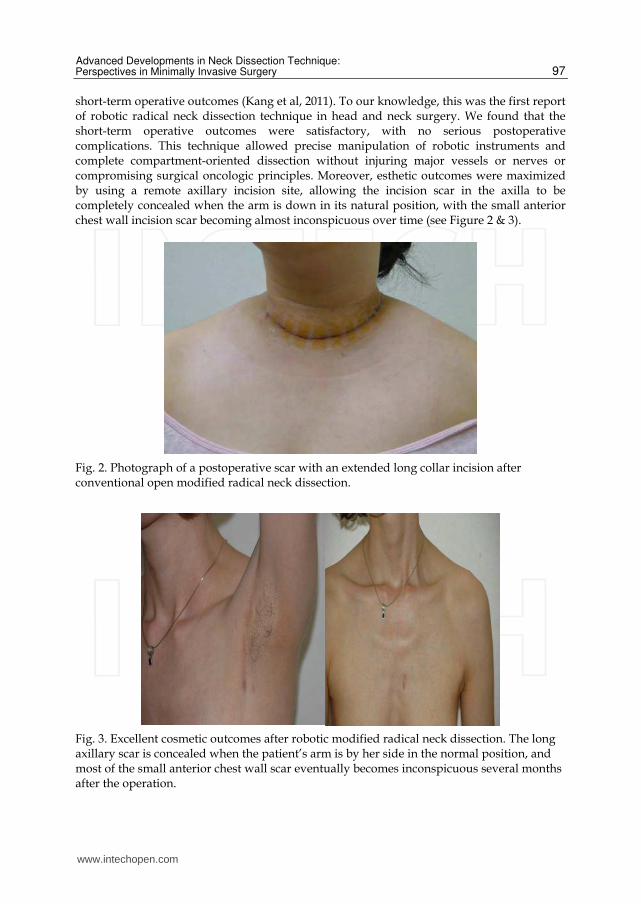
short-term operative outcomes (Kang et al, 2011). To our knowledge, this was the first report of robotic radical neck dissection technique in head and neck surgery. We found that the short-term operative outcomes were satisfactory, with no serious postoperative complications. This technique allowed precise manipulation of robotic instruments and complete compartment-oriented dissection without injuring major vessels or nerves or compromising surgical oncologic principles. Moreover, esthetic outcomes were maximized by using a remote axillary incision site, allowing the incision scar in the axilla to be completely concealed when the arm is down in its natural position, with the small anterior chest wall incision scar becoming almost inconspicuous over time (see Figure 2 & 3).
Fig. 2. Photograph of a postoperative scar with an extended long collar incision after conventional open modified radical neck dissection.
Fig. 3. Excellent cosmetic outcomes after robotic modified radical neck dissection. The long axillary scar is concealed when the patient’s arm is by her side in the normal position, and most of the small anterior chest wall scar eventually becomes inconspicuous several months after the operation.
www.intechopen.com
Neck Dissection – Clinical Application and Recent Advances
98
However, robotic neck dissection for patients with head-and-neck cancer remains at an early stage, and many unanswered questions remain; the benefits afforded by the technique require further evaluation.
2.6 Robotic neck dissection technique In robotic modified radical neck dissection technique, the complete anatomical neck lymph
node dissection, matching that of the open method, was found to be possible using excellent
robotic instruments, such as magnified and 3-dimensional operative field, a stable camera
platform, multi-articulated and tremor filtering system, and three accessible robotic arms.
We briefly introduced our robotic modified radical neck dissection technique (Kang et al,
2011).
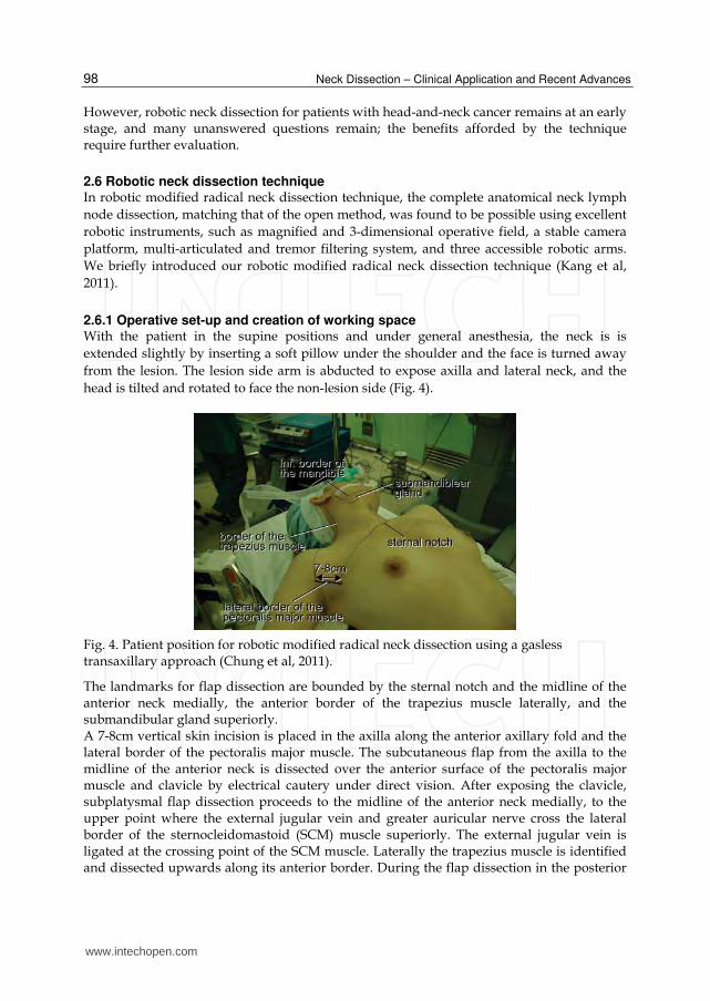
2.6.1 Operative set-up and creation of working space With the patient in the supine positions and under general anesthesia, the neck is is
extended slightly by inserting a soft pillow under the shoulder and the face is turned away
from the lesion. The lesion side arm is abducted to expose axilla and lateral neck, and the
head is tilted and rotated to face the non-lesion side (Fig. 4).
Fig. 4. Patient position for robotic modified radical neck dissection using a gasless transaxillary approach (Chung et al, 2011).
The landmarks for flap dissection are bounded by the sternal notch and the midline of the anterior neck medially, the anterior border of the trapezius muscle laterally, and the submandibular gland superiorly. A 7-8cm vertical skin incision is placed in the axilla along the anterior axillary fold and the lateral border of the pectoralis major muscle. The subcutaneous flap from the axilla to the midline of the anterior neck is dissected over the anterior surface of the pectoralis major muscle and clavicle by electrical cautery under direct vision. After exposing the clavicle, subplatysmal flap dissection proceeds to the midline of the anterior neck medially, to the upper point where the external jugular vein and greater auricular nerve cross the lateral border of the sternocleidomastoid (SCM) muscle superiorly. The external jugular vein is ligated at the crossing point of the SCM muscle. Laterally the trapezius muscle is identified and dissected upwards along its anterior border. During the flap dissection in the posterior
www.intechopen.com
Advanced Developments in Neck Dissection Technique: Perspectives in Minimally Invasive Surgery
99
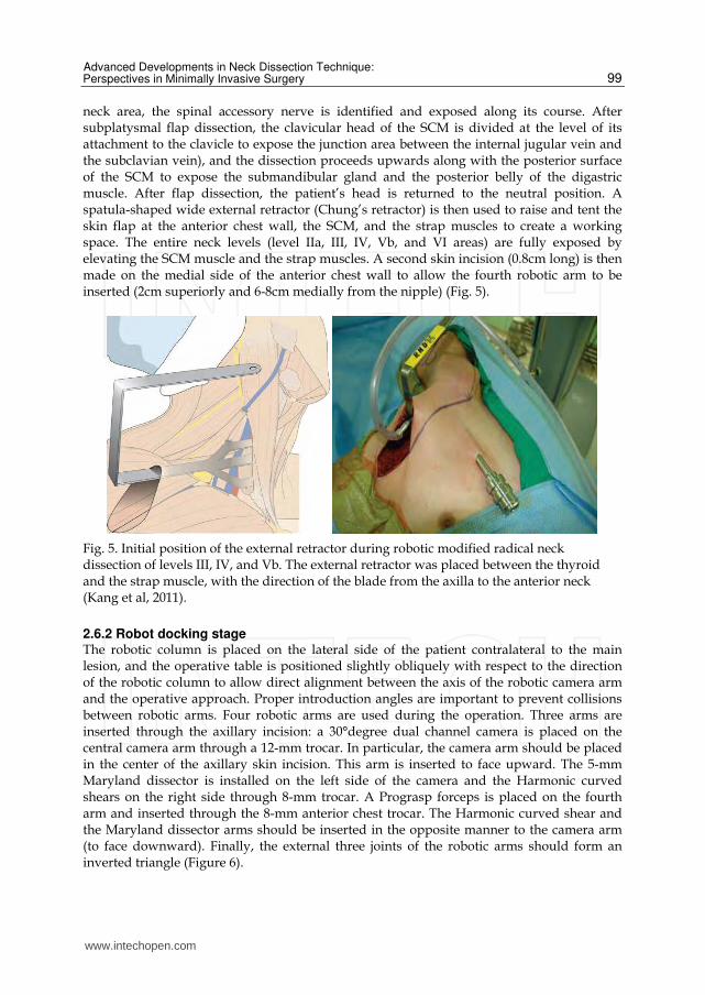
neck area, the spinal accessory nerve is identified and exposed along its course. After subplatysmal flap dissection, the clavicular head of the SCM is divided at the level of its attachment to the clavicle to expose the junction area between the internal jugular vein and the subclavian vein), and the dissection proceeds upwards along with the posterior surface of the SCM to expose the submandibular gland and the posterior belly of the digastric muscle. After flap dissection, the patient’s head is returned to the neutral position. A spatula-shaped wide external retractor (Chung’s retractor) is then used to raise and tent the skin flap at the anterior chest wall, the SCM, and the strap muscles to create a working space. The entire neck levels (level IIa, III, IV, Vb, and VI areas) are fully exposed by elevating the SCM muscle and the strap muscles. A second skin incision (0.8cm long) is then made on the medial side of the anterior chest wall to allow the fourth robotic arm to be inserted (2cm superiorly and 6-8cm medially from the nipple) (Fig. 5).
Fig. 5. Initial position of the external retractor during robotic modified radical neck dissection of levels III, IV, and Vb. The external retractor was placed between the thyroid and the strap muscle, with the direction of the blade from the axilla to the anterior neck (Kang et al, 2011).
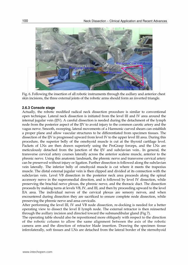
2.6.2 Robot docking stage The robotic column is placed on the lateral side of the patient contralateral to the main lesion, and the operative table is positioned slightly obliquely with respect to the direction of the robotic column to allow direct alignment between the axis of the robotic camera arm and the operative approach. Proper introduction angles are important to prevent collisions between robotic arms. Four robotic arms are used during the operation. Three arms are inserted through the axillary incision: a 30°degree dual channel camera is placed on the central camera arm through a 12-mm trocar. In particular, the camera arm should be placed in the center of the axillary skin incision. This arm is inserted to face upward. The 5-mm Maryland dissector is installed on the left side of the camera and the Harmonic curved shears on the right side through 8-mm trocar. A Prograsp forceps is placed on the fourth arm and inserted through the 8-mm anterior chest trocar. The Harmonic curved shear and the Maryland dissector arms should be inserted in the opposite manner to the camera arm (to face downward). Finally, the external three joints of the robotic arms should form an inverted triangle (Figure 6).
www.intechopen.com
Neck Dissection – Clinical Application and Recent Advances
100
Fig. 6. Following the insertion of all robotic instruments through the axillary and anterior chest skin incisions, the three external joints of the robotic arms should form an inverted triangle.
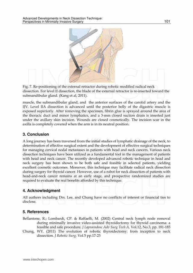
2.6.3 Console stage Actually, the robotic modified radical neck dissection procedure is similar to conventional open technique. Lateral neck dissection is initiated from the level III and IV area around the internal jugular vein (IJV). A careful dissection is needed during the detachment of the lymph node from the posterior aspect of the IJV to avoid injury to the common carotic artery and the vagus nerve. Smooth, sweeping, lateral movements of a Harmonic curved shears can establish a proper plane and allow vascular structures to be differentiated from specimen tissues. The dissection of the IJV is progressed upward from level IV to the upper level III area. During this procedure, the superior belly of the omohyoid muscle is cut at the thyroid cartilage level. Packets of LNs are then drawn superiorly using the ProGrasp forceps, and the LNs are meticulously detached from the junction of the IJV and subclavian vein.. In general, the transverse cervical artery courses laterally across the anterior scalene muscle, anterior to the phrenic nerve. Using this anatomic landmark, the phrenic nerve and transverse cervical artery can be preserved without injury or ligation. Further dissection is followed along the subclavian vein laterally. The inferior belly of omohyoid muscle is cut where it meets the trapezius muscle. The distal external jugular vein is then clipped and divided at its connection with the subclavian vein. Level VB dissection in the posterior neck area proceeds along the spinal accessory nerve in the superomedial direction, and is followed by level IV dissection, while preserving the brachial nerve plexus, the phrenic nerve, and the thoracic duct. The dissection proceeds by making turns at levels VB, IV, and III, and then by proceeding upward to the level IIA area. The individual nerves of the cervical plexus are sensory nerves, and when encountered during dissection they are sacrificed to ensure complete node dissection, while preserving the phrenic nerve and ansa cervicalis. After performing the level III, IV and VB node dissection, re-docking is needed for a better operating view to dissect the level II lymph node. The external retractor is then reinserted through the axillary incision and directed toward the submandibular gland (Fig 7). The operating table should also be repositioned more obliquely with respect to the direction of the robotic column to allow the same alignment between the axis of the robotic camera arm and the direction of retractor blade insertion. Drawing the specimen tissue inferolaterally, soft tissues and LNs are detached from the lateral border of the sternohyoid
www.intechopen.com
Advanced Developments in Neck Dissection Technique: Perspectives in Minimally Invasive Surgery
101
Fig. 7. Re-positioning of the external retractor during robotic modified radical neck dissection. For level II dissection, the blade of the external retractor is re-inserted toward the submandibular gland. (Kang et al, 2011).
muscle, the submandibular gland, and the anterior surfaces of the carotid artery and the IJV. Level IIA dissection is advanced until the posterior belly of the digastric muscle is exposed superiorly. After removing the specimen, fibrin glue is sprayed around the area of the thoracic duct and minor lymphatics, and a 3-mm closed suction drain is inserted just under the axillary skin incision. Wounds are closed cosmetically. The incision scar in the axilla is completely covered when the arm is in its neutral position.
3. Conclusion
A long journey has been traversed from the initial studies of lymphatic drainage of the neck, to determination of effective surgical extent and the development of effective surgical techniques for managing cervical nodal metastases in patients with head and neck cancers. Various neck dissection techniques have been utilized as a fundamental tool in the management of patients with head and neck cancer. The recently developed advanced robotic technique in head and neck surgery has been shown to be both safe and feasible in selected patients, yielding excellent cosmetic outcomes. Moreover, this technique may facilitate radical neck dissection during surgery for thyroid cancer. However, use of a robot for neck dissection of patients with head-and-neck cancer remains at an early stage, and prospective randomized studies are required to evaluate the real benefits afforded by this technique.
4. Acknowledgment
All authors including Drs. Lee, and Chung have no conflicts of interest or financial ties to disclose.
5. References
Bellantone, R.; Lombardi, CP. & Raffaelli, M. (2002) Central neck lymph node removal during minimally invasive video-assisted thyroidectomy for thyroid carcinoma: a feasible and safe procedure. J Laproendosc Adv Surg Tech A, Vol.12, No.3, pp. 181-185
Chung, WY. (2011) The evolution of robotic thyroidectomy: from inception to neck dissection. J Robotic Surg, Vol.5 pp.17-23
www.intechopen.com
Neck Dissection – Clinical Application and Recent Advances
102
Dulquerov, P.; Leuchter, I. & Szalay-Quinodoz, I. (2001) Endoscopic neck dissection in human cadavers. Laryngoscope, Vol.111, No.12, pp.2135-2139
Dutta, S.; Slater, B, & Butler M. (2008) “Stealth surgery”: transaxillary subcutaneous endoscopic excision of benign neck lesions. J Pediatr Surg, Vol.43, No.11, pp.2070-2074
Ferlito, A.; Rinaldo, A. & Siver, CE. (2006) Neck dissection: then and now. Auris Nasus Larynx, Vol.33, No.4, pp.365-374
Ferlito, A.; Siver, CE. & Rinaldo, A. (2008) Neck dissection: present and future? Eur Arch Otorhinolaryngol, Vol.265, No.6, pp.621-626
Ferlito, A; Robbins, KT. & Silver, CE. (2009) Classification of neck dissections: an evolving sytem. Auris Nasus Larynx, Vol.36, No.2, pp.127-134
Kang, SW.; Lee, SH. & Ryu, HR. (2010) Initial experience with robot-assisted modified radical neck dissection for the management of thyroid carcinoma with lateral neck node metastasis. Surgery, Vol.148, No.6, pp.1214-1221
Kazi, RA. (2003) The life and times of George Washinton Crile. J Postgrad Med, Vol.49, pp.289-290
Kitagawa, W.; Shimizu, K. & Akasu, H. (2003) Endoscopic neck surgery with lymph node dissection for papillary carcinoma of the thyroid using a totally gasless anterior neck skin lifting method, J Am Coll Surg, Vol.196, No.6, pp.990-994
Kowalski, LP. & Sanabria, A. (2007) Elective neck dissection in oral carcinoma: a critical review of the evidence. Acta Otorhinolaryngol Ital, Vol.27, No.3, pp.113-117
Lombardi, CP.; Raffaelli, M. & Princi, P. (2007) Minimally invasive video-assisted functional lateral neck dissection for metastatic papillary thyroid carcinoma. Am J Surg, Vol.193, No.1, pp. 114-118
Martin, H; Del Valle, B. & Ehrlich, H. (1951) Neck dissection. Cancer, Vol.4, No.3, pp.441-499 Miccoli, P. & Materazzi G. (2004) Update on endoscopic cervical surgery. Semin Laparosc
Surg, Vol.11, No.3, pp.139-145 Muenscher, A.; Dalchow, C. & Kutta, H. (2011) The endoscopic approach to the neck: a
review of the literature, and overview of the various techniques. Surg Endosc, Vol.25, No.5, pp.1358-1363
Richtsmeier, WJ. (2003) Dissecting the “endoscopic neck”. Arch Otolaryngol Head Neck Surg, Vol.129, No.6, pp.612
Rinaldo, A.; Ferlito, A. & Silver, CE. (2008) Early history of neck dissection. Eur Arch Otorhinolaryngol, Vol.265, No.12, pp.1535-1538
Robbins, KT.; Clayman, G. & Levine, PA. (2002) Neck dissection classification update: revisions proposed by the American Head and Neck Society and the American Academy of Otolaryngology-Head and Neck Surgery. Arch Otolaryngol Head neck Surg, Vol.128, No.7, pp.751-758
Robbins, KT; Shaha, AR. & Medina, JE. (2008) Consensus statement on the classification and terminology of neck dissection. Arch Otolaryngol Head Neck Surg, Vol.134, No.5, pp.536-538
Seethala, RR. (2009) Current state of neck dissection in the United States. Am J Surg Pathol, Vol.34, No.8, pp.1106-1121
Terris, DJ.; Monfared, A. & Thomas, A. (2003) Endoscopic selective neck dissection in a porcine model. Arch Otolaryngol Head Neck Surg, Vol.129, No.6, pp.613-617
Ward, GE. & Robben, JO. (1951) A composite operation for radical neck dissection and removal of cancer of the mouth. Cancer, Vol.4, No.1, pp.98-109
Werner, JA.; Sapundzhiev, NR. & Teymoortash, A. (2004) Endoscopic sentinel lymphadenectomy as a new diagnostic approach in the N0 neck. Eur Arch Otorhinolaryngol, Vol.261, No.9, pp.463-468
www.intechopen.com
Neck Dissection - Clinical Application and Recent AdvancesEdited by Prof. Raja Kummoona
ISBN 978-953-51-0104-8Hard cover, 164 pagesPublisher InTechPublished online 22, February, 2012Published in print edition February, 2012
InTech EuropeUniversity Campus STeP Ri Slavka Krautzeka 83/A 51000 Rijeka, Croatia Phone: +385 (51) 770 447 Fax: +385 (51) 686 166www.intechopen.com
InTech ChinaUnit 405, Office Block, Hotel Equatorial Shanghai No.65, Yan An Road (West), Shanghai, 200040, China
Phone: +86-21-62489820 Fax: +86-21-62489821
Neck Dissection - Clinical Application and Recent Advances is a leading book in neck surgery and representsthe recent work and experiences of a number of top international scientists. The book covers all techniques ofneck dissection and the most recent advances in neck dissection by advocating better access to all techniquesof neck dissection; e.g. Robotic surgery (de Venice) system, a technique for detection of lymph nodemetastasis by ultra sonography and CT scan, and a technique of therapeutic selective neck dissection inmultidisciplinary treatment. This book is essential to any surgeon specializing or practicing neck surgery,including Head Neck Surgeons, Maxillofacial Surgeons, ENT Surgeons, Plastic and Reconstructive Surgeons,Craniofacial Surgeons and also to all postgraduate Medical & Dental candidates in the field.
How to referenceIn order to correctly reference this scholarly work, feel free to copy and paste the following:
Jandee Lee and Woong Youn Chung (2012). Advanced Developments in Neck Dissection Technique:Perspectives in Minimally Invasive Surgery, Neck Dissection - Clinical Application and Recent Advances, Prof.Raja Kummoona (Ed.), ISBN: 978-953-51-0104-8, InTech, Available from:http://www.intechopen.com/books/neck-dissection-clinical-application-and-recent-advances/advanced-developments-in-neck-dissection-technique-perspectives-in-minimally-invasive-surgery