Embed Size (px)
Citation preview
Management of B3 lesions
Pathological viewPathological view
Abeer ShaabanAbeer ShaabanAbeer ShaabanAbeer Shaaban
Queen Elizabeth Hospital Birmingham Queen Elizabeth Hospital Birmingham
B3 lesions
� FEA
� AIDP
� In situ Lobular neoplasia
� Papilloma� Papilloma
� Radial scar
� Fibroaepithelial lesion
� Mucocoele like lesion
� Other: spindle cell lesions, apocrine atypia…
B3 category
• Includes lesions of variable significance.
•Includes lesions with and without atypia.
•Risk of upgrade increases in the presence of atypia.
•Requires further sampling: surgery/other.
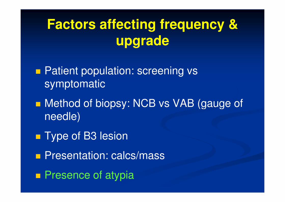
Factors affecting frequency &
upgrade
� Patient population: screening vs symptomatic
� Method of biopsy: NCB vs VAB (gauge of � Method of biopsy: NCB vs VAB (gauge of needle)
� Type of B3 lesion
� Presentation: calcs/mass
� Presence of atypia
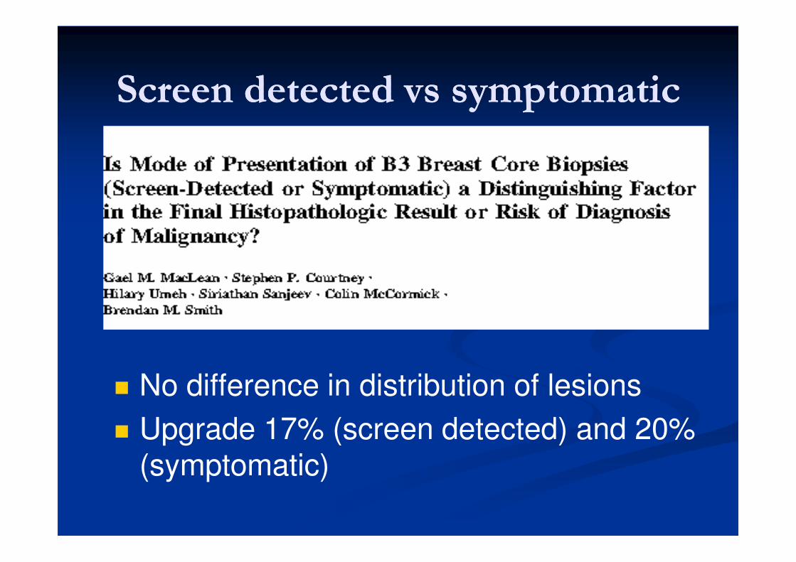
Screen detected Screen detected vsvs symptomaticsymptomatic
� No difference in distribution of lesions
� Upgrade 17% (screen detected) and 20% (symptomatic)
Atypia Atypia vsvs no atypiano atypia
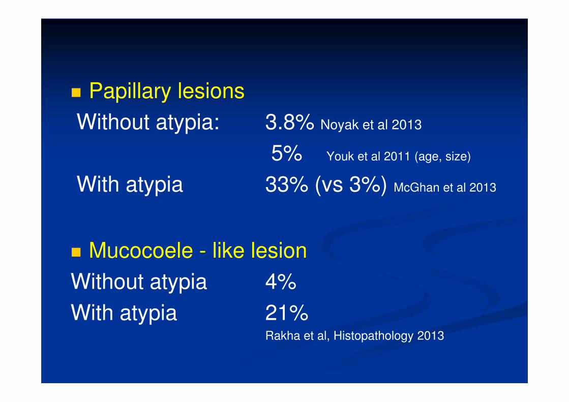
� Papillary lesions
Without atypia: 3.8% Noyak et al 2013
5% Youk et al 2011 (age, size)
With atypia 33% (vs 3%) McGhan et al 2013
� Mucocoele - like lesion
Without atypia 4%
With atypia 21%Rakha et al, Histopathology 2013
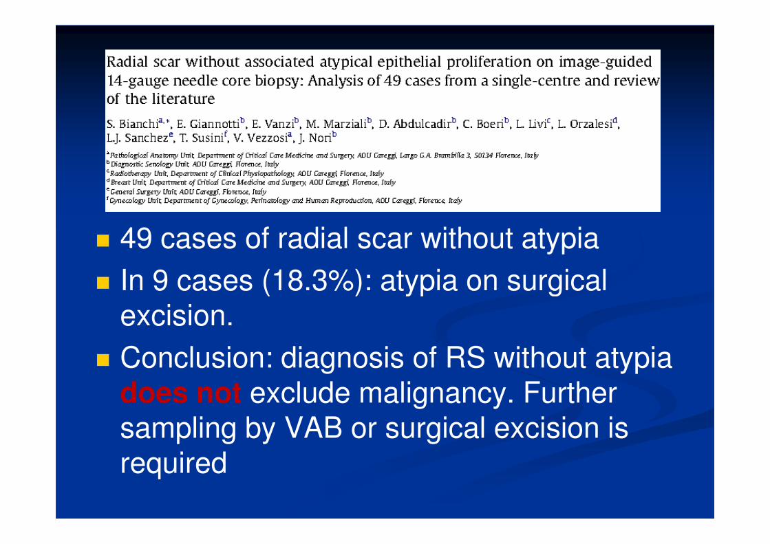
� 49 cases of radial scar without atypia
� In 9 cases (18.3%): atypia on surgical � In 9 cases (18.3%): atypia on surgical excision.
� Conclusion: diagnosis of RS without atypia does not exclude malignancy. Further sampling by VAB or surgical excision is required
Rakha et al 2014
Type of B3 lesion/combination Type of B3 lesion/combination
of lesionsof lesionsof lesionsof lesions
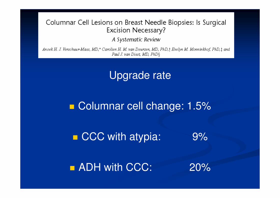
Upgrade rate
� Columnar cell change: 1.5%
� CCC with atypia: 9%
� ADH with CCC: 20%
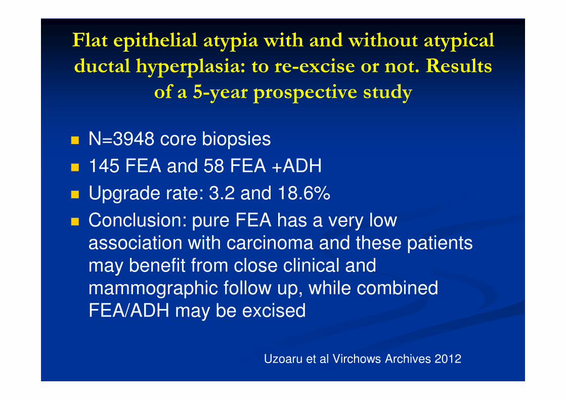
Flat epithelial atypia with and without atypical
ductal hyperplasia: to re-excise or not. Results
of a 5-year prospective study
� N=3948 core biopsies
� 145 FEA and 58 FEA +ADH
� Upgrade rate: 3.2 and 18.6%� Upgrade rate: 3.2 and 18.6%
� Conclusion: pure FEA has a very low
association with carcinoma and these patients
may benefit from close clinical and
mammographic follow up, while combined
FEA/ADH may be excised
Uzoaru et al Virchows Archives 2012
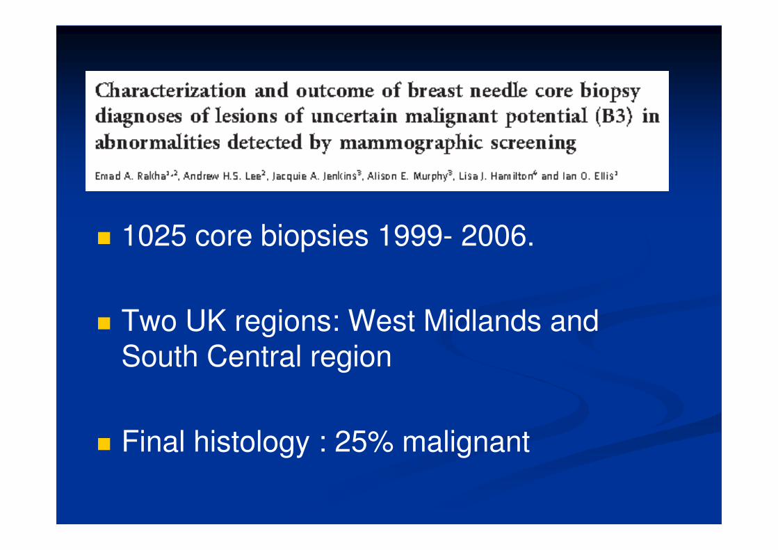
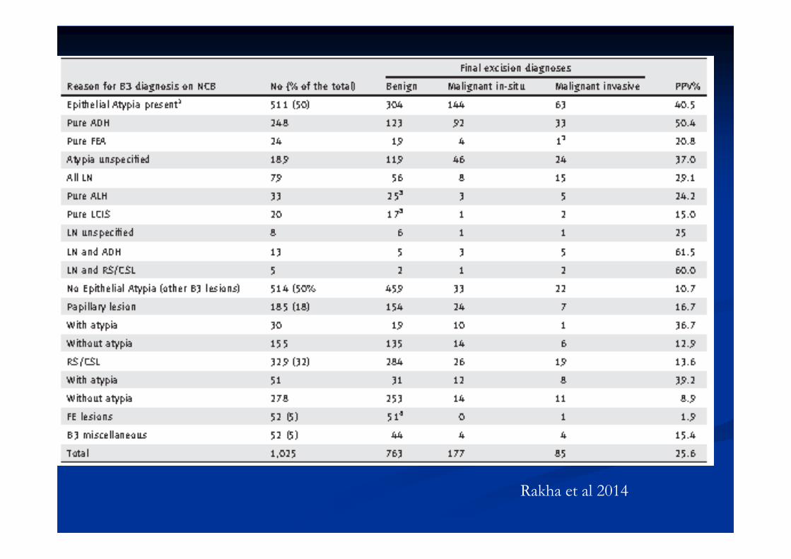
� 1025 core biopsies 1999- 2006.
� Two UK regions: West Midlands and South Central region
� Final histology : 25% malignant
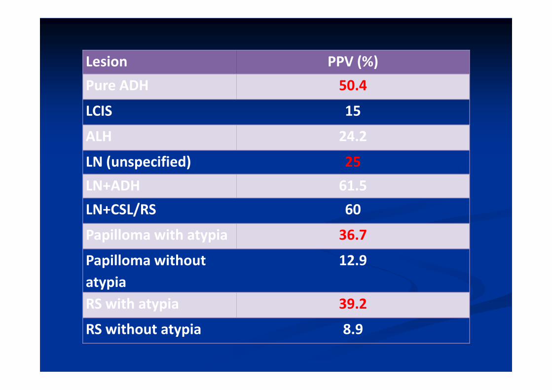
Lesion PPV (%)
Pure ADH 50.4
LCIS 15
ALH 24.2
LN (unspecified) 25
LN+ADH 61.5
LN+CSL/RS 60
Papilloma with atypia 36.7
Papilloma without
atypia
12.9
RS with atypia 39.2
RS without atypia 8.9
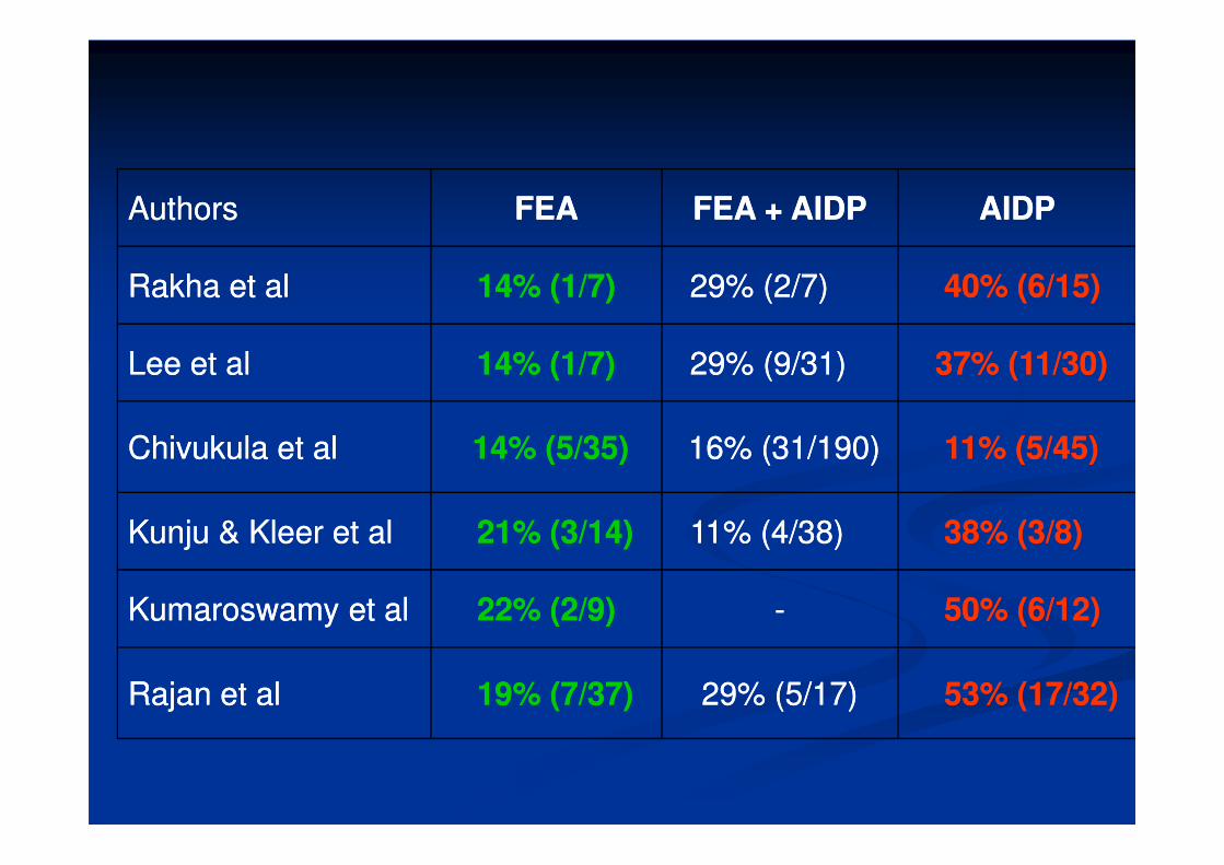
AuthorsAuthors FEAFEA FEA + AIDPFEA + AIDP AIDPAIDP
Rakha et alRakha et al 14% (1/7)14% (1/7) 29% (2/7)29% (2/7) 40% (6/15)40% (6/15)
Lee et alLee et al 14% (1/7)14% (1/7) 29% (9/31)29% (9/31) 37% (11/30)37% (11/30)
Chivukula et alChivukula et al 14% (5/35)14% (5/35) 16% (31/190)16% (31/190) 11% (5/45)11% (5/45)Chivukula et alChivukula et al 14% (5/35)14% (5/35) 16% (31/190)16% (31/190) 11% (5/45)11% (5/45)
Kunju & Kleer et alKunju & Kleer et al 21% (3/14)21% (3/14) 11% (4/38)11% (4/38) 38% (3/8)38% (3/8)
Kumaroswamy et alKumaroswamy et al 22% (2/9)22% (2/9) -- 50% (6/12)50% (6/12)
Rajan et al Rajan et al 19% (7/37)19% (7/37) 29% (5/17)29% (5/17) 53% (17/32)53% (17/32)
Conventional core Conventional core vsvs 11stst line VABline VAB
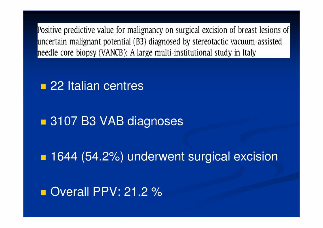
� 22 Italian centres
3107 B3 VAB diagnoses� 3107 B3 VAB diagnoses
� 1644 (54.2%) underwent surgical excision
� Overall PPV: 21.2 %

Lesion PPV (%)
Pure ADH 27.3
FEA 12.7
ALH 24.2
VAB upgrade
ALH 24.2
LIN 22
RS 10.6
All B3 21.2
Are B3 patients being over Are B3 patients being over
treated?treated?
� The majority of patients with B3 diagnosis (75%) will have a benign histology on further sampling.
� Is surgical excision necessary?
� Aim to adequately sample to rule out coexistent cancer
Follow up � Follow up
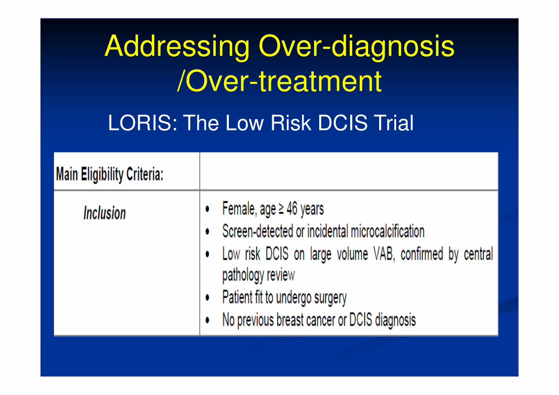
Addressing Over-diagnosis
/Over-treatment
LORIS: The Low Risk DCIS Trial
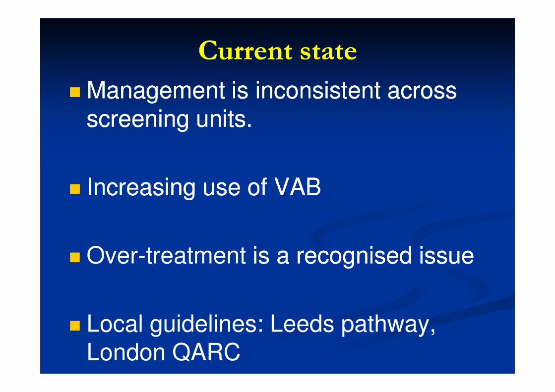
Current stateCurrent state
�� Management is inconsistent across Management is inconsistent across
screening units.screening units.
�� Increasing use of VABIncreasing use of VAB�� Increasing use of VABIncreasing use of VAB
� Over-treatment is a recognised issueis a recognised issue
� Local guidelines: Leeds pathway,
London QARC
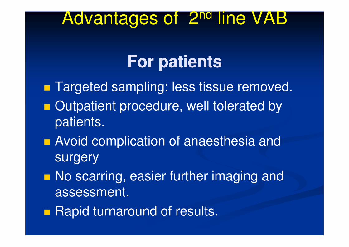
Advantages of 2nd line VAB
For patientsFor patients
� Targeted sampling: less tissue removed.
� Outpatient procedure, well tolerated by patients.patients.
� Avoid complication of anaesthesia and surgery
� No scarring, easier further imaging and assessment.
� Rapid turnaround of results.
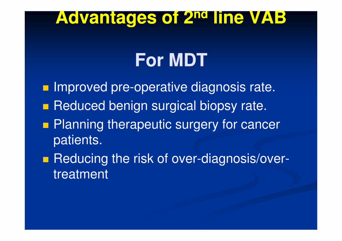
� Improved pre-operative diagnosis rate.
� Reduced benign surgical biopsy rate.
� Planning therapeutic surgery for cancer
Advantages of 2Advantages of 2ndnd line VABline VAB
For MDT For MDT
� Planning therapeutic surgery for cancer patients.
� Reducing the risk of over-diagnosis/over-treatment
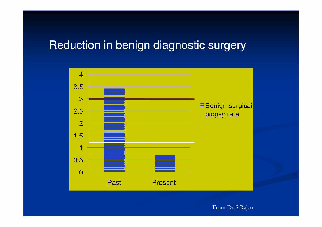
Reduction in benign diagnostic surgeryReduction in benign diagnostic surgery
From Dr S Rajan
B3 Guidelines GroupB3 Guidelines Group
Remit
�� To undertake review of the literature on To undertake review of the literature on lesions categorised as B3lesions categorised as B3lesions categorised as B3lesions categorised as B3
�� To come up with guideline document for a To come up with guideline document for a practical approach to management of practical approach to management of these for the NHS BSP.these for the NHS BSP.
Proposed planplan
� Review the literature on the upgrade rate of each of lesions categorised as B3, of uncertain malignant potential.
� Consider if these upgrade rates are influenced by presentation (screen-detected vs symptomatic vs incidental)

� Explore how radiological and histological diagnostic features may influence management approach.
� Consider any present guideline/publications.
Propose safe and nationally applicable � Propose safe and nationally applicable approaches to the different lesions.
� Write a document for publication through NHS BSP and circulate to ‘Big 18s’ for feedback/ratification.
Composition of the B3 groupComposition of the B3 group
� Chair: Prof Sarah Pinder
� Radiologists: Nisha Sharma, Louise WilkinsonWilkinson
� Surgeons: Simon pain , Desai Anil, Ashu Gandhi
� Pathologists: Sarah Pinder, Andrew Lee, Abeer Shaaban
ApproachApproach
� Management recommendations lesion by lesion.
� Use of diagrams/flow charts� Use of diagrams/flow charts
� General principles: radiological/pathological concordance
� 14g core/first line VAB : diagnostic sampling
Guidance on second line VAB adequate � Guidance on second line VAB adequate sampling
� Advice on follow up
Pathological issues
� Complete excision of lesion by VAB biopsy
(false positive)
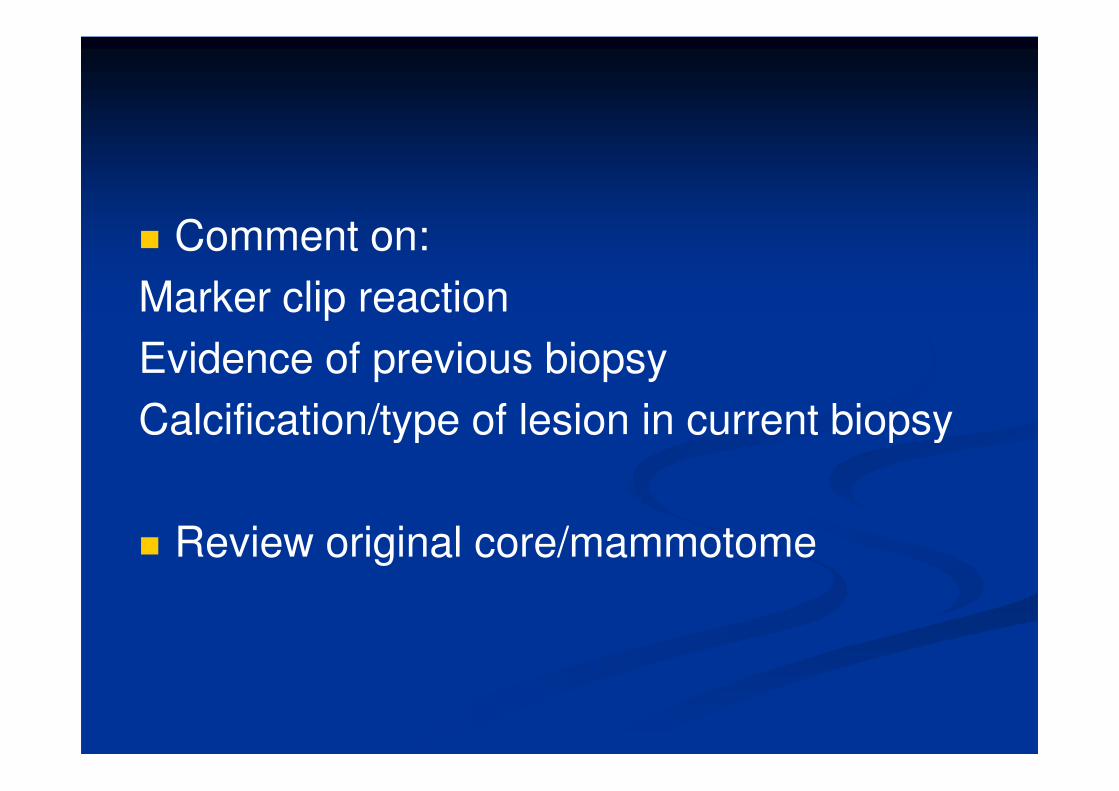
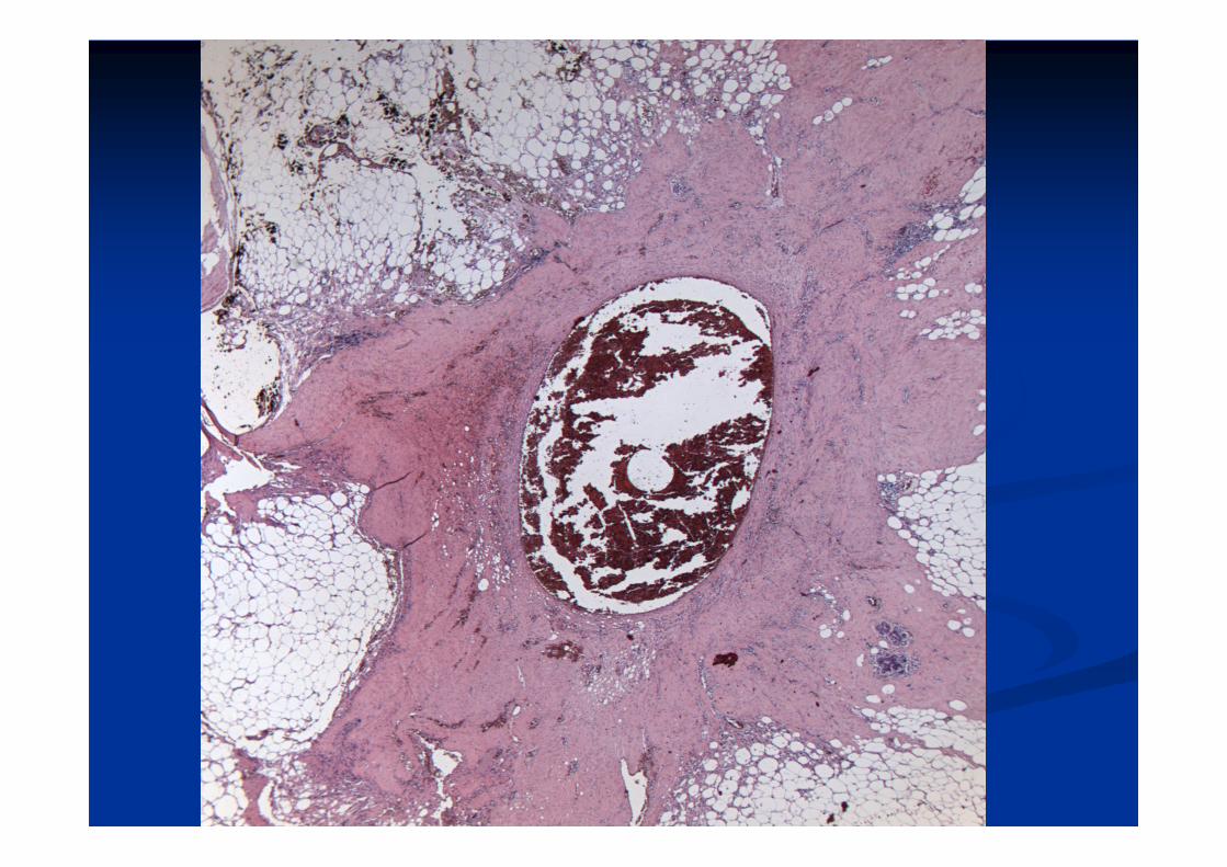
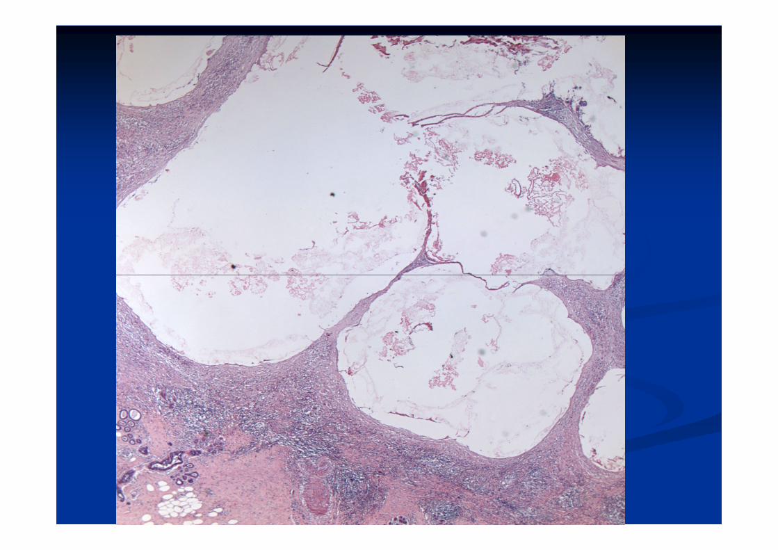
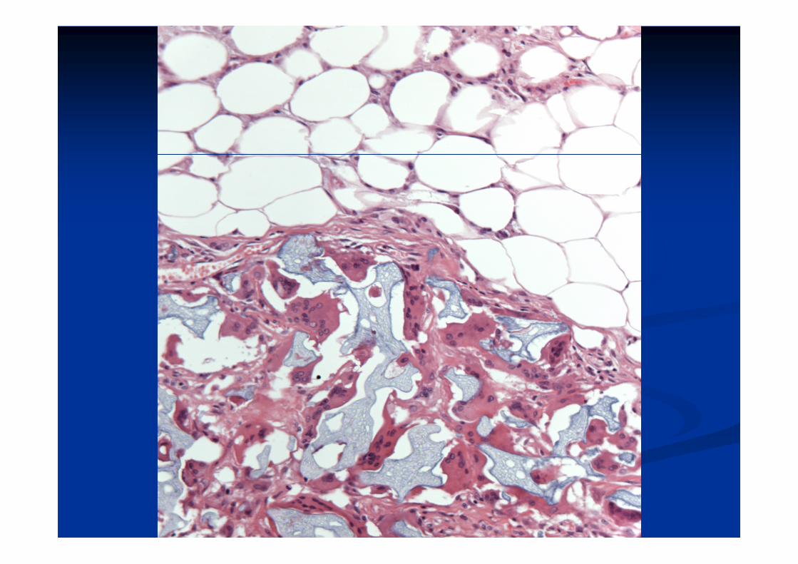
� Comment on:
Marker clip reaction
Evidence of previous biopsy
Calcification/type of lesion in current biopsyCalcification/type of lesion in current biopsy
� Review original core/mammotome
� Sizing of lesion: e.g AIDP
Pragmatic approach
Largest size on any biopsy providedLargest size on any biopsy provided
� Difficult interpretation of some lesions if piecemeal: Papilloma with atypia, fibroepithelial lesions, spindle cell lesions
Consistency in pathological
diagnosis
� Low reproducibility in diagnosing some B3 lesions as FEA, AIDP.lesions as FEA, AIDP.
� Discussion with colleagues, courses
Extensive calcification
� Sampling two ends of the lesion by 1st line VAB
� Follow by 2nd line VAB
Incidental small RS/Incidental small RS/PapillomaPapilloma
�� Lee et al 2011 examined incidental microscopic Lee et al 2011 examined incidental microscopic papillomaspapillomas ((nn=18) and radial scars (=18) and radial scars (nn=17).=17).
�� If no atypia and the lesion is fully represented on If no atypia and the lesion is fully represented on �� If no atypia and the lesion is fully represented on If no atypia and the lesion is fully represented on core/VAB, categorise as B2.core/VAB, categorise as B2.
�� If not sure is completely excised, code as B3 and If not sure is completely excised, code as B3 and discuss at MDT meeting. If confirmed wholly discuss at MDT meeting. If confirmed wholly excised, no further action is neededexcised, no further action is needed
Summary
� Current management of B3 lesions is not uniform and likely to represent over-treatment.
The majority of lesions are benign on � The majority of lesions are benign on excision.
� Radiological-pathological correlation is essential for planning management.
� There is increasing use of VAB for further sampling as alternative to diagnostic surgery.
� Guidelines for B3 management are being developed.


![[b3] Salter David_ Rtf Session b3](https://img.pdfslide.us/doc/110x75/577ce47b1a28abf1038e744e/b3-salter-david-rtf-session-b3.jpg)