Embed Size (px)
Citation preview
REVIEW
Morphological classification and definition of benign,preneoplastic and non-invasive neoplastic lesions of theurinary bladder
R Montironi, A Lopez-Beltran,1 M Scarpelli, R Mazzucchelli & L Cheng2
Section of Pathological Anatomy, School of Medicine, Polytechnic University of the Marche Region (Ancona), Ancona,
Italy, 1Unit of Anatomic Pathology, Cordoba University Medical School, Cordoba, Spain and 2Department of Pathology and
Laboratory Medicine, Indiana University School of Medicine, Indianapolis, IN, USA
Date of submission 4 January 2008Accepted for publication 25 January 2008
Montironi R, Lopez-Beltran A, Scarpelli M, Mazzucchelli R & Cheng L
(2008) Histopathology 53, 621–633
Morphological classification and definition of benign, preneoplastic and non-invasiveneoplastic lesions of the urinary bladder
The morphological classification used in this essay hasbeen based on the most recent World Health Organi-zation (WHO) classification of tumours of the urinarysystem (i.e. 2004 WHO classification). It includesepithelial abnormalities and metaplasias as well asdysplasias and carcinomas in situ. The lesions arebroadly subdivided into two major groups: benign,preneoplastic and non-invasive neoplastic lesions of the
urothelium; and benign, preneoplastic and non-inva-sive neoplastic bladder lesions other than urothelial.Each of these lesions is defined with strict morpholog-ical criteria to provide more accurate information tourologists and oncologists in managing patients. Thereis still debate in the literature as to whether the 2004WHO system should be the only one to be used andwhether the 1973 WHO system should be abandoned.
Keywords: bladder metaplasia, bladder neoplasms, carcinoma in situ, flat urothelial hyperplasia, reactive urothelialatypia, urothelial dysplasia, urothelial papillary carcinoma
Abbreviations: CEA, carcinoembryonic antigen; CIS, carcinoma in situ; CK, cytokeratin; ISUP, International Societyof Urologic Pathology; PUNLMP, papillary urothelial neoplasm of low malignant potential; WHO, World HealthOrganization
Introduction
Bladder cancer is morphologically heterogeneous;>90% of bladder cancer cases are urothelial (transi-tional cell) carcinoma, whereas primary squamouscell carcinoma, adenocarcinoma and other tumours
are less common. Several classifications for the non-invasive neoplasms have been proposed in recentyears.
For over two decades the 1973 World HealthOrganization (WHO) classification of urothelial neo-plasms1 has dominated. In the early 1990s severalfactors emerged that resulted in the need to re-evaluatethis approach:
1. The controversy of calling grade 1 papillarytumours ‘carcinoma’ arose, with several groups ledby William Murphy beginning to call all tumours in thelow-grade end papilloma.2
Address for correspondence: Rodolfo Montironi, Section of Patho-
logical Anatomy and Histopathology, Polytechnic University of the
Marche Region (Ancona), School of Medicine, United Hospitals,
I-60020 Torrette, Ancona, Italy. e-mail: [email protected]
� 2008 The Authors. Journal compilation � 2008 Blackwell Publishing Limited.
Histopathology 2008, 53, 621–633. DOI: 10.1111/j.1365-2559.2008.03025.x

2. The use of intravesical therapy as standardpractice in the treatment of high-risk non-invasivepapillary tumours demanded that high-risk tumours beidentified.
3. The 1973 WHO classification was criticized for theimprecision of the histological criteria, leading manypathologists to create five grade groups (1, 1–2, 2, 2–3and 3). The effect was confusion among clinicians.
In 1998, a system of classifying non-invasive flat andpapillary urothelial neoplasms of the urinary bladderwas proposed by the International Society of UrologicPathology (ISUP) in association with the WHO.3 Thisbecame known as the 1998 ISUP ⁄ WHO classificationsystem. In 2004, this classification system was adoptedin Pathology and genetics of tumours of the urinary systemand male genital organs, one of a series of WHO ‘BlueBooks’ for the classification of tumours.4 This is knownas the 2004 WHO classification.
The aim of this paper is to review the currentapproach to the morphological classification and def-inition of the benign, preneoplastic and non-invasiveneoplastic lesions of the urinary bladder. This reviewhas been based on the 2004 WHO classification.4 Theselesions are subdivided into two major groups:• Benign, preneoplastic and non-invasive neoplasticlesions of the urothelium; and• Benign, preneoplastic and non-invasive neoplasticbladder lesions other than urothelial.
The precise identification of these lesions requiresknowledge of the histology of the normal urotheliumand the range of variations. This serves as referencewhen lesions are evaluated for their type and degree ofalteration.
Normal urothelium
Urothelium is a multilayered epithelium composed ofbasal, intermediate and very large surface cells called‘umbrella cells’ (Figure 1A). The latter may show somedegree of nuclear pleomorphism, which should not bemisconstrued to be dysplastic. The thickness of theurothelium varies with the state of distension of thebladder (two to four cell layers when dilated and five toseven layers when contracted).5
The urothelium of the renal pelvis, urethra and thebladder neck is usually composed of slightly larger cells,which have diminished cytoplasmic clearing and hencemay be misinterpreted as dysplasia.
If the sections are thick, the urothelium may appearhyperchromatic, and this artefact, compounded bytangential sectioning, may result in changes felt torepresent dysplasia. Vagaries of staining and fixationmay also impart hyperchromasia to normal nuclei.6
Benign, preneoplastic and non-invasiveneoplastic lesions of the urothelium
This group of urothelial lesions is subdivided into threemajor subgroups, depending on the relationship withthe surface of the surrounding urothelial mucosa:7
• Flat• Papillary (exophytic)• Endophytic.Each of these three subgroups is further subdividedinto:• Lesions without cytological atypia• Lesions with cytological atypia.
flat les ions
The current classification of flat lesions of the urothe-lium was originally proposed by Amin et al.,8 wassubsequently incorporated into the 1998 ISUP ⁄ WHOand 2004 WHO classifications, and was extensivelycommented on by Lopez-Beltran et al.9
Flat lesions without cytological atypiaFlat hyperplasia. Urothelial hyperplasia is characterizedby markedly thickened mucosa with an increase inthe number of cell layers, usually ‡10. However, it isnot necessary to count the number of cell layers forthe diagnosis. The cells in urothelial hyperplasia donot show any significant cytological abnormalities,although slight nuclear enlargement may be focallypresent.9,10 Morphological evidence of maturation frombase to surface is generally evident (Figure 1B).
When seen as an isolated phenomenon, there is noevidence to suggest that primary urothelial hyperpla-sia has premalignant potential. However, molecularanalysis has shown that this lesion may be clonallyrelated to the papillary tumours in bladder cancerpatients. Flat urothelial hyperplasia has been consid-ered by some authors to be the source of papi-llary neoplasia, usually associated with low-gradetumours.9,10
Flat lesions with cytological atypiaReactive (inflammatory) atypia. In reactive atypia theepithelium may or may not be thickened. Nuclei areuniformly enlarged, vesicular, and may have promi-nent, usually centrally located nucleoli. Mitoses maybe frequent in the lower epithelial layers (Table 1).Inflammation is almost always present (Figure 1C).There is usually a history of instrumentation, infec-tion or treatment with intravesicle agents. Somepatterns of reactive atypia are associated with specificaetiologies.9,11
622 R Montironi et al.
� 2008 The Authors. Journal compilation � 2008 Blackwell Publishing Ltd, Histopathology, 53, 621–633.

AB
CD
EF
GH
IJ
KL
Fig
ure
1.
A,
No
rma
lu
roth
eliu
m.
B,
Fla
tu
roth
elia
lh
yp
erp
lasi
a.
C,
Rea
ctiv
ea
typ
ia.
D,
Uro
thel
ial
dy
spla
sia
(lef
tp
art
of
the
ima
ge)
ad
jace
nt
ton
orm
al
uro
thel
ium
(rig
ht)
(in
sert
:p
53
exp
ress
ion
ind
ysp
lasi
a).
E,
Uro
thel
ial
carc
ino
ma
insi
tu(C
IS),
pa
get
oid
typ
e.F
,U
roth
elia
lC
ISw
ith
mic
roin
va
sio
n(t
he
inv
ad
ing
cell
sa
reh
igh
lig
hte
db
yim
mu
no
his
toch
emis
try
wit
h
an
tib
od
ya
ga
inst
AE
1–
AE
3cy
tok
era
tin
s).
G,
Uro
thel
ial
pa
pil
lom
a(i
nse
rt:
ma
gn
ifica
tio
no
fth
ele
sio
n).
H,
Pa
pil
lary
uro
thel
ial
neo
pla
smw
ith
low
ma
lig
na
nt
po
ten
tia
l.I,
Uro
thel
ial
pa
pil
lary
carc
ino
ma
of
low
gra
de.
J,U
roth
elia
lp
ap
illa
ryca
rcin
om
ao
fh
igh
gra
de
(in
sert
:m
icro
inv
asi
on
det
ecte
dw
ith
an
tib
od
ya
ga
inst
AE
1–
AE
3cy
tok
era
tin
s).
K,
Sq
ua
mo
us
cell
CIS
(in
sert
1:
squ
am
ou
sce
llm
eta
pla
sia
,v
ag
ina
lty
pe;
inse
rt2
:sq
ua
mo
us
cell
met
ap
lasi
a,
ker
ati
niz
ing
typ
e).
L,
Vil
lou
sa
den
om
a(i
nse
rt:
inte
stin
al
met
ap
lasi
a).
Classification of bladder neoplasms 623
� 2008 The Authors. Journal compilation � 2008 Blackwell Publishing Ltd, Histopathology, 53, 621–633.
Atypia of unknown significance. This category wascreated to include those instances where a lesioncannot be confidently placed in the reactive versusdysplastic groups. The degree of cytological atypia isjudged to be outside of the accepted range for reactiveprocesses, although this possibility cannot be excluded.Histologically, there is usually an inflammatory back-ground. Re-evaluation after inflammation subsidesmay resolve the problem, particularly in the follow-upof patients with known urothelial neoplasia who havebeen treated with intravesicle therapy. There is noevidence supporting a premalignant nature of atypia ofunknown significance. Progression to urothelial carcin-oma has not been documented. The utility of creatingthis diagnostic category has been questioned and itsuse is discouraged.8,9,12 Reproducibility studies havedemonstrated lack of diagnostic consistency.
Urothelial dysplasia. Histologically, there is somearchitectural distortion.13 The nuclei are irregularlyenlarged with some hyperchromasia and pleomorphism.Overall, the features are those of a neoplastic atypia butfall short of the criteria for carcinoma in situ (CIS)outlined below (Table 1 and Figure 1D). This categorysuffers from a significant problem in diagnostic repro-ducibility. It is most often diagnosed in the context ofknown urothelial neoplasia.14 There is some evidence,largely genetic, that dysplasia shares some abnormali-
ties with CIS and therefore is likely to represent aprecursor lesion. One study that applied the 1998ISUP ⁄ WHO criteria indicated a 19% risk of developingcancer with a mean follow-up of 4.9 years15 (Table 2).
Urothelial carcinoma in situ. CIS is characterized byarchitectural disorder and nuclear pleomorphism9,10
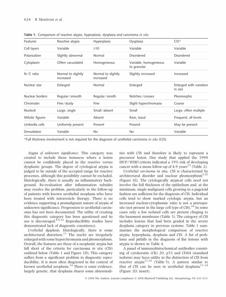
(Figure 1E). The cytologically atypical cells need notinvolve the full thickness of the epithelium and, at theminimum, single malignant cells growing in a pagetoidfashion are sufficient for the diagnosis of CIS. Individualcells tend to show marked cytologic atypia, but anincreased nuclear:cytoplasmic ratio is not a prerequi-site (not present in the large cell type of CIS).16 In somecases only a few isolated cells are present clinging tothe basement membrane (Table 3). The category of CISincludes lesions that had been graded in the severedysplasia category in previous systems. Table 1 sum-marizes the morphological comparison of reactiveatypia, hyperplasia, dysplasia and CIS. A list of prob-lems and pitfalls in the diagnosis of flat lesions withatypia is shown in Table 4.
A panel of immunohistochemical antibodies consist-ing of cytokeratin (CK) 20, p53 and CD44 (standardisoform) may have utility in the distinction of CIS fromreactive atypia17,18 (Table 5). A pattern similar tothat of CIS can be seen in urothelial dysplasia17–24
(Figure 1D, insert).
Table 1. Comparison of reactive atypia, hyperplasia, dysplasia and carcinoma in situ
Features Reactive atypia Hyperplasia Dysplasia CIS*
Cell layers Variable ‡10 Variable Variable
Polarization Slightly abnormal Normal Disordered Disordered
Cytoplasm Often vacuolated Homogeneous Variable, homogeneousto granular
Variable
N ⁄ C ratio Normal to slightlyincreased
Normal to slightlyincreased
Slightly increased Increased
Nuclear size Enlarged Normal Enlarged Enlarged with variationin size
Nuclear borders Regular ⁄ smooth Regular ⁄ smoth Notches ⁄ creases Pleomorphic
Chromatin Fine ⁄ dusty Fine Slight hyperchromasia Coarse
Nucleoli Large, single Small ⁄ absent Small Large, often multiple
Mitotic figures Variable Absent Rare, basal Frequent, all levels
Umbrella cells Uniformly present Present Present May be present
Denudation Variable No No Variable
*Full thickness involvement is not required for the diagnosis of urothelial carcinoma in situ (CIS).
624 R Montironi et al.
� 2008 The Authors. Journal compilation � 2008 Blackwell Publishing Ltd, Histopathology, 53, 621–633.
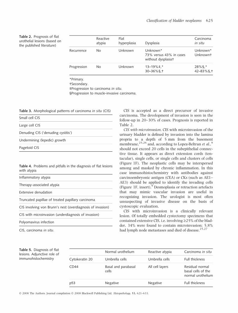
CIS is accepted as a direct precursor of invasivecarcinoma. The development of invasion is seen in thefollow-up in 20–30% of cases. Prognosis is reported inTable 2.
CIS with microinvasion. CIS with microinvasion of theurinary bladder is defined by invasion into the laminapropria to a depth of 5 mm from the basementmembrane,25,26 and, according to Lopez-Beltran et al.,9
should not exceed 20 cells in the subepithelial connec-tive tissue. It appears as direct extension cords (ten-tacular), single cells, or single cells and clusters of cells(Figure 1F). The neoplastic cells may be interspersedamong and masked by chronic inflammation. In thiscase immunohistochemistry with antibodies againstcarcinoembryonic antigen (CEA) or CKs (such as AE1–AE3) should be applied to identify the invading cells(Figure 1F, insert).9 Desmoplasia or retraction artefactsthat may mimic vascular invasion are useful inrecognizing invasion. The urologist is most oftenunsuspecting of invasive disease on the basis ofcystoscopic evaluation.
CIS with microinvasion is a clinically relevantlesion. Of totally embedded cystectomy specimens thatcontained extensive CIS, i.e. involving ‡25% of the blad-der, 34% were found to contain microinvasion; 5.8%had lymph node metastases and died of disease.25,27
Table 4. Problems and pitfalls in the diagnosis of flat lesionswith atypia
Inflammatory atypia
Therapy-associated atypia
Extensive denudation
Truncated papillae of treated papillary carcinoma
CIS involving von Brunn’s nest (overdiagnosis of invasion)
CIS with microinvasion (underdiagnosis of invasion)
Polyomavirus infection
CIS, carcinoma in situ.
Table 2. Prognosis of flaturothelial lesions (based onthe published literature)
Reactiveatypia
Flathyperplasia Dysplasia
Carcinomain situ
Recurrence No Unknown Unknown*73% versus 43% in caseswithout dysplasia†
Unknown*Unknown†
Progression No Unknown 13–19%‡,*30–36%§,†
28%§,*42–83%§,†
*Primary.
†Secondary.
‡Progression to carcinoma in situ.
§Progression to muscle-invasive carcinoma.
Table 3. Morphological patterns of carcinoma in situ (CIS)
Small cell CIS
Large cell CIS
Denuding CIS (‘denuding cystitis’)
Undermining (lepedic) growth
Pagetoid CIS
Table 5. Diagnosis of flatlesions. Adjunctive role ofimmunohistochemistry
Normal urothelium Reactive atypia Carcinoma in situ
Cytokeratin 20 Umbrella cells Umbrella cells Full thickness
CD44 Basal and parabasalcells
All cell layers Residual normalbasal cells of thenormal urothelium
p53 Negative Negative Full thickness
Classification of bladder neoplasms 625
� 2008 The Authors. Journal compilation � 2008 Blackwell Publishing Ltd, Histopathology, 53, 621–633.

papillary or exophytic les ions
Histological grading according to the 1973WHO classificationHistological grading is one of the most importantprognostic factors in bladder cancer. The first widelyaccepted grading system for papillary urothelial neo-plasms was the 1973 WHO classification system,which divided urothelial papillary tumours into fourcategories: papilloma, grade 1 carcinoma, grade 2carcinoma and grade 3 carcinoma.1 Histological grad-ing is based on the degree of anaplasia, with grade 1tumours having the least degree of anaplasia compat-ible with a diagnosis of malignancy, grade 3 tumourshave the most severe degree of anaplasia, and grade 2tumours have an intermediate degree of anaplasia.Anaplasia is defined by the authors of the 1973 WHOclassification as increased cellularity, nuclear crowd-ing, disturbed cellular polarity, failure of differentiationfrom the base to the surface, nuclear polymorphism,irregular cell size, variations in nuclear shape andchromatin pattern, displaced or abnormal mitoticfigures, and giant cells.1
The 1973 WHO histological grading of bladdercancer is one of most successful grading systemsamong all organ sites and has been validated since itsintroduction three decades ago. It has been accepted bypathologists, urologists, oncologists and cancer regis-trars in Europe and elsewhere. An enormous amount ofdata has been accumulated using this system in studiesof the morphological properties, clinical behaviour,treatment and follow-up of urothelial tumours. Becauseof its relative simplicity and its well-documentedpowerful predictive value, it has been well acceptedby urologists and used globally for several decades inmaking clinical decisions for management of patientswith urothelial cancer.
In an effort to improve understanding and tostandardize use of the 1973 WHO classification, anexpanded and refined contemporary description of thescheme was presented in 2001. This proposal is knownas the Ancona refinement of the 1973 WHO gradingsystem.28,29 This effort was inspired by discussionsduring the international consensus meeting on bladdercancer held in Ancona, Italy, 2001. The diagnosticcriteria for each of these categories were refined andoptimized for reproducibility.28,29
Histological grading according to the 2004WHO classificationAccording to its proponents, the key points of the 2004WHO classification of non-invasive urothelial tumoursare:4
1. The description of the categories has beenexpanded to improve their recognition; one group(papillary urothelial neoplasm of low malignantpotential, PUNLMP) with particularly good prognosisdoes not carry the label of ‘cancer’.
2. It avoids use of ambiguous grading such as grade1 ⁄ 2 or 2 ⁄ 3.
3. The group of non-invasive high-grade carcinomais large enough to contain virtually all those tumoursthat have biological properties (and a high level ofgenetic instability) similar to those seen in invasiveurothelial carcinoma.
Exophytic lesions without cytological atypiaPseudopapillary hyperplasia (papillary urothelial hyper-plasia). In the 1998 ISUP ⁄ WHO classification, papillaryhyperplasia was included as a category with the groupof papillary lesions. In the 2004 WHO classification,this is no longer included as a specific designation, butit is recognized that hyperplasias may be flat orpseudopapillary.4 Hyperplasia with a pseudopapillaryarchitecture refers to a slight tenting or undulationof the urothelium lacking a well-defined central fibro-vascular core, although small vessels may be presentat the base of the papillae. There is no significantcytological or architectural atypia. Pseudopapillaryhyperplasia has most often been described in thesetting of known papillary neoplasia. When identifiedde novo, the significance regarding subsequent devel-opment of neoplasia is unknown.
Urothelial papilloma. There has been long-standingcontroversy regarding the nature of papillary lesionswith minimal cytological atypia. The application of thisterm by some experts to up to 1 ⁄ 3 of all papillarylesions was a major stimulant to the re-evaluation ofthese lesions that began in 1997. The current classi-fication retains the very restrictive traditional criteria.Histologically, papilloma is characterized by a few finepapillary fronds without fusion or complexity. Individ-ual fronds are covered by an essentially normalurothelium without architectural or cytological atypia.The number of cell layers is not a criterion for diagnosis(Table 6 and Figure 1G, including insert).
Papillomas meeting these restricted criteria occur ata younger age than other urothelial bladder tumoursand often present with only one or a few papillaryprocesses. They have a low recurrence rate30–32
(Table 7).Papillary urothelial neoplasms of low malignant poten-
tial. The creation of this category represented acompromise between the ‘papilloma’ supporters andthose insisting on the use of ‘carcinoma’ for allpapillary lesions. The 1998 consensus statement
626 R Montironi et al.
� 2008 The Authors. Journal compilation � 2008 Blackwell Publishing Ltd, Histopathology, 53, 621–633.

acknowledged that the lower grade papillary neo-plasms were not intrinsically malignant, but wereassociated with significant risk for the development ofnew papillary tumours (i.e. recurrence).3 These lesions
at the lower end of the spectrum were acknowledgedto be clinically significant, with close clinical follow-up necessary but further intravesicle therapy notindicated.
Table 6. Comparison of papilloma, papillary neoplasm of low malignant potential, low-grade papillary carcinoma and high-grade papillary carcinoma
Features Papilloma
Papillary neoplasmof low malignantpotential
Low-grade papillarycarcinoma High-grade papillary carcinoma
ArchitecturePapillae Delicate Delicate. Occasional
fusedFused, branching,and delicate
Fused, branching and delicate
Organizationof cells
Identicalto normal
Polarity identicalto normal.Any thickness.Cohesive
Predominantly ordered,yet minimal crowdingand minimal loss ofpolarity. Any thickness.Cohesive
Predominantly disordered withfrequent loss of polarity.Any thickness. Often discohesive
CytologyNuclear size Identical
to normalMay be uniformlyenlarged
Enlarged with variationin size
Enlarged with variation in size
Nuclear shape Identicalto normal
Elongated, round–oval, uniform
Round–oval. Slightvariation in shapeand contour
Moderate–marked pleomorphism
Nuclear chromatin Fine Fine Mild variation withinand between cells
Moderate–marked variation bothwithin and between cells withhyperchromasia
Nucleoli Absent Absent toinconspicuous
Usually inconspicuous* Multiple prominent nucleoli maybe present
Mitoses Absent Rare, basal Occasionally at any level Usually frequent, at any level
Umbrella cells Uniformlypresent
Present Usually present May be absent
*If present, small and regular and not accompanied by other features of high-grade carcinoma.
Table 7. Prognosis of urothelial papillary lesions*
Papilloma(%)
Papillaryneoplasm oflow malignantpotential (%)
Low-gradepapillarycarcinoma (%)
High-gradepapillarycarcinoma (%)
Recurrence 0–8 27–47 48–71 55–58
Grade progression 2 11 7 Not applicable
Stage progression 0 0–4 2–12 27–61
Survival 100 93–100 82–96 74–90
*From Lopez-Beltran and Montironi.40
Classification of bladder neoplasms 627
� 2008 The Authors. Journal compilation � 2008 Blackwell Publishing Ltd, Histopathology, 53, 621–633.

Morphologically, PUNLMP largely, though not com-pletely, corresponds to grade 1 papillary carcinoma inthe old WHO system (see below). The tumour consistsof delicate papillae with little or no fusion. The coveringurothelium shows minimal architectural irregularity(Figure 1H). Nuclei lack significant nuclear hyper-chromasia or pleomorphism. The chromatin is fineand nucleoli are inconspicuous. Mitoses are infrequentand basally located.
These tumours have a significantly lower rate ofrecurrence than either low- or high-grade papillarycarcinomas and a very low rate of grade and stageprogression.33–39 In a review of published studies, Lopez-Beltran40 has found the mean tumour recurrence rate tobe 36% and stage progression rate to be 3.7%.
Exophytic lesions with cytological atypiaPapillary urothelial carcinoma, low grade. This categorycontains the intermediate group of lesions. In the 1973WHO system this would include the lower half of grade2 papillary carcinoma. Histologically, the papillae arelargely delicate and separate, but some fusion may beseen. At low magnification there is a generally orderedappearance of cells within the epithelium. The nucleitend to be uniformly enlarged, but retain the elongatedto oval shape of normal urothelial cells. The chromatinremains fine with small nucleoli (Figure 1I). Mitosesmay be present, but are few and remain basally located.
These tumours have a significantly higher recur-rence rate than for PUNLMP and similar to high-gradepapillary carcinomas. They also have a significantlyhigher rate of stage progression than PUNLMP, butsignificantly lower than for high-grade papillary carci-noma.33,37–39 A review of the literature has revealed amean recurrence rate of 50% and mean stage progres-sion rate of 10%.40
Papillary urothelial carcinoma, high grade. This cate-gory contains grade 3 and the upper half of grade 2papillary carcinoma of the 1973 WHO system. Histo-logically, the papillae are frequently fused, formingapparent solid masses. The overall impression is one ofdisordered growth (Figure 1J). The epithelium is ofvariable thickness. Individual cells are haphazardlyarranged within the epithelium and have a generallydiscohesive nature. Nuclei are hyperchromatic andpleomorphic. The chromatin is dense, irregularly dis-tributed and often clumped. Nucleoli may be single ormultiple and are often prominent. Mitoses are generallyfrequent and may be seen at any level of the epithe-lium.10 It is often associated with invasive disease atthe time of diagnosis (Figure 1J, insert).
These tumours not only have a risk of invasion buthave a significant risk of recurrence and progression.
The overall progression rate (to invasive carcinoma)ranges from 15% to 40% (Table 7).41,42 Thesetumours, when non-invasive (pTa), are all likely torequire additional intravesicle therapy. Heterogeneityof grade is recognized in papillary lesions43 and theconsensus was that tumours should be graded on theirworst part, although this needs further study.
Table 6 summarizes the morphological comparisonof papilloma, papillary neoplasm of low malignantpotential, low-grade papillary carcinoma, and high-grade papillary carcinoma. Prognosis of the papillarylesions is reported in Table 7.
Several studies have looked at a variety of biologicalmarkers in papillary tumours and their relationship tothe three groups; for the most part, these havedemonstrated significant differences of the respectivemarker in the different categories.39,44,45
Morphological comparison between papillary and flatlesionsPUNLMP, low-grade papillary carcinoma and high-grade papillary carcinoma show morphologicalsimilarities to flat hyperplasia, dysplasia and CIS,respectively.7
Relation of 1973 WHO to 2004 WHO classificationA major misconception is that there is a one-to-onetranslation between the 1973 and 2004 WHO classi-fications. Only at the extremes of grades in the 1973WHO classification does this correlation hold true.7,10
Lesions called papilloma in the 1973 WHO classifica-tion system would also be called papilloma in the 2004WHO system. At the other end of the grading extreme,lesions called WHO grade 3 are by definition high-grade carcinoma in the 2004 WHO system. However,for WHO grades 1 and 2, there is no direct translationto the 2004 WHO system. Some lesions classified asWHO grade 1 in the 1973 system, which upon reviewshow no cytological atypia, some nuclear enlargementand merely thickened urothelium, are PUNLMPs in the2004 WHO system. However, other WHO grade 1lesions showing slight cytological atypia and mitosesare diagnosed in the 2004 WHO system as low-gradepapillary urothelial carcinomas. WHO grade 2 is a verybroad category. It includes lesions that are relativelybland, which in some places are diagnosed as WHOgrade 1–2; these lesions in the 2004 WHO systemwould be called low-grade papillary urothelial carcin-oma. In other cases, WHO grade 2 lesions border onhigher grade lesions, which in many institutions arecalled WHO grade 2–3; these lesions in the 2004 WHOclassification system would be called high-grade pap-illary urothelial carcinoma.7,10
628 R Montironi et al.
� 2008 The Authors. Journal compilation � 2008 Blackwell Publishing Ltd, Histopathology, 53, 621–633.

Has the 2004 WHO classification system facilitatedchanges in clinical management of papillary urothelialneoplasms?In the past few decades it has been and still is wellunderstood by most practising urologists and oncolo-gists that non-invasive papillary urothelial tumours ofall 1973 WHO grades require follow-up to detectrecurrence or progression, despite the fact that grade 1tumours are characteristically associated with anexcellent prognosis. The length of clinical follow-up,the frequency of surveillance cystoscopy and theadjunctive use of intravesicle instillations of bacillusCalmette–Guerin or a variety of chemotherapeuticagents are influenced by many factors, includinghistological grade, tumour size, tumour multiplicity,depth of tumour invasion, recurrence history andapparent grade of migration with recurrence.
Currently, both in North America and Europe, thereis no uniformity in the clinical management of patientswith non-invasive papillary urothelial tumours diag-nosed according to the 2004 WHO grading system.Patients with PUNLMP and non-invasive low-gradecarcinoma are typically treated by transurethral resec-tion of their tumours and are subsequently monitoredfor recurrence or progression by regular cystoscopy.Although low-grade non-invasive carcinoma has beenfound to have a statistically significant higher progres-sion rate than PUNLMP in the study by Samaratungaet al. (8% for PUNLMP versus 13% for low-grade non-invasive urothelial carcinoma),46 the reported highincidence of recurrence (up to 60%)47 and progression(up to 8%)46,47 for PUNLMP suggest that it is prudentto follow patients with a diagnosis of PUNLMP in anidentical manner to those with a diagnosis of low-gradenon-invasive carcinoma. Indeed, investigators studyingthe recurrence and progression rate of PUNLMP haverecommended long-term clinical follow-up for patientswith these lesions.35,38,46–48
To our knowledge, there has not been a publishedrecommended surveillance protocol for PUNLMPtumours that differs significantly from the standardsurveillance for low-grade non-invasive urothelial car-cinoma. Nor, to our knowledge, have there been anypublished recommendations for following non-invasiveurothelial tumours diagnosed according to the 2004WHO grading system, nor for the use of intravesicletherapy, that vary significantly from the traditions longestablished for following comparable lesions diagnosedaccording to the 1973 WHO grading system. In short,those who are charged with following these lesionsappear to have gained minimal benefit from the newgrading system in terms of surveillance or intravesicletherapy protocols.49
endophytic urothelial les ions
A series of urothelial lesions, ranging from hyperplasiato carcinoma, can have an exclusively endophyticpattern of growth, thus causing problems in differentialdiagnosis and evaluation of invasion. Similarly to theflat and exophytic lesions, the endophytic changes canbe without atypia and with atypia.7
von Brunn’s nests and cystitis cysticaVon Brunn’s nests refer to small groups of basal-likecells lying in the subepithelial connective tissue andattached the basal cell layer of the urothelium. Cystitiscystica is made of cystically dilated von Brunn’s nestsacquiring a luminal space. Mild nuclear atypia andnucleolar prominence can be present. Cystitis cystica isa benign proliferative consequence of inflammation orother irritation. Urothelial CIS may rarely occur in thevon Brunn’s nests and cystitis cystica, and not bedetectable in the overlying flat urothelium.50 In thesecases it is usually associated with previously diagnosedCIS or infiltrating at other sites in the bladder.
Several patterns of invasive urothelial carcinoma(nested variant, tubular, and microcystic) are decep-tively bland and may mimic von Brunn’s nests andcystitis cystica, particularly when the proliferationbecomes florid.51,52
Inverted urothelial papillomaInverted papilloma is a distinct clinical pathologicalentity typically arising in the trigone region in ayounger patient population than papillary neoplasms.Grossly, inverted papilloma shows an exophytic polyp-oid growth pattern. Histologically, it consists of anasto-mosing trabeculae of urothelium covered by a normal orattenuated urothelium. There is no significant nuclearpleomorphism and few mitoses can be seen. Squamousor glandular differentiation may be present. In tran-surethral resection material, the fragmentation of thelesion may result in apparent true papillary structures,making diagnosis difficult. Distinction from carcinomawith an inverted growth pattern can be problematic (seebelow). Cases of synchronous inverted papilloma andpapillary carcinoma are known. Inverted papilloma isassociated with a low risk of recurrence (<5%).53,54
Endophytic growth patterns in urothelial carcinomaSome papillary urothelial carcinomas exhibit aprominent endophytic growth pattern, resulting inconsiderable difficulty in assessing invasion. Endo-phytic growth is evident either as interanastomosingcords and columns of urothelium, often with a strikingresemblance to inverted papilloma (inverted papilloma-
Classification of bladder neoplasms 629
� 2008 The Authors. Journal compilation � 2008 Blackwell Publishing Ltd, Histopathology, 53, 621–633.

like pattern), or as broad, pushing bulbous invagin-ations into the lamina propria (broad-front pattern).The endophytic growth pattern in urothelial carcinomadescribed here is similar to that originally presented byAmin and colleagues55 and by Montironi et al.7
Distinction from inverted papilloma requires atten-tion to architectural and cytological features. Archi-tectural features favouring a diagnosis of urothelialcarcinoma with an inverted growth pattern includethick columns with irregularity in their width andtransition into more solid areas. The characteristicorderly maturation, spindling, and peripheral palisad-ing seen in inverted papilloma are generally absent orinconspicuous in carcinoma with an inverted growthpattern. Unequivocal invasion into the lamina propriaor muscularis propria rules out the diagnosis ofinverted papilloma, but this feature is rarely found.Cytological atypia, including nuclear pleomorphism,irregularities of nuclear borders and chromatin distri-bution, prominent nucleoli, and appreciable mitoticrate, is an important feature for the diagnosis ofcarcinoma.
The second, and more common, histological appear-ance of urothelial carcinoma with an endophyticgrowth pattern is the pushing broad-front extensioninto the lamina propria, akin to cutaneous andmucosal verrucous carcinoma. The downward projec-tion can be so pronounced that the base of the tumourlies on the muscularis propria.
Diagnosis of invasion requires the unquestionablepresence within the lamina propria of irregularlyshaped nests or single cells that may have evoked adesmoplastic or inflammatory response. When a stro-mal response is absent, irregularity of the contours ofthe invasive nests, architectural complexity and recog-nition of single-cell invasion are helpful. Occasionally,the cells in the invading nests appear morphologicallydifferent from those at the base of the tumour, and theymay appear as smaller aggregates present withinempty spaces. These spaces may mimic vascularinvasion closely, but are believed to be retractionartefacts.7
Benign, preneoplastic and non-invasiveneoplastic bladder lesions other thanurothelial
Squamous cell lesionsSquamous papilloma. Squamous papilloma is a rarebenign neoplasm. It is the squamous counterpart ofurothelial papilloma and is unrelated to human pap-illomavirus infection. It usually occurs in elderly
women and follows a benign clinical course. Histolog-ically, it is composed of papillary cores with overlyingbenign squamous epithelium.56
Squamous metaplasia. It occurs in two variants:vaginal and keratinizing. The vaginal type is consid-ered normal in the female trigone (Figure 1K, insert 1).There is no known association with carcinoma. Thekeratinizing type (Figure 1K, insert 2) is usuallyassociated with chronic irritation, e.g. indwellingcatheters, or stones. Squamous metaplasia is a well-known phenomenon in association with bilharziainfestation in North African countries, especially inEgypt. The development of invasive squamous cellcarcinoma in the setting of extensive keratinizingsquamous metaplasia is not uncommon (see Squamouscell carcinoma in situ).57
Squamous cell carcinoma in situ. Only a few reports onsquamous cell CIS of the bladder are available.58
Histologically, it is identical to squamous cell CIS foundin other organ sites (Figure 1K). This finding is oftenassociated with subsequent or concurrent invasivecarcinoma. Wide-range human papillomavirus DNAsignal has occasionally been detected. Enhancedexpression of epidermal growth factor receptor in thesebladder squamous lesions suggests a possible thera-peutic target in cases that are difficult to manageclinically.58
Glandular lesionsGlandular (intestinal) metaplasia (cystitis glandularis). Itis characterized by the presence of epithelial cells ofcolonic type with goblet cell appearance (and rarelyPaneth, argentaffin or argyrophil cells) within thesurface epithelium (flat pattern) or ⁄ and in associationwith cystitis cystica (endophytic pattern, i.e. cystitisglandularis). It has been considered a preneoplasticcondition.9 The development of adenocarcinoma hasbeen documented in very few patients.
Mucin-secreting metaplasia with striking resem-blance to intestinal mucosa, including a crypt-likearchitecture, is called intestinal metaplasia (Figure 1L,insert). Extensive intestinal metaplasia can be seen inbladder extrophy and is associated with increased riskfor the development of adenocarcinoma.59 The appear-ance of cellular atypia, such as nuclear pleomorphismand nucleolar prominence, is considered indicative ofglandular dysplasia and adenocarcinoma in situ, i.e. anintermediate step between glandular metaplasia andinvasive adenocarcinoma.60–62
Villous adenoma. Villous adenoma is an uncommonglandular epithelial neoplasm with exophytic growththat can be associated with urachal adenocarcinoma.63
630 R Montironi et al.
� 2008 The Authors. Journal compilation � 2008 Blackwell Publishing Ltd, Histopathology, 53, 621–633.
Patients often present with haematuria and ⁄ or irrita-tive symptoms. Mucusuria may be present in rarecases. There is no apparent gender predominance. Thetumour usually occurs in elderly patients with apredilection for the urachus, dome, and trigone of theurinary bladder. Its cystoscopic appearance is that ofan exophytic tumour. Histologically, villous adenomaof the bladder is identical to villous adenoma of thecolon, with columnar mucin-filled goblet cells liningdelicate fibrovascular stalks (Figure 1L). Nuclear find-ings include pseudostratification, crowding, occasionalprominent nucleoli and nuclear hyperchromasia, as inthe colon. Villous adenoma of the bladder showsimmunopositivity for CK20 (100% of cases), CK7(56%) and CEA (89%). Patients with an isolated villousadenoma have an excellent prognosis, but progressionto adenocarcinoma appears to occur in 21–33% ofcases. Villous adenoma of the bladder may coexist within situ and invasive adenocarcinoma.63
Conclusions and future perspectives
There is still debate as to whether the 2004 WHOsystem should be the only one to be used and whetherthe 1973 WHO system should be abandoned.64
Reporting both grades has been recommended.65
In terms of clinical management of patients withnon-invasive papillary urothelial neoplasms, therehave been, to our knowledge, no published recommen-dations for following non-invasive urothelial tumoursdiagnosed according to the 2004 WHO grading system,nor for the use of intravesicle therapy, that vary fromthe traditions long established for following comparablelesions diagnosed according to the 1973 WHO gradingsystem. If the original focus in 1998 had been simply toretain the original 1973 WHO classification scheme,but to embellish it by expanding and clearly definingthe morphological characteristics of the original threegrades of carcinoma, a long and unproductive period ofcontroversy and uncertainty about the questionablemerits of the new terminology could have beenavoided.49
Enough is now known about the molecular aspectsof bladder tumours. The variance in biological behavi-our of low- versus high-grade tumours correlates withknown dual molecular lines of genetic development.The first and more common pathway (low grade) leadsto non-invasive, papillary tumours, those whose statusas a carcinoma might be legitimately challengedbecause they usually follow an indolent course. Thesecond pathway leads to the development of high-gradepapillary carcinoma or flat CIS and ultimately muscle-invasive carcinoma if left untreated. In their earliest,
but commonly progressive phase, these lesions will bestaged as CIS or Ta. Although not invasive, they sharemolecular alterations common in invasive disease andrepresent the stage before invasive disease.66
There is a need of a universally accepted newclassification of bladder non-invasive neoplasms inwhich essential morphological elements from bothWHO classifications as well as information on molec-ular marker expression are incorporated.
Acknowledgements
This review was supported by grants from the Poly-technic University of the Marche Region (Ancona) Italy(M.S. and R.M.). The content of this review is solely theresponsibility of the authors and does not necessarilyrepresent the official views of the Polytechnic Univer-sity of the Marche Region (Ancona, Italy). Presented inpart at the Centennial Congress of the InternationalAcademy of Pathology, Montreal, Canada, 16–21September 2006 (by A.L-B.) and at the Symposiumon Urological Pathology, British Division of the Inter-national Academy of Pathology, London, UK, 23–24November 2007 (by R.Mo.).
References
1. Mostofi FK, Sobin LH. Histologic typing of urinary bladder tumors.
Geneva: World Health Organization, 1973.
2. Murphy WM, Beckwith JB, Farrow GM. Atlas of tumor pathology:
tumors of the kidney, bladder, and related urinary structures. armed
forces institute of pathology, 3rd series, Fascicle 11. Washington:
AFIP, 1994.
3. Epstein JI, Amin MB, Reuter VR, Mostofi FK. The World Health
Organization ⁄ International Society of Urological Pathology
consensus classification of urothelial (transitional cell) neoplasms
of the urinary bladder. Bladder Consensus Conference Committee.
Am. J. Surg. Pathol. 1998; 22; 1435–1448.
4. Sauter G, Algaba F, Amin MB et al. Non-invasive urothelial
tumours. In Eble JN, Sauter G, Epstein JI, Sesterhenn IA eds.
World Health Organization classification of tumours: pathology and
genetics of tumours of the urinary system and male genital organ.
Lyon: IARC Press, 2004, pp. 110–123.
5. Jost SP, Gosling JA, Dixon JS. The morphology of normal human
bladder urothelium. J. Anat. 1989; 167; 103–115.
6. Epstein JI, Amin MB, Reuter VE. In Epstein JI ed. Urinary bladder
biopsy interpretatio. Philadelphia: Lippincott Williams & Wilkins,
2004.
7. Montironi R, Mazzucchelli R, Lopez-Beltran A, Cheng L, Scarpelli
M. My approach to the morphological diagnosis of the urothelial
neoplasms. J. Clin. Pathol. 2008; 61; 3–10.
8. Amin MB, Young RH. Intraepithelial lesions of the urinary
bladder with a discussion of the histogenesis of urothelial
neoplasia. Semin. Diagn. Pathol. 1997; 14; 84–97.
9. Lopez-Beltran A, Cheng L, Andersson L et al. Preneoplastic non-
papillary lesions and conditions of the urinary bladder: an update
based on the Ancona International Consultation. Virchows Arch.
2002; 440; 3–11.
Classification of bladder neoplasms 631
� 2008 The Authors. Journal compilation � 2008 Blackwell Publishing Ltd, Histopathology, 53, 621–633.
10. Montironi R, Lopez-Beltran A. The 2004 WHO classification of
bladder tumors: a summary and commentary. Int. J. Surg. Pathol.
2005; 13; 143–153.
11. Nagy GH, Frable WJ, Murphy WM. The classification of premalig-
nant urothelial abnormalities. Pathol. Ann. 1982; 17; 219–233.
12. Murphy WM, Busch C, Algaba F. Intraepithelial lesions of the
urinary bladder: morphologic considerations. Scand. J. Urol.
Nephrol. 2000; 205; 67–81.
13. Murphy WM, Soloway MS. Urothelial dysplasia. J. Urol. 1982;
127; 849–854.
14. Zuk RJ, Rogers HS, Martin JE, Baithun SI. Clinicopathological
importance of primary dysplasia of bladder. J. Clin. Pathol. 1988;
41; 1277–1280.
15. Cheng L, Cheville JC, Neumann RM, Bostwick DG. Natural
history of urothelial dysplasia of the bladder. Am. J. Surg. Pathol.
1999; 23; 443–447.
16. Owens CL, Epstein JI. Significance of denuded urothelium in
papillary urothelial lesions. Am. J. Surg. Pathol. 2007; 31; 298–
303.
17. Harnden P, Eardley I, Joyce AD, Southgate J. Cytokeratin 20 as
an objective marker of urothelial dysplasia. Br. J. Urol. 1996; 78;
870–875.
18. Mallofre C, Castillo M, Morente V, Sole M. Immunohistochemical
expression of CK20, p53, and Ki-67 as objective markers of
urothelial dysplasia. Mod. Pathol. 2003; 16; 187–191.
19. Desai S, Lim SD, Jimenez RE et al. Relationship of cytokeratin 20
and CD44 protein expression with WHO ⁄ ISUP grade in pTa and
pT1 papillary urothelial neoplasia. Mod. Pathol. 2000; 13; 1315–
1323.
20. Kunju LP, Lee CT, Montie J, Shah RB. Utility of cytokeratin 20
and Ki-67 as markers of urothelial dysplasia. Pathol. Int. 2005;
55; 248–254.
21. McKenney JK, Amin MB. The role of immunohistochemistry in
the diagnosis of urinary bladder neoplasms. Semin. Diagn. Pathol.
2005; 22; 69–87.
22. McKenney JK, Desai S, Cohen C, Amin MB. Discriminatory
immunohistochemical staining of urothelial carcinoma in situ
and non neoplastic urothelium. An analysis of cytokeratin 20,
p53, and CD44 antigens. Am. J. Surg. Pathol. 2001; 25; 1074–
1078.
23. Parker DC, Folpe AL, Bell J et al. Potential utility of uroplakin III,
thrombomodulin, high molecular weight cytokeratin, and cyto-
keratin 20 in noninvasive, invasive, and metastatic urothelial
(transitional cell) carcinomas. Am. J. Surg. Pathol. 2003; 27; 1–
10.
24. Sarkis AS, Dalbagni G, Cordon-Cardo C et al. Association of p53
nuclear overexpression and tumor progression in carcinoma in
situ of the bladder. J. Urol. 1994; 152; 388–392.
25. Farrow GM, Utz DC. Observations on microinvasive transitional
cell carcinoma of the urinary bladder. Clin. Oncol. 1982; 1; 609–
615.
26. McKenney JK, Gomez JA, Desai S, Lee MW, Amin MB. Morphologic
expressions of urothelial carcinoma in situ. A detailed evaluation of
its histologic patterns with emphasis on carcinoma in situ with
microinvasion. Am. J. Surg. Pathol. 2001; 25; 356–362.
27. Farrow GM, Utz DC, Rife CC. Clinical observations on 69 cases of
in situ carcinoma of the urinary bladder. Cancer Res. 1977; 37;
2794–2802.
28. Bostwick DG, Mikuz G. Urothelial papillary (exophytic) neo-
plasms. Virchows Arch. 2002; 441; 109–116.
29. Bostwick DG, Ramnani DM, Cheng L. Diagnosis and grading of
bladder cancer and associated lesions. Urol. Clin. North Am.
1999; 26; 493–507.
30. Cheng L, Darson M, Cheville JC et al. Urothelial papilloma of the
bladder: clinical and biologic implications. Cancer 1999; 86;
2098–2101.
31. Eble JN, Young RH. Benign and low grade papillary lesions of the
urinary bladder: a review of the papilloma–papillary carcinoma
controversy and a report of 5 typical papillomas. Semin. Diagn.
Pathol. 1987; 6; 351–371.
32. McKenney JK, Amin MB, Young RH. Urothelial (transitional cell)
papilloma of the urinary bladder: a clinicopathologic study of 26
cases. Mod. Pathol. 2003; 16; 623–629.
33. Alvarez-Kindelan J, Lopez-Beltran A, Anglada-Curado F et al.
Clinico-pathologic differences between bladder neoplasms with
low malignant potential and low-grade carcinomas. Actas Urol.
Esp. 2001; 25; 645–650.
34. Campbell PA, Conrad RJ, Campbell CM, Nicol DL, MacTaggart P.
Papillary urothelial neoplasm of low malignant potential: reliabil-
ity of diagnosis and outcome. BJU Int. 2004; 93; 1228–1291.
35. Cheng L, Neumann RL, Bostwick DG. Papillary urothelial
neoplasms of low malignant potential: clinical and biological
implications. Cancer 1999; 86; 2102–2108.
36. Holmang S, Andius P, Hedelin H, Wester K, Busch C, Johansson
SL. Stage progression in Ta papillary urothelial tumors: relation-
ship to grade, immunohistochemical expression of tumor mark-
ers, mitotic frequency and DNA ploidy. J. Urol. 2001; 165;
1124–1128.
37. Malmstrom PU, Busch C, Norlen BJ. Recurrence, progression and
survival in bladder cancer: a retrospective analysis of 232
patients with greater than or equal to 5-year follow-up. Scand. J.
Urol. Nephrol. 1987; 21; 185–195.
38. Oosterhuis JW, Schapers RF, Janssen-Heijnen ML, Pauwels RP,
Newling DW, ten Kate F. Histological grading of papillary
urothelial carcinoma of the bladder: prognostic value of the 1998
WHO ⁄ ISUP classification system and comparison with conven-
tional grading systems. J. Clin. Pathol. 2002; 55; 900–905.
39. Yin H, Leong AS-Y. Histologic grading of noninvasive papillary
urothelial tumors: validation of the 1998 WHO ⁄ ISUP system by
immunophenotyping and follow up. Am. J. Clin. Pathol. 2004;
121; 679–687.
40. Lopez-Beltran A, Montironi R. Non-invasive urothelial neo-
plasms: according to the most recent WHO classification.
Eur. Urol. 2004; 46; 170–176.
41. Herr HW. Tumor progression and survival of patients with high-
grade, noninvasive papillary (TaG3) bladder tumors: 15-year
outcome. J. Urol. 2000; 163; 79–80.
42. Saika T, Tsushima T, Nasu Y et al. Clinical study of G3 superficial
bladder cancer without concomitant CIS treated with conserva-
tive therapy. Jpn. J. Clin. Oncol. 2002; 32; 461–465.
43. Cheng L, Neumann RM, Nehra A, Spotts BE, Weaver AL,
Bostwick DG. Cancer heterogeneity and its biological implica-
tions in the grading of urothelial carcinoma. Cancer 2000; 88;
1663–1670.
44. Cina SJ, Lancaster-Weiss KJ, Lecksell K, Epstein JI. Correlation of
Ki67 and p53 with the new World Health Organization ⁄ Inter-
national Society of Urological Pathology classification system for
urothelial neoplasms. Arch. Pathol. Lab. Med. 2001; 125; 646–
651.
45. Pich A, Chiusa L, Formiconi A, Galliano D, Bortolin P, Navone R.
Biological differences between noninvasive papillary urothelial
neoplasms of low malignant potential and low-grade (grade 1)
papillary carcinoma of the bladder. Am. J. Surg. Pathol. 2001; 25;
1528–1533.
46. Samaratunga H, Makarov DV, Epstein JI. Comparison of
WHO ⁄ ISUP and WHO classification of noninvasive papillary
632 R Montironi et al.
� 2008 The Authors. Journal compilation � 2008 Blackwell Publishing Ltd, Histopathology, 53, 621–633.

urothelial neoplasms for risk of progression. Urology 2002; 60;
315–319.
47. Fujii Y, Kawakami S, Koga F, Nemoto T, Kihara K. Long-term
outcome of bladder papillary urothelial neoplasms of low
malignant potential. BJU Int. 2003; 92; 559–562.
48. Holmang S, Hedelin H, Anderstrom C, Holmberg E, Busch C,
Johansson SL. Recurrence and progression in low grade papillary
urothelial tumors. J. Urol. 1999; 162; 702–707.
49. MacLennan GT, Kirkali Z, Cheng L. Histologic grading of
noninvasive papillary urothelial neoplasms. Eur. Urol. 2007;
51; 889–898.
50. Melicow M, Hollowell J. Intra-urothelial cancer: carcinoma
in situ, Bowen’s disease of the urinary system. Discussion of 30
cases. J. Urol. 1952; 68; 763–771.
51. Volmar KE, Chan TY, De Marzo AM, Epstein JI. Florid von Brunn
nests mimicking urothelial carcinoma: a morphologic and
immunohistochemical comparison to the nested variant of
urothelial carcinoma. Am. J. Surg. Pathol. 2003; 27; 1243–1252.
52. Humphrey PA. Urinary bladder pathology 2004: an update.
Ann. Diagn. Pathol. 2004; 6; 380–389.
53. Cameron KM, Lupton CH. Inverted papilloma of the lower
urinary tract. Br. J. Urol. 1976; 48; 567–577.
54. Cheng CW, Chan LW, Chan CK et al. Is surveillance necessary for
inverted papilloma in the urinary bladder and urethra? ANZ J.
Surg. 2005; 75; 213–217.
55. Amin MB, Gomez JA, Young RH. Urothelial transitional cell
carcinoma with endophytic growth patterns: a discussion of
patterns of invasion and problems associated with assessment of
invasion in 18 cases. Am. J. Surg. Pathol. 1997; 21; 1057–1068.
56. Cheng L, Leibovich BC, Cheville JC et al. Squamous papilloma of
the urinary tract is unrelated to condyloma acuminata. Cancer
2000; 88; 1679–1686.
57. Bessette PL, Abell MR, Herwinig KR. A clinicopathologic study of
squamous cell carcinoma of the bladder. J. Urol. 1974; 112; 66–
67.
58. Guo CC, Fine SW, Epstein JI. Noninvasive squamous lesions
in the urinary bladder: a clinicopathologic analysis of 29 cases.
Am. J. Surg. Pathol. 2006; 30; 883–891.
59. Elem B, Alam SZ. Total intestinal metaplasia with focal adeno-
carcinoma in a schistosoma-infested defunctioned urinary
bladder. Br. J. Urol. 1984; 56; 331–343.
60. Chan TY, Epstein JI. In situ adenocarcinoma of the bladder. Am. J.
Surg. Pathol. 2001; 25; 892–899.
61. Corica FA, Husmann DA, Churchill BM et al. Intestinal
metaplasia is not a strong risk factor for bladder cancer: study
of 53 cases with long-term follow up. Urology 1997; 50; 427–
431.
62. Shaw JL, Gislason GJ, Imbriglia JE. Transition of cystitis gland-
ularis to primary adenocarcinoma of the bladder. J. Urol. 1958;
79; 815.
63. Cheng L, Montironi R, Bostwick DG. Villous adenoma of
the urinary tract: a report of 23 cases, including 8 with
coexistent adenocarcinoma. Am. J. Surg. Pathol. 1999; 23; 764–
771.
64. Harnden P. A critical appraisal of the classification of the
urothelial tumours: time for a review of the evidence and a
radical change? BJU Int. 2007; 99; 723–725.
65. Lopez-Beltran A, Bassi PF, Pavone-Macaluso M, Montironi R.
Handling and pathology reporting of specimens with carcinoma
of the urinary bladder, ureter, and renal pelvis. Eur. Urol. 2004;
45; 257–266.
66. Hensel DE, Jones JS. A critical appraisal of the classification of the
urothelial tumours: time for a change but not the change
proposed. BJU Int. 2007; 100; 1213–1214.
Classification of bladder neoplasms 633
� 2008 The Authors. Journal compilation � 2008 Blackwell Publishing Ltd, Histopathology, 53, 621–633.