Embed Size (px)
Citation preview
1 3
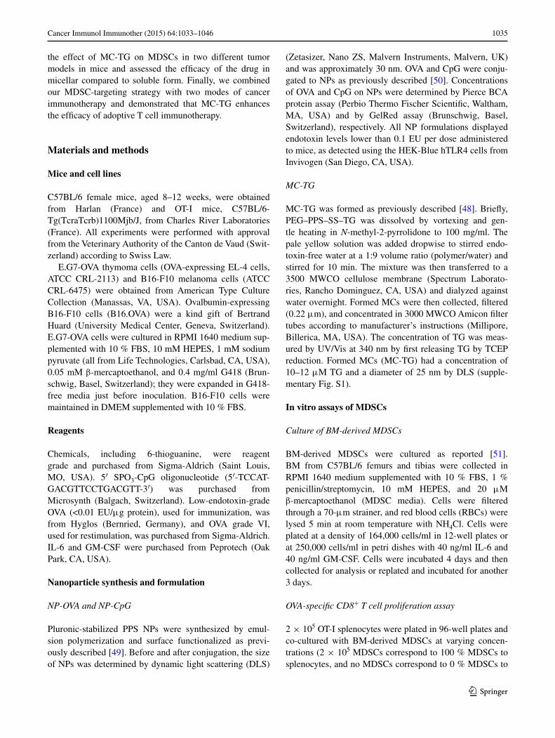
Cancer Immunol Immunother (2015) 64:1033–1046DOI 10.1007/s00262-015-1702-8
ORIGINAL ARTICLE
6‑Thioguanine‑loaded polymeric micelles deplete myeloid‑derived suppressor cells and enhance the efficacy of T cell immunotherapy in tumor‑bearing mice
Laura Jeanbart1,2 · Iraklis C. Kourtis1 · André J. van der Vlies1 · Melody A. Swartz1,2,3,4 · Jeffrey A. Hubbell1,3,4,5
Received: 1 October 2014 / Accepted: 16 April 2015 / Published online: 16 May 2015 © The Author(s) 2015. This article is published with open access at Springerlink.com
as well as of Ly6chi macrophages, for up to 7 days follow-ing a single administration. MDSC depletion was dose dependent and more effective with MC-TG than with equal doses of free TG. Finally, we tested whether this MDSC-depleting strategy might enhance cancer immunotherapies in the B16-F10 melanoma model. We found that MC-TG significantly improved the efficacy of adoptively trans-ferred, OVA-specific CD8+ T cells in melanoma cells expressing OVA. These findings highlight the capacity of MC-TG in depleting MDSCs in the tumor microenviron-ment and show promise in promoting anti-tumor immunity when used in combination with T cell immunotherapies.
Keywords MDSC depletion · 6-Thioguanine · Cancer · T cell therapy
AbbreviationsBM Bone marrowDC Dendritic cellG (-MDSC) Granulocytic
Abstract Myeloid-derived suppressor cells (MDSCs) are a heterogeneous population of immature myeloid cells that suppress effector T cell responses and can reduce the effi-cacy of cancer immunotherapies. We previously showed that ultra-small polymer nanoparticles efficiently drain to the lymphatics after intradermal injection and target antigen-presenting cells, including Ly6chi Ly6g− mono-cytic MDSCs (Mo-MDSCs), in skin-draining lymph nodes (LNs) and spleen. Here, we developed ultra-small polymer micelles loaded with 6-thioguanine (MC-TG), a cytotoxic drug used in the treatment of myelogenous leukemia, with the aim of killing Mo-MDSCs in tumor-bearing mice and thus enhancing T cell-mediated anti-tumor responses. We found that 2 days post-injection in tumor-bearing mice (B16-F10 melanoma or E.G7-OVA thymoma), MC-TG depleted Mo-MDSCs in the spleen, Ly6clo Ly6g+ granulo-cytic MDSCs (G-MDSCs) in the draining LNs, and Gr1int Mo-MDSCs in the tumor. In both tumor models, MC-TG decreased the numbers of circulating Mo- and G-MDSCs,
Melody A. Swartz and Jeffrey A. Hubbell contributed equally to this work.
Electronic supplementary material The online version of this article (doi:10.1007/s00262-015-1702-8) contains supplementary material, which is available to authorized users.
* Melody A. Swartz [email protected]
* Jeffrey A. Hubbell [email protected]
1 Institute of Bioengineering, School of Life Sciences and School of Engineering, Ecole Polytechnique Fédérale de Lausanne (EPFL), Lausanne, Switzerland
2 Swiss Institute for Experimental Cancer Research (ISREC), School of Life Sciences, EPFL, Lausanne, Switzerland
3 Institute for Chemical Sciences and Engineering, School of Basic Sciences, EPFL, Lausanne, Switzerland
4 Institute for Molecular Engineering, University of Chicago, Chicago, IL, USA
5 Materials Science Division, Argonne National Laboratory, Argonne, IL, USA
1034 Cancer Immunol Immunother (2015) 64:1033–1046
1 3
i.d. Intradermal(ly)i.v. Intravenous(ly)LN Lymph nodeMC MicelleMDSC Myeloid-derived suppressor cellMo (-MDSC) MonocyticMΦ MacrophageNP NanoparticleOVA OvalbuminPEG Poly(ethylene glycol)PPS Poly(propylene sulfide)p.i. Post-tumor inoculationRA Retinoic acidRBC Red blood cellTG 6-Thioguanine
Introduction
Over the past decades, many novel cancer immunothera-pies have been developed to boost anti-tumor immunity, targeting a variety of mechanisms including tumor anti-gen presentation by dendritic cells (DCs), anti-tumor T cell priming, overall T cell activation status, immune sup-pression, and T cell infiltration in the tumor [1, 2]. Strat-egies have included cell-based therapies such as transfer of ex vivo activated DCs or engineered T cells as well as antibody-based therapies that target specific T cell inhibi-tory pathways including CTLA-4 or PD-1/PD-L1 [3–6]. Despite these efforts, many therapeutic modalities encoun-ter limited success because of tumor-induced immune sup-pression and evasion mechanisms [6–9]. It has been shown that targeting these immune suppressive mechanisms can lead to enhanced immunotherapy efficacy in cancer [10–14].
Myeloid-derived suppressor cells (MDSCs) are a het-erogeneous population of immature myeloid cells, charac-terized by their expression of CD11b and Gr1 and lack of MHCII; they comprise a Ly6chi Ly6g− Gr1int monocytic subset (Mo-MDSCs) and a Ly6clo Ly6g+ Gr1hi granulo-cytic subset (G-MDSCs) [15]. MDSCs are induced by tumor-mediated inflammation [16–18], recruited to the circulation via tumor-derived factors such as IL-1, IL-6, GM-CSF, G-CSF, and VEGF [19–21], and accumulate in the tumor, tumor-draining lymph node (LN), and spleen, with MDSC numbers increasing with tumor load [16, 21]. MDSCs play a major role in anti-tumor immunity by inhib-iting both CD8+ and CD4+ T cell activation, proliferation, and homing [16, 17, 22, 23]. G-MDSCs infiltrate and exert their suppressive activity in an antigen-specific manner in the LNs, while Mo-MDSCs, considered as the more sup-pressive ones, infiltrate and suppress T cell responses in the spleen and tumor [18, 24–26].
Strategies to target MDSCs, and thereby improve local T cell function, include depletion (affecting both recruitment and expansion in the tumor), functional inhibition, and dif-ferentiation into mature antigen-presenting cells [27, 28]. Ly6chi monocytes and Mo-MDSCs traffic from the bone marrow (BM) to sites of inflammation via CCR2-signaling [29], and therapeutic strategies based on CCR2-siRNA showed significant reduction in inflammatory monocyte effects in murine models of atherosclerosis, cancer, and diabetes [30]. Also, all-trans-retinoic acid (RA), which is important for hematopoietic stem cell development [31], was shown to eliminate immature myeloid cells in tumor-bearing mice and drive their differentiation into mature myeloid cells in cancer patients [10, 32, 33], although without affecting tumor growth. The chemotherapeutic pyrimidine analogs gemcitabine and 5-fluorouracil, which prevent DNA replication and lead to apoptosis, have shown efficacy in depleting MDSCs in tumors and lymphoid organs, leading to expansion of tumor-specific T cells and delayed tumor growth in mice [34, 35]. High doses of the TLR9 agonist CpG have been shown to impact both Ly6g+ and Ly6chi MDSCs in tumor-bearing mice by decreasing their suppressive function and leading to their differentia-tion into mature myeloid cells [36, 37]. Finally, combining adoptive T cell therapy or DC transfer with cytotoxic drugs or small molecule inhibitors has shown some promise in slowing tumor growth [38–41].
The purine analog 6-thioguanine (TG) is an effective anti-inflammatory and anticancer drug [42]. It is used in pediatric and adult leukemias, including myeloid and mye-logenous leukemias [43, 44], where it can “freeze” myelo-blasts in an immature state to prevent their differentiation into mature myeloid cells, including monocytes and gran-ulocytes [18, 45]. We hypothesized that TG might be an interesting drug candidate to deplete Mo-MDSCs based on its ability to target the myeloid cell lineage.
MDSC-directed strategies have been shown to enhance anti-tumor immunity and to synergize with anticancer vac-cines [10, 11, 40]. Our laboratory has developed nano-particles (NPs) that drain from i.d. administration sites through lymphatics to target skin-draining LNs, where they are taken up by resident antigen-presenting cells [46, 47]. In a biodistribution study, we showed that NPs are taken up remarkably effectively by Mo-MDSCs in LNs, spleen, and tumor [47]. Based on this observation, we developed a nanoscale polymeric micelle (MC) capable of sequestering and releasing TG, with the intention of depleting MDSCs in a more targeted manner than might be achievable with free TG. MCs consist of TG chemically conjugated via a disulfide bond to a block copolymer of the hydrophilic polyethylene glycol (PEG) and the hydrophobic polypro-pylene sulfide (PPS) [48]. After first finding that MC-TG entirely depleted BM-derived Mo-MDSCs, we then tested
1035Cancer Immunol Immunother (2015) 64:1033–1046
1 3
the effect of MC-TG on MDSCs in two different tumor models in mice and assessed the efficacy of the drug in micellar compared to soluble form. Finally, we combined our MDSC-targeting strategy with two modes of cancer immunotherapy and demonstrated that MC-TG enhances the efficacy of adoptive T cell immunotherapy.
Materials and methods
Mice and cell lines
C57BL/6 female mice, aged 8–12 weeks, were obtained from Harlan (France) and OT-I mice, C57BL/6-Tg(TcraTcrb)1100Mjb/J, from Charles River Laboratories (France). All experiments were performed with approval from the Veterinary Authority of the Canton de Vaud (Swit-zerland) according to Swiss Law.
E.G7-OVA thymoma cells (OVA-expressing EL-4 cells, ATCC CRL-2113) and B16-F10 melanoma cells (ATCC CRL-6475) were obtained from American Type Culture Collection (Manassas, VA, USA). Ovalbumin-expressing B16-F10 cells (B16.OVA) were a kind gift of Bertrand Huard (University Medical Center, Geneva, Switzerland). E.G7-OVA cells were cultured in RPMI 1640 medium sup-plemented with 10 % FBS, 10 mM HEPES, 1 mM sodium pyruvate (all from Life Technologies, Carlsbad, CA, USA), 0.05 mM β-mercaptoethanol, and 0.4 mg/ml G418 (Brun-schwig, Basel, Switzerland); they were expanded in G418-free media just before inoculation. B16-F10 cells were maintained in DMEM supplemented with 10 % FBS.
Reagents
Chemicals, including 6-thioguanine, were reagent grade and purchased from Sigma-Aldrich (Saint Louis, MO, USA). 5′ SPO3-CpG oligonucleotide (5′-TCCAT-GACGTTCCTGACGTT-3′) was purchased from Microsynth (Balgach, Switzerland). Low-endotoxin-grade OVA (<0.01 EU/μg protein), used for immunization, was from Hyglos (Bernried, Germany), and OVA grade VI, used for restimulation, was purchased from Sigma-Aldrich. IL-6 and GM-CSF were purchased from Peprotech (Oak Park, CA, USA).
Nanoparticle synthesis and formulation
NP‑OVA and NP‑CpG
Pluronic-stabilized PPS NPs were synthesized by emul-sion polymerization and surface functionalized as previ-ously described [49]. Before and after conjugation, the size of NPs was determined by dynamic light scattering (DLS)
(Zetasizer, Nano ZS, Malvern Instruments, Malvern, UK) and was approximately 30 nm. OVA and CpG were conju-gated to NPs as previously described [50]. Concentrations of OVA and CpG on NPs were determined by Pierce BCA protein assay (Perbio Thermo Fischer Scientific, Waltham, MA, USA) and by GelRed assay (Brunschwig, Basel, Switzerland), respectively. All NP formulations displayed endotoxin levels lower than 0.1 EU per dose administered to mice, as detected using the HEK-Blue hTLR4 cells from Invivogen (San Diego, CA, USA).
MC‑TG
MC-TG was formed as previously described [48]. Briefly, PEG–PPS–SS–TG was dissolved by vortexing and gen-tle heating in N-methyl-2-pyrrolidone to 100 mg/ml. The pale yellow solution was added dropwise to stirred endo-toxin-free water at a 1:9 volume ratio (polymer/water) and stirred for 10 min. The mixture was then transferred to a 3500 MWCO cellulose membrane (Spectrum Laborato-ries, Rancho Dominguez, CA, USA) and dialyzed against water overnight. Formed MCs were then collected, filtered (0.22 μm), and concentrated in 3000 MWCO Amicon filter tubes according to manufacturer’s instructions (Millipore, Billerica, MA, USA). The concentration of TG was meas-ured by UV/Vis at 340 nm by first releasing TG by TCEP reduction. Formed MCs (MC-TG) had a concentration of 10–12 μM TG and a diameter of 25 nm by DLS (supple-mentary Fig. S1).
In vitro assays of MDSCs
Culture of BM‑derived MDSCs
BM-derived MDSCs were cultured as reported [51]. BM from C57BL/6 femurs and tibias were collected in RPMI 1640 medium supplemented with 10 % FBS, 1 % penicillin/streptomycin, 10 mM HEPES, and 20 μM β-mercaptoethanol (MDSC media). Cells were filtered through a 70-μm strainer, and red blood cells (RBCs) were lysed 5 min at room temperature with NH4Cl. Cells were plated at a density of 164,000 cells/ml in 12-well plates or at 250,000 cells/ml in petri dishes with 40 ng/ml IL-6 and 40 ng/ml GM-CSF. Cells were incubated 4 days and then collected for analysis or replated and incubated for another 3 days.
OVA‑specific CD8+ T cell proliferation assay
2 × 10⁵ OT-I splenocytes were plated in 96-well plates and co-cultured with BM-derived MDSCs at varying concen-trations (2 × 10⁵ MDSCs correspond to 100 % MDSCs to splenocytes, and no MDSCs correspond to 0 % MDSCs to
1036 Cancer Immunol Immunother (2015) 64:1033–1046
1 3
splenocytes). Cells were cultured in MDSC medium and incubated 24 h with 250 μg/ml OVA grade VI. 0.5 μCi of 3H thymidine was added to each well, and cells were fur-ther incubated for 18 h. Cells were then stored at −20 °C before collection on filter plates and analysis by a scintilla-tion counter to determine thymidine incorporation.
Tumor inoculation and injections
Mice were anesthetized with isoflurane (5 % for induction and 2 % for maintenance) and injected with 106 E.G7-OVA cells, 5 × 10⁵ B16-F10 cells, or 2.5 × 10⁵ B16.OVA cells in 30 μl 0.9 % saline solution intradermally (i.d.) on the left side of the back. E.G7-OVA, B16-F10, or B16.OVA tumor-bearing mice were injected 7, 5, or 4 days post-tumor inoculation (p.i.), respectively, with 10 mg/kg MC-TG or free TG injected i.d. in all four footpads (unless otherwise specified) in the following experiments:
Biodistribution MC-TG was labeled with the fluoro-phore Dy649; mice were killed on day 9;Time course blood was sampled every 2–3 days starting on injection day;Multiple doses mice were boosted on day 13 with 5 mg/kg MC-TG;Dosage mice were injected with 2, 5, or 10 mg/kg MC-TG on day 7 and killed on day 14;NP-vaccine mice were immunized on days 3 and 10 with 10 μg NP-OVA and 1 μg NP-CpG (NP-vaccine) i.d. in the front footpad draining the tumor; mice were injected with 10 mg/kg MC-TG on day 13;Adoptive T cell transfer 10 mg/kg MC-TG or free TG was injected i.d. on day 4 p.i., and 2 days later (day 6 p.i.), 106 OT-I CD8+ T cells were transferred i.v. in the tail vein.
Blood was sampled from the submandibular vein of the cheek with a 4-mm lancet at indicated time points. Tumors were measured starting 5 days p.i. with a digital caliper, and volumes (V) were calculated as an ellipsoid (V = π/6 · l ·w · h, where l is length, w width, and h height). Mice were killed by CO2 asphyxiation. Experiments were stopped when tumor volumes reached 1 cm3 or earlier if necrotic.
Adoptive CD8+ T cell transfer
Splenic CD8+ T cells from OT-I mice cells were isolated by immunomagnetic negative selection (EasySep Mouse CD8+ T Cell Isolation Kit) and CD11c+ by positive selection (EasySep Mouse CD11c Positive Selection Kit), both from Stemcell Technologies (Vancouver, BC, Canada). CD8+ and CD11c+ cells were co-cultured 72 h at a ratio of 10:1 with 1 nM OVA257-
264 peptide (Genscript, Piscataway, NJ, USA) and 10 U/ml recombinant mouse IL-2 (Roche, Rotkreuz, Switzerland). Cells
were then collected, washed in basal medium, and resuspended to 107 cells/ml prior to tail vein injection.
Tissue and cell preparation
Spleens, LNs (brachial, axillary, inguinal), and tumors were harvested at time of killing. LNs and tumors were digested 20 and 60 min, respectively, in DMEM supplemented with 1 mg/ml collagenase D (Roche). Single-cell suspensions were obtained by gently disrupting the organs through a 70-μm cell strainer. Spleen and blood RBCs were lysed with NH4Cl 5 min. Cells were counted and resuspended in IMDM supplemented with 10 % FBS and 1 % penicillin/streptomycin (full medium) (all from Life Technologies).
Flow cytometry
Cells were washed and stained with surface antibodies in staining buffer [HBSS (Life Technologies) supplemented with 0.5 % bovine serum albumin]. Cell viability was deter-mined by propidium iodide incorporation in staining buffer after surface antibody staining or with live/dead fixable cell viability reagent (Life Technologies) in PBS before anti-body staining. Cells were stained with PE-labeled H-2Kb/OVA257–264 pentamer (Proimmune, Oxford, UK) according to manufacturer’s instructions.
AccuCount cell counting beads (Spherotech, Lake For-est, IL, USA) were added to blood samples. Samples were acquired on CyAn ADP analyzer (Beckman Coulter, Brea, CA, USA), and data were analyzed with FlowJo software (v9.4; Tree Star, Ashland, OR, USA). Antibodies against mouse CD8, CD3, MHCII, B220, CD45, CD11b, Gr1, Ly6c, Ly6g, and CD11c were purchased from eBioscience or BioLegend (San Diego, CA, USA). Pacific orange-con-jugated and Alexa Fluor 647-conjugated streptavidins were from Life Technologies.
Statistical analysis
Statistically significant differences between experimental groups were determined by one-way analysis of variance (ANOVA) followed by Bonferroni posttest correction with Prism software (v5, GraphPad, San Diego, CA, USA). *, **, and *** indicate P values <0.05, 0.01, and 0.001, respectively.
Results
MC‑TG depletes BM‑derived Mo‑MDSCs in vitro
Based on the hypothesis that TG in a nanoparticulate for-mulation may be more readily targeted to MDSCs than in
1037Cancer Immunol Immunother (2015) 64:1033–1046
1 3
soluble form [47], we formulated TG as a 25-nm micelle (MC-TG) by linking TG to a PEG–PPS chain via a disulfide bond (supplementary Fig. S1) [48]. We generated MDSCs in vitro following a well-established protocol [51] (supplementary Fig. S2 A) using IL-6 and GM-CSF, two factors secreted by tumors that recruit MDSCs from the BM to the circulation in tumor-bearing mice [19–21]. After 4 days of culture, BM cells were skewed toward a CD11b+ MHCII− CD11c− immature myeloid phenotype character-istic of MDSCs, with Ly6chi Ly6g− Mo-MDSC and Ly6clo Ly6g+ G-MDSC subsets (supplementary Fig. S2 B) [16].
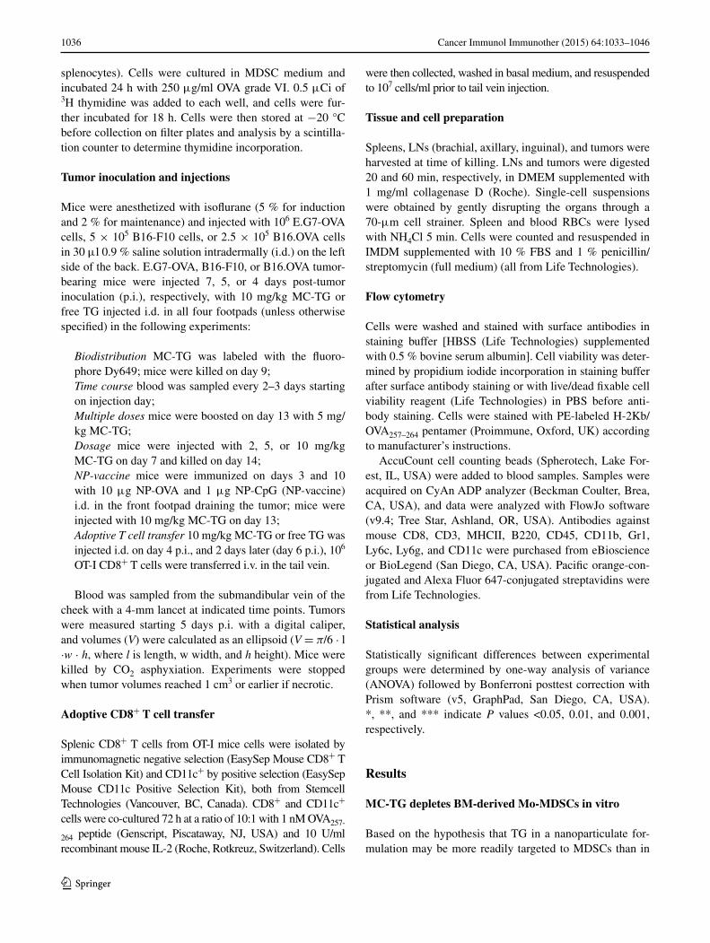
TG, in both free and micellar forms, depleted Mo-MDSCs in vitro (Fig. 1a). Mo-MDSCs were reduced from 5.6 ± 0.5 % of the culture to 0.1 % (**) of the culture with free TG and MC-TG. While control MDSCs efficiently prevented OT-I T cell proliferation, adding either free TG or MC-TG to MDSCs rendered them less suppressive (Fig. 1b). Furthermore, both MC-TG and free TG depleted already differentiated Mo-MDSCs by day 7 in vitro (Fig. 1c), from 3.9 ± 0.7 to 0.2 and 0.3 %, respectively, and rendered BM-derived MDSCs less suppressive than control MDSCs (Fig. 1d). While a ratio of approximately 1:8 of control MDSCs/splenocytes (12 %) was needed to achieve a 50 % reduction in T cell proliferation, approximately
40 % TG-treated MDSCs were needed to achieve equiva-lent inhibition of T cell proliferation (Fig. 1d). These results show that TG, in both free and micellar forms, can deplete BM-derived Mo-MDSCs in vitro and can render MDSCs overall less suppressive against T cell proliferation.
MC‑TG targets and depletes MDSCs in the spleen, LNs, and tumor after 2 days
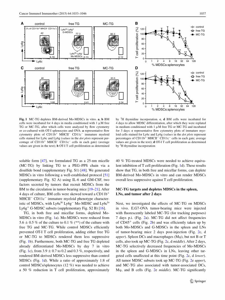
Next, we investigated the effects of MC-TG on MDSCs in vivo. E.G7-OVA tumor-bearing mice were injected with fluorescently labeled MC-TG (for tracking purposes) 7 days p.i. (Fig. 2a). MC-TG did not affect frequencies of CD45+ cells (Fig. 2b) and was efficiently taken up by both Mo-MDSCs and G-MDSCs in the spleen and LNs of tumor-bearing mice 2 days post-injection (Fig. 2c, d upper). Spleen DCs and macrophages (Mϕ), but not B or T cells, also took up MC-TG (Fig. 2c, d middle). After 2 days, MC-TG selectively decreased frequencies of Mo-MDSCs in the spleen and G-MDSCs in LNs, leaving other tar-geted cells unaffected at this time point (Fig. 2c, d lower). All tumor MDSC subsets took up MC-TG (Fig. 2e upper), and MC-TG also associated with tumor-associated DCs, Mϕ, and B cells (Fig. 2e middle). MC-TG significantly
control free TG MC-TGA B
Ly6c
Ly6g
T c
ell p
rolif
erat
ion
(%)
% MDSCs:splenocytescontrol free TG MC-TG
T c
ell p
rolif
erat
ion
(%)
% MDSCs:splenocytes
MC-TGfree TGcontrol
MC-TGfree TGcontrol
C D
Ly6c
Ly6g
6.19
42
0.120.13
77.3
3.66
46
0.41
63
0.34
63.5
(% of CD11b+ MHCII- CD11c-)
Ly6chi Ly6g- Mo-MDSCs
Ly6clo Ly6g+ G-MDSCs
80.3
1 2 4 8 16 32 64 12800
20
40
60
80
100
120
1 2 4 8 16 32 64 12800
20
40
60
80
100
120
*
** **
**
*
*
Fig. 1 MC-TG depletes BM-derived Mo-MDSCs in vitro. a, b BM cells were incubated for 4 days in media conditioned with 1 μM free TG or MC-TG, after which cells were analyzed by flow cytometry or co-cultured with OT-I splenocytes and OVA: a representative flow cytometry plots of CD11b+ MHCII− CD11c− immature myeloid cells stained for Ly6c and Ly6g (values in the dot plots represent per-centage of CD11b+ MHCII− CD11c− cells in each gate) (average values are given in the text); b OT-I T cell proliferation as determined
by 3H thymidine incorporation. c, d BM cells were incubated for 4 days to allow MDSC differentiation, after which they were replated in medium conditioned with 1 μM free TG or MC-TG and incubated for 3 days: c representative flow cytometry plots of immature mye-loid cells stained for Ly6c and Ly6g (values in the dot plots represent percentages of CD11b+ MHCII− CD11c− cells in each gate; average values are given in the text); d OT-I T cell proliferation as determined by 3H thymidine incorporation
1038 Cancer Immunol Immunother (2015) 64:1033–1046
1 3
decreased the frequency of tumor-infiltrating Gr1int Mo-MDSCs and increased Gr1hi G-MDSCs, but did not affect other targeted cells (Fig. 2e lower). Taken together, these results show that MC-TG acts on Mo-MDSCs in the spleen, G-MDSCs in the LNs, and Gr1int Mo-MDSCs in the tumor. MC-TG and TG seemed well tolerated.
MC‑TG depletes circulating Mo‑MDSCs and G‑MDSCs in tumor‑bearing mice
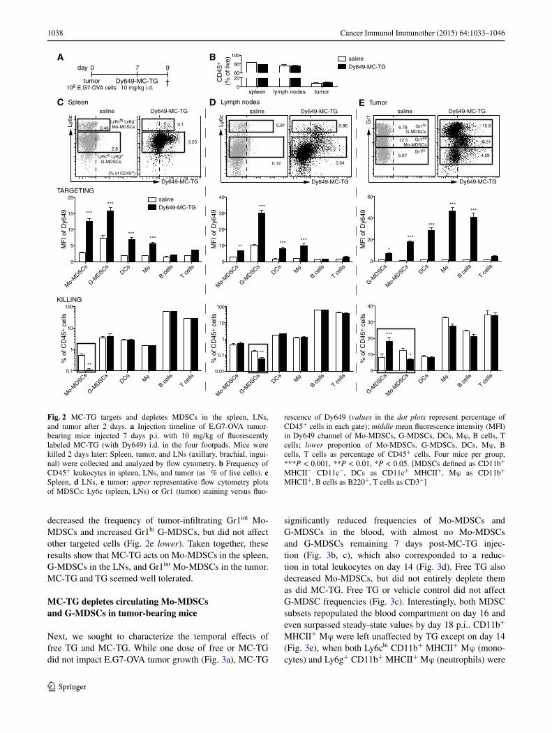
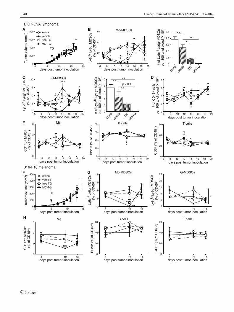
Next, we sought to characterize the temporal effects of free TG and MC-TG. While one dose of free or MC-TG did not impact E.G7-OVA tumor growth (Fig. 3a), MC-TG
significantly reduced frequencies of Mo-MDSCs and G-MDSCs in the blood, with almost no Mo-MDSCs and G-MDSCs remaining 7 days post-MC-TG injec-tion (Fig. 3b, c), which also corresponded to a reduc-tion in total leukocytes on day 14 (Fig. 3d). Free TG also decreased Mo-MDSCs, but did not entirely deplete them as did MC-TG. Free TG or vehicle control did not affect G-MDSC frequencies (Fig. 3c). Interestingly, both MDSC subsets repopulated the blood compartment on day 16 and even surpassed steady-state values by day 18 p.i.. CD11b+ MHCII+ Mϕ were left unaffected by TG except on day 14 (Fig. 3e), when both Ly6chi CD11b+ MHCII+ Mϕ (mono-cytes) and Ly6g+ CD11b+ MHCII+ Mϕ (neutrophils) were
Csaline Dy649-MC-TG
Spleen Lymph nodes
Ly6c
Mo-
MDSCs
G-MDSCs
DCsMφ
B cells
T cells
0
5
10
15
20
Mo-
MDSCs
G-MDSCs
DCsMφ
B cells
T cells
0.1
1
10
100
Mo-
MDSCs
G-MDSCs
DCsMφ
B cells
T cells
0
10
20
30
40
Mo-
MDSCs
G-MDSCs
DCsMφ
B cells
T cells
0.01
0.1
1
10
100
D
TARGETING
KILLING
0 7
tumor Dy649-MC-TG
day 9
†106 E.G7-OVA cells 10 mg/kg i.d.
TumorE
Gr1hi
G-MDSCs
Gr1lo
Gr1int
Mo-MDSCs
Ly6clo Ly6g+ G-MDSCs
Ly6chi Ly6g- Mo-MDSCs
***
***
****** ******
***
**
**
**
0.1
3.23
2.8
0.460.81
0.12 0.04
0.86 6.78
12.5
5.07
15.8
4.59
6.51
A
Dy649-MC-TG
saline Dy649-MC-TG
Ly6c
Dy649-MC-TG
saline Dy649-MC-TG
Gr1
Dy649-MC-TG
salineDy649-MC-TG
MF
I of D
y649
% o
f CD
45+ c
ells
MF
I of D
y649
% o
f CD
45+ c
ells
MF
I of D
y649
% o
f CD
45+ c
ells
(% of CD45+)
0
2060
80
100
CD
45+
(% o
f liv
e)
spleen lymph nodes tumor
B salineDy649-MC-TG
0
10
20
30
40
***
*
0
20
40
60
*
***
***
******
G-MDSCs
Mo-
MDSCs
DCsMφ
B cells
T cells
G-MDSCs
Mo-
MDSCs
DCsMφ
B cells
T cells
Fig. 2 MC-TG targets and depletes MDSCs in the spleen, LNs, and tumor after 2 days. a Injection timeline of E.G7-OVA tumor-bearing mice injected 7 days p.i. with 10 mg/kg of fluorescently labeled MC-TG (with Dy649) i.d. in the four footpads. Mice were killed 2 days later: Spleen, tumor, and LNs (axillary, brachial, ingui-nal) were collected and analyzed by flow cytometry. b Frequency of CD45+ leukocytes in spleen, LNs, and tumor (as % of live cells). c Spleen, d LNs, e tumor: upper representative flow cytometry plots of MDSCs: Ly6c (spleen, LNs) or Gr1 (tumor) staining versus fluo-
rescence of Dy649 (values in the dot plots represent percentage of CD45+ cells in each gate); middle mean fluorescence intensity (MFI) in Dy649 channel of Mo-MDSCs, G-MDSCs, DCs, Mϕ, B cells, T cells; lower proportion of Mo-MDSCs, G-MDSCs, DCs, Mϕ, B cells, T cells as percentage of CD45+ cells. Four mice per group, ***P < 0.001, **P < 0.01, *P < 0.05. [MDSCs defined as CD11b+ MHCII− CD11c−, DCs as CD11c+ MHCII+, Mϕ as CD11b+ MHCII+, B cells as B220+, T cells as CD3+]
1039Cancer Immunol Immunother (2015) 64:1033–1046
1 3
substantially reduced and Ly6clo/− Ly6g− Mϕ were left unaffected (supplementary Fig. S3 A-C). MC-TG and solu-ble TG had no impact on frequencies of circulating B and T cells (Fig. 3e).
The above experiment was reproduced in the B16-F10 melanoma model, which is an orthotopic, more immu-nosuppressive, and more aggressive cancer model [52]. As in the E.G7-OVA model, MC-TG did not affect B16-F10 tumor growth (Fig. 3f) and depleted circulating Mo-MDSCs 5 days post-injection (Fig. 3g left); free TG also reduced Mo-MDSCs but did not deplete them. G-MDSCs levels, not affected by free TG, were significantly reduced by MC-TG (Fig. 3g right). Mϕ, B, and T cells were not affected by MC-TG (Fig. 3h). Together, these results show that a single injection of MC-TG depleted Mo-MDSCs and G-MDSCs for 7 days in both E.G7-OVA and B16-F10 cancer models. MC-TG also depleted cir-culating monocytic Ly6chi Mϕ (CD11b+ MHCII+ mature myeloid cells). Finally, MC-TG was more effective than free TG in depleting MDSCs without affecting tumor growth.
Dose and schedule of MC‑TG delivery modulate MDSC depletion
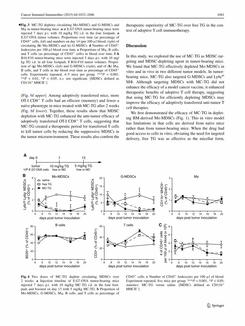
Since circulating MDSCs were restored 1 week post-TG injection, we sought to extend the MDSC-free window through multiple injections, namely on days 7 and 13 with 10 and 5 mg/kg MC-TG or free TG, respectively (Fig. 4a), doses that are cumulatively under the toxic threshold [43]. In both free and MC-TG-treated mice, Mo-MDSCs and G-MDSCs were reduced for approximately 10 days before starting to repopulate the blood by day 20 p.i. (Fig. 4b). As in Fig. 3, Mϕ, B cells, and T cells were not affected by MC-TG or TG, as were total numbers of circulating CD45+ leukocytes (Fig. 4b, c).
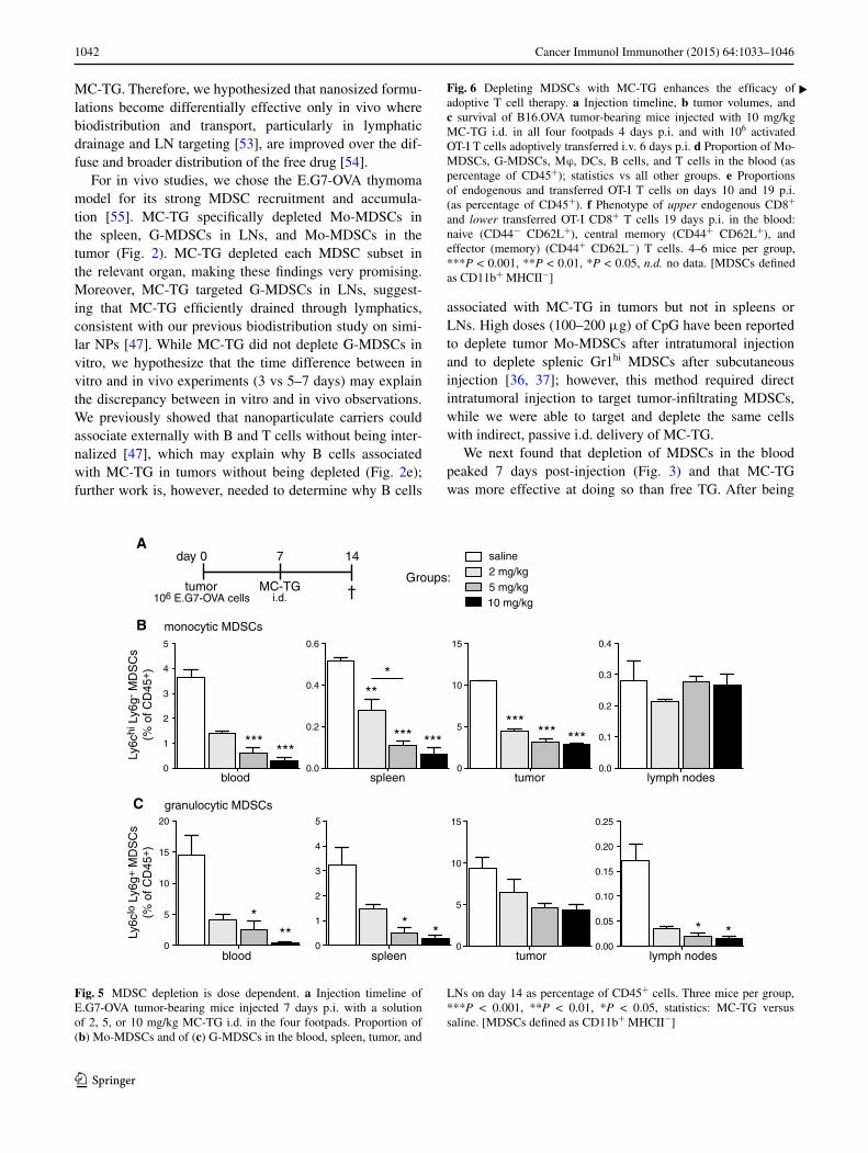
Given the efficacy of the doses used above, we asked whether lower doses of MC-TG could provide similar effi-cacy. E.G7-OVA-bearing mice were injected with MC-TG 7 days p.i. with 2, 5, or 10 mg/kg (Fig. 5a). After 14 days, MC-TG-treated mice showed significantly reduced fre-quencies of Mo-MDSCs in the blood, spleen, and tumor, but not in LNs (Fig. 5b). In contrast, MC-TG led to a sig-nificant reduction in G-MDSCs levels in blood, spleen, and LNs, but not in the tumor (Fig. 5c). We observed a dose response to MC-TG, with the 10 mg/kg dose leading to a stronger reduction in MDSCs than the 5 mg/kg dose, which itself was more potent than the 2 mg/kg dose. These results show that two doses of MC-TG prolong MDSC depletion in the blood, that lower doses of MC-TG were effective at reducing MDSCs systemically and locally, and that the magnitude of MDSC depletion was dose dependent, with 10 mg/kg being the most effective dose.
Depleting MDSCs with MC‑TG enhances the efficacy of adoptive T cell therapy
We next asked whether MDSC depletion with MC-TG could improve the efficacy of a model cancer vaccine com-posed of OVA-conjugated and CpG-conjugated NPs [50]. E.G7-OVA-bearing mice were immunized 3 and 10 days p.i. with NP-OVA + NP-CpG and treated with 10 mg/kg MC-TG 13 days p.i. (supplementary Fig. S4 A). We chose this TG dose for its ability to deplete MDSCs systemi-cally. After regressing, none of the tumors recurred in the mice receiving MC-TG, while 25 % of tumors recurred in immunized mice that did not receive MC-TG (supplemen-tary Fig. S4 B). Five days post-MC-TG treatment, mice had almost a threefold reduction in OVA-specific CD8+ T cells compared to mice that did not receive MC-TG (supplemen-tary Fig. S4 C). Immunized mice had reduced Mo-MDSC frequencies compared to control mice, and the addition of MC-TG rendered their levels undetectable by day 5 post-treatment (supplementary Fig. S4 D). MC-TG also reduced the frequencies of circulating G-MDSCs and Mϕ compared to vaccine-only mice (supplementary Fig. S4 D).
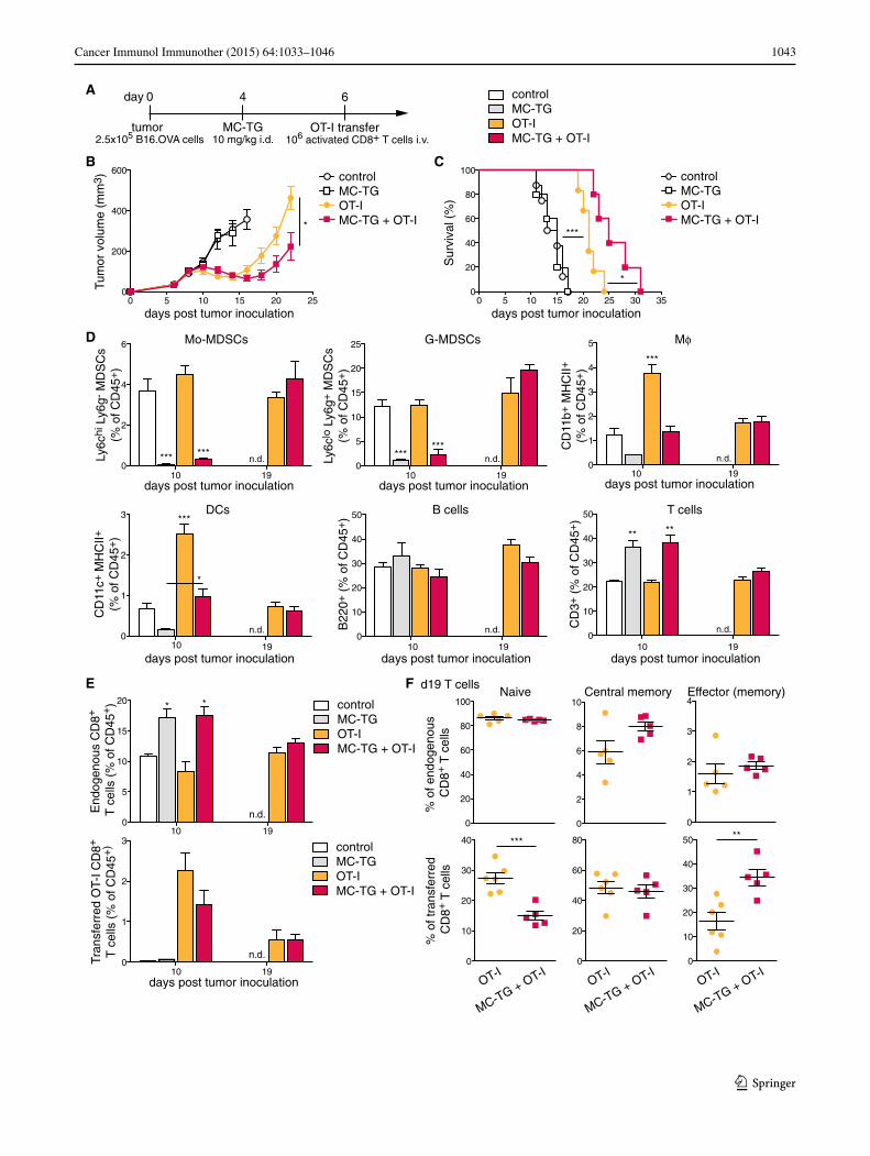
Because MDSC depletion with MC-TG did not enhance anti-tumoral adaptive immunity to a vaccine targeted to tumor-draining LNs, we sought to assess potential benefits of depleting MDSCs in adoptive T cell therapy. Four days p.i., B16.OVA-bearing mice were injected with 10 mg/kg MC-TG, followed by adoptive T cell therapy on day 6 (Fig. 6a). B16.OVA tumors regressed 4 days after adoptive transfer (Fig. 6b). While MC-TG had no effect on B16.OVA tumor growth, a single dose of MC-TG prolonged tumor regression and delayed tumor growth in response to OT-I adoptive transfer, leading to significantly smaller tumors by day 22 p.i. (Fig. 6b). As a consequence, MC-TG + OT-I-treated mice demonstrated significantly enhanced survival compared to OT-I-treated mice without MDSC depletion (Fig. 6c). Free TG, on the other hand, did not affect tumor regression beyond the effect of OT-I transfer and did not enhance survival of tumor-bearing mice (supplementary Fig. S5 A–B). As in Fig. 3, MC-TG transiently reduced proportions of circulating Mo-MDSCs and G-MDSCs, as well as of Mϕ and DCs, and all cell subsets repopulated the blood compartment 2 weeks post-MC-TG administration (day 19 p.i.) (Fig. 6d); free TG did not significantly reduce Mo- and G-MDSC levels (supplementary Fig. S5 C-D). B cells were not affected by MC-TG treatment, and propor-tions of T cells were higher in mice that received MC-TG compared to other groups (Fig. 6d). Consistent with this observation, frequencies of endogenous CD8+ T cells were more elevated in groups receiving MC-TG 10 days p.i., and no difference was observed between OT-I trans-ferred mice 19 days p.i. (Fig. 6e). No differences in endog-enous (non-OT-I) CD8+ T cell phenotype were detected
1040 Cancer Immunol Immunother (2015) 64:1033–1046
1 3
6 8 10 12 14 16 18 200
2
4
6
6 8 10 12 14 16 18 200
5
10
15
20
Ly6c
hi L
y6g-
MD
SC
s (%
of C
D45
+)
Ly6c
lo L
y6g+
MD
SC
s (%
of C
D45
+)
* *** **
*
**
**
****
*
0 5 10 15 200
200
400
600
800
Tum
or v
olum
e (m
m3 )
days post tumor inoculation days post tumor inoculation
days post tumor inoculation
BA
MC-TGfree TGvehiclesaline
6 8 10 12 14 16 18 200
1
2
3
CD
11b+
MH
CII+
(% o
f CD
45+)
*
days post tumor inoculation6 8 10 12 14 16 18 20
0
20
40
60
80
6 8 10 12 14 16 18 200
20
40
60
B22
0+ (
% o
f CD
45+)
CD
3+ (
% o
f CD
45+)
***
***
*
days post tumor inoculation days post tumor inoculation
E.G7-OVA lymphoma
B16-F10 melanoma
TG
Mo-MDSCs
G-MDSCs
Mφ B cells T cells
D
6 8 10 12 14 16 18 200
8
6
4
2
# of
CD
45+ c
ells
per
100
µl o
f blo
od (
x 10
5 )
*
days post tumor inoculation
salin
e
vehic
leTG
MC-T
G0
0.5
1.0
1.5
2.0
2.5
salin
e
vehic
leTG
MC-T
G0
2
4
6
8
10
# of
Ly6
chi L
y6g-
MD
SC
spe
r 10
0 µl
of b
lood
(x
104 )
# of
Ly6
clo L
y6g+
MD
SC
spe
r 10
0 µl
of b
lood
(x
104 )C
E
n.s.
***
n.s.p < 0.1
n.s.
**
0 5 10 150
100
200
300
400
500
4 10 130
2
4
6
8
******
4 10 130
5
10
15
20
25
*
4 10 130
1
2
3
n.s.
4 10 130
20
40
60
4 10 130
20
40
60
days post tumor inoculation
Ly6c
hi L
y6g-
MD
SC
s(%
of C
D45
+)
days post tumor inoculation
Tum
or v
olum
e (m
m3 )
F G
Ly6c
lo L
y6g+
MD
SC
s(%
of C
D45
+)
days post tumor inoculation
CD
11b+
MH
CII+
(% o
f CD
45+)
days post tumor inoculation
B22
0+ (
% o
f CD
45+)
CD
3+ (
% o
f CD
45+)
days post tumor inoculation days post tumor inoculation
Mo-MDSCs G-MDSCs
Mφ B cells T cellsH
MC-TGfree TGvehiclesaline
TG
1041Cancer Immunol Immunother (2015) 64:1033–1046
1 3
(Fig. 6f upper). Among adoptively transferred mice, more OT-I CD8+ T cells had an effector (memory) and fewer a naïve phenotype in mice treated with MC-TG after 2 weeks (Fig. 6f lower). Together, these results show that MDSC depletion with MC-TG enhanced the anti-tumor efficacy of adoptively transferred OT-I CD8+ T cells, suggesting that MC-TG created a therapeutic period for transferred T cells to kill tumor cells by reducing the suppressive MDSCs in the tumor microenvironment. These results also confirm the
therapeutic superiority of MC-TG over free TG in the con-text of adoptive T cell immunotherapy.
Discussion
In this study, we explored the use of MC-TG as MDSC-tar-geting and MDSC-depleting agent in tumor-bearing mice. We found that MC-TG effectively depleted Mo-MDSCs in vitro and in vivo in two different tumor models. In tumor-bearing mice, MC-TG also targeted G-MDSCs and Ly6chi MΦ. Although targeting MDSCs with MC-TG did not enhance the efficacy of a model cancer vaccine, it enhanced therapeutic benefits of adoptive T cell therapy, suggesting that using MC-TG for efficiently depleting MDSCs may improve the efficacy of adoptively transferred anti-tumor T cell therapies.
We first demonstrated the efficacy of MC-TG in deplet-ing BM-derived Mo-MDSCs (Fig. 1). This in vitro model has limitations in that cells are derived from naïve mice rather than from tumor-bearing mice. When the drug had good access to cells in vitro, obviating the need for targeted delivery, free TG was as effective as the micellar form,
0 13
tumor 5 mg/kg TG
day
106 E.G7-OVA cells
7
10 mg/kg TGfree or MC free or MC
6 8 10 12 14 16 18 200
2
4
6
6 8 10 12 14 16 18 200
5
10
15
20
6 8 10 12 14 16 18 200
1
2
3
6 8 10 12 14 16 18 200
20
40
60
80
6 8 10 12 14 16 18 200
20
40
60
6 8 10 12 14 16 18 200
8
6
4
2
Ly6c
hi L
y6g-
MD
SC
s(%
of C
D45
+)
Ly6c
lo L
y6g+
MD
SC
s(%
of C
D45
+)
CD
11b+
MH
CII+
(% o
f CD
45+)
B22
0+ (
% o
f CD
45+)
CD
3+ (
% o
f CD
45+)
# of
CD
45+ c
ells
per
100
µl o
f blo
od (
x 10
5 )
A
B
C
* ***
***
**
***
days post tumor inoculation days post tumor inoculation days post tumor inoculation
days post tumor inoculation days post tumor inoculation days post tumor inoculation
MC-TGfree TG
salineMo-MDSCs G-MDSCs Mφ
B cells T cells
Fig. 4 Two doses of MC-TG deplete circulating MDSCs over 2 weeks. a Injection timeline of E.G7-OVA tumor-bearing mice injected 7 days p.i. with 10 mg/kg MC-TG i.d. in the four foot-pads and boosted on day 13 with 5 mg/kg MC-TG. b Proportion of Mo-MDSCs, G-MDSCs, Mϕ, B cells, and T cells as percentage of
CD45+ cells. c Number of CD45+ leukocytes per 100 μl of blood. Experiment repeated, five mice per group. ***P < 0.001, *P < 0.05; statistics: MC-TG versus saline. [MDSCs defined as CD11b+ MHCII−]
Fig. 3 MC-TG depletes circulating Mo-MDSCs and G-MDSCs and Mϕ in tumor-bearing mice. a–e E.G7-OVA tumor-bearing mice were injected 7 days p.i. with 10 mg/kg TG i.d. in the four footpads. a E.G7-OVA tumor volumes. Proportions over time (as percentage of CD45+ cells, left) and numbers on day 14 (per 100 μl blood, right) of circulating (b) Mo-MDSCs and (c) G-MDSCs. d Number of CD45+ leukocytes per 100 μl blood over time. e Proportions of Mϕ, B cells, and T cells (as percentage of CD45+ cells) in blood over time. f–h B16-F10 tumor-bearing mice were injected 5 days p.i. with 10 mg/kg TG i.d. in all four footpads. f B16-F10 tumor volumes. Propor-tion of (g) Mo-MDSCs (left) and G-MDSCs (right), and of (h) Mϕ, B cells, and T cells in the blood over time as percentage of CD45+ cells. Experiments repeated, 4–5 mice per group. ***P < 0.001, **P < 0.01, *P < 0.05, n.s. not significant. [MDSCs defined as CD11b+ MHCII−]
◂
1042 Cancer Immunol Immunother (2015) 64:1033–1046
1 3
MC-TG. Therefore, we hypothesized that nanosized formu-lations become differentially effective only in vivo where biodistribution and transport, particularly in lymphatic drainage and LN targeting [53], are improved over the dif-fuse and broader distribution of the free drug [54].
For in vivo studies, we chose the E.G7-OVA thymoma model for its strong MDSC recruitment and accumula-tion [55]. MC-TG specifically depleted Mo-MDSCs in the spleen, G-MDSCs in LNs, and Mo-MDSCs in the tumor (Fig. 2). MC-TG depleted each MDSC subset in the relevant organ, making these findings very promising. Moreover, MC-TG targeted G-MDSCs in LNs, suggest-ing that MC-TG efficiently drained through lymphatics, consistent with our previous biodistribution study on simi-lar NPs [47]. While MC-TG did not deplete G-MDSCs in vitro, we hypothesize that the time difference between in vitro and in vivo experiments (3 vs 5–7 days) may explain the discrepancy between in vitro and in vivo observations. We previously showed that nanoparticulate carriers could associate externally with B and T cells without being inter-nalized [47], which may explain why B cells associated with MC-TG in tumors without being depleted (Fig. 2e); further work is, however, needed to determine why B cells
associated with MC-TG in tumors but not in spleens or LNs. High doses (100–200 μg) of CpG have been reported to deplete tumor Mo-MDSCs after intratumoral injection and to deplete splenic Gr1hi MDSCs after subcutaneous injection [36, 37]; however, this method required direct intratumoral injection to target tumor-infiltrating MDSCs, while we were able to target and deplete the same cells with indirect, passive i.d. delivery of MC-TG.
We next found that depletion of MDSCs in the blood peaked 7 days post-injection (Fig. 3) and that MC-TG was more effective at doing so than free TG. After being
0.0
0.2
0.4
0.6
0
1
2
3
4
5
0
5
10
15
0
1
2
3
4
5
0
5
10
15
20
saline2 mg/kg5 mg/kg10 mg/kg
blood spleen tumor0
5
10
15
0.0
0.1
0.2
0.3
0.4
0.00
0.05
0.10
0.15
0.20
0.25
lymph nodes
Ly6c
lo L
y6g+
MD
SC
s(%
of C
D45
+)
Ly6c
hi L
y6g-
MD
SC
s(%
of C
D45
+)
0 7
tumor MC-TG
day
106 E.G7-OVA cells i.d.
14
†
A
B
C
monocytic MDSCs
granulocytic MDSCs
blood spleen tumor lymph nodes
Groups:
****** ******
****** ***
****
***
***
Fig. 5 MDSC depletion is dose dependent. a Injection timeline of E.G7-OVA tumor-bearing mice injected 7 days p.i. with a solution of 2, 5, or 10 mg/kg MC-TG i.d. in the four footpads. Proportion of (b) Mo-MDSCs and of (c) G-MDSCs in the blood, spleen, tumor, and
LNs on day 14 as percentage of CD45+ cells. Three mice per group, ***P < 0.001, **P < 0.01, *P < 0.05, statistics: MC-TG versus saline. [MDSCs defined as CD11b+ MHCII−]
Fig. 6 Depleting MDSCs with MC-TG enhances the efficacy of adoptive T cell therapy. a Injection timeline, b tumor volumes, and c survival of B16.OVA tumor-bearing mice injected with 10 mg/kg MC-TG i.d. in all four footpads 4 days p.i. and with 106 activated OT-I T cells adoptively transferred i.v. 6 days p.i. d Proportion of Mo-MDSCs, G-MDSCs, Mϕ, DCs, B cells, and T cells in the blood (as percentage of CD45+); statistics vs all other groups. e Proportions of endogenous and transferred OT-I T cells on days 10 and 19 p.i. (as percentage of CD45+). f Phenotype of upper endogenous CD8+ and lower transferred OT-I CD8+ T cells 19 days p.i. in the blood: naive (CD44− CD62L+), central memory (CD44+ CD62L+), and effector (memory) (CD44+ CD62L−) T cells. 4–6 mice per group, ***P < 0.001, **P < 0.01, *P < 0.05, n.d. no data. [MDSCs defined as CD11b+ MHCII−]
▸
1043Cancer Immunol Immunother (2015) 64:1033–1046
1 3
0 4
tumor MC-TG
day
2.5x10⁵ B16.OVA cells 10 mg/kg i.d.
6
OT-I transfer106 activated CD8+ T cells i.v.
A
CB
D
E
0
1
2
3
0
10
20
30
40
50
0
10
20
30
40
50
0
2
4
6
0
5
10
15
20
25
0
1
2
3
4
5
Ly6c
hi L
y6g-
MD
SC
s(%
of C
D45
+)
Ly6c
lo L
y6g+
MD
SC
s(%
of C
D45
+)
CD
11b+
MH
CII+
(% o
f CD
45+)
10 19days post tumor inoculation
Tum
or v
olum
e (m
m3 )
days post tumor inoculation
Sur
viva
l (%
)
days post tumor inoculation
controlMC-TGOT-IMC-TG + OT-I
CD
11c+
MH
CII+
(% o
f CD
45+)
B22
0+ (
% o
f CD
45+)
CD
3+ (
% o
f CD
45+)
Naive Central memory Effector (memory)
% o
f end
ogen
ous
CD
8+ T
cel
ls%
of t
rans
ferr
ed C
D8+
T c
ells
0
2
4
6
8
10
0
1
2
3
4
0
20
40
60
80
100
0
10
20
30
40
0
20
40
60
80
0
10
20
30
40
50
10 19days post tumor inoculation
10 19days post tumor inoculation
10 19days post tumor inoculation
10 19days post tumor inoculation
10 19days post tumor inoculation
n.d.
n.d.
n.d.
n.d.
n.d.
n.d.
*****
** **
*** *** ******
controlMC-TGOT-IMC-TG + OT-I
***
***
*
0 5 10 15 20 25 30 350
20
40
60
80
100
***
*
0 5 10 15 20 250
200
400
600
*
controlMC-TGOT-IMC-TG + OT-I
MφsCSDM-GsCSDM-oM
sllecTsllecBDCs
OT-I
MC-TG + OT-IOT-I
MC-TG + OT-IOT-I
MC-TG + OT-I
d19 T cells
10 190
5
10
15
20
n.d.
0
1
2
3
n.d.
controlMC-TGOT-IMC-TG + OT-I
controlMC-TGOT-IMC-TG + OT-I
10 19
End
ogen
ous
CD
8+
T c
ells
(%
of C
D45
+)
Tran
sfer
red
OT-
I CD
8+
T c
ells
(%
of C
D45
+)
F
days post tumor inoculation
**
1044 Cancer Immunol Immunother (2015) 64:1033–1046
1 3
depleted, MDSCs repopulated the blood to finally surpass their control populations, suggesting a compensatory mech-anism in hematopoiesis. We hypothesized that Mo-MDSCs were more readily depleted because they can further divide and proliferate, while G-MDSCs cannot [56]. These results were reproducible in the B16-F10 melanoma model, which also recruits significant MDSC numbers [55]. Similarly to what has been reported with RA [10], depleting MDSCs with MC-TG did not affect tumor growth (Figs. 3, 6), sug-gesting that MDSC depletion on its own did not sufficiently impact ongoing anti-tumor immunity. It has been shown that depletion of MDSCs with other drugs leads to delayed tumor growth, suggesting that MDSC depletion with MC-TG acts differently on E.G7-OVA and B16-F10 tumor growth than with other drugs [10, 11, 34, 35, 37]. While we aimed to specifically deplete MDSCs, the observation that MΦ were also targeted is not surprising given the use of TG and other thiopurine drugs as chemotherapeutics for myeloid and myelogenous leukemias, where monocyte and granulocyte precursors are targeted [42, 44].
Finally, we combined MC-TG treatment with two dif-ferent modalities of cancer immunotherapy and found that our MDSC-depleting strategy enhanced adoptive T cell therapy and led to an enhanced effector phenotype of transferred OT-I CD8+ T cells (Fig. 6). This suggests that MC-TG created a period of time that enabled transferred T cells to infiltrate the tumor and kill tumor cells without being immune suppressed by the tumor microenvironment. Although other groups have reported that targeting MDSCs in combination with a cancer vaccine can improve immune outcomes [10, 11, 57], we found no therapeutic benefit in combining MC-TG treatment with a potent anti-tumor vac-cine that delivered antigen and adjuvant to LNs (supple-mentary Fig. S4) [50]. The lack of response to a LN-tar-geting vaccine may be related to the transient reduction in other myeloid cells, namely MΦ and DCs, which may thus inhibit adaptive immunity to vaccination; this indeed cor-related with a decrease in circulating OVA-specific CD8+ T cells. In contrast, the efficacy of adoptively transferred effector T cells, which do not require antigen presentation and priming steps, was enhanced when combined with MC-TG-mediated MDSC depletion (Fig. 6).
In summary, these data suggest that MC-TG can be used to efficiently target and deplete Mo-MDSCs and G-MDSCs, as well as monocytic MΦ. We further show that MC-TG was more efficacious than equivalent doses of free TG in depleting MDSCs in vivo, with a peak response after 7 days. When used in combination with adoptive transfer of activated, anti-tumor effector CD8+ T cells, MC-TG, but not free TG, could significantly improve therapeutic outcome by depleting suppressive MDSCs, thus allowing the T cells to be more effective in the tumor microenvironment.
Acknowledgments The authors are grateful to David Scott Wilson, Alexandre de Titta, Thomas Maurissen, and Miguel Garcia for help-ful advice and technical assistance. This work was funded in part by Grants from the Swiss Cancer League (Oncosuisse, #02114-08-2007 and #02696-08-2010 to Melody A. Swartz), the European Research Commission (#206653 to Melody A. Swartz, NanoImmune to Jeffrey A. Hubbell), and the Swiss National Science Foundation (#31-13576 to Melody A. Swartz).
Conflict of interest The authors declare no conflicts of interest in the work.
Open Access This article is distributed under the terms of the Creative Commons Attribution 4.0 International License (http://crea-tivecommons.org/licenses/by/4.0/), which permits unrestricted use, distribution, and reproduction in any medium, provided you give appropriate credit to the original author(s) and the source, provide a link to the Creative Commons license, and indicate if changes were made.
References
1. Vanneman M, Dranoff G (2012) Combining immunotherapy and targeted therapies in cancer treatment. Nat Rev Cancer 12(4):237–251. doi:10.1038/nrc3237
2. Chen DS, Mellman I (2013) Oncology meets immunology: the cancer-immunity cycle. Immunity 39(1):1–10. doi:10.1016/j.immuni.2013.07.012
3. Gajewski TF (2012) Cancer immunotherapy. Mol Oncol 6(2):242–250. doi:10.1016/j.molonc.2012.01.002
4. Palucka K, Banchereau J (2013) Dendritic-cell-based thera-peutic cancer vaccines. Immunity 39(1):38–48. doi:10.1016/j.immuni.2013.07.004
5. Restifo NP, Dudley ME, Rosenberg SA (2012) Adoptive immu-notherapy for cancer: harnessing the T cell response. Nat Rev Immunol 12(4):269–281. doi:10.1038/nri3191
6. Mellman I, Coukos G, Dranoff G (2011) Cancer immunother-apy comes of age. Nature 480(7378):480–489. doi:10.1038/nature10673
7. Sharma P, Wagner K, Wolchok JD, Allison JP (2011) Novel cancer immunotherapy agents with survival benefit: recent suc-cesses and next steps. Nat Rev Cancer 11:805–812. doi:10.1038/nrc3153
8. Zitvogel L, Tesniere A, Kroemer G (2006) Cancer despite immu-nosurveillance: immunoselection and immunosubversion. Nat Rev Immunol 6(10):715–727. doi:10.1038/nri1936
9. Motz GT, Coukos G (2013) Deciphering and reversing tumor immune suppression. Immunity 39(1):61–73. doi:10.1016/j.immuni.2013.07.005
10. Kusmartsev S, Cheng F, Yu B, Nefedova Y, Sotomayor E, Lush R, Gabrilovich D (2003) All-trans-retinoic acid eliminates imma-ture myeloid cells from tumor-bearing mice and improves the effect of vaccination. Cancer Res 63(15):4441–4449
11. Nagaraj S, Youn JI, Weber H, Iclozan C, Lu L, Cotter MJ, Meyer C, Becerra CR, Fishman M, Antonia S, Sporn MB, Liby KT, Rawal B, Lee JH, Gabrilovich DI (2010) Anti-inflammatory triterpenoid blocks immune suppressive function of MDSCs and improves immune response in cancer. Clin Cancer Res 16(6):1812–1823. doi:10.1158/1078-0432.CCR-09-3272
12. Hou D, Muller A, Sharma M, DuHadaway J, Banerjee T, John-son M, Mellor A, Prendergast G, Munn D (2007) Inhibition of indoleamine 2, 3-dioxygenase in dendritic cells by stere-oisomers of 1-methyl-tryptophan correlates with antitumor
1045Cancer Immunol Immunother (2015) 64:1033–1046
1 3
responses. Cancer Res 67(2):792–801. doi:10.1158/0008-5472.CAN-06-2925
13. Li X, Kostareli E, Suffner J, Garbi N, Hämmerling GJ (2010) Efficient Treg depletion induces T-cell infiltration and rejection of large tumors. Eur J Immunol 40(12):3325–3335. doi:10.1002/eji.201041093
14. Marabelle A, Kohrt H, Sagiv-Barfi I, Ajami B, Axtell RC, Zhou G, Rajapaksa R, Green MR, Torchia J, Brody J, Luong R, Rosenblum MD, Steinman L, Levitsky HI, Tse V, Levy R (2013) Depleting tumor-specific Tregs at a single site eradicates dis-seminated tumors. J Clin Invest 123(6):2447–2463. doi:10.1172/JCI64859DS1
15. Ding ZC, Lu X, Yu M, Lemos H, Huang L, Chandler P, Liu K, Walters M, Krasinski A, Mack M, Blazar BR, Mellor AL, Munn DH, Zhou G (2014) Immunosuppressive myeloid cells induced by chemotherapy attenuate antitumor CD4+ T-cell responses through the PD-1–PD-L1 axis. Cancer Res 74(13):3441–3453. doi:10.1158/0008-5472.CAN-13-3596
16. Movahedi K, Guilliams M, Van den Bossche J, Van den Bergh R, Gysemans C, Beschin A, De Baetselier P, Van Ginderachter JA (2008) Identification of discrete tumor-induced myeloid-derived suppressor cell subpopulations with distinct T cell-suppressive activity. Blood 111(8):4233–4244. doi:10.1182/blood-2007-07-099226
17. Gabrilovich DI, Ostrand-Rosenberg S, Bronte V (2012) Coordi-nated regulation of myeloid cells by tumours. Nat Rev Immunol 12(4):253–268. doi:10.1038/nri3175
18. Gabrilovich D, Nagaraj S (2009) Myeloid-derived suppressor cells as regulators of the immune system. Nat Rev Immunol 9(3):162–174. doi:10.1038/nri2506
19. Condamine T, Gabrilovich DI (2011) Molecular mechanisms regulating myeloid-derived suppressor cell differentiation and function. Trends Immunol 32(1):19–25. doi:10.1016/j.it.2010.10.002
20. Peranzoni E, Zilio S, Marigo I, Dolcetti L, Zanovello P, Mandru-zzato S, Bronte V (2010) Myeloid-derived suppressor cell het-erogeneity and subset definition. Curr Opin Immunol 22(2):238–244. doi:10.1016/j.coi.2010.01.021
21. Serafini P, Borrello I, Bronte V (2006) Myeloid suppressor cells in cancer: recruitment, phenotype, properties, and mecha-nisms of immune suppression. Semin Cancer Biol 16(1):53–65. doi:10.1016/j.semcancer.2005.07.005
22. Nagaraj S, Youn JI, Gabrilovich DI (2013) Reciprocal relation-ship between myeloid-derived suppressor cells and T cells. J Immunol 191(1):17–23. doi:10.4049/jimmunol.1300654
23. Solito S, Bronte V, Mandruzzato S (2011) Antigen specificity of immune suppression by myeloid-derived suppressor cells. J Leu-koc Biol 90(1):31–36. doi:10.1189/jlb.0111021
24. Ugel S, Peranzoni E, Desantis G, Chioda M, Walter S, Wein-schenk T, Ochando JC, Cabrelle A, Mandruzzato S, Bronte V (2012) Immune tolerance to tumor antigens occurs in a spe-cialized environment of the spleen. Cell Rep 2(3):628–639. doi:10.1016/j.celrep.2012.08.006
25. Wynn TA (2013) Myeloid-cell differentiation redefined in can-cer. Nat Immunol 14(3):197–199. doi:10.1038/ni.2539
26. Watanabe S, Deguchi K, Zheng R, Tamai H, Wang L-X, Cohen PA, Shu S (2008) Tumor-induced CD11b+Gr-1+ myeloid cells suppress T cell sensitization in tumor-draining lymph nodes. J Immunol 181(5):3291–3300. doi:10.4049/jimmunol.181.5.3291
27. Najjar YG, Finke JH (2013) Clinical perspectives on targeting of myeloid derived suppressor cells in the treatment of cancer. Front Oncol 3:49. doi:10.3389/fonc.2013.00049
28. Lechner MG, Epstein AL (2011) A new mechanism for block-ing myeloid-derived suppressor cells by CpG. Clin Cancer Res 17(7):1645–1648. doi:10.1158/1078-0432.CCR-11-0024
29. Shi C, Pamer EG (2011) Monocyte recruitment during infec-tion and inflammation. Nat Rev Immunol 11(11):762–774. doi:10.1038/nri3070
30. Leuschner F, Dutta P, Gorbatov R, Novobrantseva TI, Dona-hoe JS, Courties G, Lee KM, Kim JI, Markmann JF, Marinelli B, Panizzi P, Lee WW, Iwamoto Y, Milstein S, Epstein-Barash H, Cantley W, Wong J, Cortez-Retamozo V, Newton A, Love K, Libby P, Pittet MJ, Swirski FK, Koteliansky V, Langer R, Ander-son DG, Weissleder R, Nahrendorf M (2011) Therapeutic siRNA silencing in inflammatory monocytes in mice. Nat Biotechnol 29(11):1005–1010. doi:10.1038/nbt.1989
31. Chanda B, Ditadi A, Iscove NN, Keller G (2013) Reti-noic acid signaling is essential for embryonic hematopoietic stem cell development. Cell 155(1):215–227. doi:10.1016/j.cell.2013.08.055
32. Mirza N, Fishman M, Fricke I, Dunn M, Neuger A, Frost T, Lush R, Antonia S, Gabrilovich D (2006) All-trans-retinoic acid improves differentiation of myeloid cells and immune response in cancer patients. Cancer Res 66(18):9299–9307. doi:10.1158/0008-5472.CAN-06-1690
33. Nefedova Y, Fishman M, Sherman S, Wang X, Beg A, Gabrilovich D (2007) Mechanism of all-trans retinoic acid effect on tumor-associated myeloid-derived suppressor cells. Cancer Res 67(22):11021–11028. doi:10.1158/0008-5472.CAN-07-2593
34. Le HK, Graham L, Cha E, Morales JK, Manjili MH, Bear HD (2009) Gemcitabine directly inhibits myeloid derived suppressor cells in BALB/c mice bearing 4T1 mammary carcinoma and aug-ments expansion of T cells from tumor-bearing mice. Int Immu-nopharmacol 9(7–8):900–909. doi:10.1016/j.intimp.2009.03.015
35. Vincent J, Mignot G, Chalmin F, Ladoire S, Bruchard M, Chevriaux A, Martin F, Apetoh L, Rebe C, Ghiringhelli F (2010) 5-Fluorouracil selectively kills tumor-associated myeloid-derived suppressor cells resulting in enhanced T cell-dependent antitu-mor immunity. Cancer Res 70(8):3052–3061. doi:10.1158/0008-5472.CAN-09-3690
36. Zoglmeier C, Bauer H, Norenberg D, Wedekind G, Bittner P, Sandholzer N, Rapp M, Anz D, Endres S, Bourquin C (2011) CpG blocks immunosuppression by myeloid-derived suppressor cells in tumor-bearing mice. Clin Cancer Res 17(7):1765–1775. doi:10.1158/1078-0432.CCR-10-2672
37. Shirota Y, Shirota H, Klinman DM (2012) Intratumoral injec-tion of CpG oligonucleotides induces the differentiation and reduces the immunosuppressive activity of myeloid-derived suppressor cells. J Immunol 188(4):1592–1599. doi:10.4049/jimmunol.1101304
38. Alizadeh D, Trad M, Hanke NT, Larmonier CB, Janikashvili N, Bonnotte B, Katsanis E, Larmonier N (2013) Doxorubicin eliminates myeloid-derived suppressor cells and enhances the efficacy of adoptive T cell transfer in breast cancer. Cancer Res 74(1):104–118. doi:10.1158/0008-5472.CAN-13-1545
39. Kodumudi KN, Weber A, Sarnaik AA, Pilon-Thomas S (2012) Blockade of myeloid-derived suppressor cells after induction of lymphopenia improves adoptive T cell therapy in a murine model of melanoma. J Immunol 189(11):5147–5154. doi:10.4049/jimmunol.1200274
40. Srivastava MK, Zhu L, Harris-White M, Kar U, Huang M, John-son MF, Lee JM, Elashoff D, Strieter R, Dubinett S, Sharma S (2012) Myeloid suppressor cell depletion augments antitumor activity in lung cancer. PLoS One 7(7):e40677. doi:10.1371/journal.pone.0040677.t001
41. Mok S, Koya RC, Tsui C, Xu J, Robert L, Wu L, Graeber TG, West BL, Bollag G, Ribas A (2014) Inhibition of CSF-1 receptor improves the antitumor efficacy of adoptive cell transfer immu-notherapy. Cancer Res 74(1):153–161. doi:10.1158/0008-5472.CAN-13-1816
1046 Cancer Immunol Immunother (2015) 64:1033–1046
1 3
42. Karran P, Attard N (2008) Thiopurines in current medical prac-tice: molecular mechanisms and contributions to therapy-related cancer. Nat Rev Cancer 8(1):24–36. doi:10.1038/nrc2292
43. Aubrecht J, Goad ME, Schiestl RH (1997) Tissue specific toxicities of the anticancer drug 6-thioguanine is dependent on the Hprt status in transgenic mice. J Pharmacol Exp Ther 282(2):1102–1108
44. LePage GA, Whitecar JPJ (1971) Pharmacology of 6-thiogua-nine in Man. Cancer Res 31:1627–1631
45. Goldman JM, Melo JV (2003) Chronic myeloid leukemia—advances in biology and new approaches to treatment. N Engl J Med 349(15):1451–1464. doi:10.1056/NEJMra020777
46. Reddy S, Rehor A, Schmoekel H, Hubbell J, Swartz M (2006) In vivo targeting of dendritic cells in lymph nodes with poly(propylene sulfide) nanoparticles. J Control Release 112(1):26–34. doi:10.1016/j.jconrel.2006.01.006
47. Kourtis IC, Hirosue S, de Titta A, Kontos S, Stegmann T, Hub-bell JA, Swartz MA (2013) Peripherally administered nanoparti-cles target monocytic myeloid cells, secondary lymphoid organs and tumors in mice. PLoS One 8(4):e61646. doi:10.1371/jour-nal.pone.0061646.s006
48. van der Vlies AJ, Hasegawa U, Hubbell JA (2012) Reduction-sensitive tioguanine prodrug micelles. Mol Pharm 9(10):2812–2818. doi:10.1021/mp3001183
49. van der Vlies AJ, O’neil CP, Hasegawa U, Hammond N, Hubbell JA (2010) Synthesis of pyridyl disulfide-functionalized nanopar-ticles for conjugating thiol-containing small molecules, peptides, and proteins. Bioconjug Chem 21(4):653–662. doi:10.1021/bc9004443
50. Jeanbart L, Ballester M, de Titta A, Corthesy P, Romero P, Hub-bell JA, Swartz MA (2014) Enhancing efficacy of anti-cancer vaccines by targeted delivery to tumor-draining lymph nodes. Cancer Immunol Res 2(5):436–447. doi:10.1158/2326-6066.CIR-14-0019-T
51. Marigo I, Bosio E, Solito S, Mesa C, Fernandez A, Dolcetti L, Ugel S, Sonda N, Bicciato S, Falisi E, Calabrese F, Basso G, Zanovello P, Cozzi E, Mandruzzato S, Bronte V (2010) Tumor-induced tolerance and immune suppression depend on the C/EBPβ transcription factor. Immunity 32(6):790–802. doi:10.1016/j.immuni.2010.05.010
52. Overwijk WW, Restifo NP (2001) B16 as a mouse model for human melanoma. Curr Protoc Immunol Chapter 20:1–29. doi:10.1002/0471142735.im2001s39
53. Swartz MA, Hirosue S, Hubbell JA (2012) Engineering approaches to immunotherapy. Sci Transl Med 4(148):148rv149. doi:10.1126/scitranslmed.3003763
54. Irvine DJ, Swartz MA, Szeto GL (2013) Engineering syn-thetic vaccines using cues from natural immunity. Nat Mater 12(11):978–990. doi:10.1038/nmat3775
55. Youn J-I, Nagaraj S, Collazo M, Gabrilovich DI (2008) Subsets of myeloid-derived suppressor cells in tumor-bearing mice. J Immunol 181(8):5791–5802. doi:10.4049/jimmunol.181.8.5791
56. Youn J-I, Kumar V, Collazo M, Nefedova Y, Condamine T, Cheng P, Villagra A, Antonia S, McCaffrey JC, Fishman M, Sar-naik A, Horna P, Sotomayor E, Gabrilovich DI (2013) Epigenetic silencing of retinoblastoma gene regulates pathologic differen-tiation of myeloid cells in cancer. Nat Immunol 14(3):211–220. doi:10.1038/ni.2526
57. Veltman JD, Lambers ME, van Nimwegen M, Hendriks RW, Hoogsteden HC, Aerts JG, Hegmans JP (2010) COX-2 inhibi-tion improves immunotherapy and is associated with decreased numbers of myeloid-derived suppressor cells in mesothelioma. Celecoxib influences MDSC function. BMC Cancer 10(1):464. doi:10.1186/1471-2407-10-464