Embed Size (px)
Citation preview
149
6
Sensitivity Enhancement by Inverse Detection in Solids
Kay Saalwachter and Ayyalusamy Ramamoorthy
CONTENTS
6.1 Introduction ..................................................................................................1496.2 Basic Concepts and Techniques...................................................................150
6.2.1 Techniques Based on CPMAS.........................................................1526.2.2 REDOR-Based Techniques ..............................................................1556.2.3 Inverse Detection in Static Samples ................................................1606.2.4 Which Experiment to Use?..............................................................161
6.3 Applications .................................................................................................1636.3.1 Applications to Study Small Molecules and Materials...................1636.3.2 Biomolecular Applications...............................................................165
References..............................................................................................................170
6.1 INTRODUCTION
Solid-state nuclear magnetic resonance (NMR) spectroscopy has proven to be apowerful method to obtain atomistic-level information from a variety of crystalline,noncrystalline, and amorphous samples.
1
Recent technique and instrumentaladvancements further strengthened the scope of the technique. For example, theadvent of higher magnetic fields, the art of sample preparation and fast MAScapabilities dramatically increased the sensitivity and resolution of spectra even frombiological complexes such as membranes and amorphous materials.
2–4
Althoughspectral resolution rendered by solid-state NMR experiments is on par with that ofhigh-resolution solution NMR experiments, the sensitivity is still a major concern.The techniques demand a rather large (milligrams) quantity of biological solids inspite of their capability to provide impressive “solution-like” spectra from mostsolids. However, the mandatory requirement of long experiments to enhance thesensitivity suffers because of instabilities of the spectrometer and the sample. Forexample, typically, signal acquisition for more than a day is necessary to obtain atwo-dimensional PISEMA
3,5–11
spectrum of a mechanically aligned lipid bilayer
Au: Pls. pro-vide abstract and key-words for this chapter.
Au: Pls. write out PISEMA.
DK3302_C006.fm Page 149 Wednesday, May 18, 2005 12:31 PM
150
NMR Spectroscopy of Biological Solids
sample containing a few milligrams of
15
N-labeled membrane-associated protein.
12–16
Thus, the development of techniques that further enhances the sensitivity of solid-state NMR experiments is of great importance.
Highly abundant nuclei with large gyromagnetic ratios (such as
1
H and
19
F) arevery NMR sensitive and therefore should be the first choice for detection in NMRexperiments. Although this has become true for solution NMR experiments in which
1
H-detected (or inversely detected) multidimensional techniques are routinelyapplied,
17–20
it is still under development for solid-state studies. The key factors thathave been preventing the application of
1
H-detected solid-state NMR are the large
1
H-
1
H dipolar interaction, short relaxation (T
2
and T
1
ρ
) times, and need for high rfpower. Some of these difficulties have been overcome by the recent developmentsin the field. As a result, several reports have demonstrated the feasibility of
1
H-detected solid-state NMR experiments as well as significant gain in sensitivity.
3,21–31
Because this remarkable methodology, based on coherence transfer (CT) betweencoupled nuclear spins, can in principle be applied to any heteronuclear spin system,in this chapter we refer to it as “inverse detection” instead of “
1
H-detection.”Although most of these inverse-detected techniques are efficient under fast MASconditions,
21,22,26,30,31
some of the techniques for static or slow spinning speeds arealso reported.
3,23–25,27,29
Most of these techniques employ a reverse polarization trans-fer step based on ramp
32,33
or Lee-Goldburg (LG) CP,
34
as well as other coherencetransfers, possibly involving some type of recoupling.
26,35
This happy circumstance is naturally a major advancement in the field and
certainly will have a major effect on NMR studies of biological solids. In addition,the development of a plethora of new multidimensional solid-state NMR techniquesbased on inverse detection is also in progress in several laboratories. In this chapter,
1
H-detected methods reported in the literature are reviewed with a special emphasison the basic concepts that led to the development of
1
H-detected solid-state NMRexperiments under static and MAS conditions. Advantages, disadvantages, and hard-ware requirements for each method are briefly discussed with some examples fromthe literature. In addition, possible applications of these remarkable techniques toinvestigate small molecules and large proteins are highlighted.
6.2 BASIC CONCEPTS AND TECHNIQUES
The sensitivity of an NMR experiment depends on the gyromagnetic ratio (
γ
) of thenuclei that are being prepared and the
γ
of the detected nuclei. This sensitivity isfurther increased if the nuclei under preparation and detection are highly abundant.The potential to significantly enhance the sensitivity of heteronuclear correlationexperiments via inverse detection, particularly of low-
γ
nuclei such as
15
N and
13
C,was recognized at the early stages of the development of two-dimensional NMR.
17–19
The basic idea is to make use of the large
γ
, which, first of all, provides a largemagnetic moment that leads to a large induction voltage in the receiver coil. Second,most solution-state inverse-detected experiments take advantage of the high equi-librium magnetization of protons by coherent transfer of polarization.
18,19
Becausethe transverse relaxation has negligible effects on the timescale needed for CT viaisotropic scalar couplings, transfer efficiencies close to 100% are possible in solution
Au: Pls. write out “rf.”
Au: Pls. write out “CP.”
DK3302_C006.fm Page 150 Wednesday, May 18, 2005 12:31 PM
Sensitivity Enhancement by Inverse Detection in Solids
151
samples.
36
Therefore, the magnitude of the acquired signal in an optimized inversedetected experiment will not depend on the low gyromagnetic ratio (
γ
S
) of theheteronucleus, and thus inverse detection may lead to a gain proportional to (
γ
H
/
γ
S
)
3/2
. A serious limitation of this approach in
1
H-detected solution NMR spectroscopyarises when the heteronucleus is not highly abundant. This results in receiver satu-ration resulting from unwanted (uncoupled and also solvent) proton signal. However,this problem can be overcome by employing a suitable phase cycle to suppress thesolvent signal. As the wanted signal is relatively weak and appears as a differenceof large numbers, significant amounts of noise are introduced into the spectrum,thus often spoiling the sensitivity gain because of inverse detection. This difficultywas solved by the use of pulsed field gradients for coherence selection as well asby dephasing unwanted coherences,
37
which allow the receiver to be optimized forthe wanted signal.
In solid-state NMR spectroscopy, however, inverse detection was not regardedas a useful procedure for a long time. First, the high filling factor of solid ratherthan solution samples, along with polarization enhancement by CP, line narrowingby moderate MAS, high-power dipolar decoupling, and possibly isotopic labeling,makes low-abundance heteronucleus spectroscopy quite feasible in many relevantcases. Second, the signal-to-noise ratio (
S
/
N
) gained by an inverse detection
21
depends on the quality factor of the detection circuits,
Q
; the effective line width,
W
, of the two heteronuclei and the efficiency of the additional CT step,
f
XH
, as givenby the following equation
(6.1)
Although
Q
H
/Q
X
is usually greater than unity even in most commonly usedcommercial double-resonance probes (and can certainly be further improved), theproton line width was traditionally considered prohibitively large (i.e., on the orderof 40 kHz even at moderate MAS). In addition, CT via scalar couplings in solids isnot as efficient as in solution. Although full CT via
J
couplings is theoreticallypossible in solids and has experimentally been demonstrated,
38
it is subject to largelosses resulting from strong
T
2
relaxation. All other alternatives rely on the use oforientation-dependent dipolar couplings.
39,40
Apart from experiments in ordered sam-ples or single (liquid) crystals, the inevitable random orientation of polycrystalliteswill always limit the efficiency of CT to values on the order of 50%.
39,41
As analternative, adiabatic transfer schemes may be considered,
42
but these often sufferfrom the need to have short and selective single-bond transfers.
Even though first reports of
1
H-detected double-resonance experiments date backmuch further than the above-mentioned milestones in solution-state NMR,
43
fewapplications were reported, mainly in the field of CP, involving quadrupolar nuclei.
44
A first notable gain in sensitivity was described for the indirect detection of rare-spin resonances such as
113
Cd,
77
Se, and
29
Si via their
J
coupling to
31
P in MASNMR of inorganic solids,
45
using a method that is essentially a variant of the original
ξ γγ
=( )( ) ∝
S N
S Nf
W
Wid
dd
XHH
X
X
H
/
/
/3 2 11 2 1 2/ /Q
QH
X
DK3302_C006.fm Page 151 Wednesday, May 18, 2005 12:31 PM
152
NMR Spectroscopy of Biological Solids
HMQC experiment.
18
31
P represents a favorable case of a comparatively sensitivenucleus with rather weak
T
2*
relaxation times, and thus narrow lines in MAS spectra. Renewed interest in sensitivity enhancement of solid-state experiments has
mainly been spawned by the growing interest in studying biological solids, whereeven when the heteronuclei of interest are isotopically labeled and very low
S
/
N
arecommon because of the high dilution of specific nuclei in a given macromolecule.Also, in the case of membrane-associated proteins, labeled proteins are often needto be reconstituted in artificial membranes, possibly stacked between thin glassplates, which further lowers the effective filling factor. Significant technologicaladvances, particularly the possibility to do fast MAS (spinning speed > 20 kHz) andthe development of other efficient proton line-narrowing techniques have finallyopened avenues to successful inverse detection via protons in solids.
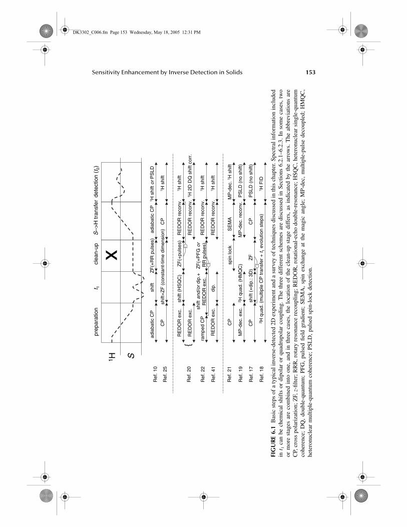
A schematic representation of various types of inverse-detection experiment isdepicted in Figure 6.1. During the first stage, initial
1
H polarization is transferred tothe heteronucleus (
S
) either using CP or some other CT scheme. In the second step,spectral information on
S
, most commonly its chemical shift but also dipolar couplingsor quadrupole coupling, is encoded in an indirect dimension (
t
1
). In addition, whensignificant polarization is on the
S
spin (and possibly stored along the
z
-axis to avoidloss resulting from
T
2
), specific measures can be taken to remove any unwanted protonmagnetization. This is particularly important when isotopically dilute systems areinvestigated, in which leakage of such signals into the detection stage is often delete-rious. Finally, the
S
spin polarization needs to be transferred to protons for detectionon the proton channel, possibly under heteronuclear decoupling.
Most approaches discussed in the next three subsections follow this scheme.Sections 6.2.1 and 6.2.2 comprise techniques designed to work under fastMAS,
21,22,26,28,30,31
where the CP-based techniques (given in Section 6.2.1)
23,27,29
aremostly used to establish chemical-shift correlations and semiquantitative distanceconstraints. Although the REDOR-based
46
heteronuclear CT (Section 6.2.2) givehigh-precision information on heteronuclear dipolar couplings,
26,28
it is also usefulfor the investigation of local structure and dynamics.
35
More specialized approachesapplicable in static samples are summarized in Section 6.2.3.
6.2.1 T
ECHNIQUES
B
ASED
ON
CPMAS
A sensitivity gain in a solid-state inverse
15
N-
1
H shift correlation experiment wassuccessfully demonstrated under fast MAS conditions.
21
For both heteronuclearpolarization transfer steps in the pulse sequence (Figure 6.1), a standard CP sequencewas used. An optimized polarization transfer can be achieved by the use of adiabaticspin-lock pulses during CP on one of the rf channels.
42
This is particularly importantat fast MAS frequencies of around 30 kHz, which are crucial, as the observed gainis only possible with rather narrow proton lines [Equation 6.1]. Although an adiabatictransfer is certainly recommendable for the initial enhancement of
15
N polarization,actual applications like the resonance assignment in proteins might require a morespecific transfer at the second stage; in such applications, polarization transfer amongdirectly dipolar coupled spin pairs is desired, as relayed polarization transfer or spindiffusion defeat the purpose of heteronuclear correlation experiments. In many cases,
DK3302_C006.fm Page 152 Wednesday, May 18, 2005 12:31 PM
Sensitivity Enhancement by Inverse Detection in Solids
153
FIG
UR
E 6.
1
Bas
ic s
teps
of
a ty
pica
l inv
erse
-det
ecte
d 2D
exp
erim
ent a
nd a
sur
vey
of te
chni
ques
dis
cuss
ed in
this
cha
pter
. Spe
ctra
l inf
orm
atio
n in
clud
edin
t
1
can
be
chem
ical
shi
fts
or d
ipol
ar o
r qu
adru
pola
r co
uplin
g. T
he t
hree
dif
fere
nt s
chem
es a
re d
iscu
ssed
in
Sect
ions
6.2
.1–6
.2.3
. In
som
e ca
ses,
tw
oor
mor
e st
ages
are
com
bine
d in
to o
ne,
and
in t
hree
cas
es,
the
loca
tion
of t
he c
lean
-up
stag
e di
ffer
s, a
s in
dica
ted
by t
he a
rrow
s. T
he a
bbre
viat
ions
are
CP,
cro
ss p
olar
izat
ion;
ZF,
z
-filte
r; R
RR
, rot
ary
reso
nanc
e re
coup
ling;
RE
DO
R, r
otat
iona
l-ec
ho d
oubl
e-re
sona
nce;
HSQ
C, h
eter
onuc
lear
sin
gle-
quan
tum
cohe
renc
e; D
Q,
doub
le-q
uant
um;
PFG
, pu
lsed
fiel
d gr
adie
nt;
SEM
A,
spin
exc
hang
e at
the
mag
ic a
ngle
; M
P-de
c, m
ultip
le-p
ulse
dec
oupl
ed;
HM
QC
,he
tero
nucl
ear
mul
tiple
-qua
ntum
coh
eren
ce;
PSL
D,
puls
ed s
pin-
lock
det
ectio
n.
RE
DO
Rre
conv
.
1 H2D
DQ
shift
corr
.R
ED
OR
reco
nv.
RE
DO
Rex
c.
1 Hsh
iftsh
ift(H
SQ
C)
1 H S
prep
arat
ion
clea
n-up
t 1S
-->
Htr
ansf
erde
tect
ion
(t2)
Ref
.10
adia
batic
CP
shift
adia
batic
CP
1 Hsh
iftor
PS
LD
RE
DO
Rex
c.
Ref
.20
ZF
(+R
Rpu
lses
) MP
-dec
.rec
onv.
1 HF
ID2 H
quad
.(m
ultip
leC
Ptr
ansf
er+
t 1ev
olut
ion
step
s)R
ef.1
8
PS
LD(n
osh
ift)
MP
-dec
.exc
.R
ef.1
92 H
quad
.(H
MQ
C)
MP
-dec
.1H
shift
SE
MA
spin
lock
CP
Ref
.21
ZF
(+pu
lses
)
RE
DO
Rre
conv
.1 H
shift
shift
and/
ordi
p.+
RE
DO
Rex
c.ra
mpe
dC
PR
ef.2
2Z
F(+
PF
Gor
RR
puls
es)
RE
DO
Rre
conv
.1 H
shift
Ref
.41
PS
LD(n
osh
ift)
CP
shift
(+di
p.:3
D)
CP
Ref
.17
ZF
RE
DO
Rex
c.
Ref
.25
CP
shift
+Z
F(c
onst
ant-
time
dim
ensi
on)
CP
1 Hsh
ift
x
dip.
{
DK3302_C006.fm Page 153 Wednesday, May 18, 2005 12:31 PM
154
NMR Spectroscopy of Biological Solids
a short, ramped CP
32
has proven to be suitable at fast MAS. However, an off-resonance spin-lock
33,34
can be used for effective polarization transfer among directlydipolar coupled spin pairs under static or slow spinning conditions.
When this technique is to be applied at low isotopic abundance, surplus protonsignals from uncoupled protons that are bound or close to
14
N or
12
C must beremoved. A simple and also rather robust approach was presented later for the caseof
13
C-
1
H correlation at natural abundance,
22
where two half-millisecond pulses atthe rotary resonance recoupling condition (
ν
1
= n
ν
R
) were applied during a
13
C
z
-filter delay to convert the dipolar-coupled
1
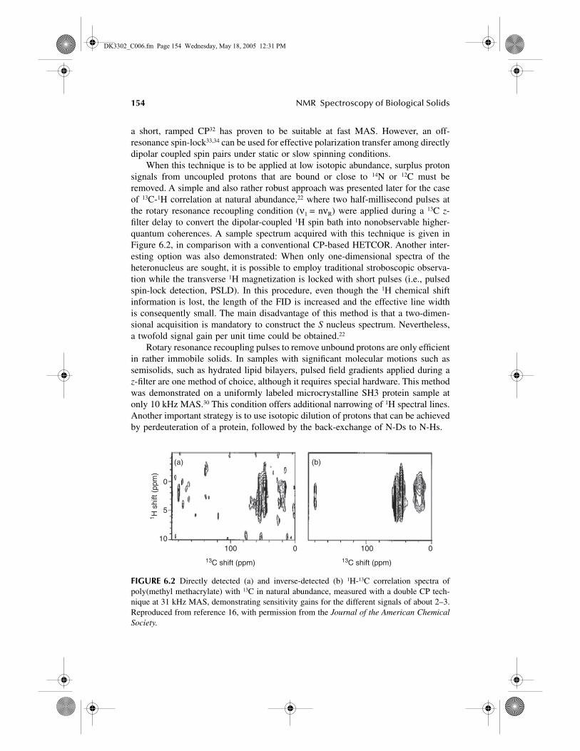
H spin bath into nonobservable higher-quantum coherences. A sample spectrum acquired with this technique is given inFigure 6.2, in comparison with a conventional CP-based HETCOR. Another inter-esting option was also demonstrated: When only one-dimensional spectra of theheteronucleus are sought, it is possible to employ traditional stroboscopic observa-tion while the transverse
1
H magnetization is locked with short pulses (i.e., pulsedspin-lock detection, PSLD). In this procedure, even though the
1
H chemical shiftinformation is lost, the length of the FID is increased and the effective line widthis consequently small. The main disadvantage of this method is that a two-dimen-sional acquisition is mandatory to construct the
S
nucleus spectrum. Nevertheless,a twofold signal gain per unit time could be obtained.
22
Rotary resonance recoupling pulses to remove unbound protons are only efficientin rather immobile solids. In samples with significant molecular motions such assemisolids, such as hydrated lipid bilayers, pulsed field gradients applied during a
z
-filter are one method of choice, although it requires special hardware. This methodwas demonstrated on a uniformly labeled microcrystalline SH3 protein sample atonly 10 kHz MAS.
30
This condition offers additional narrowing of
1
H spectral lines.Another important strategy is to use isotopic dilution of protons that can be achievedby perdeuteration of a protein, followed by the back-exchange of N-Ds to N-Hs.
FIGURE 6.2 Directly detected (a) and inverse-detected (b) 1H-13C correlation spectra ofpoly(methyl methacrylate) with 13C in natural abundance, measured with a double CP tech-nique at 31 kHz MAS, demonstrating sensitivity gains for the different signals of about 2–3.Reproduced from reference 16, with permission from the Journal of the American ChemicalSociety.
(a) (b)
100 0
13C shift (ppm)
100 0
13C shift (ppm)
1 H s
hift
(ppm
)
0
5
10
DK3302_C006.fm Page 154 Wednesday, May 18, 2005 12:31 PM
Sensitivity Enhancement by Inverse Detection in Solids 155
A recent study demonstrated that pulsed field gradients are not even necessaryto remove excess water signal.31 Introduction of a constant-time interval with aprolonged heteronuclear decoupling in the pulse sequence (Figure 6.1) effectivelydephases the water signal. The constant-time interval comprises the t1 evolution timeand the z-filter, where simply the filter store pulse is moved from t1 = 0 to t1,max. Asall pulses on the proton channel stay exactly the same during this procedure, anyresidual water signal is suppressed by a simple two-step phase cycle on the S-channelpulse. In fact, this study showed that it is possible to obtain a two-dimensional 15N-1H chemical shift correlation spectrum of a uniformly 15N labeled protein in about10 min with no additional phase cycling. At 20 kHz MAS, proton line widths asnarrow as 0.2 ppm were reported.
6.2.2 REDOR-BASED TECHNIQUES
A second family of inverse-detection schemes was devised using a quantifiabledipolar transfer step at the very fast spinning speeds necessary to achieve high protonresolution. Earlier work has shown that REDOR, originally designed to recoupledipolar interactions46 and transfer polarization35 between isolated pairs of heteronu-clei, is surprisingly efficient even for 1H-S systems.48,49 REDOR-type CT among 1Hand S nuclei was made possible mainly because fast MAS on the order of 30 kHzeffectively suppresses 1H-1H dipolar couplings. In this limit, the 1H-S spin systemcan, to a very good approximation, be analyzed, using simple expressions derivedusing product operator formalism in conjunction with the simple REDOR averageHamiltonian and a complete neglect of 1H homonuclear couplings.
Apart from the replacement of free scalar-coupling evolution periods by REDORπ pulses spaced by half the rotor period, this whole family of techniques closelyresembles the structure of solution-state inverse-detected experiments. In liquid-state, CT via the scalar coupling is usually restricted to protons directly bound tothe S nucleus, such that Hn → S and S → Hn transfer periods are considered equivalentand are optimized for a specific J value and the multiplet type (or a compromisewhen different kinds of multiplets are measured at the same time). However, moresubtleties arise in the solid-state: although the evolution of S-spin transverse mag-netization is influenced by the joint dipolar field of differently positioned 1H nucleithat are close to the S nucleus, an evolving 1H transverse coherence typically feelsthe dipolar field of only one S nucleus. The former situation is commonly referredto as separated local field (SLF), and the latter is termed proton-detected local field(PDLF) and has the advantage that the theoretical description embodies a simplesummation over spin pairs.50 For this reason, the details of the CT process in a solid-state REDOR experiment depend sensitively on the nucleus that represents thetransverse part of the coherence evolving under REDOR recoupling. The modulationof the final signal intensities observed as a function of the recoupling time or t1 rotorencoding (see following) can be very sensitive to the coupling constants as well asthe local coupling topology, and the choice of transfer pathway should not just bemade on the basis of sensitivity considerations. In fact, when directly detected andinverse-detected experiments are combined, valuable information on the local cou-pling topology (“spin triangulation”) becomes accessible.47
DK3302_C006.fm Page 155 Wednesday, May 18, 2005 12:31 PM

156 NMR Spectroscopy of Biological Solids
There are essentially four possible permutations of transfer pathways, which areshown in Figure 6.3 for the specific case of HSQC experiments, in which evolutionof a heteronuclear antiphase coherence is probed in t1. An HSQC experiment derivedfrom the traditional REDOR scheme is shown in Figure 6.3a. The intensities of thecross signals in such an experiment can be analyzed in terms of the strongestcouplings of S-spin to its surrounding protons,49 and as the S-spin coherence isalways transverse during this experiment, it has the lowest T2
* losses during recou-pling. When the channels are switched, the initial CP can be omitted, however, atthe expense of larger losses by T2
* of protons during recoupling (Figure 6.3b). Thisexperiment is conceptually identical to one of the first inverse HSQC experimentsin solution,19 and its use in solid-state shift correlation has been demonstrated.26 Ithas been proven to provide 5–10-fold sensitivity enhancements over a directlydetected version (Figure 6.3c), which also does not require an initial CP. It wasfurther demonstrated that one could omit t1 and use the sequence only as a hetero-nuclear editing filter in front of a 1H homonuclear DQ shift correlation experiment.
FIGURE 6.3 Variants of REDOR-based heteronuclear single-quantum shift correlation(HSQC) experiments that differ in their SH coherence transfer pathways. Panels (a) and (b)are “symmetric” with respect to the spin which is transverse during the two recoupling periods,and (c) and (d) are “asymmetric” and embody a net polarization transfer. Panel (a) representsthe classic SLF configuration, (b) is a PDLF experiment, and (c) and (d) feature both typesof spin configuration in separate recoupling periods. Panels (b) and (d) are inverse-detectedexperiments.
yx
(a)
(b)
(c)
(d)
t1 detectionexcitation reconversionpreparation
x x
DDH
S
y
x xy
2IxSzCP
CP
DD
y
yx
H
S
x x
CP
CP
y
H
S
y x x
y
y
y
x
H
S
y
y
SS
HH
HS
SHx
DD
DD! τR
τexc τrec
2IxSz
2IzSx
2IzSx
DK3302_C006.fm Page 156 Wednesday, May 18, 2005 12:31 PM
Sensitivity Enhancement by Inverse Detection in Solids 157
The experiment in Figure 6.3c is very similar to the TEDOR experiment,35 theonly difference being that HSQC coherence is monitored in the middle of the transferprocess as opposed to observing single 1H-spin coherence before excitation andreconversion. It combines PDLF and SLF coupling topologies, and calculations showthat this type of CT is only efficient for single S-H moieties at the shortest possiblerecoupling times (one or two rotor periods at 30 kHz MAS). Under these conditions,its total performance is still somewhat inferior to a well-optimized CP, but thisdisadvantage is compensated for by the possibility to determine the actual hetero-nuclear coupling with high precision.48
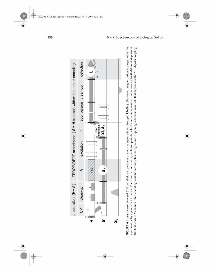
Finally, the experiment in Figure 6.3d is the only one that was successfully usedto obtain 15N-1H correlation spectra and determine their distances in small moleculeswith 15N natural abundance.28,47 A more detailed scheme can be inspected in Figure6.4. The initial CP is essential in that it creates 15N polarization, which can be storedalong the z-axis during the time needed for the removal of the overhead 1H magne-tization. There are two important differences: first, the chemical shift modulationoccurs before the REDOR excitation period, and second, the coherence presentduring a second indirect dimension t1 is heteronuclear dipolar order, which is rotor-encoded when t1 is incremented in fractions of the rotor period. Moving the chemicalshift dimension up front has the advantage that the t1 increment can be chosen freelyto suit the required spectral width. This is not possible in the HSQC variants, inwhich increments are restricted to integer rotor periods. This is because nonintegerrotor period increments between REDOR excitation and reconversion lead to theappearance of a special kind of side-band spectra with spinning side-bands separatedby 2νR. These increments depend sensitively on the heteronuclear dipolar couplingconstant.47 Their large frequency separation is not compatible with the simultaneousencoding of chemical shifts, which would require an extremely high number of slicesin the indirect dimension when considering the concept in the case of HSQC. Thisproblem is overcome by encoding heteronuclear dipolar order (which does notevolve) during t1 simultaneously with the initial t1, but with different increments. Inthis way, the side-band pattern is folded into the chemical shift range, and its apparentfrequency spread is scaled by the ratio of ∆t1/∆t1.
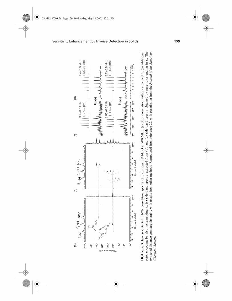
Results obtained using this experiment are shown in Figure 6.5. Note thatheteronuclear dipolar order rotor encoding (HDOR) can also be performed withoutthe additional chemical shift dimension (Figure 6.5d). Efficient processing strategieshelp to further minimize the acquisition time and yield precise coupling informationwithin reasonable experimental times.48 Because the combined shift–side band spec-trum of Figure 5b takes about a day of acquisition time on a 700-MHz spectrometer,this technique, when applied at natural abundance, is clearly limited to the study ofsmall molecules. Experiments on labeled proteins are certainly feasible and promiserich structural insights via the exact measurement of amide-NH distances, whichprovides information on hydrogen bonds.
Finally, we note that probably the most promising inverse experiment of Figure6.3b has not been applied at high isotopic dilution. It does not require an initial CPand features an efficient symmetric REDOR transfer pathway, which is expected tomore than compensate its increased losses resulting from proton T2
*. The HSQCdimension is easily split into a separate HSQC t1 and an HDOR t1. The only major
DK3302_C006.fm Page 157 Wednesday, May 18, 2005 12:31 PM
158 NMR Spectroscopy of Biological Solids
FIG
UR
E 6.
4A
n in
vers
e-de
tect
ed S
-H c
orre
latio
n ex
peri
men
t to
stu
dy s
ampl
es w
ithou
t is
otop
ic l
abel
ing.
Unw
ante
d m
agne
tizat
ion
is p
urge
d ei
ther
by
a gr
adie
nt o
r by
a p
air
of R
RR
pul
ses.
The
re a
re t
wo
sepa
rate
t1
evol
utio
n pe
riod
s, w
hich
can
be
incr
emen
ted
sim
ulta
neou
sly
(with
dif
fere
nt s
tep
size
s).
The
firs
t lea
ds to
S c
hem
ical
shi
ft e
ncod
ing,
and
the
seco
nd s
plits
the
sign
al in
to a
spi
nnin
g si
de-b
and
man
ifol
d th
at d
epen
ds o
n th
e S-
H d
ipol
ar c
oupl
ing.
xy
DDt 1
dete
ctio
nex
cita
tion
clea
n-up
CP
H S
clea
n-up
GZ
prep
arat
ion
(HS
)
φ
TE
DO
R/R
EP
Tex
perim
ent
(S
Htr
ansf
er)
with
/with
outr
otor
-enc
odin
g
x x
y
Sx
I x
reco
nver
sion
t 1'
roto
ren
cod
ing
2IzS
z
DK3302_C006.fm Page 158 Wednesday, May 18, 2005 12:31 PM
Sensitivity Enhancement by Inverse Detection in Solids 159
FIG
UR
E 6.
5In
vers
e-de
tect
ed 1 H
-15N
cor
rela
tion
spec
tra
of L
-his
tidin
e·H
Cl·H
2O a
t 70
0 M
Hz.
(a)
Shi
ft c
orre
latio
n w
ith i
ncre
men
ted
t 1,
(b)
addi
tiona
lro
tor
enco
ding
by
also
inc
rem
entin
g t 1
, (c
) si
de-b
and
spec
tra
extr
acte
d fr
om (
b),
and
(d)
side
-ban
d sp
ectr
a ob
tain
ed b
y pu
re r
otor
end
ing
in t
1. T
heex
trac
ted
dist
ance
com
pare
fav
orab
ly w
ith r
esul
ts f
rom
oth
er m
etho
ds. R
epro
duce
d fr
om r
efer
ence
22,
with
per
mis
sion
fro
m th
e Jo
urna
l of t
he A
mer
ican
Che
mic
al S
ocie
ty.
ppm
2420
1612
84
0pp
m
-150
-200
-250
-300
-350
-400
2420
1612
84
0pp
m
NH
3+ε 2-
NH
δ 1-N
HN
H3+
ε 2-N
Hδ 1-
NH
(a)
(b)
(c)
N
NCH
CO
O
NH
3 H
H
+
+
-
δ 1
ε 2
(d)
ω/ω
R-7
-5-3
-11
35
7
δ 1-N
H
ε 2-N
H
ppm
-50
-150
-250
-350
-100 -50
1 H c
hem
ical
shi
ft1 H
che
mic
al s
hift
15N chemical shift
8.2±
0.9
kHz
(114
±4
pm)
9.4±
0.9
kHz
(109
±4
pm)
9.9±
0.5
kHz
(107
±2
pm)
8.85
±0.
5 kH
z(1
11±
2pm
)
DK3302_C006.fm Page 159 Wednesday, May 18, 2005 12:31 PM
160 NMR Spectroscopy of Biological Solids
requirement would be the application of pulsed field gradients for coherence path-ways selection, as 15N is never present in a pure magnetization state, and a simplez-period for clean up cannot be implemented.
6.2.3 INVERSE DETECTION IN STATIC SAMPLES
The foregoing sections have presented widely useful experiments that yield high-resolution shift and dipolar coupling spectra under fast MAS. In this section, wereview the inverse-detection techniques that are specifically designed for studiesunder static or slow spinning conditions. In one of the techniques, an off-resonancespin-lock34 (i.e., using LG51,52 or flip-flop Lee-Goldburg [FFLG]53,54 pulse sequence)is used to transfer the amide-15N transverse magnetization to its dipolar coupled 1Hspin. This polarization transfer step via SEMA (spin exchange at magic angle) avoidsthe relay of polarization transfer as well as suppresses spin diffusion via 1H-1Hdipolar couplings.34,54 Employing this step in an inverse-detection experiment (Figure6.1), the selective acquisition of amide 1H chemical shifts under multipulse decou-pling (or CRAMPS-type detection1) has been demonstrated.27 A spin-lock pulse inthe S-spin channel after the t1 period is used to suppress the 1H channel before thesecond polarization transfer step. This sequence can also be used to obtain S nucleichemical shift and heteronuclear dipolar coupling in the t1 dimension. Because thismethod involves CRAMPS-type acquisition of the 1H signal,1 it is applicable tosolids under slow spinning condition and to study static aligned samples such asmechanically/magnetically aligned bilayers and liquid crystalline materials.7
A recent study has demonstrated that the PISEMA sequence can be modifiedusing the inverse-detection procedure.7 Because transverse magnetization isexchanged between I and S nuclei during the t1 period (or SEMA sequence) of thePISEMA sequence, a stroboscopic observation of the 1H signal can be used toobserve the heteronuclear dipolar coupling. This method has been successfullydemonstrated on site-specifically 15N-labeled single-crystalline and polycrystallinepeptide samples. The main disadvantage of this sequence is the requirement of thesampling window during a multiple-pulse sequence like FFLG. Because FFLG is awindowless sequence, the insertion of rf-free delays reduces the efficiency of thesequence. In addition, the bulk magnetization from residual solvent (mainly water)or from protons that are not involved in the polarization transfer process result in azero-frequency peak in the IS dipolar coupling spectrum. To avoid this difficulty, itis possible to use a two-dimensional version of this technique that will suppress thenonparticipating 1H magnetization as well as use the spin-lock detection in theacquisition dimension. Such sequences will be useful in structural studies of mem-brane proteins.
A proton inverse-detected deuteron (PRIDE) NMR experiment is demonstratedfor the measurement of 2H wide line spectra from a small amount of sample (on themilligram scale) within 2 h.25 This technique is expected to be useful in studyingmolecular motions in complex organic solids. Evolution under heteronuclear dipolarcoupling although suppressing the homonuclear dipolar couplings, is used to createand reconvert HMQC coherences, which are modulated by the 2H quadrupolarcoupling in an indirect dimension. Proton magnetization is detected using PSLD,
DK3302_C006.fm Page 160 Wednesday, May 18, 2005 12:31 PM
Sensitivity Enhancement by Inverse Detection in Solids 161
which is essential for the observed enhancements on the order of 10–20. An impor-tant feature is that the shape of the 2H spectra is free of artifacts and independentof the HMQC excitation/reconversion times. This would be expected for a powder-average dependent transfer via single 1H-2H dipolar couplings but is not observedbecause the 2H obtains its polarization from many surrounding protons. No specificclean-up stage was introduced, yet good suppression of large amounts of mobilesignal components was also demonstrated. Its suitability to do 2H spectroscopy innatural abundance will need to be examined.
Two-dimensional correlation of 1H-15N dipolar coupling with 15N chemical shiftusing inverse detection was demonstrated for a static sample.23 It employs a simpledouble CP, between which a conventional SLF-type 15N-1H dipolar coupling dimen-sion featuring a partially multipulse-decoupled Hahn echo is combined with asecond, isotropic 15N chemical shift dimension. PSLD is the preferred choice for 1Hdetection, resulting in a three-dimensional measurement protocol, in which, ofcourse, only two dimensions are of interest. Proton inverse-detected nitrogen static(PRINS) NMR has been demonstrated on a labeled peptide and a membrane channeldomain protein, with about twofold sensitivity enhancements. Successful applica-tions in the field of aligned biological samples are anticipated.
Another recent study24 has revived the early experiments of Grannell et al.,43 inwhich the whole process of cross-polarization to an S nucleus, its chemical shiftevolution, and the back transfer to protons is combined within a single extendedproton spin lock period, during which an S spin lock is interleaved with multiple t1
evolution periods. A phase cycling on the S spin lock pulses is apparently sufficientto remove large overheads of uncoupled proton magnetization, such that it providesstatic 2H wide line spectra in natural abundance (0.015%). This is remarkableconsidering that the broad 1H signal was detected directly, without taking advantageof the large potential gain of PSLD used in other cases when 1H chemical shiftinformation is of minor importance. Line shape distortions are apparent in theresultant spectra, but it is proposed that use of modern hardware would eliminatesuch artifacts. The main drawback of this sequence is the use of long spin-lockperiods, which restricts the applications to samples with a long T1ρ.
6.2.4 WHICH EXPERIMENT TO USE?
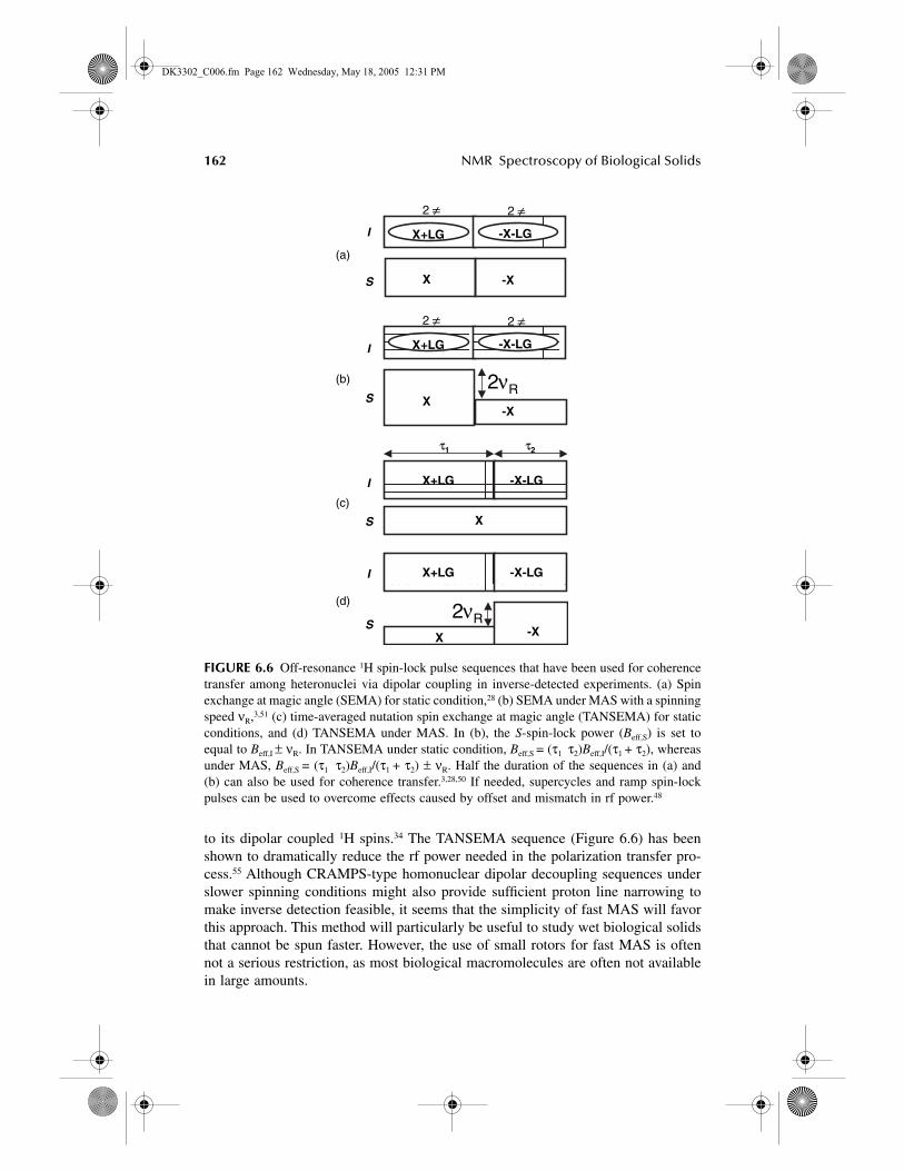
The above sections provided an overview over published approaches to inversedetection in different samples, in which different types of polarization transfer stepshave been employed. Apart from the examples concerning static samples, all othertechniques benefit from fast MAS conditions in excess of 20 kHz, for which specialequipment is mandatory. The choice between CP- and REDOR-based techniques issimple: Given a reasonable spectrometer stability, CP approaches appear to be themost efficient ones and are the methods of choice when qualitative distance con-straints are sought. LG-CP certainly represents a very promising alternative for aquantifiable transfer,34,43,56 but a slight modification to meet the Hartmann-Hahncondition under MAS will be useful for studies under fast spinning frequencies(Figure 6.6).7,57 Because FFLG is efficient in suppressing 1H-1H dipolar couplings,SEMA-type sequence is preferred to selectively transfer coherence from an S nucleus
Au: Pls. specify: 10–20 what?
DK3302_C006.fm Page 161 Wednesday, May 18, 2005 12:31 PM
162 NMR Spectroscopy of Biological Solids
to its dipolar coupled 1H spins.34 The TANSEMA sequence (Figure 6.6) has beenshown to dramatically reduce the rf power needed in the polarization transfer pro-cess.55 Although CRAMPS-type homonuclear dipolar decoupling sequences underslower spinning conditions might also provide sufficient proton line narrowing tomake inverse detection feasible, it seems that the simplicity of fast MAS will favorthis approach. This method will particularly be useful to study wet biological solidsthat cannot be spun faster. However, the use of small rotors for fast MAS is oftennot a serious restriction, as most biological macromolecules are often not availablein large amounts.
FIGURE 6.6 Off-resonance 1H spin-lock pulse sequences that have been used for coherencetransfer among heteronuclei via dipolar coupling in inverse-detected experiments. (a) Spinexchange at magic angle (SEMA) for static condition,28 (b) SEMA under MAS with a spinningspeed νR,3,51 (c) time-averaged nutation spin exchange at magic angle (TANSEMA) for staticconditions, and (d) TANSEMA under MAS. In (b), the S-spin-lock power (Beff,S) is set toequal to Beff,I ± νR. In TANSEMA under static condition, Beff,S = (τ1 τ2)Beff,I/(τ1 + τ2), whereasunder MAS, Beff,S = (τ1 τ2)Beff,I/(τ1 + τ2) ± νR. Half the duration of the sequences in (a) and(b) can also be used for coherence transfer.3,28,50 If needed, supercycles and ramp spin-lockpulses can be used to overcome effects caused by offset and mismatch in rf power.48
I
S X -X
2 ≠ 2 ≠X+LG -X-LG
(a)
2 ≠ 2 ≠X+LG -X-LG
X-X
I
S
(b) 2νR
X+LG -X-LG
X -X
I
S
(d)2νR
X
X+LG -X-LGI
S
(c)
τ1 τ2
DK3302_C006.fm Page 162 Wednesday, May 18, 2005 12:31 PM
Sensitivity Enhancement by Inverse Detection in Solids 163
Pulsed field gradients have been used in some cases to remove mobile watersignals and surplus uncoupled 1H magnetization in samples with a low abundant Snucleus. However, both problems can be solved with conventional probes and clean-up based on rf irradiation (i.e., prolonged 1H decoupling during a z-filter in a constanttime protocol and RRR pulses, respectively). Gradients might, however, prove indis-pensable when recoupling pulse sequences, such as the symmetric REDOR-HSQC,do not allow for the introduction of S nucleus z-filter delays, during which clean-up rf schemes can be applied.
The REDOR-based schemes are at present the best choice when precise dipolarcouplings between the heteronuclei are to be determined. Also, in this case, we stressthe fact that such studies do not depend on a scaling factor, as REDOR is ratherrobust and forgiving of experimental errors resulting from nonideality of π pulses.This holds in particular when the rotor-encoding spinning side-bands, instead ofspectral intensities, are used for the determinations. In the following section, wepresent a model study in which not only precise distances but also a full couplingtopology was elucidated by these methods.
6.3 APPLICATIONS
In general, most of the techniques discussed herein have just recently been devel-oped, and not many actual applications have emerged until now. The sensitivity gainis the key factor that will enable many studies to be completed within a fraction ofthe time needed to do the experiments in the conventional way. We here append afew further examples that highlight the great potential of the new methods, whichwe expect to have a significant effect on the routine toolbox of the solid-state NMRspectroscopists.
6.3.1 APPLICATIONS TO STUDY SMALL MOLECULES AND MATERIALS
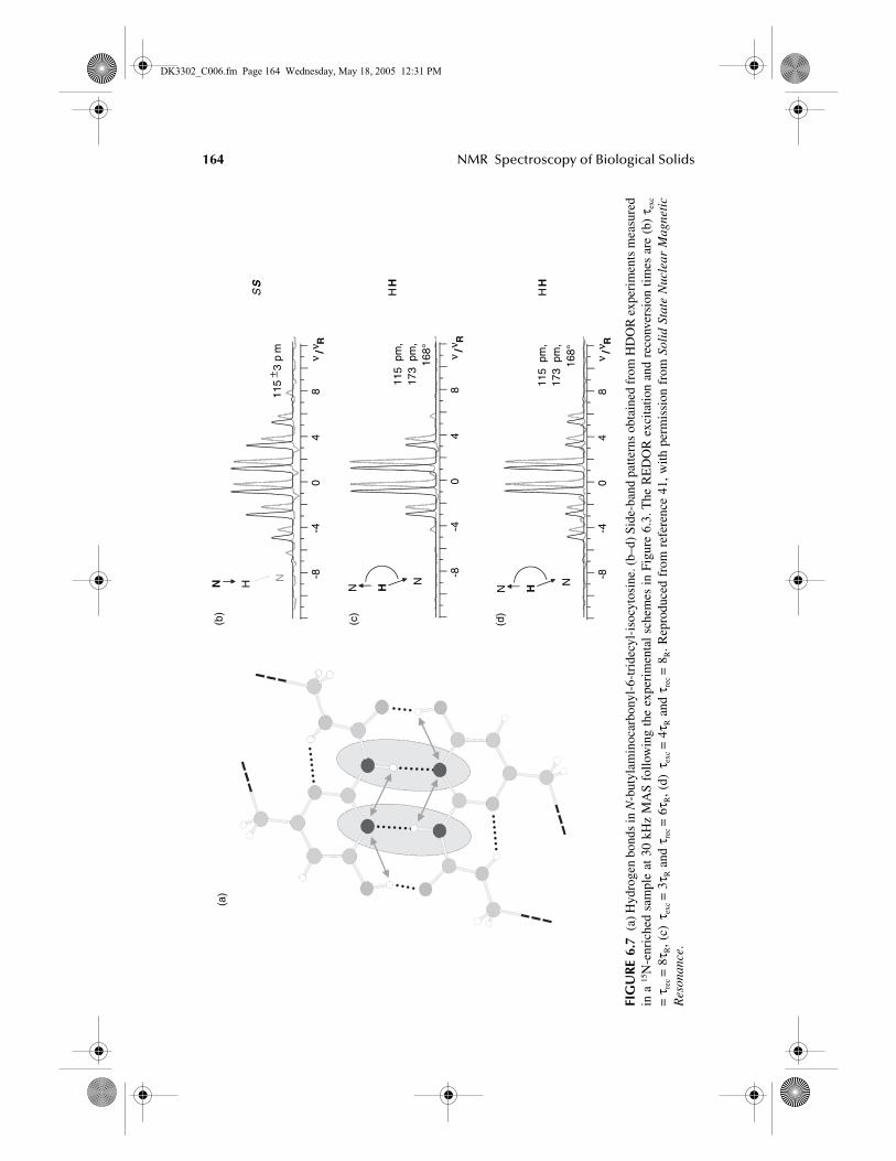
In Section 6.2.2, it was indicated that the REDOR-based approaches not only holdpromise for remarkable sensitivity enhancements by inverse detection but also serveto elucidate local coupling geometries by combining inverse- and directly detectedexperiments and making use of the different influence of SLF- and PDLF-typecoupling topologies on the measured signals. We highlight this aspect here byreviewing a study concerned with the structure of N-H…N hydrogen bonds in theenol form of N-butylaminocarbonyl-6-tridecyl-isocytosine. A dimer structure (Figure6.7a) is stabilized by four hydrogen bonds, and we focus on the two equivalentcentral ones between a urea-N and a pyrimidine-N. Figure 6.7b shows a directlydetected side-band spectrum using the pulse scheme of Figure 6.3a, in which HDORrather than HSQC was selected and rotor-encoded during t1 and was then Fourier-transformed. Each of the urea-N has a relatively tightly bound 1H with secondarycouplings being comparably weak; thus, the bond length is easily determined fromthe side-band intensities using a simple spin-pair solution for the fit. When analogousside-band spectra are taken with the inverse-detected version of the same experiment(Figures 6.7c and 6.7d), the local coupling topology around the evolving and detected1H coherence is strongly influenced by both nitrogens in the hydrogen bond. The
Au: Pls. write out “RRR.”
DK3302_C006.fm Page 163 Wednesday, May 18, 2005 12:31 PM
164 NMR Spectroscopy of Biological Solids
FIG
UR
E 6.
7(a
) Hyd
roge
n bo
nds
in N
-but
ylam
inoc
arbo
nyl-
6-tr
idec
yl-i
socy
tosi
ne. (
b–d)
Sid
e-ba
nd p
atte
rns
obta
ined
from
HD
OR
exp
erim
ents
mea
sure
din
a 15
N-e
nric
hed
sam
ple
at 3
0 kH
z M
AS
follo
win
g th
e ex
peri
men
tal
sche
mes
in
Figu
re 6
.3. T
he R
ED
OR
exc
itatio
n an
d re
conv
ersi
on t
imes
are
(b)
τex
c
= τ
rec =
8τ R
, (c)
τex
c =
3τ R
and
τre
c =
6τ R
, (d)
τex
c =
4τ R
and
τre
c =
8R. R
epro
duce
d fr
om r
efer
ence
41,
with
per
mis
sion
fro
m S
olid
Sta
te N
ucle
ar M
agne
tic
Res
onan
ce.
-8-4
04
8ν
/ν R
(d)
(c)
N H N
N H NN H N
115
± 3p
m
115
pm,
173
pm,
168°
115
pm,
173
pm,
168°
-8-4
04
8ν
/ν R
-8-4
04
8ν
/ν R
(b)
(a)
SS
HH
HH
DK3302_C006.fm Page 164 Wednesday, May 18, 2005 12:31 PM
Sensitivity Enhancement by Inverse Detection in Solids 165
side-band modulation then depends not only on the two distances but also on theangle between the two coupling tensors. This unique correlation between distancesand angles can be further amplified by using different REDOR recoupling times forexcitation and reconversion. This leads to some intensity loss (as this is not thecondition for optimum transfer), but still the obtained side-band spectra are consid-erably less noisy than the one in Figure 6.7b as a result of inverse detection.
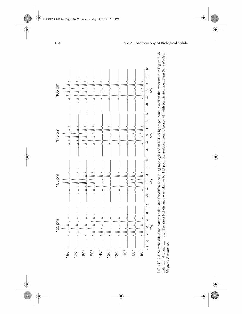
The missing longer distance and the hydrogen bond angle can by deduced bycomparison with simulated side-band spectra plotted in Figure 6.8. The accuracy ofthe determination can be increased by analyzing further side-band spectra takenunder different recoupling conditions and fitting them to the same set of angle anddistance. Obviously, this approach bears a large potential for applications in thebiological and material sciences, in which hydrogen bonds are probably the mostimportant supramolecular structure-directing interaction. NMR thus provides accessto structure–function relationships in materials that do not need to be crystalline.
6.3.2 BIOMOLECULAR APPLICATIONS
Most of the presently used solid-state NMR experiments require a large quantity ofsample or a long acquisition time to obtain a reasonable S/N multidimensionalspectrum from biological solids. Because most of the interesting biological mole-cules, such as membrane-associated proteins and RNA, are not available in largequantities, applications of existing NMR techniques to such systems are highlyrestricted. However, in many cases, even when obtaining large quantities of thesystem of interest is feasible, the use of a large quantity of such a system does notprovide biologically relevant information. For example, in the structural studies ofmembrane-permeating peptides (such as antimicrobial peptides, toxins, fusion pep-tides, and channel forming peptides), there is considerable interest in obtaining datafrom biologically relevant concentrations of the peptide in lipid bilayers, as theincrease in the peptide concentration can lead to the disruption of the lipid bilayerstructure.58,59 Ligand-binding studies are another example in which the concentrationof the ligand cannot be increased to acquire signal from the bound ligand. In addition,the mandatory sample size restriction is a heavy price to pay for static as well asMAS experiments with a higher magnetic field. Long signal acquisitions used toavoid these concerns suffer from spectrometer instability and sample denaturingresulting from rf heat dissipation. Therefore, the remarkable sensitivity gainedthrough an inverse-detected experiment could be the long-awaited “magic wand”for NMR applications on biological solids.
Inverse-detected NMR experiments for studies on biosolids fall under two cat-egories: aligned or unaligned crystalline samples. Experiments on aligned samplesare usually performed under static condition, whereas unaligned samples are studiedunder MAS. Experiments need to be chosen on the basis of the nature of the sampleunder study. The advantages of using fast sample spinning and REDOR-basedcoherence transfer cannot be used for experiments on aligned samples or on samplesthat cannot be spun faster. However, the rate of CT among heteronuclear spins andthe extent of line broadening caused by homo- and heteronuclear dipolar couplingsdepend on the rigidity of the sample. For example, because of molecular motions
DK3302_C006.fm Page 165 Wednesday, May 18, 2005 12:31 PM
166 NMR Spectroscopy of Biological Solids
FIG
UR
E 6.
8Sa
mpl
e si
de-b
and
patte
rns
calc
ulat
ed f
or d
iffe
rent
cou
plin
g to
polo
gies
of
an N
-H-N
hyd
roge
n bo
nd, b
ased
on
the
expe
rim
ent i
n Fi
gure
6.3
bw
ith τ
exc =
4τ R
and
τre
c =
8τ R
. The
sho
rt N
H d
ista
nce
was
tak
en t
o be
115
ppm
. Rep
rodu
ced
from
ref
eren
ce 4
1, w
ith p
erm
issi
on f
rom
Sol
id S
tate
Nuc
lear
Mag
neti
c R
eson
ance
.
-12
-8-4
04
812
ν /ν R
90°
100°
110°
120°
130°
140°
150°
160°
170°
180°
185
pm17
5 pm
165
pm15
5 pm
-8-4
04
812
ν /ν R
-8-4
04
812
ν /ν R
-8-4
04
812
ν /ν R
DK3302_C006.fm Page 166 Wednesday, May 18, 2005 12:31 PM
Sensitivity Enhancement by Inverse Detection in Solids 167
in wet samples, dipolar couplings are partially averaged out, and therefore the contacttime for optimum cross polarization needs to be carefully chosen. In addition, theuse of constant time period is not possible if T2 is not favorable.
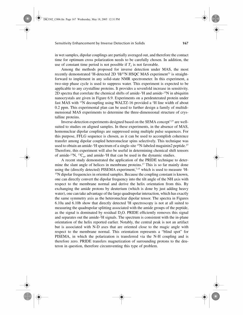
Among the methods proposed for inverse detection under MAS, the mostrecently demonstrated 1H-detected 2D 1H/15N HSQC MAS experiment31 is straight-forward to implement in any solid-state NMR spectrometer. In this experiment, atwo-step phase cycle is used to suppress water. This experiment is expected to beapplicable to any crystalline proteins. It provides a sevenfold increase in sensitivity.2D spectra that correlate the chemical shifts of amide-1H and amide-15N in ubiquitinnanocrystals are given in Figure 6.9. Experiments on a perdeuterated protein underfast MAS with 15N decoupling using WALTZ-16 provided a 1H line width of about0.2 ppm. This experimental plan can be used to further design a family of multidi-mensional MAS experiments to determine the three-dimensional structure of crys-talline proteins.
Inverse-detection experiments designed based on the SEMA concept7,27 are well-suited to studies on aligned samples. In these experiments, in the absence of MAS,homonuclear dipolar couplings are suppressed using multiple pulse sequences. Forthis purpose, FFLG sequence is chosen, as it can be used to accomplish coherencetransfer among dipolar coupled heteronuclear spins selectively. This technique wasused to obtain an amide-1H spectrum of a single-site 15N-labeled magainin2 peptide.27
Therefore, this experiment will also be useful in determining chemical shift tensorsof amide-15N, 13Cα, and amide-1H that can be used in the dynamic studies.
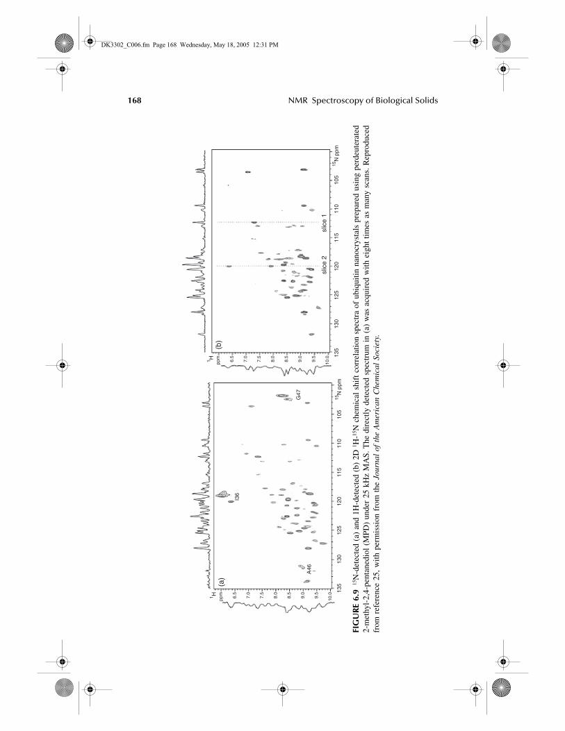
A recent study demonstrated the application of the PRIDE technique to deter-mine the slant angle of helices in membrane proteins.17 This is so far mainly doneusing the (directly detected) PISEMA experiment,7,15 which is used to measure 1H-15N dipolar frequencies in oriented samples. Because the coupling constant is known,one can directly convert the dipolar frequency into the tilt angle of the NH axis withrespect to the membrane normal and derive the helix orientation from this. Byexchanging the amide protons by deuterium (which is done by just adding heavywater), one can take advantage of the large quadrupolar interaction, which has exactlythe same symmetry axis as the heteronuclear dipolar tensor. The spectra in Figures6.10a and 6.10b show that directly detected 2H spectroscopy is not at all suited tomeasuring the quadrupolar splitting associated with the amide groups of the peptide,as the signal is dominated by residual D2O. PRIDE efficiently removes this signaland separates out the amide-2H signals. The spectrum is consistent with the in-planeorientation of the helix reported earlier. Notably, the central peak is not an artifactbut is associated with N-D axes that are oriented close to the magic angle withrespect to the membrane normal. This orientation represents a “blind spot” forPISEMA, in which the polarization is transferred via the N-H coupling and istherefore zero. PRIDE transfers magnetization of surrounding protons to the deu-teron in question, therefore circumventing this type of problem.
DK3302_C006.fm Page 167 Wednesday, May 18, 2005 12:31 PM
168 NMR Spectroscopy of Biological Solids
FIG
UR
E 6.
915
N-d
etec
ted
(a)
and
1H-d
etec
ted
(b)
2D 1 H
-15N
che
mic
al s
hift
cor
rela
tion
spec
tra
of u
biqu
itin
nano
crys
tals
pre
pare
d us
ing
perd
eute
rate
d2-
met
hyl-
2,4-
pent
aned
iol
(MPD
) un
der
25 k
Hz
MA
S. T
he d
irec
tly d
etec
ted
spec
trum
in
(a)
was
acq
uire
d w
ith e
ight
tim
es a
s m
any
scan
s. R
epro
duce
dfr
om r
efer
ence
25,
with
per
mis
sion
fro
m t
he J
ourn
al o
f th
e A
mer
ican
Che
mic
al S
ocie
ty.
1 H
15N
ppm
ppm 6.5
7.0
7.5
8.0
8.5
9.0
9.5
10.0
135
130
125
120
115
110
105
1 H
15N
ppm
ppm 6.5
7.0
7.5
8.0
8.5
9.0
9.5
10.0 13
513
012
512
011
511
010
5
I36
A46
G47
(a)
(b)
slic
e 2
slic
e 1
DK3302_C006.fm Page 168 Wednesday, May 18, 2005 12:31 PM
Sensitivity Enhancement by Inverse Detection in Solids 169
FIG
UR
E 6.
10St
atic
2 H s
pect
ra o
f 2.
5 m
g ba
ck-e
xcha
nged
ovi
spir
in,
cons
titut
ed i
n m
embr
ane
lipid
s (m
ole
ratio
1:2
7).
(a)
Dir
ectly
det
ecte
d sp
ectr
um,
(b)
the
sam
e, m
agni
fied
1000
tim
es v
ertic
ally
, an
d (c
) pr
oton
inv
erse
-det
ecte
d sp
ectr
um,
expe
rim
enta
l tim
e: 6
h.
Rep
rodu
ced
from
ref
eren
ce 2
3, w
ithpe
rmis
sion
fro
m t
he J
ourn
al o
f M
agne
tic
Res
onan
ce.
(a)
(b)
(c)
200
0–2
002 H
qua
drup
olar
cou
plin
g (k
Hz)
200
0–2
002 H
qua
drup
olar
cou
plin
g (k
Hz)
200
0–2
002 H
qua
drup
olar
cou
plin
g (k
Hz)
× 10
00
DK3302_C006.fm Page 169 Wednesday, May 18, 2005 12:31 PM
170 NMR Spectroscopy of Biological Solids
REFERENCES
1. Schmidt-Rohr, K. and Spiess, H.W., Multidimensional Solid-State NMR and Poly-mers, Academic Press, New York, 1994.
2. Castellani, F., van Rossum, B., Diehl, A., Schubert, M., Rehbein, K. and Oschkinat,H., Nature, 420, 98, 2002.
3. Baldus, M., Prog. NMR Spectrosc., 41, 1, 2002.4. Saito, H., Tuzi, S., Tanio, M., Naito, K., Ann. Rep. NMR Spectrosc., 47, 41, 2002.5. Saito, H., Yamaguchi, S., Ogawa, K., Tuzi, S., Marquez, M., Sanz, C., Biophys. J.,
86, 1673, 2004.6. Fujiwara, T., Todokoro, Y., Yanagishita, H., Tawarayama, M., Kohno, T., Wakamatsu,
K. and Akutsu, H., J. Biomol. NMR, 28, 311, 2004. 7. Ramamoorthy, A., Wei, Y. and Lee, D.K., Ann. Rep. NMR Spectrosc., 52, 1, 2004.8. Marassi, F.M. and Opella, S.J., Chem. Rev., 104, 3587, 2004.9. Wu, C.H., Ramamoorthy, A. and Opella, S.J.J., Magn. Reson., A109, 270, 1994.
10. Ramamoorthy, A. and Opella, S.J., Solid State NMR Spectrosc. 4, 387, 1995 11. Ramamoorthy, A., Marassi, F. and Opella, S.J., Applications of multidimensional
solid-state NMR spectroscopy to membrane proteins, in Proceedings of the Int. Schoolof Biological Magnetic Resonance, 2nd course, Dynamics and the Problem of Rec-ognition in Biological Macromolecules (Jardetsky O. and Lefeure, J. Eds.), Plenum,New York, 1996, p. 238.
12. Jelinek, R., Ramamoorthy, A. and Opella, S.J., High-resolution three-dimensionalsolid-state NMR spectroscopy of a uniformly 15N-labeled protein, J. Am. Chem. Soc.,117, 12348, 1995.
13. Marassi, F.M., Ramamoorthy, A. and Opella, S.J., Proc. Natl. Acad. Sci. USA, 94,8551, 1997.
14. Wang, J., Denny, J., Tian, C., Kim, S., Mo, Y., Kovacs, F., Song, Z., Nishimura, K.,Gan, Z., Fu, R., Quine, J.R. and Cross, T.A., J. Magn. Reson., 144, 162, 2000.
15. Marassi, F.M. and Opella, S.J., Protein Sci., 12, 403, 2003.16. Thiriot, D.S., Nevzorov, A.A., Zagyanskiy, L., Wu, C. and Opella, S.J., J. Mol. Biol.,
341, 869, 2004.17. Maudsley, A.A., Müller, L. and Ernst, R.R., J. Magn. Reson., 28, 463, 1977.18. Müller, L., J. Am. Chem. Soc., 101, 4481, 1979.19. Bodenhausen, G. and Ruben, D.J., Chem. Phys. Lett., 69, 185, 1980.20. Bax, A., Curr. Opin. Struct. Biol., 4, 738, 1994.21. Ishii, Y. and Tycko, R., J. Magn. Reson., 142, 199, 2000.22. Ishii, Y., Yesinowski, J.P. and Tycko, R., J. Am. Chem. Soc., 123, 2921, 2001.23. Hong, M. and Yamaguchi, S., J. Magn. Reson., 150, 43, 2001.24. Khitrin, A.K. and Fung, B.M., J. Magn. Reson., 152, 185, 2001.25 Schmidt-Rohr, K., Saalwächter, K., Liu, S.-F. and Hong, M., J. Am. Chem. Soc.,
123, 7168, 2001.26. Schnell, I., Langer, B., Sontjens, S.H.M., van Genderen, M.H.P., Sijbesma, R.P. and
Spiess, H.W., J. Magn. Reson., 150, 57, 2001.27. Wei, Y., Lee, D.-K., Hallock, K.J. and Ramamoorthy, A., Chem. Phys. Lett., 351, 42,
2002.28. Schnell, I. and Saalwächter, K., J. Am. Chem. Soc., 124, 10938, 2002.29. Yamaguchi, S. and Hong, M., J. Magn. Reson., 155, 244, 2002.30. Chevelkov, V., van Rossum, B.J., Castellani, F., Rehbein, K., Diel, A., Hohwy, M.H.,
Steuernagel, S., Engelke, F., Oschkinat, H. and Reif, B., J. Am. Chem. Soc., 125,7788, 2003.
Au: Pls. pro-vide titles for all articles.
DK3302_C006.fm Page 170 Wednesday, May 18, 2005 12:31 PM
Sensitivity Enhancement by Inverse Detection in Solids 171
31. Paulson, E.K., Morcombe, C.R., Gaponenko, V., Dancheck, B., Byrd, R.A. andZilm.K.W., J. Am. Chem. Soc., 125, 15831, 2003.
32. Metz, G. Wu, X. and Smith, S.O., J. Magn. Reson. A, 110, 219, 1994.33. Shekar, S.C., Lee, D.K. and Ramamoorthy, A., J. Magn. Reson., 157, 223, 2002.34. Ramamoorthy, A., Wu, C.H. and Opella, S.J., J. Magn. Reson., 140, 131, 1999.35. Hing, A.W., Vega, S. and Schaefer, J., J. Magn. Reson., 96, 205, 1992.36. Ramamoorthy, A. and Chandrakumar, N., J. Magn. Reson., 100, 60, 1992 37. Sattler, M., Schleucher, J. and Griesinger, C., Progr. Nucl. Magn. Reson. Spectrosc.,
34, 93, 1999.38. Lesage, A., Steuernagel, S. and Emsley, L., J. Am. Chem. Soc., 120, 7095, 1998.39. Taylor, D.A. and Ramamoorthy, A., J. Magn. Reson., 141, 18, 1999.40. Shekar S.C. and Ramamoorthy, A., Chem. Phys. Lett., 342, 127, 2001.41. Taylor, D.A. and Ramamoorthy, A., J. Mol. Struct., 602, 115, 2001.42. Hediger, S., Meier, B.H. and Ernst.R.R., Chem. Phys. Lett., 240, 449, 1995.43. Grannell, P.K., Mansfield, P. and Whitaker, M.A.B., Phys. Rev. B, 8, 4149, 1973. 44. Vega, S., Shattuck, T.W. and Pines, A. Phys. Rev. A, 22, 638, 1980.45. Franke, D., Hudalla, C. and Eckert, H., Solid State Nucl. Magn. Reson., 1, 33, 1992.46. Gullion, T. and Schaefer, J., J. Magn. Reson., 81, 196, 1989.47. Saalwächter, K. and Schnell, I. Solid State Nucl. Magn. Reson., 22, 154, 2002.48. Saalwächter, K., Graf, R. and Spiess, H.W., J. Magn. Reson., 148, 398, 2001.49. Saalwächter, K. and Spiess, H.W., J. Chem. Phys., 114, 5707, 2001.50. Schmidt-Rohr, K., Nanz, D., Emsley, L. and Pines, A., J. Phys. Chem., 98, 6668, 1994.51. Lee, M. and Goldburg, W.J., Phys. Rev. A, 140, 1261, 1965. 52. Ravikumar, M. and Ramamoorthy, A., Chem. Phys. Lett., 286, 199, 1998.53. Mehring, M. and Waugh, J.S., Phys. Rev. B, 5, 3459, 1972.54. Bielecki, A., Kolbert, A.C., de Groot, H.S.M., Griffin, R.G. and Levitt, M.H., Adv.
Magn. Reson., 14, 111, 1990.55. Lee, D.K., Narasimhaswamy, T. and Ramamoorthy, A., Chem. Phys. Lett., 399, 359,
2004 56. van Rossum, B.-J., de Groot, C.P., Ladizhanski, V., Vega, S. and de Groot, H.J.M.,
J. Am. Chem. Soc., 122, 3465, 2000. 57. Dvinskikh, S.V., Zimmermann, H., Maliniak, A. and Sandstrom, D., J. Magn. Reson.,
164, 165, 2003.58. Lee, D.K., Henzler-Wildman, K.A. and Ramamoorthy, A., J. Am. Chem. Soc., 126,
2318, 2004.59. Henzler-Wildman, K.A. Biochemistry, 43, 8459, 2004.
DK3302_C006.fm Page 171 Wednesday, May 18, 2005 12:31 PM
DK3302_C006.fm Page 172 Wednesday, May 18, 2005 12:31 PM


![SAMPLING-BASED RBDO USING STOCHASTIC SENSITIVITY …inverse reliability analysis using HMV+ and design optimization using PMA+ [14,15]. The propagation of the input uncertainty into](https://img.pdfslide.us/doc/110x75/5ede3c05ad6a402d66698d10/sampling-based-rbdo-using-stochastic-sensitivity-inverse-reliability-analysis-using.jpg)