Embed Size (px)
Citation preview

6 Indian Journal of Contemporary Dentistry, July-December 2021, Vol.9, No.2
Post Covid-19 Mucormycosis (Black Fungus) and Its Prosthodontic Considerations
Gayatri Deokar1, Babita Yeshwante2, Nazish Baig3, Vivek Jadhav4, Rasika Patil1
1Postgraduate Student, 2Professor and Head Dept.of Prosthodontics, 3Professor, 4Associate Professor, Dept.of Prosthodontics, Crown & Bridge and Implantology, CSMSS Dental college & Hospital,Aurangabad, Maharashtra
AbstractWhile our country battles with COVID-19, the issue of post COVID-19 mucormycosis termed as black fungus has emerged as a significant problem. India bears the dubious distinction of being both the diabetes, as well as the mucormycosis, ‘capital’ of the world.
Mucormycosis is one of the several opportunistic fungi that becomes invasive and pathogenic in patients with altered metabolic status in those who are immunologically compromised.It is of particular interest to the dentists because of the presenting symptoms and the oral and facial defects that result from the course of the disease and the treatment.
Keywords: Mucormycosis, POST-COVID 19, Obturator.
Introduction
With an estimated 77 million cases in the adult population, diabetes is India’s fastest growing epidemic. A recent cross-sectional study from all states of India, revealed that 47% of Indians are unaware of their diabetic status and only a quarter of all patients achieved adequate glycemic control on treatment. The unholy association between diabetes and the severity of SARS-CoV-2 infection has been repeatedly established in various studies from across the world.
Mucormycosis sometimes appears as the diabetes-defining illness, and remains one of the most devastating complications in uncontrolled diabetics with mortality rates ranging between 40-80%. India contributes to 40% of the global burden of this “rare mould” infection as it is called in western literature, with an estimated prevalence of 140 cases per million population.
Post COVID-19 sepsis is what occurs after SARS- CoV-2 has had a rampage in the human body and we are literally left picking up the pieces. It leads to a dysregulated innate immune response, ciliary dysfunction, cytokine storm, thrombo-inflammation,
microvascular coagulation and eventual immune exhaustion. This cascade of events facilitates secondary bacterial and fungal infections especially in critically ill patients subjected to emergency invasive procedures, mechanical ventilation, CRRT, ECMO, poor nursing ratios, prolonged hospital stays and breaches in asepsis. Further, the use of corticosteroid treatment and anti-IL-6-directed strategies in these highly susceptible hosts along with high fungal spore counts in the environment creates the perfect setting for mould infections.
While COVID-19-associated pulmonary aspergillosis (CAPA) has received much international attention, the Indian epidemiology of invasive mould infections in the ICU reveals a significant burden of invasive mucormycosis.This has recently emerged as a life threatening complication of COVID-19 in our country. Although the predisposing factors and pathogenesis are somewhat similar to that of other mould infections, certain unique characteristics and key distinguishing factors must be kept in mind in order to promptly suspect the infection, confirm the diagnosis and offer timely therapeutic intervention.1
Indian Journal of Contemporary Dentistry, July-December 2021, Vol.9, No.2 7
POST COVID-19 MUCORMYCOSIS:-
Mucorales are ubiquitous moulds, abundantly found in the environment on decaying organic matter. Various studies from hospitals across the country have revealed heavy mould spore counts even in hospital air due to predominantly hot, humid conditions in our tropical climate.2
Unlike CAPA, invasive mucormycosis has been observed even in patients with mild to moderate SARS- CoV-2 infections. The strongest predisposing factor appears to be hyperglycemia in undiagnosed or uncontrolled diabetics. Hyperglycemia leads to increased expression of the endothelial receptor GRP78, resulting in polymorphonuclear dysfunction, impaired chemotaxis and defective intracellular killing. An important virulence trait of Mucorales is the ability to acquire iron from the host which is an essential element for its growth. In conditions of ketoacidosis, free iron becomes readily available in the serum. This excess endogenous iron is efficiently taken up by the Mucorales through siderophores or iron permeases, further enhancing their virulence. These effects are greatly amplified by the use of corticosteroids and immunosuppressants in susceptible hosts. Corticosteroids themselves cause impairment in the neutrophil migration, ingestion, and phagolysosome fusion. Coupled with the potential implications of steroid-induced hyperglycemia, the diabetic COVID 19 patient receiving corticosteroids or other immunosuppressants is exceptionally vulnerable to the development of mucormycosis.3,4
The landmark RECOVERY trial published in June 2020 has served as a ‘license’ to use steroids in patients with COVID-19. However, the fine print clearly revealed some important messages that we seem to have overlooked. Benefit was specifically shown with low dose, short duration dexamethasone in moderate to severe illness. Although, higher doses and longer durations may be used in exceptional cases due to compelling reasons, such patients should be evaluated for undiagnosed diabetes, checked for strict glycemic control and closely monitored for secondary infections. A cavalier attitude to the use of steroids should be
discouraged at all costs.
The two most important manifestations of Mucormycosis in this setting are rhino-orbital-cerebral and pulmonary. Suspicion is based on subtle clinical and imaging clues, risk factors and disease development or progression while on any antibacterial or antifungal therapy that does not cover Mucor. Physicians need to have seen a ‘critical’ number of cases to recognize the signature of Mucor.1
POST COVID-19 CLINICAL MANIFESTATIONS AND DIAGNOSIS:-
The clinical hallmark is tissue necrosis manifested as a necrotic lesion, eschar or black discharge in the nasal or oral cavity. Orbital, ocular and cranial nerve involvement are ominous signs that must be taken seriously. Alternative erroneous diagnoses lead to antibacterial and further steroid use which add fuel to the fire. Pulmonary Mucormycosis has certain radiologic findings which help to distinguish it from Aspergillosis. There is no biomarker for mucormycosis and hence a negative galactomannan and beta-d-glucan are useful pointers to rule out other mould infections. A false positive galactomannan due to generic piperacillin tazobactam use etc. can lead to the erroneous diagnosis of invasive aspergillosis. Although challenging, the need to distinguish Mucor from bacterial infections and from aspergillosis in a timely fashion is of essence. Treatment with voriconazole for suspected invasive aspergillosis increases the pathogenicity of Mucor with obvious dire consequences.
Rapid diagnostic methods include biopsy, KOH mount and Calcofluor stain. Mucor is difficult to routinely culture. Biopsy remains the mainstay of diagnosis and the benefits of the procedure outweigh the risk, even in a ‘difficult to access’ location or in the presence of coagulopathy.1
POST COVID-19 TREATMENT
Treatment principles include antifungal agents, surgical debridement, reversal of underlying predisposing factors and adjuvant therapy. Amphotericin B has been the standard of treatment for invasive

8 Indian Journal of Contemporary Dentistry, July-December 2021, Vol.9, No.2
mucormycosis. COVID-19 patients may have developed acute on chronic renal failure which may be mitigated by switching to a less- or non-nephrotoxic alternative. Therefore Posaconazole or Isavuconazole may have to be used. The latter has the added advantage of shortening the QT interval which may have been affected by HCQ, Azithromycin which many patients still continue to receive. Surgical debridement, the earlier the better, is pivotal in the management of mucormycosis. The optimal time of surgery to reduce the operative risk to the patient with COVID-19 and the risk of transmission to the operating team is a contentious issue. Replication competent virus has not been recovered from patients
with mild to moderate illness after ten days, from patients with severe illness after fifteen days or from any critically ill patient after twenty days.5
Adjuvant therapy with caspofungin, deferasirox, statins, aspirin, and hyperbaric oxygen may have to be considered. Mucormycosis needs to be actively managed by a team which includes members from almost all departments in the hospital. Therapy is toxic and very resource intensive. In a recent Indian study, 24.3% patients left the hospital against medical advice due to the anticipated cost, morbidity of surgery and prognosis.6
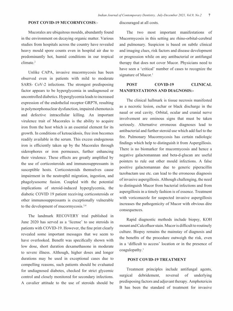
Fig 1: Evidence based advisory for Mucormycosis in the time of covid (ICMR approved)

Indian Journal of Contemporary Dentistry, July-December 2021, Vol.9, No.2 9
MUCORMYCOSIS
Opportunistic fungal infections such as mucormycosis usually occur in immunocompromised patients, but can infect healthy individuals as well. The predisposing factors for mucormycosis are uncontrolled diabetes (particularly in patients with ketoacidosis), malignancies such as lymphomas and leukemia’s, renal failure, organ transplant, long‐term corticosteroid and immunosuppressive therapy, cirrhosis, burns, protein energy malnutrition and acquired immune deficiency syndrome.7
PATHOGENESIS-
The infection begins in the nose and para nasal sinuses due to inhalation of fungal spores. The infection can spread to orbital and intracranial structures either by direct invasion or through the blood vessels.The fungus invades the arteries leading to thrombosis that subsequently causes necrosis of hard and soft tissues.
Mucormycosis primarily affects immunocompromised, bone marrow–transplanted, hematological malignancies, or poorly controlled diabetic individuals Rhizopus is the chief pathogenic mycotic organism in cases with rhinocerebral mucormycosis. Mucormycoses are identified on bread mold, soil, decaying vegetation, or animal manure. Cultures of swab obtained from the oral cavity, nasal cavity, throat, and stools of healthy persons may show mucormycoses. Fungal spore germinates to form hyphae in the host system upon entering tissues. Hyphae are responsible for the initiation of clinical symptoms, and persons with defective phagocytic function are at risk for developing an infection. Impaired phagocytic functions increase the hyphae levels in the blood vessels, which results in ischemia, thrombosis, and finally infarction and tissue necrosis. In diabetic patients with ketoacidosis, the binding of iron to transferrin is inhibited and results in elevated iron levels, which promotes the growth of mucormycoses.8
CLINICAL FEATURES-
Six well recognized clinical forms of mucormycosis are the pulmonary, cutaneous, gastrointestinal,
rhinocerebral, central nervous system, and disseminated. Oral mucormycosis occurs usually in paranasal sinuses or nasal areas. Serious involvement of paranasal sinuses leads to palatal necrosis and/or ulceration.8
Mucormycosis must be considered in the diagnosis when a patient presents with unilateral proptosis, swelling of the periorbital and perinasal tissues, dilation and fixation of the pupil, paranasal sinusitis,andcranialnerveinvolvement)
Other important symptoms include blood-tingednasaldischarge,black encrustations-of the palate,turbinates,or alveolarridge and unilateral face pain or numbness .Occasionally, the-most significant presenting symptoms are necroticulcerationor sloughing of the maxillary or palatal mucosa.Unless con- trolled,the organism inevitably advances along the arterial pathways to the brain.9
RADIOGRAPHY-
Radiographic examination reveals a spectrum of findings, from thickening of the mucosal lining and cloudiness of the sinuses to extensive bone destruction .Focal bone destruction,nodular thickening of the sinus mucosa,and absence of fluid levels in erect radiographs are considered diagnostic of mucormycosis.The maxillary and ethmoid sinuses are invariably involved,but the frontal and sphenoid sinuses are seldom involved in the disease process,thickening can appear uniform, resembling acute suppurative sinusitis; but, in most patients, it can be differentiated from sinusitis by frank destruction of bone.
Mucormycosis can be differentiated from carcinoma by the multiple sinus involvement. The disease is usually unilateral but will occasionally involve both ethmoid sinuses.Although radiography can sometimes be suggestive,definitive diagnosis is made only by biopsy of the affected tissues and identification of the organism.The radiographic appearance of mucormycosisis often normal, even though the organism may have destroyed tissues and denuded large areas of the bone.

10 Indian Journal of Contemporary Dentistry, July-December 2021, Vol.9, No.2
DIFFERENTIAL DIAGNOSIS
The differential diagnosis includes carcinoma, sinusitis,cavernous sinus thrombosis. Presenting symptoms are complicated by predisposing conditions, such as renal dialysis, and in some instances by the more chronic, less explosive course of the disease. Other entities have been included in the differential: retro-orbital abscess, uremic frost , gangrenous stromatitis ,bacterial sepsis,diabetic polyneuritis, and abscessed tooth.The increasing incidence reportedly in the literature has led to a heightened degree of suspicion towards mucormycosis.Diagnosis and initiation of treatment are made on the basis of clinical findings and have resulted in a more favourable outcome.
DIAGNOSIS-
Clinical presentation of mucormycosis usually provides an invasive picture of perforation into bony areas. Cases have been documented with oroantral communication or perforation extending to facial tissues. Confirmation of clinical diagnosis requires microscopic examination of the biopsied tissue. The histopathological examination of tissue shows broad, non-septate type of hyphae with the pathognomonic nature of hyphae branching at right angles. The hyphae will be observed with a deeper connective tissue invasion. The special staining method, Grocott-Gomori methylamine silver stain will help in confirmation of the non-septate hyphae. Cases with bony perforations may show the presence of fungal organisms in marrow areas during histopathological examination. It is worth noting that cytological specimens collected from cases with histological diagnosis of mucormycosis may show the absence of organisms in fungal culture. A thorough clinical examination of the oral cavity in invasive lesions is recommended to achieve a clinical diagnosis, since not all the cases with mucormycosis will show the classical diagnostic interpretation in imaging studies like radiographs, computerized tomography (CT), magnetic resonance (MR), culture studies, or serological tests. Thus, a good clinical evaluation and histopathological examination remains the gold standard in diagnosis.7
A definitive diagnosis of mucormycosis can be made by tissue biopsy that identifies the characteristic hyphae, by positive culture or both. Initial culture of diseased tissue may be negative and histopathologic examination is essential for early diagnosis. In this case, the fungus was identified and confirmed with periodic acid schiff stain. The tissue involved in this section showed variable
amount of necrosis. The organism appears non‐septate hyphae with branching at obtuse angle.
THE ORGANISM
Phycomycetesis an obsolete,broad classification of fungi that has recently been divided into six classes,one of which is Zygomycetes.Two orders of Zygomycetes ( Mucorales and (Entomophthorales)contain all seven major human pathogens.The pathogens in the order Mucoralesare Rhizopus,Mucor, Absidia, Saksenaea, and Cunninghamella. The term mucormycosis includes infections caused by any of these pathogens. Mucorales is a ubiquitous, opportunistic bread mold that is pathogenic only in severely compromised patients,because it is a common laboratory contaminant, culturing the organism is not adequate for diagnosis; identification of the organism is made by histologic examination of multiple biopsy specimens. Because the organism invades by extension along arterial pathways,biopsy specimens should be taken from the edge of infarcted region so that the organism cane identified in its characteristic position within the arterioles.Necrotic tissue may not contain identifiable segments of the organism. Initial diagnosis of the organism can be made on a direct wet -mount preparation by mixing necrotic specimens with 10% to 20% aqueousKOH) identification of the organism by this method-is adequate to allow the initiation of treatment; in fact, therapy should be initiated as soon as possible on the basis of clinical signs and symptoms to prevent massive tissue destruction. Tissue specimens-are stained-with hematoxylin and eosin.
THERAPEUTIC MEASURES-
Successful treatment of mucormycosis requires immediate control of the underlying metabolic disorder,initiation of antifungal therapy, and surgical

Indian Journal of Contemporary Dentistry, July-December 2021, Vol.9, No.2 11
debridement of the affected tissues.In the rhinocerebral form of mucormycosis ,diabetic acidosis is the most common predisposing factor in compliance with the prescribed insulin therapy is often found in patients with Mucor. Correcting the acidosis limits the pathogenicity of the organism,but it does not eradicate it.
AMPHOTERICIN B
Amphotericin B (Fungizone)is the only antifungal agent known to control mucormycosis. Amphotericin B is a broad-spectrum anti fungal antibiotic first isolated from a strain of Streptomycis in 1955.When administered intravenously,amphotericinB has been effective in treating mucormycosis. Irrigation of the paranasal sinuses with amphotericinB has been suggested but is not considered effective controlling the organism.
As the disease advances,the affected tissues become necrotic, necessitating surgical debridement procedure, which are often required until the patient’s metabolic and immunologic status improve and the systemic amphotericin B level is adequate to contain the invading organism.Indication for surgery is the removal of necrotic tissue, not containment of the organism.
DISABILITY
The degree of morbidity in patients who have survived mucormycosis depends primarily on how quickly the disease is diagnosed and treated.
The disease has the potential to advance throughout the entire midfacial region~causing destruction of both the maxilla and orbital contents and then advancing further into the cranium.It is significant ,however,that the patients who have survived the disease suffered either orbital destructions, or maxillary destruction but not both.Residual defects include decreased vision and decreased hearing, exenterations, blindness and ophthalmoplegia,oral- antral fistula,palatal defect,maxillectomyv, and bilateral maxillary sequestration.
MANAGEMENT
Both medication and surgical management strategies are employed in mucormycosis cases.
Amphotericin B (liposomal) is the most commonly used drug in the management of mucormycosis. Combination management of liposomal amphotericin B and posaconazole showed synergistic effects against fungal hyphae formation.Neutropenic patients or individuals with graft-versus-host disease should be recommended for oral posaconazole medication as prophylactic management against mucormycosis, whereas mucormycosis cases in neutropenia or graft-versus-host disease patients should be managed by oral administration of fluconazole, while itraconazole and voriconazole are administered as prophylactic doses.
PROSTHODONTIC CONSIDERATIONS
The defects caused by mucormycosis are-both intra- oral and facial. A significant number of patients suffered total or partial loss of the hard palate.Defects resulting from surgical debridement or disease extension from Mucormycosis differ significantly from the defects that result from tumor resection because of the unpredictable,in definable advancements of the fungus clinically and because of the probability that additional debridement procedures will be required. In addition, the marginal tissues may be ischemic, pre- cluding the use of skin grafts to covert the raw tissue surfaces,at least during the treatment phrase.These defects are allowed to epithelialize spontaneously, generally resulting in a non keratinised mucous mem- brane that provides a poor stress-bearing surface.
A surgically induced defect secondary to tumor resection can be designed and modified to maximise the potential for prosthodontic rehabilitation . Important raw surfaces such as the lateral cheek flap in a Weber-Ferguson incision can be lined with split- thickness skin, thereby improving the retention of the defect as well as providing a keratinized denture- bearing surface.Normal palatal mucosa can also be used to cover the freshly cut surface of the palatal bone,creating an additional keratinised bearing surfacefor obturator prosthesis. Because most tumours are unilateral and the boundaries definable through diagnostic studies,key palatal structures such as the premaxillary segment on the resection side may be retained,thereby improving the

12 Indian Journal of Contemporary Dentistry, July-December 2021, Vol.9, No.2
support for the future obturator prosthesis.
Most of these surgical modifications of the defect useful for prosthetic rehabilitation in tumor resection cannot be accomplished in a patient with mucormycosis. The difficulty faced in providing prosthodontic treatment for maxillectomy patients is thus compounded in mucormycosis patients ,especially when they are also edentulous,because the resultant defect often cannot be used effectively to retain, support, or stabilise the obturator prosthesis.
The needs of the patient,both physical and psychological ,are best served if an an immediate surgical obturator is provided.9
IMMEDIATE SURGICAL OBTURATOR
An immediate surgical obturator allows normal speech, deglutition, and protection for the surgical site during healing. Because of the unpredict- able course of the disease,preparation should be made for modification of the immediate surgical obturator at surgery and after subsequent debridement procedures.
Adequate clasping should be provided if teeth are present because,after the surgical packs and sutures are removed, retention might be essentially dependent on the teeth. After the surgical packs are removed, the obturator is modified with a soft treatment liner. The patient is seen weekly after debridement procedures for evaluation of fit. Close tissue adaptation of the prosthesis is essential for stability. Modification of the prosthesis can and should be made immediately after debridement procedures. It has been our experience that, because of cranial nerve involvement during the acute phase,the patient suffers no discomfort from this type
of manipulation and benefits considerably. It is important to note that, even though the organism is actively destroying tissue in the metabolically or immunologically compromised patient, it is not a hazard to others not similarly compromised.9
DEFINITIVE PROSTHESIS
Definitive prosthodontic treatment should be considered only when healing is complete because the
configuration of the permanent defect will be determined by the healing process and scar contraction will be a prominent feature.
Facial defects also heal by secondary intention, with considerable scarring, contracture, and distortion of adjacent facial tissues. The defects that result are very difficult to restore prosthetically. The defect that occurs because of orbital exenteration for tumor control tends to be relatively discrete, the margins of the prosthesis can often be masked within the framework of spectacles.The mucormycosis defect, however, is not confined within convenient boundaries and usually involves the cheek and ala of the nose on the affected side.9
Conclusion
Thus the black fungus infection can be life threatening.
The key to effective treatment of mucormycosis is prompt detection, accurate diagnosis and immediate and aggressive treatment.Patients suffering from COVID-19 or those just recovering must practise impeccable personal hygiene to prevent mucormycosis.
Conflict of Interest- Nil
Source of Funding- Self
Ethical Clearance- Taken from committee
References 1) Rajeev Soman, Ayesha Sunavala Post COVID-19
Mucormycosis - from the Frying Pan into the Fire Journal of the Association of Physicians of India 2021 January.
2) Rudramurthy SM, Singh G, Hallur V et al. High fungal spore burden with predominance of Aspergillus in hospital air of a tertiary care hospital in Chandigarh. Indian J Med Microbiol2016; 34:529-532.
3) Ibrahim AS, Spellberg B, Walsh TJ, et al. Pathogenesis of mucormycosis. Clin Infect Dis 2012; 54 (Suppl 1):S16-22.
4) Kathy H, Tony A, Matthew J, et al. A case of invasive pulmonary mucormycosis resulting from short courses of corticosteroids in a well-controlled

Indian Journal of Contemporary Dentistry, July-December 2021, Vol.9, No.2 13
diabetic patient. Medical Mycology Case Reports 2020; 29:22-24,
5) ASA and APSF Joint Statement on Elective Surgery and Anesthesia for Patients after COVID-19 Infection
6) Patel A, Kaur H, Xess I, et al. Clin Micro Inf 2020; 944.e15
7) Jayaraman Arunkumar,Parthiban Babu,Komagan Prabhu,Prem Kumar Mucormycosis in maxilla. Rehabilitation of facial defects using interim removable prostheses: A clinical case report Journal
of Pharmacy and Bioallied Sciences July 2013. 8) https://link.springer.com/article/10.1007/s42399-
021-00873-9#auth-Arvind_Babu-Rajendra_Santosh Fungal Infections of Oral Cavity: Diagnosis, Management, and Association with COVID-19 March 2021.
9) Madeline Kurrasch, John Beumer,Takumi Kagawa Mucormycosis:oral and prosthodontic implications. A report of 14 patients The Journal of Prosthetic Dentistry April 1982.