Embed Size (px)
Citation preview


56th Annual Meeting
American Society for Radiation Oncology
Meet-the-Expert:
“Technology and Biology: The Next
Generation of Progress”
Moderator: Laura A. Dawson, MD
Wednesday, Sept. 16, 2014
7 a.m. (PT)
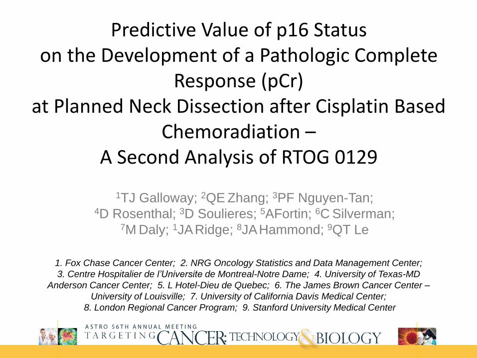
Predictive Value of p16 Status on the Development of a Pathologic Complete
Response (pCr) at Planned Neck Dissection after Cisplatin Based
Chemoradiation – A Second Analysis of RTOG 0129
1TJ Galloway; 2QE Zhang; 3PF Nguyen-Tan; 4D Rosenthal; 3D Soulieres; 5AFortin; 6C Silverman;
7M Daly; 1JA Ridge; 8JA Hammond; 9QT Le
1. Fox Chase Cancer Center; 2. NRG Oncology Statistics and Data Management Center;
3. Centre Hospitalier de l’Universite de Montreal-Notre Dame; 4. University of Texas-MD
Anderson Cancer Center; 5. L Hotel-Dieu de Quebec; 6. The James Brown Cancer Center –
University of Louisville; 7. University of California Davis Medical Center;
8. London Regional Cancer Program; 9. Stanford University Medical Center
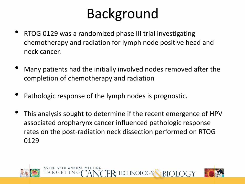
Background • RTOG 0129 was a randomized phase III trial investigating
chemotherapy and radiation for lymph node positive head and neck cancer.
• Many patients had the initially involved nodes removed after the completion of chemotherapy and radiation
• Pathologic response of the lymph nodes is prognostic.
• This analysis sought to determine if the recent emergence of HPV associated oropharynx cancer influenced pathologic response rates on the post-radiation neck dissection performed on RTOG 0129
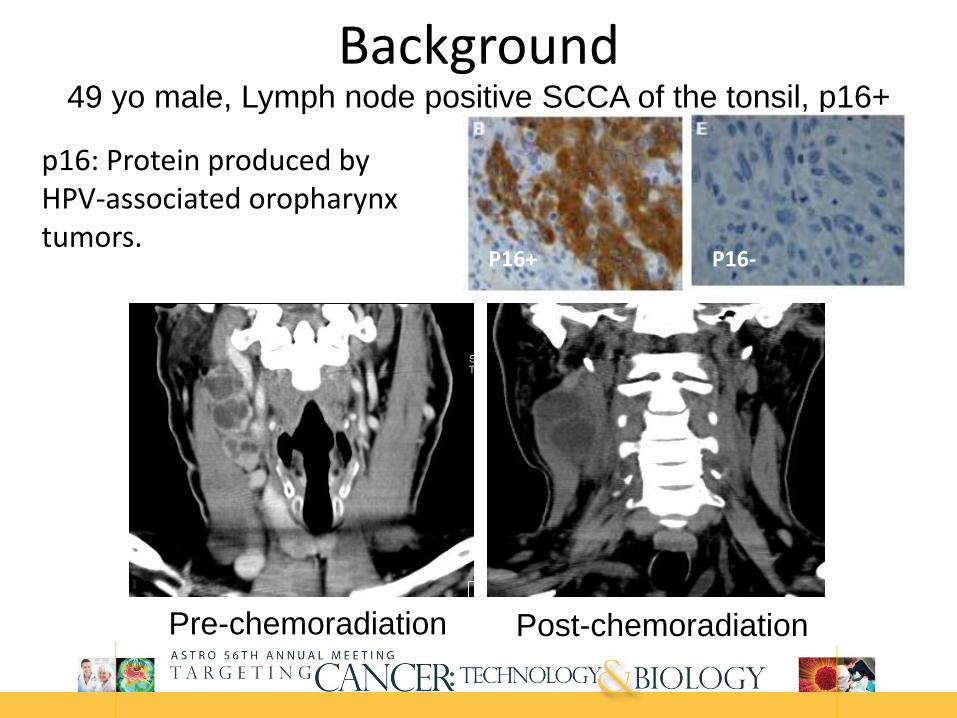
Background 49 yo male, Lymph node positive SCCA of the tonsil, p16+
Pre-chemoradiation Post-chemoradiation
p16: Protein produced by HPV-associated oropharynx tumors.
P16+ P16-

Background RTOG 0129 Neck Dissection Analysis
433 oropharynx cancer patients
721 patients
247 with an N2a,
N2b, N2c, N3 neck
99 neck dissections
within 180 days of
the completion of
chemoradiotherapy
23 with an
N0 neck
46 with an
N1 neck
193 initially involved
necks observed
316 patients with a known p16 status
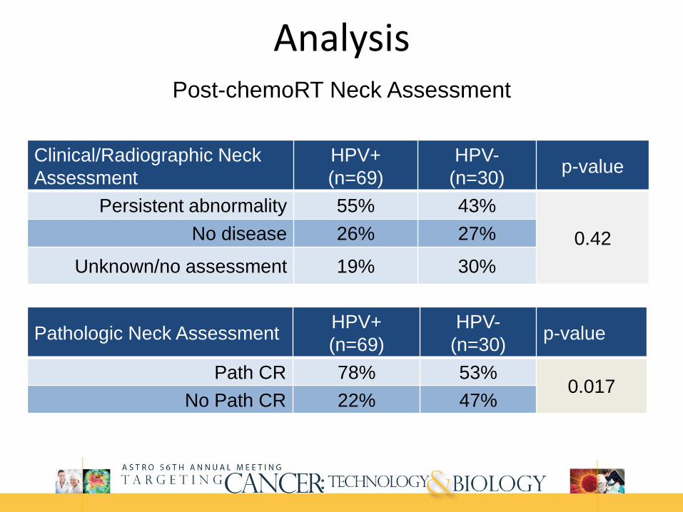
Analysis Post-chemoRT Neck Assessment
Pathologic Neck Assessment HPV+
(n=69)
HPV-
(n=30) p-value
Path CR 78% 53% 0.017
No Path CR 22% 47%
Clinical/Radiographic Neck
Assessment
HPV+
(n=69)
HPV-
(n=30) p-value
Persistent abnormality 55% 43%
0.42 No disease 26% 27%
Unknown/no assessment 19% 30%

Analysis Local-regional Failure after Neck Dissection
(Cox model)
P-value for interaction between pathologic CR and p16 status: 0.3669
Parameter Hazard Ratio p-value
Pathologic CR (yes v no) If HPV- 0.31 (0.11-0.88) 0.028
Pathologic CR (yes v no) If HPV+ 0.68 (0.18-2.59) 0.57
HPV status (pos v neg) If not path CR 0.13 (0.03 – 0.48) 0.0025
HPV status (pos v neg) If pathCR 0.28 (0.10 – 0.80) 0.018
Comparison of
Neck Dissection v. Neck Observation
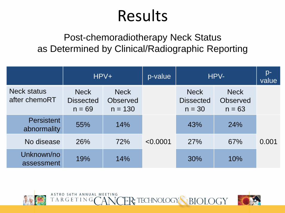
Results Post-chemoradiotherapy Neck Status
as Determined by Clinical/Radiographic Reporting
HPV+ p-value HPV- p-
value
Neck status
after chemoRT Neck
Dissected
n = 69
Neck
Observed
n = 130
Neck
Dissected
n = 30
Neck
Observed
n = 63
Persistent
abnormality 55% 14%
<0.0001
43% 24%
0.001 No disease 26% 72% 27% 67%
Unknown/no
assessment 19% 14% 30% 10%

Results
2-year Local-Regional Failure (HPV-)
Patients Estimate (%)
Observed neck w/ clinical CR 42 19.2 (8.9-32.5)
pCR neck dissection 16 26.0 (7.5-49.6)
Persistent tumor neck dissection 14 75.0 (36.0-92.2)
2-year Local-Regional Failure (HPV+)
Patients Estimate (%)
Observed neck w/ clinical CR 94 9.9 (4.8-17.1)
pCR neck dissection 54 7.5 (2.4-16.7)
Persistent tumor neck dissection 15 20.0 (4.5-43.3)
Local-regional Failure

Conclusions • Patients with p16+ tumors had significantly higher complete
pathologic response rates than those with p16- tumors.
• Patients with a complete clinical/radiographic response in the neck were more likely to be managed without a neck dissection, and had a failure rate similar to patients with a complete pathologic response.
The Significance of p16 and p53 Expression on Clinical Outcome in Patients with Anal Cancer
Treated with Chemoradiotherapy: An Analysis of RTOG 98-11
Corinne M. Doll; Kathryn Winter; Jaffer Ajani; Alexander Klimowicz;
Christopher H. Crane; Lisa A. Kachnic; Gordon Okawara;
Himanshu Lukka; Lawrence Berk; Kevin Roof; Mark Becker;
David L Grisell; Chandan Guha; and Anthony M. Magliocco
Supported by NCI grants:
U10 CA21661, U10 CA37422, and U24 CA114734

Background
• Several studies show prognostic significance of human papillomavirus (HPV) and outcome in patients with squamous oropharyngeal cancers - Improved outcome in HPV+ tumors and
chemoradiotherapy (CRT)
• Impact of HPV pathway activation on prognosis in patients with anal cancer not as well defined
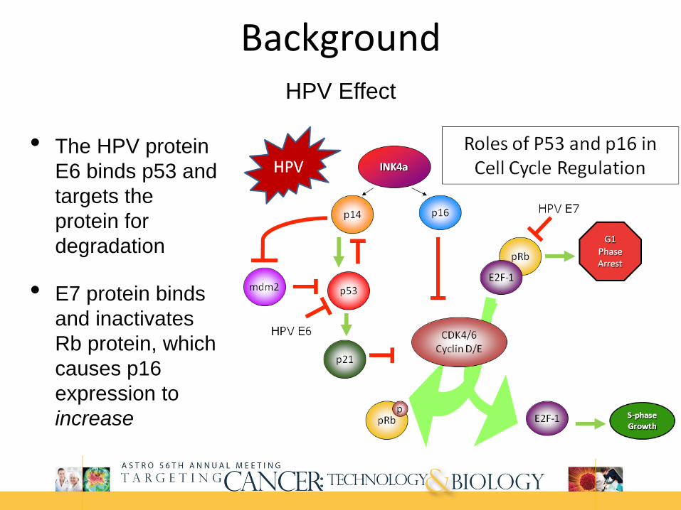
Background HPV Effect
• The HPV protein
E6 binds p53 and
targets the
protein for
degradation
• E7 protein binds
and inactivates
Rb protein, which
causes p16
expression to
increase

Background RTOG 98-11
• Phase III RCT: 5FU, MMC and RT vs. 5FU, cisplatin and RT for carcinoma of the anal canal, n=682
• Concurrent chemoradiation with 5FU-MMC has a statistically significant impact on DFS and OS vs. induction + concurrent 5FU-CDDP
• Potential strategies to improve outcomes include treatment intensification/modification and individualized molecular-based treatment
Gunderson et al, J Clin Oncol 2012; 30(35): 4344-4351.

Aims
• Measure expression of p16 (surrogate for HPV) and p53 (tumor suppressor protein) in pre-treatment tumor biopsies of anal cancer patients enrolled on RTOG 98-11
• Correlate expression with patient outcome

Methodology • Retrospective analysis of RTOG 98-11, pre-treatment anal
cancers, n=155 analyzed
• Quantitative immunohistochemistry expression of p16 and p53 proteins for each tumor sample (core)
• Protein expression was analyzed using X-tile cut-points defining high (H) vs. low (L) expression status, based on overall survival
• Associations between the tumor marker categories and clinical outcome parameters Cox proportional hazards models

Results Fluorescence IHC and AQUA® Results
for p16 and p53 in RTOG 98-11 Anal Cancers
Examples of p16 and p53 staining in:
Normal tissue
RTOG 98-11 cores with high p16/low p53 expression
RTOG 98-11 cores with low p16/high p53 expression

Results Patient/tumor characteristics
by p16 and p53 status
Patients whose tumors had high p16/low p53 status were more likely: Female, better performance status, smaller tumors
Ov
er
all
S
ur
viv
al
(%
)
0
2 5
5 0
7 5
1 0 0
Y e a rs fro m R a n d o m iz a tio n
0 1 2 3 4 5 6
P a tie n ts a t R is k
H -p 1 6 /L -p 5 3
O th e r
1 2 6
2 9
1 2 0
2 6
1 1 4
2 2
1 0 7
1 8
9 9
1 6
9 4
1 6
7 7
1 1
F a ile d
1 91 2
T o ta l
1 2 62 9
L o g -ra n k p -v a lu e < 0 .0 0 0 1
H -p 1 6 /L -p 5 3O th e r
4-year OS for patients with high p16/low p53
was 88% (95% CI: 80%, 92%) vs.
60% (95% CI: 39%, 75%) for other

Multivariate Analyses Patients with high p16/low p53
expression had better outcomes
Comparison HR
95% C.I.
LL
95% C.I.
UL p-value†
H-p16/L-p53
Other
1.00
3.80
-
2.02
-
7.15
-
<0.0001
Comparison HR
95% C.I.
LL
95% C.I.
UL p-value†
H-p16/L-p53
Other
1.00
2.15
-
1.23
-
3.75
-
0.0075
OS
DFS
†p-value from Chi-square test using the Cox proportional hazards model

Conclusions
• In this exploratory analysis of a subset of patients treated with CRT on the RTOG 98-11 protocol: High p16/low p53 tumor status was associated with
better clinical outcomes o Independently associated with better OS, DFS, LRF
• Further exploration of the optimal biologic cut-point should be evaluated
• Differential treatment strategies could be considered for patients with these distinct tumor subtypes


Metabolic Tumor Volume on FDG-PET Predicts Clinical Outcomes Following Chemoradiotherapy
for Locally Advanced Non-small Cell Lung Cancer:
A Secondary Analysis of ECOG-ACRIN 6668 / RTOG 0235
Nitin Ohri; Fenghai Duan; Mitchell Machtay;
Jeremy Gorelick; Bradley Snyder; Abass Alavi;
Barry Siegel; Douglas Johnson; Jeffrey Bradley;
Albert DeNittis; Maria Werner-Wasik

Background
• Approximately 1/3 of the 220,000 patients diagnosed with non-small cell lung cancer (NSCLC) in the U.S. each year have stage III disease.
• 18F-FDG positron emission tomography (PET) is an important tool for the staging and radiotherapy planning for patients with NSCLC.
• ECOG-ACRIN 6668 / RTOG 0235: A prospective, multi-institutional trial that evaluated the prognostic value of
PET for patients treated with definitive chemoradiotherapy for locally advanced NSCLC.
PET was performed before radiotherapy and 12-16 weeks after radiotherapy.
Enrolled 251 patients.

Background ECOG-ACRIN 6668 / RTOG 0235: Primary Analysis
Post-treatment Peak SUV
J Clin Oncol 31:3823-3830
Purpose
• To evaluate associations between pre- and post-treatment PET metrics and clinical outcomes for stage III NSCLC patients treated with definitive, concurrent chemoradiotherapy.

Methodology
• PET Metrics: Maximum SUV (SUVmax)
Metabolic Tumor Volume (MTV)
Total Glycolytic Activity (TGA)
Results
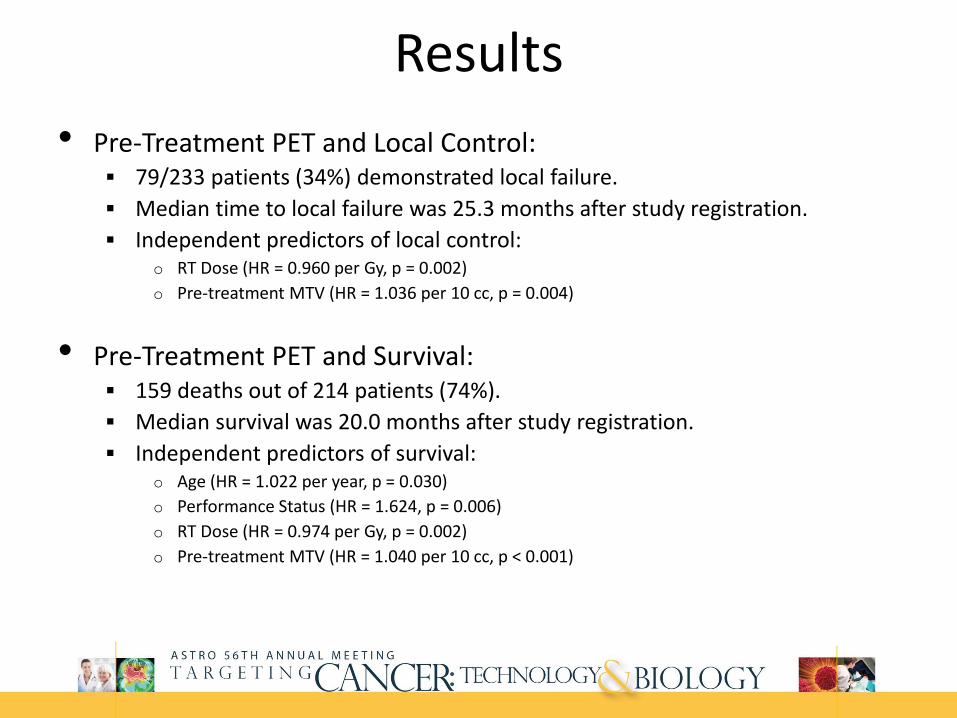
• Pre-Treatment PET and Local Control: 79/233 patients (34%) demonstrated local failure.
Median time to local failure was 25.3 months after study registration.
Independent predictors of local control: o RT Dose (HR = 0.960 per Gy, p = 0.002)
o Pre-treatment MTV (HR = 1.036 per 10 cc, p = 0.004)
• Pre-Treatment PET and Survival: 159 deaths out of 214 patients (74%).
Median survival was 20.0 months after study registration.
Independent predictors of survival: o Age (HR = 1.022 per year, p = 0.030)
o Performance Status (HR = 1.624, p = 0.006)
o RT Dose (HR = 0.974 per Gy, p = 0.002)
o Pre-treatment MTV (HR = 1.040 per 10 cc, p < 0.001)

Results
• Post-Treatment PET and Local Control: 68/164 patients (41%) demonstrated local failure.
Median time to local failure was 18.6 months after post-treatment PET.
Independent predictors of local control: o Post-treatment SUVmax (HR = 1.135, p = 0.002)
• Post-Treatment PET and Survival: 119 deaths out of 170 patients (70%).
Median survival was 18.2 months after post-treatment PET.
Independent predictors of survival: o Age (HR = 1.034 per year, p = 0.006)
o Performance Status (HR = 1.585, p = 0.021)
o Pre-treatment MTV (HR = 1.029 per 10 cc, p = 0.019)
o Post-treatment SUVmax (HR = 1.106, p < 0.001)
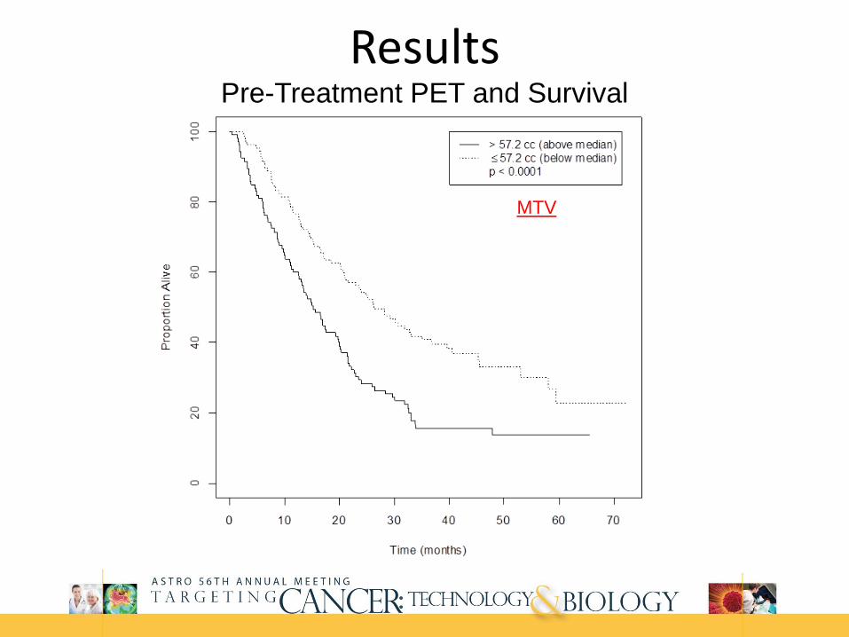
Results Pre-Treatment PET and Survival
MTV
Conclusion • PET metrics are strong predictors of clinical outcomes for stage III
NSCLC patients treated with definitive chemoradiotherapy. Pre-treatment MTV
Post-treatment SUVmax

Questions?
Contact ASTRO’s Press Office
In San Francisco, Sept. 14-17, 2014:
415-978-3503
Via email: [email protected]
The online press kit:
www.astro.org/AMPress