Embed Size (px)
Citation preview
Acta Medica International| Jul - Dec 2014 | Vol 1 | Issue 2 | 74
Studies on the Protoscoleces and Hooks of Echinococcus Granulosus from Libya by Scanning Electron Microscope
Layla O. Elmajdoub1, Wahab A. Rahman2, Mustafa Fadzil3 , Siti Azizah Mohd2
1Lecturer, Molecular parasitology, Science College, Misurata University, Libya, 2 Professor, School of Biological Sciences, University Sains Malaysia, 11800 Minden, Penang, 3Assistant Professor, School of Biological Sciences, University Sains Malaysia, 11800 Minden, Penang, Malaysia
ABSTRACTIntroduction: Protoscolex and hook morphology had been used to identify different strains of Echinococcus granulosus. Method: In the present study, hydatid sand was isolated from infected organs such as liver, lung and spleen from slaughtered animals in several abattoirs in the selected study areas in Libya. Scanning Electron Microscope (SEM) was used to describe the outer surface of the Protoscoleces and the hooks. Result: SEM pictures showed different morphological characters of the outer surface of the different parts of the same protoscolex. Also, the buds on the outer surface of brood capsule, and showed some differences between the large and small hooks. These findings appeared the that there was a correlation with significant statistical difference between the measurements of large as well as small hooks from the same organs of each host as well as between different hosts. Conclusion: Finally, it was difficult to determine whether these cases are not real distortions in the morphology of E. granulosus. They possibly could be caused by mutation or related to a special strain of E.granulosus. Thus, the findings of the present study would suggest that there is a need to consider relationships between the different characters of hooks with different strains of E.granulosus.
Keywords: Echinococcus granulosus, Liver, Lung, SEM
with the hooks of the lower row.10 Until now, there were limited studies using SEM.10-14 Criteria used in recognition of specific strains have all emphasized the use of morphological details, particularly that of the protoscoleces and hooks.15
In Libya, sheep- dog strain of E. granulosus is widespread in a farmed area.16 Criteria used in the recognition of specific strains have all emphasized the use of morphological details, particularly of the Protoscoleces and hooks.17 The morphology of the Protoscoleces isolated from Libyan infections using the Scanning Electron Microscope, they presented a pioneer study in this field, but called for confirmation of their results and stated that identification of different strains may be in epidemiological investigations.5
The aim of the present work, therefore, is the study of the morphological characters of the Protoscoleces and brood capsules isolated from liver, lung and spleen of slaughtered animals by Scanning Electron Microscope. Variations in morphometric measurements of rostellar hooks in the different strains of E. granulosus were determined using Computer Image Analysis System (CIAS) to find out their possible role in strain recognition.
INTRODUCTION
Cystic echinococcosis is amongst the most important parasitic zoonoses and remains a public health and economic problem of global concern.1 Hydatid disease is endemic in Libya, where many domestic animals act as intermediate hosts of Echinococcus granulosus.2,3
Recently still have considered the disease as zoonotic disease in Libya.4-6
The larva of E. granulosus develops as a fluid bladder by a cyst wall. Protoscoleces have derived from germinal layer, which grew toward the cyst cavity, the method of the derived of the buds from the germinal layer become Protoscoleces is not known.7 Usually the larval hooks characters remain unchanged by passage through the final host.8 Normally the protoscolex is invaginated into the posterior region9 by external factors stimulate evagination, the body of evaginated Protoscoleces consisted of three parts rostellum with two rows of hooks, non- rostellar part bearing four suckers and the posterior part called Samoa.8
Recent studies by Scanning Electron Microscope (SEM) detected that hooks of the upper row are long compared
Corresponding Author: 1Dr. Layla Elmajdoub, Lecturer, Molecular parasitology, Science College, Misurata University E‑mail: [email protected]
or
IGIn
al a
rt
Icl
e
Elmajdoub, et al.: Electron Microscopic Study of Echinococcus Granulosus
Acta Medica International | Jul - Dec 2014 | Vol 1 | Issue 2 |75
METHODS
Collection of Hydatid CystsThese were collected from sheep (0-10 years old), camels (3-10 years old) from different areas in Libya.
Preparation of Hydatid SandHydatid fluid was aspirated from 16 cysts in sheep, and 12 from camels. Hydatid sand was prepared by repeated centrifugations, and washed several times in phosphate buffer, and fixed in 3% (V/V) glutaraldehyde in 0.1 M of the phosphate buffer; the resuspended samples were centrifuged two times for 5 min at 1000 RPM. The supernatant was discarded, and the pellet was resuspended in 1% osmium tetroxide prepared at room temperature on the stubs with a double-sided sticky tape, and then, sputter coated with gold. The preparations were viewed in Leo Supra 50 VP field Emission SEM (Model Carl-Ziess SMT, Oberkochen, Germany). In terms of estimating the differences in the shape of hooks, computer image software was used to detect any significant difference between the hooks as in Figure 1.
Lastly, the guard angle was measured as the angle between the blade and guard regions. In addition, this measurement was used to determine the correlation between the measurements of different parts in large and small hooks in the same and different organs as Figure 2.
Data AnalysisStatistical analysis was based on correlating all the measurements between large and small hooks from the same organ from the same host and between different hosts by SEM. Morphometric data was analyzed using Computer Image Analysis System (CIAS) and SPSS software v.19.
RESULTS
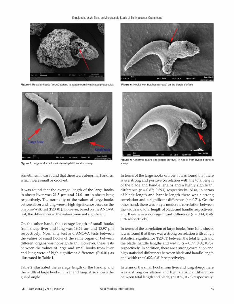
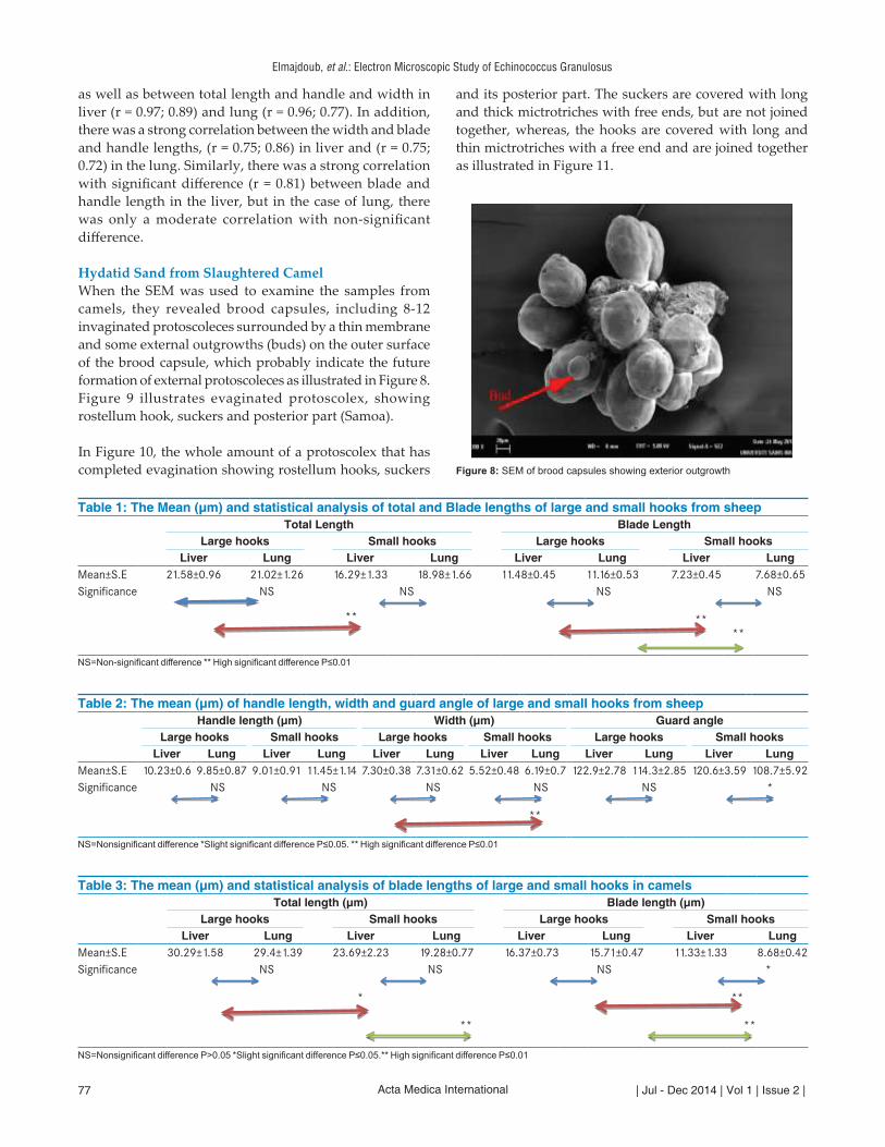
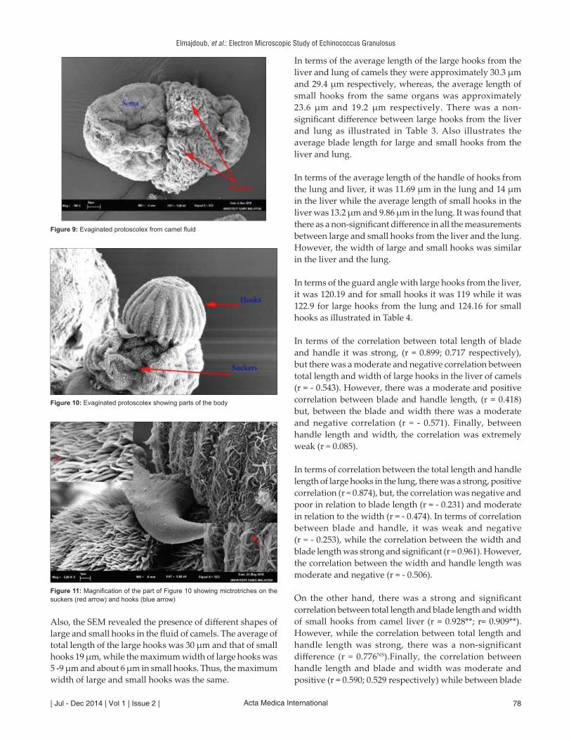
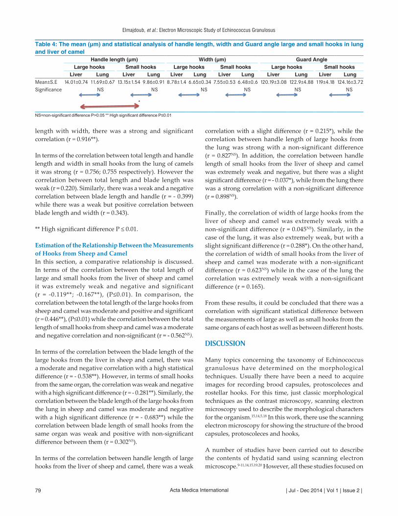
Hydatid Sand from Slaughtered SheepFigure 3 illustrates an invaginated protoscolex with a hole from sheep liver, where suckers start to appear during the process of evagination, while Figure 4 illustrates the rostellar hook starting to appear during the process of evaginating of protoscolex from sheep lung. The hooks were covered by microtriches. In addition, different types of hooks were detected by SEM. As illustrated in Figure 5 some hooks were of normal appearance, whereby the outer surface of the hooks was smooth. However, sometimes there were notches on the dorsal surface or in the space between the guard and handle, as illustrated in Figure 6. Normally, the guard area of these hooks was attached to the body of the hook and extended deeper into the rostellum region. It was found that there were different shapes of guards on the hooks and sometimes the hooks had abnormal guards or did not have a guard, as illustrated in Figure 7. In addition, since the hooks used the handle to attach their body to the rostellum, they were pointed slightly and curved. However,
Figure 1: Measurements of the hook lengths using computer image analysis system Key: TL = Total length, BL = blade length, HL = handle length and W = width
Figure 2: Measurements of the guard angle were described by the red arrow
Figure 3: Invaginated protoscolex with a hole (arrow) from sheep liver
Elmajdoub, et al.: Electron Microscopic Study of Echinococcus Granulosus
Acta Medica International| Jul - Dec 2014 | Vol 1 | Issue 2 | 76
sometimes, it was found that there were abnormal handles, which were small or crooked.
It was found that the average length of the large hooks in sheep liver was 21.5 µm and 21.0 µm in sheep lung respectively. The normality of the values of large hooks between liver and lung were of high significance based on the Shapiro-Wilk test (P≤0. 01). However, based on the ANOVA test, the differences in the values were not significant.
On the other hand, the average length of small hooks from sheep liver and lung was 16.29 µm and 18.97 µm respectively. Normality test and ANOVA tests between the values of small hooks of the same organ or between different organs was non-significant. However, these tests between the values of large and small hooks from liver and lung were of high significant difference (P≤0.01) as illustrated in Table 1.
Table 2 illustrated the average length of the handle, and the width of large hooks in liver and lung. Also shown the guard angle.
In terms of the large hooks of liver, it was found that there was a strong and positive correlation with the total length of the blade and handle lengths and a highly significant difference (r = 0.87; 0.893) respectively. Also, in terms of blade length and handle length there was a strong correlation and a significant difference (r = 0.71). On the other hand, there was only a moderate correlation between the width and total length of blade and handle respectively, and there was a non-significant difference (r = 0.44; 0.46; 0.36 respectively).
In terms of the correlation of large hooks from lung sheep, it was found that there was a strong correlation with a high statistical significance (P≤0.01) between the total length and the blade, handle lengths and width, (r = 0.77; 0.88; 0.78), respectively. In addition, there are a strong correlation and high statistical differences between blade and handle length and width (r = 0.622; 0.819 respectively).
In terms of the small hooks from liver and lung sheep, there was a strong correlation and high statistical differences between total length and blade, (r = 0.89; 0.75) respectively,
Figure 4: Rostellar hooks (arrow) starting to appear from invaginated protoscolex
Figure 5: Large and small hooks from hydatid sand in sheep
Figure 6: Hooks with notches (arrows) on the dorsal surface
Figure 7: Abnormal guard and handle (arrows) in hooks from hydatid sand in sheep
Elmajdoub, et al.: Electron Microscopic Study of Echinococcus Granulosus
Acta Medica International | Jul - Dec 2014 | Vol 1 | Issue 2 |77
as well as between total length and handle and width in liver (r = 0.97; 0.89) and lung (r = 0.96; 0.77). In addition, there was a strong correlation between the width and blade and handle lengths, (r = 0.75; 0.86) in liver and (r = 0.75; 0.72) in the lung. Similarly, there was a strong correlation with significant difference (r = 0.81) between blade and handle length in the liver, but in the case of lung, there was only a moderate correlation with non-significant difference.
Hydatid Sand from Slaughtered CamelWhen the SEM was used to examine the samples from camels, they revealed brood capsules, including 8-12 invaginated protoscoleces surrounded by a thin membrane and some external outgrowths (buds) on the outer surface of the brood capsule, which probably indicate the future formation of external protoscoleces as illustrated in Figure 8. Figure 9 illustrates evaginated protoscolex, showing rostellum hook, suckers and posterior part (Samoa).
In Figure 10, the whole amount of a protoscolex that has completed evagination showing rostellum hooks, suckers
and its posterior part. The suckers are covered with long and thick mictrotriches with free ends, but are not joined together, whereas, the hooks are covered with long and thin mictrotriches with a free end and are joined together as illustrated in Figure 11.
Table 1: The Mean (µm) and statistical analysis of total and Blade lengths of large and small hooks from sheepTotal Length Blade Length
Large hooks Small hooks Large hooks Small hooksLiver Lung Liver Lung Liver Lung Liver Lung
Mean±S.E 21.58±0.96 21.02±1.26 16.29±1.33 18.98±1.66 11.48±0.45 11.16±0.53 7.23±0.45 7.68±0.65Significance NS
NS
**
NS
NS
****
NS=Non‑significant difference ** High significant difference P≤0.01
Table 2: The mean (µm) of handle length, width and guard angle of large and small hooks from sheepHandle length (µm) Width (µm) Guard angle
Large hooks Small hooks Large hooks Small hooks Large hooks Small hooksLiver Lung Liver Lung Liver Lung Liver Lung Liver Lung Liver Lung
Mean±S.E 10.23±0.6 9.85±0.87 9.01±0.91 11.45±1.14 7.30±0.38 7.31±0.62 5.52±0.48 6.19±0.7 122.9±2.78 114.3±2.85 120.6±3.59 108.7±5.92Significance NS
NS
NS
NS
**
NS
*
NS=Nonsignificant difference *Slight significant difference P≤0.05. ** High significant difference P≤0.01
Table 3: The mean (µm) and statistical analysis of blade lengths of large and small hooks in camelsTotal length (µm) Blade length (µm)
Large hooks Small hooks Large hooks Small hooksLiver Lung Liver Lung Liver Lung Liver Lung
Mean±S.E 30.29±1.58 29.4±1.39 23.69±2.23 19.28±0.77 16.37±0.73 15.71±0.47 11.33±1.33 8.68±0.42Significance NS
NS
*
**
NS
*
**
**
NS=Nonsignificant difference P>0.05 *Slight significant difference P≤0.05.** High significant difference P≤0.01
Figure 8: SEM of brood capsules showing exterior outgrowth
Elmajdoub, et al.: Electron Microscopic Study of Echinococcus Granulosus
Acta Medica International| Jul - Dec 2014 | Vol 1 | Issue 2 | 78
Also, the SEM revealed the presence of different shapes of large and small hooks in the fluid of camels. The average of total length of the large hooks was 30 µm and that of small hooks 19 µm, while the maximum width of large hooks was 5 -9 µm and about 6 µm in small hooks. Thus, the maximum width of large and small hooks was the same.
In terms of the average length of the large hooks from the liver and lung of camels they were approximately 30.3 µm and 29.4 µm respectively, whereas, the average length of small hooks from the same organs was approximately 23.6 µm and 19.2 µm respectively. There was a non-significant difference between large hooks from the liver and lung as illustrated in Table 3. Also illustrates the average blade length for large and small hooks from the liver and lung.
In terms of the average length of the handle of hooks from the lung and liver, it was 11.69 µm in the lung and 14 µm in the liver while the average length of small hooks in the liver was 13.2 µm and 9.86 µm in the lung. It was found that there as a non-significant difference in all the measurements between large and small hooks from the liver and the lung. However, the width of large and small hooks was similar in the liver and the lung.
In terms of the guard angle with large hooks from the liver, it was 120.19 and for small hooks it was 119 while it was 122.9 for large hooks from the lung and 124.16 for small hooks as illustrated in Table 4.
In terms of the correlation between total length of blade and handle it was strong, (r = 0.899; 0.717 respectively), but there was a moderate and negative correlation between total length and width of large hooks in the liver of camels (r = - 0.543). However, there was a moderate and positive correlation between blade and handle length, (r = 0.418) but, between the blade and width there was a moderate and negative correlation (r = - 0.571). Finally, between handle length and width, the correlation was extremely weak (r = 0.085).
In terms of correlation between the total length and handle length of large hooks in the lung, there was a strong, positive correlation (r = 0.874), but, the correlation was negative and poor in relation to blade length (r = - 0.231) and moderate in relation to the width (r = - 0.474). In terms of correlation between blade and handle, it was weak and negative (r = - 0.253), while the correlation between the width and blade length was strong and significant (r = 0.961). However, the correlation between the width and handle length was moderate and negative (r = - 0.506).
On the other hand, there was a strong and significant correlation between total length and blade length and width of small hooks from camel liver (r = 0.928**; r= 0.909**). However, while the correlation between total length and handle length was strong, there was a non-significant difference (r = 0.776NS).Finally, the correlation between handle length and blade and width was moderate and positive (r = 0.590; 0.529 respectively) while between blade
Figure 9: Evaginated protoscolex from camel fluid
Figure 10: Evaginated protoscolex showing parts of the body
Figure 11: Magnification of the part of Figure 10 showing mictrotriches on the suckers (red arrow) and hooks (blue arrow)
Elmajdoub, et al.: Electron Microscopic Study of Echinococcus Granulosus
Acta Medica International | Jul - Dec 2014 | Vol 1 | Issue 2 |79
length with width, there was a strong and significant correlation (r = 0.916**).
In terms of the correlation between total length and handle length and width in small hooks from the lung of camels it was strong (r = 0.756; 0.755 respectively). However the correlation between total length and blade length was weak (r = 0.220). Similarly, there was a weak and a negative correlation between blade length and handle (r = - 0.399) while there was a weak but positive correlation between blade length and width (r = 0.343).
** High significant difference P ≤ 0.01.
Estimation of the Relationship Between the Measurements of Hooks from Sheep and CamelIn this section, a comparative relationship is discussed. In terms of the correlation between the total length of large and small hooks from the liver of sheep and camel it was extremely weak and negative and significant (r = -0.119**; -0.167**), (P≤0.01). In comparison, the correlation between the total length of the large hooks from sheep and camel was moderate and positive and significant (r = 0.446**), (P≤0.01) while the correlation between the total length of small hooks from sheep and camel was a moderate and negative correlation and non-significant (r = - 0.562NS).
In terms of the correlation between the blade length of the large hooks from the liver in sheep and camel, there was a moderate and negative correlation with a high statistical difference (r = - 0.538**). However, in terms of small hooks from the same organ, the correlation was weak and negative with a high significant difference (r = - 0.281**). Similarly, the correlation between the blade length of the large hooks from the lung in sheep and camel was moderate and negative with a high significant difference (r = - 0.683**) while the correlation between blade length of small hooks from the same organ was weak and positive with non-significant difference between them (r = 0.302NS).
In terms of the correlation between handle length of large hooks from the liver of sheep and camel, there was a weak
correlation with a slight difference (r = 0.215*), while the correlation between handle length of large hooks from the lung was strong with a non-significant difference (r = 0.827NS). In addition, the correlation between handle length of small hooks from the liver of sheep and camel was extremely weak and negative, but there was a slight significant difference (r = - 0.037*), while from the lung there was a strong correlation with a non-significant difference (r = 0.898NS).
Finally, the correlation of width of large hooks from the liver of sheep and camel was extremely weak with a non-significant difference (r = 0.045NS). Similarly, in the case of the lung, it was also extremely weak, but with a slight significant difference (r = 0.288*). On the other hand, the correlation of width of small hooks from the liver of sheep and camel was moderate with a non-significant difference (r = 0.623NS) while in the case of the lung the correlation was extremely weak with a non-significant difference (r = 0.165).
From these results, it could be concluded that there was a correlation with significant statistical difference between the measurements of large as well as small hooks from the same organs of each host as well as between different hosts.
DISCUSSION
Many topics concerning the taxonomy of Echinococcus granulosus have determined on the morphological techniques. Usually there have been a need to acquire images for recording brood capsules, protoscoleces and rostellar hooks. For this time, just classic morphological techniques as the contrast microscopy, scanning electron microscopy used to describe the morphological characters for the organism.15,14,5,18 In this work, there use the scanning electron microscopy for showing the structure of the brood capsules, protoscoleces and hooks,
A number of studies have been carried out to describe the contents of hydatid sand using scanning electron microscope.9-11,14,15,19,20 However, all these studies focused on
Table 4: The mean (µm) and statistical analysis of handle length, width and Guard angle large and small hooks in lung and liver of camel
Handle length (µm) Width (µm) Guard AngleLarge hooks Small hooks Large hooks Small hooks Large hooks Small hooks
Liver Lung Liver Lung Liver Lung Liver Lung Liver Lung Liver LungMean±S.E 14.01±0.74 11.69±0.67 13.15±1.54 9.86±0.91 8.78±1.4 6.65±0.34 7.55±0.53 6.48±0.6 120.19±3.08 122.9±4.88 119±4.18 124.16±3.72Significance NS
NS
*
NS
NS
NS
NS
NS=non‑significant difference P>0.05 ** High significant difference P≤0.01
Elmajdoub, et al.: Electron Microscopic Study of Echinococcus Granulosus
Acta Medica International| Jul - Dec 2014 | Vol 1 | Issue 2 | 80
the general morphology of protoscoleces and hooks without taking into account the organ and host factors. In contrast, the present study used SEM to investigate the size, shape of hooks from liver and lung from sheep and camels.
Firstly, the findings of the present study revealed, for the first time that in terms of the measurements of large and small hooks between liver and lung were of no significant difference. This indicated that the spread of the strain of E. granulosus was not dependant on the organs, but only on the hosts. Thus, between sheep and camels there was a high statistical difference only between all measurements of large hooks (P≤0.01), but not so between small hooks. This revealed that large hooks could be used as an important indicator to distinguish between strains of E. granulosus. However, it should be noted that these results were obtained from limited measurements of large and small hooks using SEM. Thus, there is a further need to use a large number of hooks from different organs from sheep and camels using Computer Image Analysis System (CIAS).
Secondly, in terms of the description of the morphology of large and small hooks of sheep origin, the large hooks had a slender guard, but the guard in small hooks was solid and spherical shaped. Scanning electron studies detected that large hooks were long, pointed, curved and less robust in comparison with small hooks. The present findings corroborated those,14 but their study did not use SEM to determine the intermediate hosts. In terms of the description of the morphology of large and small hooks of camel origin, the guard of large hooks was strong and oval shaped, and the distance between the guard and handle was almost similar to those from sheep. In addition, the guard of small hooks from camels was flat and the edges between guard and handle were serrated for reduced movement due to increased attrition. This finding corresponded to that.11
Thirdly, it was found that there were little projections on the surface of the germinal layer. However, the role of these projections could not be determined. It was found that the protoscolex of camel had mictrotriches that covered the hooks and appeared shorter and thinner than thus appear that covered the suckers. In addition, the free ends of these mictrotriches of suckers were thicker and had a loop-like appearance. This description of mictrotriches of the present study corporate that,14 where they described two types of mictrotriches in the same location but between invaginated and evaginated protoscolex. However, the findings of this present study described these mictrotriches on the same evaginated protoscolex. These mictrotriches play an important role in sensing the environment and determining the viability of protoscoleces.
REFERENCES
1. Thompson R.C.A., Lymbery A.J. and Constantine C.C. Variation in Echinococus towards a taxonomic revision of the genus. Adv. Parasitol., 1995; 35: 145-176.
2. Dar F.K. and Taguri S.C. Epidemiology and epizootiology of hydatidosis in the Libyan Jamahiriya, and recommendations for a programme of surveillance and control of the disease. Garyonis Medical Journal, 1979; 2: 11-15.
3. Shaafie L.A., Khan A.H. and Rambabu K. Biochemical profiles of hydatid cyst fluids of Echinococcus granulosus of human and animal origin in Libya. J. Helmin., 1999; 73:255-258.
4. Tashani O.A., Zhang, L.H. Jegi, A. and McManus D.P. Epidemiology and strain characteristics of Echinococcus granulosus in the Benghazi area of eastern Libya. Annals of Tropical Medicine and Parasitology. 2002; 96(4): 369-381.
5. Khalifa, R., N.A. Mazen, A.M.A. Marwan and L.O. Elmajdoub. Abnormal Preasentations of Cystic Hydatid Cysts from Livestock in Misurata, Libya. Bulletin of the Faculty of Science Assiut University, 2004; 33(2): 71-79.
6. Elmajdoub L.O., Elhoti K.M. and Hadid N. M. Prevalence of hydatid disease in slaughtered livestock animals from Misurata abattoirs, Libya. J. Union. Arab. Biol, 2007; 28: 163-174.
7. Galindo M.R., Paredes C., Marchant V. Miao and N. Galanti. Regionalization of DNA and protein synthesis in developing stages of the parasitic platyhelminth Echinococcus granulosus. Journal of Cellular Biochemistry, 2003; 90(2):294-303.
8. Yildiz K. and Gurca, S. The detection of Echinococcus granulosus strains using larval rostellar hook morphology. Turkiye Parazitoloji Dergist, 2009; 33(2): 199-202.
9. Rogan M.T. and Richards K.S. Echinococcus granulosus: changes in the surface ultrastructure during protoscolex formation. Parasitol, 1987; 94: 359-367.
10. Antoniou M. and Y Tselentis. Studies on Echinococcus granulosus using the scanning electron microscope. II. The hooks. Parasitology Research, 1993; 79(7):543-546.
11. Khalifa R., N.A. Mazen A.M.A. Marwan and L.O. Elmajdoub. Light and Scanning Electron Microscopy of the Hooklets of Protoscolices of Hydatid Cysts Infecting Sheep and Camels from Misurata, (Libya) and their Possible Role in “Parasite Strain” Recognition. Journal of the Egyption German Socity of Zoology, 2004; 44): 1-19.
12. Xiao N., Qiu J., Nakao M., Li T., Yang W., Chen X., Schantz P.M., Craig P.S. and Ito A. Echinococcus shiquicus n. sp., a taeniids cestode from Tibetan fox and plateau pika in China. International Journal for Parasitology, 2005; 35: 693-701.
13. Almeida F. B., R. Rodrigues-Silva R. H. Neves E. L. S. Romani and J. R. Machado-Silva. Intraspecific variation of Echinococcus granulosus in livestock from Peru. Veterinary Parasitology 2007; 143(1):50-58.
14. Antoniou M. and Y. Tselentis. Studies on Echinococcus granulosus using the scanning electron microscope. I. Preparations of the parasite for infection of the final host. Parasitology Research,1993; 79(7):537-542.
15. Smith S.A. and Richards K.S. ultrastructure and microanalyses of the protoscolex hooks of Echinococcus granulosus. Parasitol, 1991; 103: 267-274.
16. Gusbi A.M., Awan M.A.Q. and Bessely W.N. Prevalence of hydatidosis (E. granulosus) in sheep. Ann. Trop. Med. Parasitol., 1987; 81(1): 35-41.
17. Thompson, R.C.A. Biology and speciation of Echinococcus granulosus. Aust. Vet. J., 1979; 55:93-98.
Elmajdoub, et al.: Electron Microscopic Study of Echinococcus Granulosus
Acta Medica International | Jul - Dec 2014 | Vol 1 | Issue 2 |81
18. Almeida, F.B., Siva, R., Neves, R.H., Goncalves, M.M., Romani, E.L. and Silva, J. Morphological and morphometric studies on protoscoleces rostellar hooks of Echinococcus granulosus from Peru visualized by several microscopic techniques. Neotrop. Helminthol, 2009; 3(2): 65-72.
19. Swiderski, Z. Echinococcus granulosus: Hook-muscle systems and cellular organisation of infective oncospheres. International Journal for Parasitology,1983; 13(3):289-299.
20. Yones, D.A. Some parasitological studies on hydatid cyst in Assiut
How to Cite: Layla O. Elmajdoub, Wahab A. Rahman Mustafa Fadzil, Siti Azizah Mohd. Studies on the protoscoleces and hooks of echinococcus granulosus from Libya by scanning electron microscope. Acta Medica International. 2014;1(2):74‑81.
Source of Support: Nil, Conflict of Interest: None declared.
Governorate: M.Sc. Thesis in Parasitology, Faculty Medicine, Assiut University. 2002.


![Rachmaninov 3rd Piano Concerto [First Movement] · PDF file53-g e5 = 5 !5 = 5 5 5 5 5 4 5 5 =5 5 = 5e5 5 5 5 5 5 5 5e5 5 5!55 5 5 5 5 5e5 5 5 5 5 5 5! 5 $3e55 5 5: 5 5 5 55 5e 55 5](https://img.pdfslide.us/doc/110x75/5a78944a7f8b9a1f128d15db/rachmaninov-3rd-piano-concerto-first-movement-53-g-e5-5-5-5-5-5-5-5-4-5.jpg)


![[XLS] · Web view1 5 2 5 3 5 4 5 5 5 6 5 7 5 8 5 9 5 10 5 11 5 12 5 13 5 14 5 15 3 16 5 17 5 18 5 19 5 20 5 21 5 22 3 23 5 24 3 25 5 26 3 27 3 28 5 29 5 30 5 31 5 32 5 33 5 34 5 35](https://img.pdfslide.us/doc/110x75/5b0121497f8b9ad85d8da2f2/xls-view1-5-2-5-3-5-4-5-5-5-6-5-7-5-8-5-9-5-10-5-11-5-12-5-13-5-14-5-15-3-16-5.jpg)