Embed Size (px)
DESCRIPTION
5.4.1 X-Rays. (a) describe the nature of X-rays. X-rays - nature. Forms of electromagnetic radiation Short wavelength High frequency Wavelengths 10 -8 m to 10 -13 m Same as gamma rays. (b) describe in simple terms how X-rays are produced. X-rays - production. - PowerPoint PPT Presentation
Citation preview
5.4.1 X-Rays
(a) describe the nature of X-rays
Sto
wm
arke
t Phy
sics X-rays - nature
Forms of electromagnetic radiation
Short wavelength
High frequency
Wavelengths 10-8m to 10-13m
Same as gamma rays
(b) describe in simple terms how X-rays are produced
Sto
wm
arke
t Phy
sics X-rays - production
Produced when fast-moving electrons are rapidly decelerated
As the electrons slow down, their kinetic energy is transformed to photons of electromagnetic radiation
Less energy than gamma rays
Sto
wm
arke
t Phy
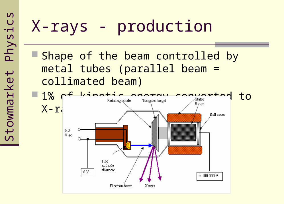
sics X-rays - production
Evacuated tube containing Cathode – heated filament emits electrons Anode – rotating – made from tungsten
External power supply – 200kV
Beam of electrons accelerates across the gap between anode and cathode
Electron arrives at 200keV Electrons lose kinetic energy as X-ray photons
Sto
wm
arke
t Phy
sics X-rays - production
Shape of the beam controlled by metal tubes (parallel beam = collimated beam)
1% of kinetic energy converted to X-rays
(c) describe how X-rays interact with matter (limited to photoelectric effect, Compton Effect and pair production)
Sto
wm
arke
t Phy
sics
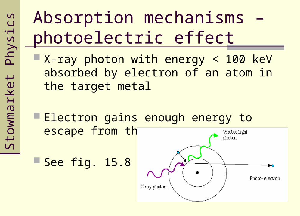
Absorption mechanisms – photoelectric effect X-ray photon with energy < 100 keV absorbed
by electron of an atom in the target metal
Electron gains enough energy to escape from the atom
See fig. 15.8
Sto
wm
arke
t Phy
sics
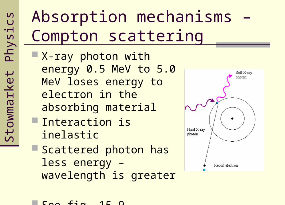
Absorption mechanisms – Compton scattering X-ray photon with energy
0.5 MeV to 5.0 MeV loses energy to electron in the absorbing material
Interaction is inelastic Scattered photon has less
energy – wavelength is greater
See fig. 15.9
Sto
wm
arke
t Phy
sics
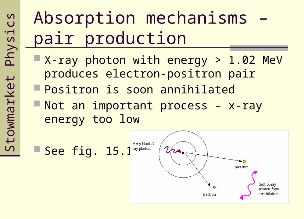
Absorption mechanisms – pair production X-ray photon with energy > 1.02 MeV produces
electron-positron pair Positron is soon annihilated Not an important process – x-ray energy too
low
See fig. 15.10
(d) define intensity as the power per unit cross-sectional area
Sto
wm
arke
t Phy
sics
The intensity of a beam of radiation indicates the rate at which energy is transferred across unit cross-sectional area.
Intensity is defined:
Intensity is the power per unit cross-sectional area
Intensity I (W m-2) = Power P (W) / Cross-sectional area A (m-2)
Intensity
(e) select and use the equation I = I0 e−μx to show how the intensity I of a collimated X ray beam varies with thickness x of medium
Sto
wm
arke
t Phy
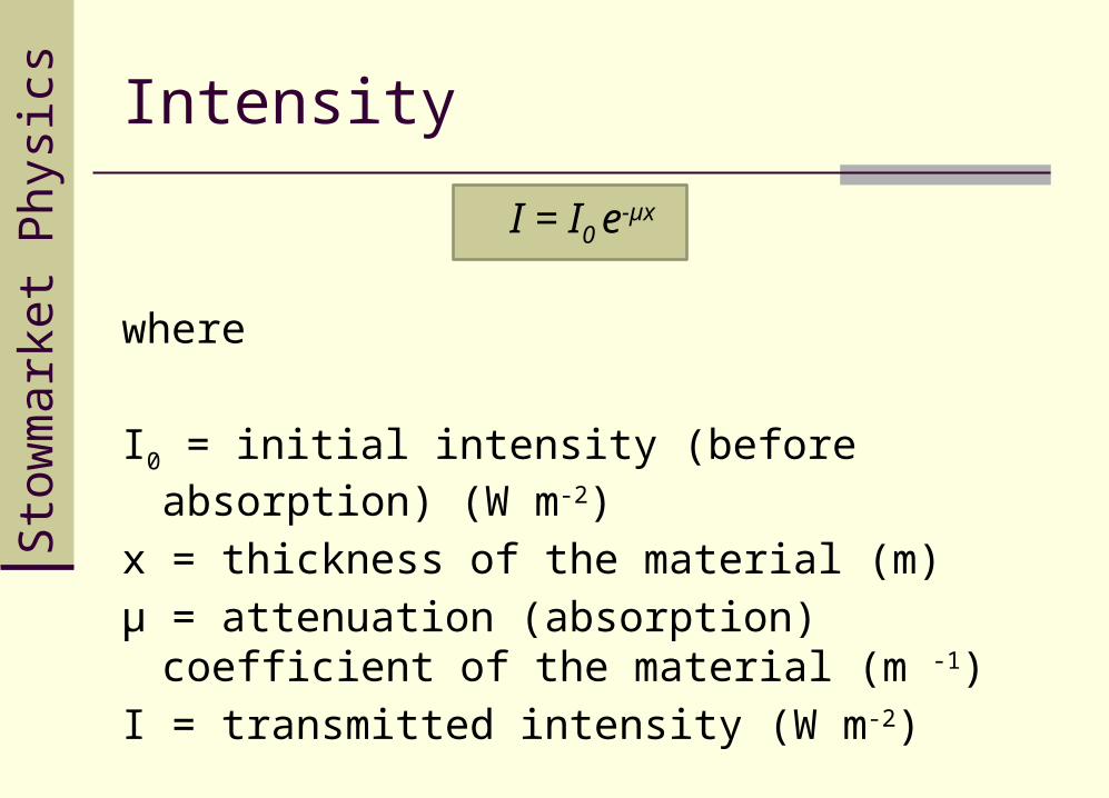
sics Intensity
I = I0 e-µx
where
I0 = initial intensity (before absorption) (W m-2)
x = thickness of the material (m)
µ = attenuation (absorption) coefficient of the material (m -1)
I = transmitted intensity (W m-2)
Sto
wm
arke
t Phy
sics Intensity
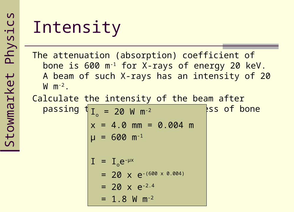
The attenuation (absorption) coefficient of bone is 600 m-1 for X-rays of energy 20 keV. A beam of such X-rays has an intensity of 20 W m-2.
Calculate the intensity of the beam after passing through a 4.0 mm thickness of bone
Io = 20 W m-2
x = 4.0 mm = 0.004 m
µ = 600 m-1
I = Ioe-µx
= 20 x e-(600 x 0.004)
= 20 x e-2.4
= 1.8 W m-2
Sto
wm
arke
t Phy
sics Intensity
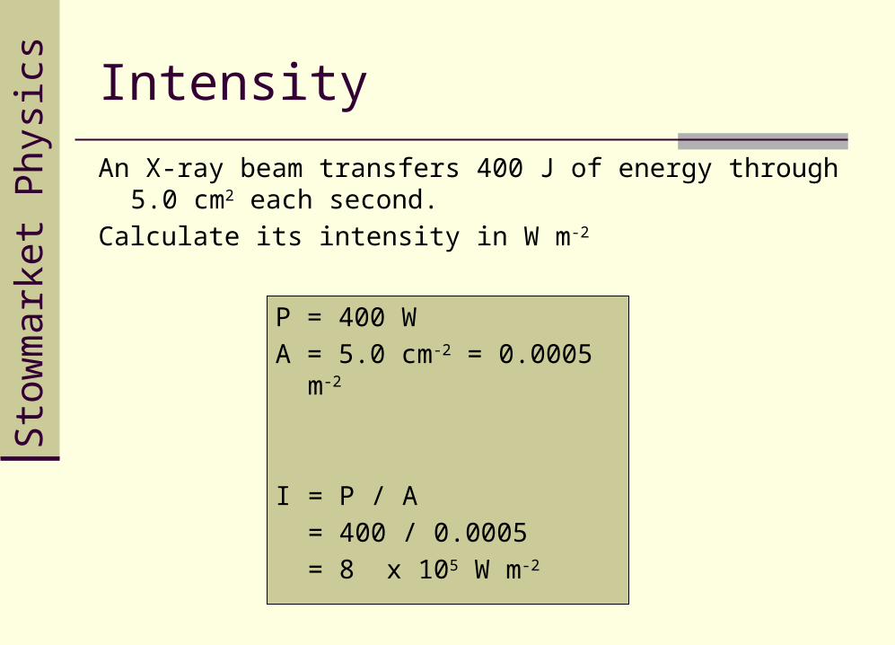
An X-ray beam transfers 400 J of energy through 5.0 cm2 each second.
Calculate its intensity in W m-2
P = 400 W
A = 5.0 cm-2 = 0.0005 m-2
I = P / A
= 400 / 0.0005
= 8 x 10⁵ W m-2
Sto
wm
arke
t Phy
sics Intensity
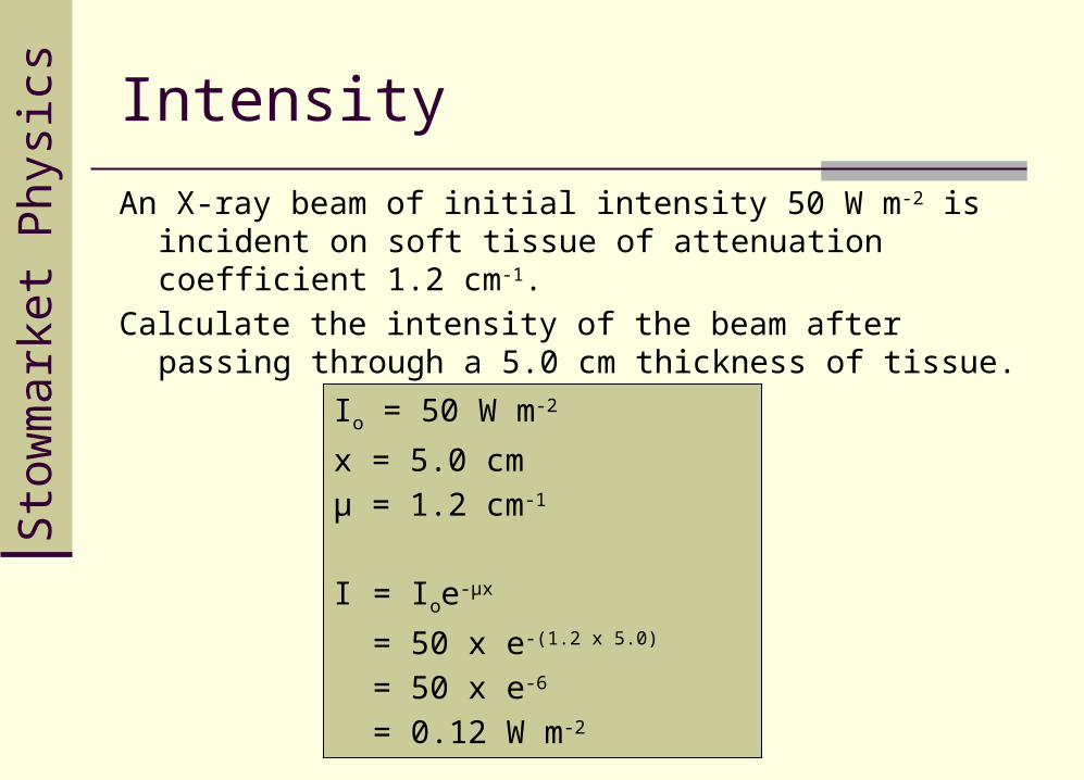
An X-ray beam of initial intensity 50 W m-2 is incident on soft tissue of attenuation coefficient 1.2 cm-1.
Calculate the intensity of the beam after passing through a 5.0 cm thickness of tissue.
Io = 50 W m-2
x = 5.0 cm
µ = 1.2 cm-1
I = Ioe-µx
= 50 x e-(1.2 x 5.0)
= 50 x e-6
= 0.12 W m-2
(f) describe the use of X-rays in imaging internal body structures including the use of image intensifiers and of contrast media (HSW 3, 4c and 6);
(g) explain how soft tissues like the intestines can be imaged using barium meal
(h) describe the operation of a computerised axial tomography (CAT) scanner
(i) describe the advantages of a CAT scan compared with an X-ray image (HSW 4c, 6)
Sto
wm
arke
t Phy
sics Assessment
Complete questions 1 to 5 on pages 236 and 237 of Physics 2
Sto
wm
arke
t Phy
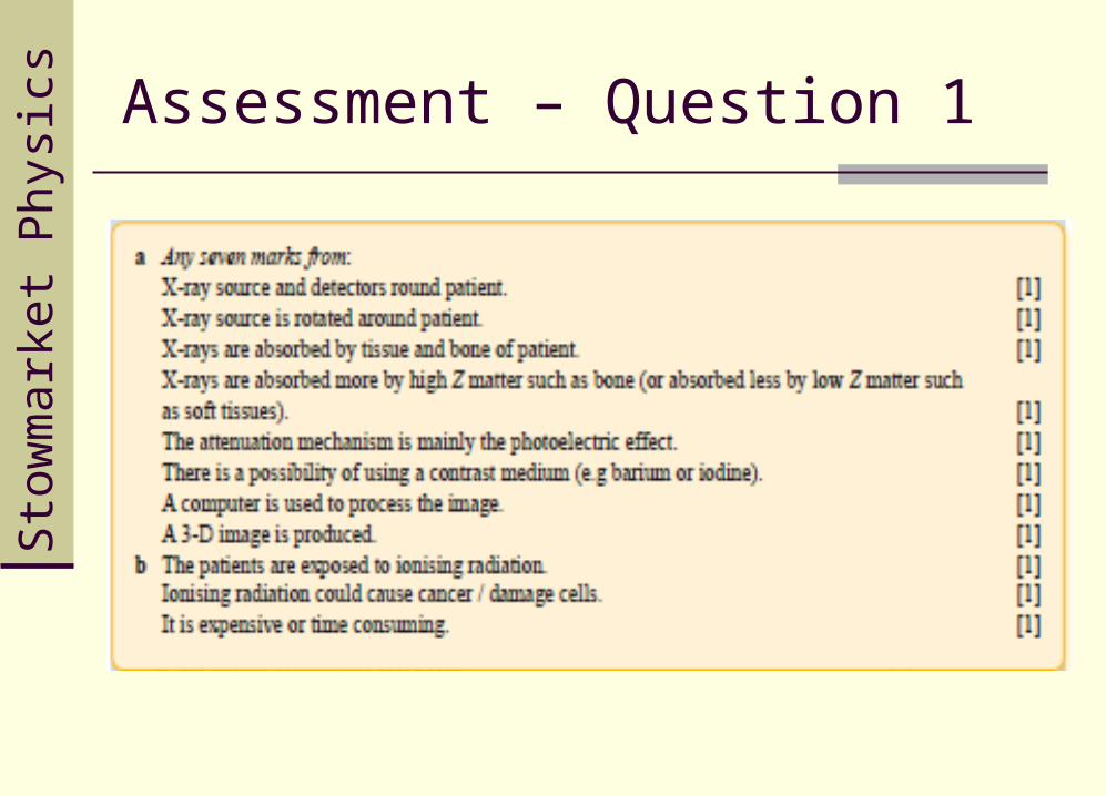
sics Assessment – Question 1
Sto
wm
arke
t Phy
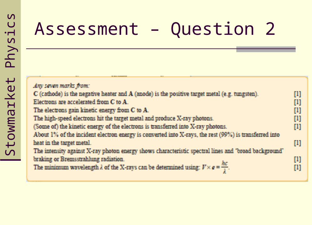
sics Assessment – Question 2
Sto
wm
arke
t Phy
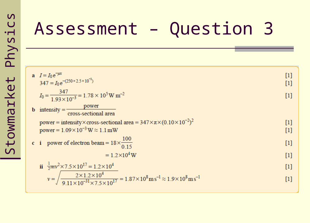
sics Assessment – Question 3
Sto
wm
arke
t Phy
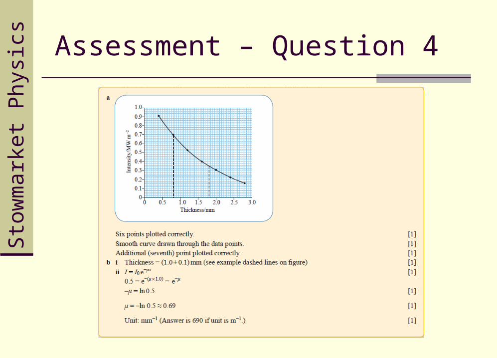
sics Assessment – Question 4
Sto
wm
arke
t Phy
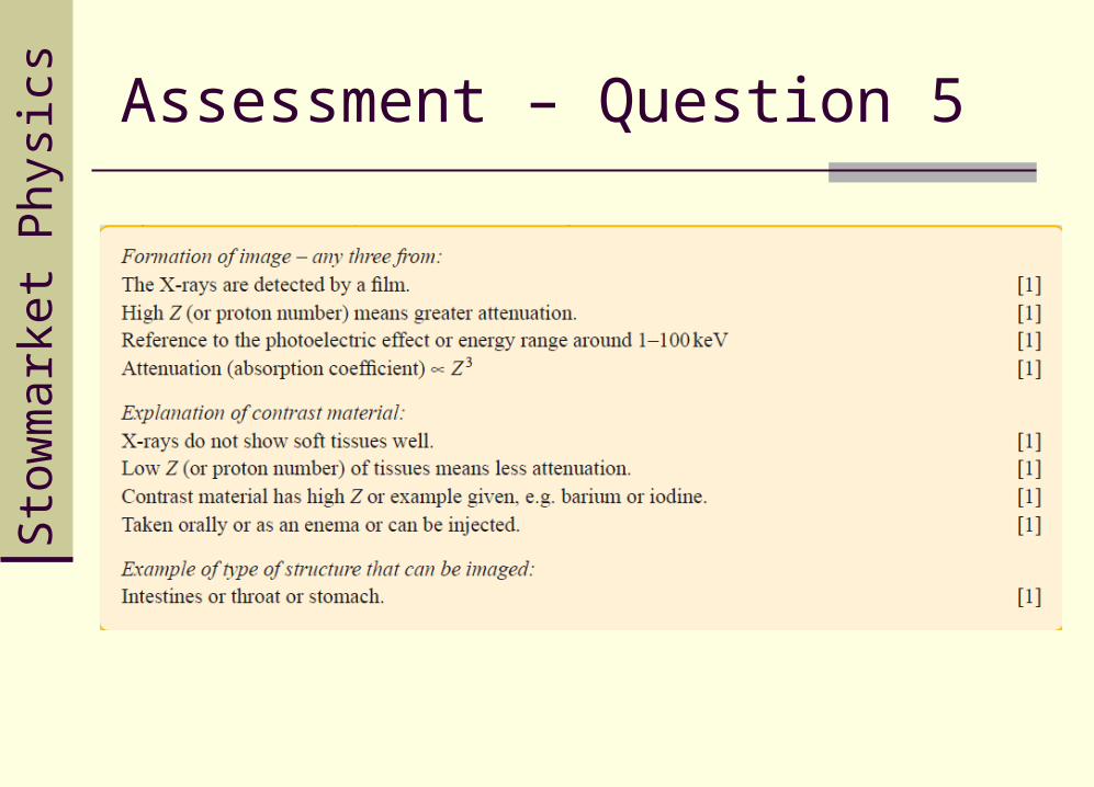
sics Assessment – Question 5

Sto
wm
arke
t Phy
sics End of Chapter Test – Mark Scheme