Embed Size (px)
Citation preview
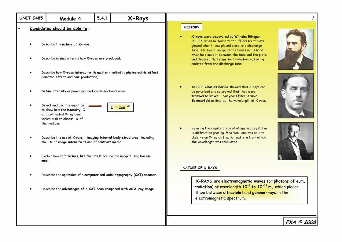
UNIT G485 Module 4 5.4.1 X-Rays Candidates should be able to :
Describe the nature of X-rays. Describe in simple terms how X-rays are produced.
Describe how X-rays interact with matter (limited to photoelectric effect, Compton effect and pair production). Define intensity as power per unit cross-sectional area. Select and use the equation to show how the intensity, I of a collimated X-ray beam varies with thickness, x of the medium. Describe the use of X-rays in imaging internal body structures, including the use of image intensifiers and of contrast media. Explain how soft tissues, like the intestines, can be imaged using barium meal. Describe the operation of a computerised axial topography (CAT) scanner. Describe the advantages of a CAT scan compared with an X-ray image.
X-rays were discovered by Wilhelm Röntgen in 1865, when he found that a fluorescent plate glowed when it was placed close to a discharge tube. He saw an image of the bones in his hand when he placed it between the tube and the plate and deduced that some sort radiation was being emitted from the discharge tube. In 1906, Charles Barkla showed that X-rays can be polarised and so proved that they were transverse waves. Six years later, Arnold Sommerfeld estimated the wavelength of X-rays. By using the regular array of atoms in a crystal as a diffraction grating, Max Von Laue was able to observe an X-ray diffraction pattern from which the wavelength was calculated.
FXA © 2008
I = I0e-μx
1
HISTORY
NATURE OF X-RAYS
X-RAYS are electromagnetic waves (or photons of e.m. radiation) of wavelength 10-8 to 10-13 m, which places them between ultraviolet and gamma-rays in the electromagnetic spectrum.
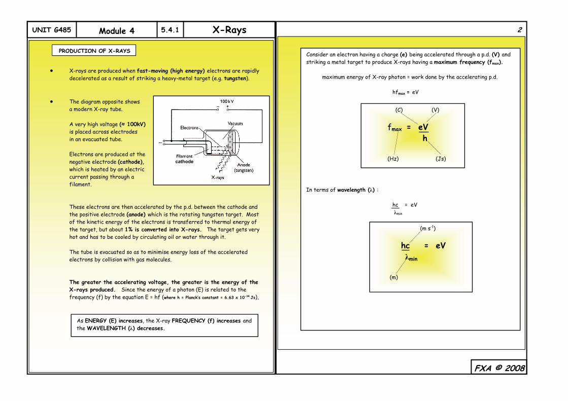
UNIT G485 Module 4 5.4.1 X-Rays
X-rays are produced when fast-moving (high energy) electrons are rapidly decelerated as a result of striking a heavy-metal target (e.g. tungsten). The diagram opposite shows a modern X-ray tube. A very high voltage (≈ 100kV) is placed across electrodes in an evacuated tube. Electrons are produced at the negative electrode (cathode), which is heated by an electric current passing through a filament. These electrons are then accelerated by the p.d. between the cathode and the positive electrode (anode) which is the rotating tungsten target. Most of the kinetic energy of the electrons is transferred to thermal energy of the target, but about 1% is converted into X-rays. The target gets very hot and has to be cooled by circulating oil or water through it. The tube is evacuated so as to minimise energy loss of the accelerated electrons by collision with gas molecules. The greater the accelerating voltage, the greater is the energy of the X-rays produced. Since the energy of a photon (E) is related to the frequency (f) by the equation E = hf (where h = Planck’s constant = 6.63 x 10⁻³⁴ Js),
FXA © 2008
PRODUCTION OF X-RAYS
cathode
As ENERGY (E) increases, the X-ray FREQUENCY (f) increases and the WAVELENGTH (λ) decreases.
Consider an electron having a charge (e) being accelerated through a p.d. (V) and striking a metal target to produce X-rays having a maximum frequency (fmax). maximum energy of X-ray photon = work done by the accelerating p.d. hfmax = eV In terms of wavelength (λ) : hc = eV λmin
(C) (V)
fmax = eV h (Hz) (Js)
(m s-1)
hc = eV λmin
(m)
2
UNIT G485 Module 4 5.4.1 X-Rays
The X-ray photons are produced by two different processes :
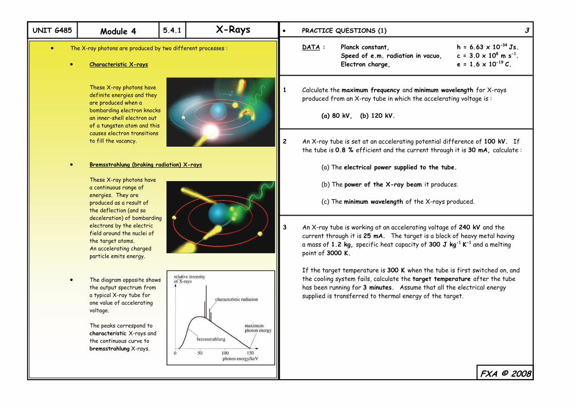
Characteristic X-rays These X-ray photons have definite energies and they are produced when a bombarding electron knocks an inner-shell electron out of a tungsten atom and this causes electron transitions to fill the vacancy. Bremsstrahlung (braking radiation) X-rays These X-ray photons have a continuous range of energies. They are produced as a result of the deflection (and so deceleration) of bombarding electrons by the electric field around the nuclei of the target atoms. An accelerating charged particle emits energy. The diagram opposite shows the output spectrum from a typical X-ray tube for one value of accelerating voltage. The peaks correspond to characteristic X-rays and the continuous curve to bremsstrahlung X-rays.
PRACTICE QUESTIONS (1) 3 DATA : Planck constant, h = 6.63 x 10⁻³⁴ Js. Speed of e.m. radiation in vacuo, c = 3.0 x 10⁸ m s-1. Electron charge, e = 1.6 x 10⁻¹⁹ C. 1 Calculate the maximum frequency and minimum wavelength for X-rays produced from an X-ray tube in which the accelerating voltage is : (a) 80 kV, (b) 120 kV. 2 An X-ray tube is set at an accelerating potential difference of 100 kV. If the tube is 0.8 % efficient and the current through it is 30 mA, calculate : (a) The electrical power supplied to the tube. (b) The power of the X-ray beam it produces. (c) The minimum wavelength of the X-rays produced. 3 An X-ray tube is working at an accelerating voltage of 240 kV and the current through it is 25 mA. The target is a block of heavy metal having a mass of 1.2 kg, specific heat capacity of 300 J kg-1 K-1 and a melting point of 3000 K. If the target temperature is 300 K when the tube is first switched on, and the cooling system fails, calculate the target temperature after the tube has been running for 3 minutes. Assume that all the electrical energy supplied is transferred to thermal energy of the target.
FXA © 2008
UNIT G485 Module 4 5.4.1 X-Rays
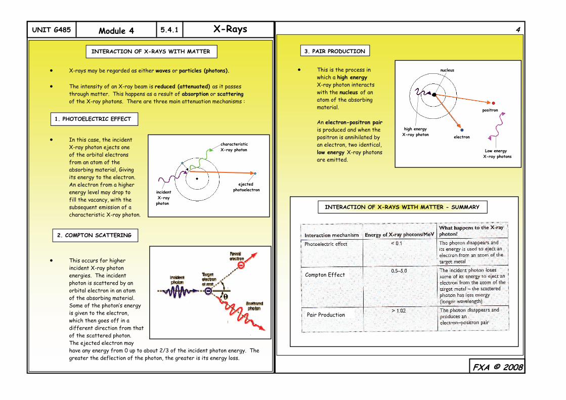
X-rays may be regarded as either waves or particles (photons). The intensity of an X-ray beam is reduced (attenuated) as it passes through matter. This happens as a result of absorption or scattering of the X-ray photons. There are three main attenuation mechanisms :
In this case, the incident X-ray photon ejects one of the orbital electrons from an atom of the absorbing material, Giving its energy to the electron. An electron from a higher energy level may drop to fill the vacancy, with the subsequent emission of a characteristic X-ray photon. This occurs for higher incident X-ray photon energies. The incident photon is scattered by an orbital electron in an atom of the absorbing material. Some of the photon’s energy is given to the electron, which then goes off in a different direction from that of the scattered photon. The ejected electron may have any energy from 0 up to about 2/3 of the incident photon energy. The greater the deflection of the photon, the greater is its energy loss.
This is the process in which a high energy X-ray photon interacts with the nucleus of an atom of the absorbing material. An electron-positron pair is produced and when the positron is annihilated by an electron, two identical, low energy X-ray photons are emitted.
FXA © 2008
INTERACTION OF X-RAYS WITH MATTER
1. PHOTOELECTRIC EFFECT
incident X-ray photon
characteristic X-ray photon
ejected photoelectron
2. COMPTON SCATTERING
4
3. PAIR PRODUCTION
high energy X-ray photon
Low energy X-ray photons
nucleus
positron
electron
INTERACTION OF X-RAYS WITH MATTER - SUMMARY
Compton Effect
Pair Production
UNIT G485 Module 4 5.4.1 X-Rays
The intensity of a collimated X-ray beam decreases as it passes through a substance. The amount of absorption varies considerably with the frequency of the X-rays. The absorption of low frequency (low energy) X-rays is mainly due to the photoelectric effect. For higher frequency X-rays, Compton scattering is the dominant absorption mechanism and for very high frequency X-rays it is pair production. The intensity (I) of an X-ray beam decreases exponentially with the thickness (x) of the substance through which it passes and it is given by the equation :
FXA © 2008
INTENSITY OF X-RAYS
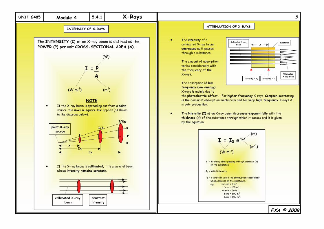
The INTENSITY (I) of an X-ray beam is defined as the POWER (P) per unit CROSS-SECTIONAL AREA (A). (W)
I = P A
(W m-2) (m2)
NOTE If the X-ray beam is spreading out from a point source, the inverse–square law applies (as shown in the diagram below). If the X-ray beam is collimated, it is a parallel beam whose intensity remains constant.
x 2x
3x
I
I/9
I/4 point X-ray source
collimated X-ray beam
Constant intensity
5
ATTENUATION OF X-RAYS
x
Collimated X-ray beam
Intensity = I0 Intensity = I
Attenuated X-ray beam
substance
(m)
I = I0 e-μx
(m-1) (W m-2) I = intensity after passing through distance (x) of the substance. I0 = initial intensity. μ = a constant called the attenuation coefficient which depends on the substance. e.g. vacuum = 0 m-1. flesh = 100 m-1. muscle = 50 m-1. bone = 300 m-1. Lead = 600 m-1.
UNIT G485 Module 4 5.4.1 X-Rays
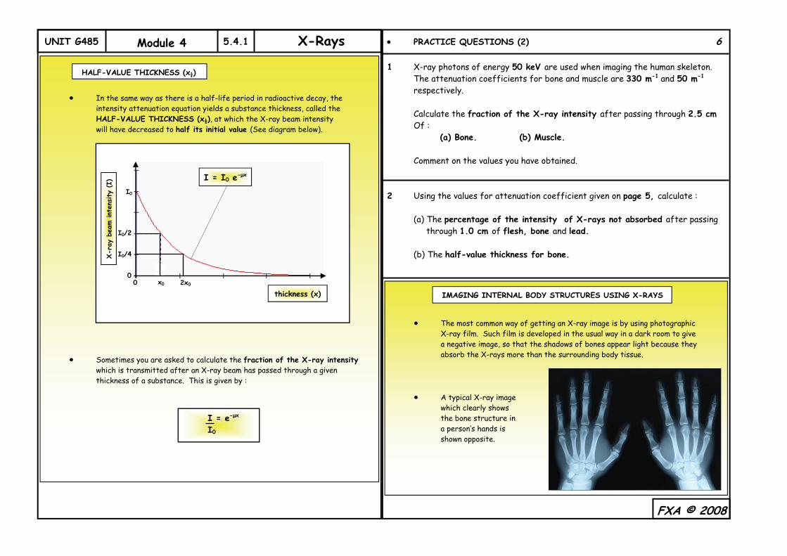
In the same way as there is a half-life period in radioactive decay, the intensity attenuation equation yields a substance thickness, called the
HALF-VALUE THICKNESS (x½), at which the X-ray beam intensity will have decreased to half its initial value (See diagram below).
Sometimes you are asked to calculate the fraction of the X-ray intensity which is transmitted after an X-ray beam has passed through a given thickness of a substance. This is given by :
PRACTICE QUESTIONS (2) 6 1 X-ray photons of energy 50 keV are used when imaging the human skeleton. The attenuation coefficients for bone and muscle are 330 m-1 and 50 m-1 respectively. Calculate the fraction of the X-ray intensity after passing through 2.5 cm Of : (a) Bone. (b) Muscle. Comment on the values you have obtained. 2 Using the values for attenuation coefficient given on page 5, calculate : (a) The percentage of the intensity of X-rays not absorbed after passing through 1.0 cm of flesh, bone and lead. (b) The half-value thickness for bone.
FXA © 2008
HALF-VALUE THICKNESS (x½)
X-r
ay b
eam int
ensity
(I)
thickness (x)
I = I0 e-μx
I0
I0/2
I0/4
0 0 x0 2x0
I = e-μx
I0
The most common way of getting an X-ray image is by using photographic X-ray film. Such film is developed in the usual way in a dark room to give a negative image, so that the shadows of bones appear light because they absorb the X-rays more than the surrounding body tissue. A typical X-ray image which clearly shows the bone structure in a person’s hands is shown opposite.
IMAGING INTERNAL BODY STRUCTURES USING X-RAYS
UNIT G485 Module 4 5.4.1 X-Rays
The X-ray image on the right shows how much the image quality has improved over the years. It is a false-colour X-ray image of the hands of a patient suffering from rheumatoid arthritis. Methods used to improve image quality and reduce the patient’s X-ray exposure include the use of :
Ultra-sensitive X-ray photographic film. A fluorescent intensifier screen placed behind the X-ray film.
Contrast media. Because different types of soft body tissue have very similar μ-values, they will absorb the X-rays by more or less the same amount. This means that there is little contrast between different structures and so the X-ray image would be of limited use. In order to make them more visible, contrast media, such as iodine or barium, are used. The patient swallows a liquid rich in barium (known as a barium meal) and since it has a large μ-value it will readily absorb X-rays. The contrast X-ray above shows the intestines. The barium meal has coated the wall of the tract, enabling its outline to be seen.
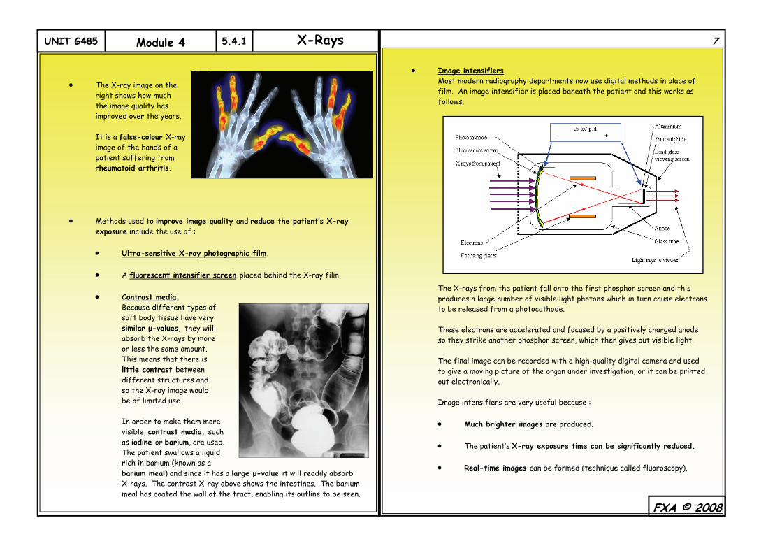
Image intensifiers Most modern radiography departments now use digital methods in place of film. An image intensifier is placed beneath the patient and this works as follows. The X-rays from the patient fall onto the first phosphor screen and this produces a large number of visible light photons which in turn cause electrons to be released from a photocathode. These electrons are accelerated and focused by a positively charged anode so they strike another phosphor screen, which then gives out visible light. The final image can be recorded with a high-quality digital camera and used to give a moving picture of the organ under investigation, or it can be printed out electronically. Image intensifiers are very useful because :
Much brighter images are produced. The patient’s X-ray exposure time can be significantly reduced. Real-time images can be formed (technique called fluoroscopy).
FXA © 2008
7
UNIT G485 Module 4 5.4.1 X-Rays
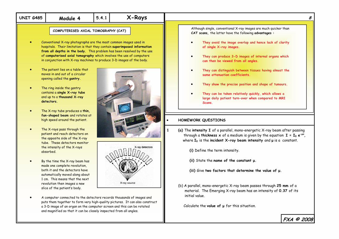
Conventional X-ray photographs are the most common images used in hospitals. Their limitation is that they contain superimposed information from all depths in the body. This problem has been resolved by the use of computerised axial tomography which involves the use of computers in conjunction with X-ray machines to produce 3-D images of the body. The patient lies on a table that moves in and out of a circular opening called the gantry. The ring inside the gantry contains a single X-ray tube and up to a thousand X-ray detectors. The X-ray tube produces a thin, fan-shaped beam and rotates at high speed around the patient. The X-rays pass through the patient and reach detectors on the opposite side of the X-ray tube. These detectors monitor the intensity of the X-rays absorbed. By the time the X-ray beam has made one complete revolution, both it and the detectors have automatically moved along about 1 cm. This means that the next revolution then images a new slice of the patient’s body. A computer connected to the detectors records thousands of images and puts them together to form very high-quality pictures. It can also construct a 3-D image of an organ on the computer screen and this can be rotated and magnified so that it can be closely inspected from all angles.
FXA © 2008
COMPUTERISED AXIAL TOMOGRAPHY (CAT)
8
Although single, conventional X-ray images are much quicker than CAT scans, the latter have the following advantages : They avoid the image overlap and hence lack of clarity of single X-ray images. They can produce 3-D images of internal organs which can then be viewed from all angles. They can distinguish between tissues having almost the same attenuation coefficients. They show the precise position and shape of tumours. They can be taken relatively quickly, which allows a large daily patient turn-over when compared to MRI Scans.
HOMEWORK QUESTIONS 1 (a) The intensity I of a parallel, mono-energetic X-ray beam after passing through a thickness x of a medium is given by the equation I = I0 e-μx, where I0 is the incident X-ray beam intensity and μ is a constant. (i) Define the term intensity. (ii) State the name of the constant μ. (iii) Give two factors that determine the value of μ. (b) A parallel, mono-energetic X-ray beam passes through 25 mm of a material. The Emerging X-ray beam has an intensity of 0.37 of its initial value. Calculate the value of μ for this situation.
UNIT G485 Module 4 5.4.1 X-Rays
2 In an X-ray tube, electrons accelerated to high speeds impact with a tungsten target, losing their energy to produce high-energy electromagnetic radiation, or X-rays. (a) For a tube voltage of 150 kV, calculate : (i) The maximum energy Emax in J of the X-ray photons produced. (ii) The minimum wavelength λmin of these photons. (b) The graph below shows the relative distribution of photon energies from The X-ray tube. (i) Explain the origin of the characteristic lines at about 60 keV. (ii) State the changes, if any, to Emax, λmin and the shape of the intensity curve, when each of the following is increased independently :
The tube voltage. The tube current.
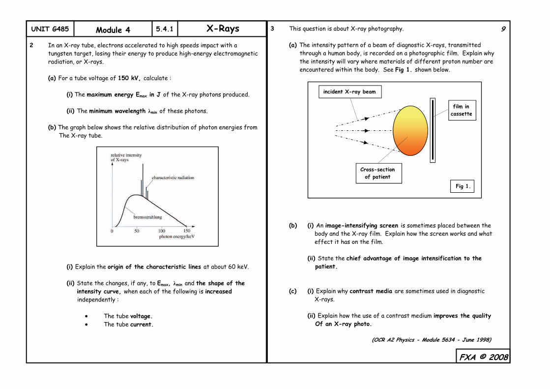
3 This question is about X-ray photography. (a) The intensity pattern of a beam of diagnostic X-rays, transmitted through a human body, is recorded on a photographic film. Explain why the intensity will vary where materials of different proton number are encountered within the body. See Fig 1. shown below. (b) (i) An image-intensifying screen is sometimes placed between the body and the X-ray film. Explain how the screen works and what effect it has on the film. (ii) State the chief advantage of image intensification to the patient. (c) (i) Explain why contrast media are sometimes used in diagnostic X-rays. (ii) Explain how the use of a contrast medium improves the quality Of an X-ray photo. (OCR A2 Physics - Module 5634 - June 1998)
FXA © 2008
9
incident X-ray beam
film in cassette
Cross-section of patient
Fig 1.
UNIT G485 Module 4 5.4.1 X-Rays
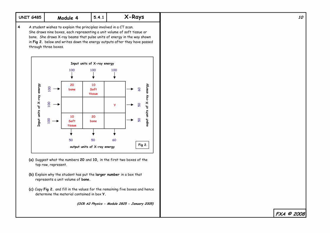
4 A student wishes to explain the principles involved in a CT scan. She draws nine boxes, each representing a unit volume of soft tissue or bone. She draws X-ray beams that pulse units of energy in the way shown in Fig 2. below and writes down the energy outputs after they have passed through three boxes. (a) Suggest what the numbers 20 and 10, in the first two boxes of the top row, represent. (b) Explain why the student has put the larger number in a box that represents a unit volume of bone. (c) Copy Fig 2. and fill in the values for the remaining five boxes and hence determine the material contained in box Y. (OCR A2 Physics - Module 2825 - January 2005)
FXA © 2008
20 bone
10 Soft tissue
Y
10 Soft tissue
20 bone
Input units of X-ray energy
oupu
t un
its
of X
-ray
ene
rgy
Inpu
t un
its
of X
-ray
ene
rgy
output units of X-ray energy
100
50
100 100
60 50
100
100
100
50
60
50
Fig 2.
10