Embed Size (px)
Citation preview
52 OstomyWound Management
FEATURE
Burn wound management is determined by the
depth and site of the burn.1 At the Prince of
Wales Hospital, Shatin, Hong Kong, effective
management strategies are based on a descriptive clas-
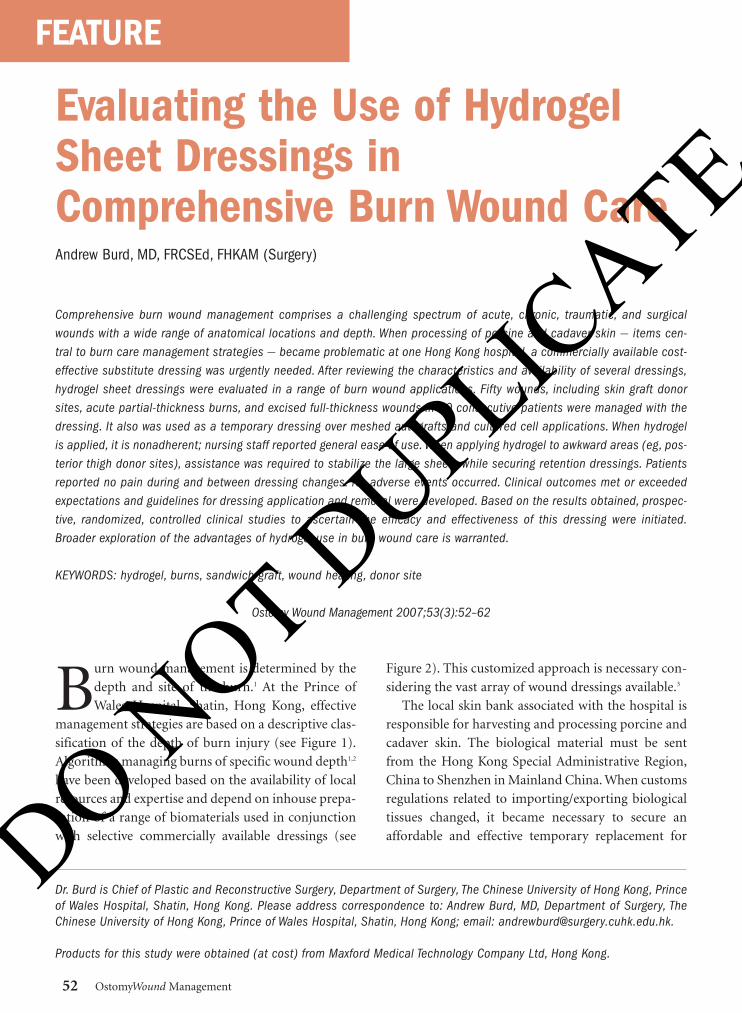
sification of the depth of burn injury (see Figure 1).
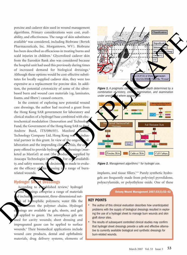
Algorithms managing burns of specific wound depth1,2
have been developed based on the availability of local
resources and expertise and depend on inhouse prepa-
ration of a range of biomaterials used in conjunction
with selective commercially available dressings (see
Figure 2). This customized approach is necessary con-
sidering the vast array of wound dressings available.3
The local skin bank associated with the hospital is
responsible for harvesting and processing porcine and
cadaver skin. The biological material must be sent
from the Hong Kong Special Administrative Region,
China to Shenzhen in Mainland China. When customs
regulations related to importing/exporting biological
tissues changed, it became necessary to secure an
affordable and effective temporary replacement for
Evaluating the Use of HydrogelSheet Dressings inComprehensive Burn Wound Care Andrew Burd, MD, FRCSEd, FHKAM (Surgery)
Comprehensive burn wound management comprises a challenging spectrum of acute, chronic, traumatic, and surgicalwounds with a wide range of anatomical locations and depth. When processing of porcine and cadaver skin — items cen-tral to burn care management strategies — became problematic at one Hong Kong hospital, a commercially available cost-effective substitute dressing was urgently needed. After reviewing the characteristics and availability of several dressings,hydrogel sheet dressings were evaluated in a range of burn wound applications. Fifty wounds, including skin graft donorsites, acute partial-thickness burns, and excised full-thickness wounds in 30 consecutive patients were managed with thedressing. It also was used as a temporary dressing over meshed autografts and cultured cell applications. When hydrogelis applied, it is nonadherent; nursing staff reported general ease of use. When applying hydrogel to awkward areas (eg, pos-terior thigh donor sites), assistance was required to stabilize the large sheets while securing retention dressings. Patientsreported no pain during and between dressing changes. No adverse events occurred. Clinical outcomes met or exceededexpectations and guidelines for dressing application and removal were developed. Based on the results obtained, prospec-tive, randomized, controlled clinical studies to ascertain the efficacy and effectiveness of this dressing were initiated.Broader exploration of the advantages of hydrogel use in burn wound care is warranted.
KEYWORDS: hydrogel, burns, sandwich graft, wound healing, donor site
Ostomy Wound Management 2007;53(3):52–62
Dr. Burd is Chief of Plastic and Reconstructive Surgery, Department of Surgery, The Chinese University of Hong Kong, Princeof Wales Hospital, Shatin, Hong Kong. Please address correspondence to: Andrew Burd, MD, Department of Surgery, TheChinese University of Hong Kong, Prince of Wales Hospital, Shatin, Hong Kong; email: [email protected].
Products for this study were obtained (at cost) from Maxford Medical Technology Company Ltd, Hong Kong.
52-62_OWM0307_Burd.qxd 3/5/07 6:21 PM Page 52
DO NOT D
UPLICATE
porcine and cadaver skin used in wound management
algorithms. Primary considerations were cost, avail-
ability, and effectiveness. The range of skin substitutes
available4 was considered, including Biobrane (Bertek
Pharmaceuticals, Inc. Morgantown, WV). Biobrane
has been described as efficacious in treating burns and
scald injuries in children.5 Glycerolized cadaver skin
from the Euroskin Bank also was considered because
the hospital unit had used this previously during times
of increased demand for biological dressings.6
Although these options would be cost-effective substi-
tutes for locally supplied cadaver skin, they were too
expensive as a replacement for porcine skin. In addi-
tion, the potential cytotoxicity of some of the silver-
based burn and wound care materials (eg, laminates,
foams, and fibers7) caused concern.
In the context of exploring new potential wound
care dressings, the author had received a grant from
the Hong Kong SAR government for laboratory and
clinical studies of a hydrogel base combined with elec-
trochemical modulation (Innovation and Technology
Fund, the Government of the Hong Kong SAR to Prof.
Andrew Burd, ITS/086/03). Maxford Medical
Technology Company Ltd, Hong Kong was an indus-
trial partner in this grant. In view of the ongoing col-
laboration and the impending clinical crisis, the com-
pany offered to provide hydrogel sheet dressings (mar-
keted as MaxGel) at cost (the product is supplied by
Anacapa Technologies in the US). For cost, availabili-
ty, and safety reasons, the decision was made to evalu-
ate the efficacy of this dressing in a range of burn-
related wounds.
Hydrogel Sheet Dressings According to a published review,8 hydrogel
wound dressings comprise a range of materials
and include a permanent, three-dimensional net-
work of hydrophilic polymers; water fills the
space between the polymer chains. Hydrogel
dressings are available as gels, sheets, and gels
pre-applied to gauze. The amorphous gels are
used for cavity wounds; sheet dressing and
impregnated gauze can be applied to surface
wounds.8 Their biomedical applications include
wound care products, dental and ophthalmic
materials, drug delivery systems, elements of
implants, and tissue fillers.9,10 Purely synthetic hydro-
gels are frequently made from polyvinyl pyrrolidone,
polyacrylamide, or polyethylene oxide. One of these
March 2007 Vol. 53 Issue 3 53
KEY POINTS• The author of this clinical evaluation describes how unanticipated
problems with the supply of biological dressings resulted in explor-ing the use of a hydrogel sheet to manage burn wounds and skingraft donor sites.
• The results of subsequent controlled clinical studies may confirmthat hydrogel sheet dressings provide a safe and effective alterna-tive to currently available biological and synthetic dressings forburn-related wounds.
Ostomy Wound Management 2007;53(3):52–62
Figure 1. A pragmatic classification with depth determined by acombination of history, clinical examination, and examinationunder anesthesia where appropriate.
Figure 2. Management algorithms1,2 for hydrogel use.
52-62_OWM0307_Burd.qxd 3/5/07 6:21 PM Page 53
DO NOT D
UPLICATE
basic constituents, polyacrylamide, has gained some
recent notoriety because of its association with
injectable hydrogel fillers and the theoretical possi-
bility that the product can degrade to a neurotoxic
and/or carcinogenic monomer.11-13 The hydrogel
used in the current evaluation contains polyvinyl
pyrolidine, polyethylene glycol, and agar. The sterile
permanent hydrogel forms a transparent sheet 3
mm to 4 mm thick. Hydrogel is Conformité
Européene marked (European Conformity — CE)
and cleared for marketing by the US Food and Drug
Administration (FDA).
Several options regarding sheet hydrogels were (or
have been) available. As reviewed by Eisenbud,8 a
number of studies have compared amorphous
hydrogels with other dressing strategies, particularly
in the treatment of chronic wounds such as pressure
ulcers.8 However, there is a paucity of reports on clin-
ical trials regarding sheet hydrogel dressings and
burn care. Product options include Vigilon® (C.R.
Bard, Inc, Covington, Ga), a commercially available
sheet hydrogel marketed in the US for almost 25
years. This gelatinous sheet consists of an insoluble
cross-linked polyethylene oxide copolymer with
water as the dispersion medium. Its tensile strength
and low-mass configuration make it susceptible to
rapid evaporative loss (a removable, polyethylene
film applied to one side of the dressing controls the
rate of moisture loss). The product’s clinical indica-
tions are limited to skin tears, minor chemical and
thermal burns, cuts, abrasions, postoperative inci-
sions and, most frequently, radiation dermatitis.
Related literature limits its clinical applications to
radiation dermatitis and postoperatively for cosmet-
ic surgery. The product has not been prescribed for
more extensive burn wounds. Other brands of
hydrogel sheets — Nu-Gel™ (Johnson and Johnson,
New Brunswick, NJ), Clear Site (Conmed
Corporation, Utica, NY), Aquasorb (DeRoyal,
Powell, Tenn), and Flexderm (Bertek [Dow Hickam]
Sugar Land, Tex) — are used mainly as primary
dressings for shallow wounds in specific anatomical
areas. No reports about their use in major burn
wounds are available. The study product choice was
made with consideration to this information and the
availability of the study product.
MethodsTo address facility need, a clinical evaluation of
sheet hydrogel used in place of porcine and cadaver
skin was conducted. Senior medical and nursing staff
in the burn unit made the decision to use hydrogel on
all patients admitted to the burn service who would
otherwise have been treated with either porcine or
cadaver skin according to established protocols. The
use of hydrogel sheets on other wounds — eg, skin
graft donor sites — also was evaluated. In the context
of evaluating a product already approved for wound
care, IRB permission was not required. When appro-
priate, patients were informed that the dressing was
used because of the lack of availability of the regular
products. All dressing applications were performed by
the burn unit staff (including burn unit nursing staff
for ward dressings and burn unit medical staff for
operating theater dressings). Objective and subjective
pain assessment tools were used to monitor patient
feedback: a visual analogue scale14 with a range of 1 to
10 was used for adults; the Wong Baker FACES Pain
rating scale was used for children.15 The only change
to existing protocol was the substitution of hydrogel
sheets where porcine or cadaver skin would other-
wise be used. Senior medical and nursing staff
(including the author, Ward Manager, and Clinical
Nurse Specialist) maintained oversight of all care and
documentation procedures, which were detailed in
the patients’ medical records. Wounds were deter-
mined to be healed when the dressing could be
detached without pain and the underlying skin was
completely re-epithelialized.
Clinical Evaluation The clinical evaluation of hydrogel in the author’s
facility included five types of traumatic and iatrogenic
wound care challenges commonly encountered in
clinical burn care: dressings for skin graft donor sites
and acute partial-thickness burns as well as temporary
dressings for excised full-thickness wounds, meshed
autografts, and cultured cell applications.
Donor site dressing. In this evaluation, sheet
hydrogel was first used on a split-thickness skin
donor site on the left lower leg. The 14-year-old
female patient had a history of extensive burn recon-
structive surgery, which has previously been
54 OstomyWound Management
52-62_OWM0307_Burd.qxd 3/5/07 6:21 PM Page 54
DO NOT D
UPLICATE
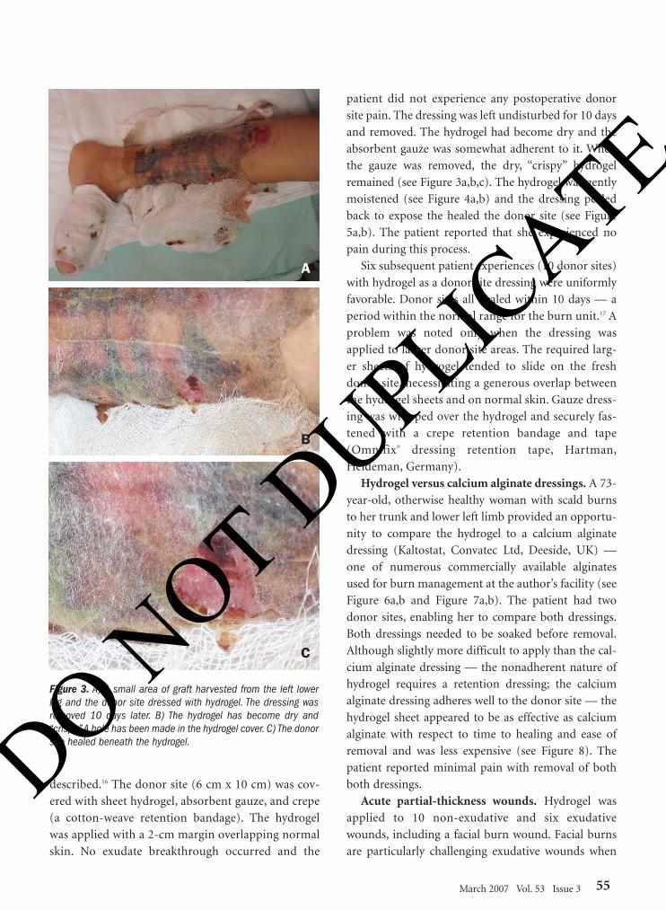
described.16 The donor site (6 cm x 10 cm) was cov-
ered with sheet hydrogel, absorbent gauze, and crepe
(a cotton-weave retention bandage). The hydrogel
was applied with a 2-cm margin overlapping normal
skin. No exudate breakthrough occurred and the
patient did not experience any postoperative donor
site pain. The dressing was left undisturbed for 10 days
and removed. The hydrogel had become dry and the
absorbent gauze was somewhat adherent to it. When
the gauze was removed, the dry, “crispy” hydrogel
remained (see Figure 3a,b,c). The hydrogel was gently
moistened (see Figure 4a,b) and the dressing pealed
back to expose the healed the donor site (see Figure
5a,b). The patient reported that she experienced no
pain during this process.
Six subsequent patient experiences (10 donor sites)
with hydrogel as a donor site dressing were uniformly
favorable. Donor sites all healed within 10 days — a
period within the normal range for the burn unit.17 A
problem was noted only when the dressing was
applied to larger donor site areas. The required larg-
er sheets of hydrogel tended to slide on the fresh
donor site, necessitating a generous overlap between
the hydrogel sheets and on normal skin. Gauze dress-
ing was wrapped over the hydrogel and securely fas-
tened with a crepe retention bandage and tape
(Omnifix® dressing retention tape, Hartman,
Heideman, Germany).
Hydrogel versus calcium alginate dressings. A 73-
year-old, otherwise healthy woman with scald burns
to her trunk and lower left limb provided an opportu-
nity to compare the hydrogel to a calcium alginate
dressing (Kaltostat, Convatec Ltd, Deeside, UK) —
one of numerous commercially available alginates
used for burn management at the author’s facility (see
Figure 6a,b and Figure 7a,b). The patient had two
donor sites, enabling her to compare both dressings.
Both dressings needed to be soaked before removal.
Although slightly more difficult to apply than the cal-
cium alginate dressing — the nonadherent nature of
hydrogel requires a retention dressing; the calcium
alginate dressing adheres well to the donor site — the
hydrogel sheet appeared to be as effective as calcium
alginate with respect to time to healing and ease of
removal and was less expensive (see Figure 8). The
patient reported minimal pain with removal of both
both dressings.
Acute partial-thickness wounds. Hydrogel was
applied to 10 non-exudative and six exudative
wounds, including a facial burn wound. Facial burns
are particularly challenging exudative wounds when
March 2007 Vol. 53 Issue 3 55
Figure 3. A) A small area of graft harvested from the left lowerleg and the donor site dressed with hydrogel. The dressing wasremoved 10 days later. B) The hydrogel has become dry and“crispy.”A hole has been made in the hydrogel cover. C) The donorsite healed beneath the hydrogel.
A
B
C
52-62_OWM0307_Burd.qxd 3/5/07 6:21 PM Page 55
DO NOT D
UPLICATE
patients have inhalation injuries and are intubated. It
was found through experience that large sheets of
hydrogel conform well to the wound beds and are
quick and easy to apply. The wound can be readily
inspected through the dressing and, in the uncon-
scious ventilated patient, no retention dressing is nec-
essary (see Figure 9a). In the case of a 23-year-old
male patient who sustained a 70% body surface area
(BSA) burn as a result of an explosion, the dressing
was left in place for 48 hours. After 48 hours, the dress-
ing had become swollen and exhibited a slightly yellow
discoloration from absorbing the exudate. The under-
lying wound bed itself was dry (see Figure 9b). The
patient’s face went on to heal spontaneously and sur-
gery was not needed.
More frequent dressing changes may be necessary
in the highly exudative wound. In the author’s expe-
rience, the dressing should be changed daily until it
becomes adherent to the underlying bed. In the
superficial partial-thickness burn with little or no
exudate, the first hydrogel dressing applied becomes
dry and adheres to the wound. As the wound heals,
the overlying hydrogel can be trimmed away (see
Figure 10). The non-exudative wounds in this evalu-
ation were all superficial partial-thickness wounds
and healed within 10 days. The exudative wounds
56 OstomyWound Management
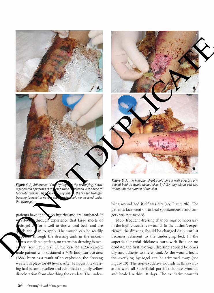
Figure 4. A) Adherence of dry hydrogel to the underlying, newlyregenerated epidermis is reversed when moistened with saline tofacilitate removal. B) Following rehydration, the “crisp” hydrogelbecame “plastic” in nature and a finger could be inserted underthe hydrogel.
A
BFigure 5. A) The hydrogel sheet could be cut with scissors andpeeled back to reveal healed skin. B) A flat, dry, blood clot wasevident on the surface of the skin.
A
B
52-62_OWM0307_Burd.qxd 3/5/07 6:21 PM Page 56
DO NOT D
UPLICATE
were assessed clinically (no biopsy or laser Doppler
used) every 24 hours using wound history, appear-
ance, and dressing change interval to predict healing,
which is ultimately a function of depth of injury.1 The
exudative wounds were found to be deeper but all
healed within 14 days following burn injury. Table 1
details the clinical management protocol for exudative
and non-exudative partial-thickness burns that was
developed during this evaluation period.
Full-thickness burn: temporary dressing. The
author’s facility utilizes a full-thickness burn care pro-
tocol of early excision (debridement that may involve
removal of unburned tissue), requiring appropriate
dressings to cover the surgical wound. The sandwich
technique is used when enough autologous donor skin
is available. This involves the application of widely
meshed autograft to the wound bed following a full-
thickness excision. Typically, the meshed skin is cov-
ered with cadaver allogenic skin to prevent desicca-
tion of the wound bed in the interstices of the
meshed graft. Hydrogel dressing also may be used in
such situations (see Figure 11a,b,c). In the case illus-
trated (see Figure 12a,b), a 35-year-old female
patient involved in an explosion had a 70% body sur-
face area burn; 1:6 meshed autograft was applied and
covered with hydrogel. Although 2 weeks later the
hydrogel exhibited a yellow appearance, more than
70% of the graft had taken and the interstices of the
March 2007 Vol. 53 Issue 3 57
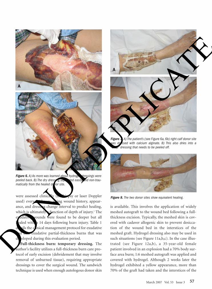
Figure 6. A) As more was learned about hydrogel, dressings werepeeled back. B) The dry dressing separated easily and non-trau-matically from the healed donor site.
A
B
Figure 7. A) The patient’s (see Figure 6a, 6b) right calf donor sitewas dressed with calcium alginate. B) This also dries into a“crisp” dressing that needs to be peeled off.
A
B
Figure 8. The two donor sites show equivalent healing.
52-62_OWM0307_Burd.qxd 3/5/07 6:21 PM Page 57
DO NOT D
UPLICATE
autograft had re-epithelialized. When used with lower
expansion ratios, graft take was found to be even more
complete. In the same patient, hydrogel was placed
directly onto an excised wound bed — a situation
where cadaver skin would typically be used. The
hydrogel was stapled to the wound bed to prevent
movement. The hydrogel was not incorporated into
the wound but it became swollen, indicating that it
had absorbed exudate from the wound bed; in this cir-
cumstance, the cadaver skin yielded better results.
Infection. In the first few cases of full-thickness
wound excision, positive cultures, primarily for
Pseudomonas, were collected from the wound bed(s).
Because the hydrogel contains no intrinsic antibacter-
ial agent, betadine or chlorhexidine-soaked gauze was
applied over the hydrogel in subsequent cases. This
modification was based on unpublished data from the
author’s laboratory experiments that examined the
susceptibility of the bacterial flora found in the burns
unit to topical antibacterial agents. Dressings were
changed every 2 to 3 days and the wound bed
remained healthy, viable, and non-infected, allowing
further sandwich grafting once the first donor site
wounds healed.
Cultured cells. Although cell suspensions are not
often used in the author’s practice, culture facilities are
58 OstomyWound Management
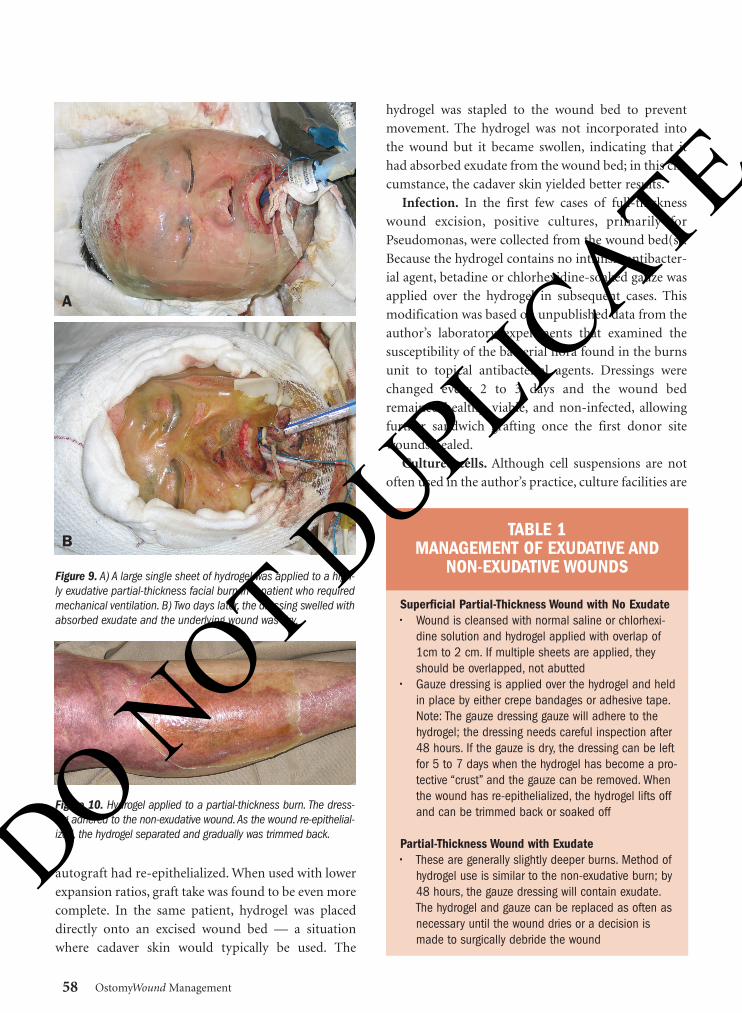
Figure 9. A) A large single sheet of hydrogel was applied to a high-ly exudative partial-thickness facial burn in a patient who requiredmechanical ventilation. B) Two days later, the dressing swelled withabsorbed exudate and the underlying wound was dry.
A
B
Figure 10. Hydrogel applied to a partial-thickness burn. The dress-ing adhered to the non-exudative wound.As the wound re-epithelial-ized, the hydrogel separated and gradually was trimmed back.
TABLE 1MANAGEMENT OF EXUDATIVE AND
NON-EXUDATIVE WOUNDS
Superficial Partial-Thickness Wound with No Exudate• Wound is cleansed with normal saline or chlorhexi-
dine solution and hydrogel applied with overlap of1cm to 2 cm. If multiple sheets are applied, theyshould be overlapped, not abutted
• Gauze dressing is applied over the hydrogel and heldin place by either crepe bandages or adhesive tape.Note: The gauze dressing gauze will adhere to thehydrogel; the dressing needs careful inspection after48 hours. If the gauze is dry, the dressing can be leftfor 5 to 7 days when the hydrogel has become a pro-tective “crust” and the gauze can be removed. Whenthe wound has re-epithelialized, the hydrogel lifts offand can be trimmed back or soaked off
Partial-Thickness Wound with Exudate• These are generally slightly deeper burns. Method of
hydrogel use is similar to the non-exudative burn; by48 hours, the gauze dressing will contain exudate.The hydrogel and gauze can be replaced as often asnecessary until the wound dries or a decision ismade to surgically debride the wound
52-62_OWM0307_Burd.qxd 3/5/07 6:21 PM Page 58
DO NOT D
UPLICATE
available. Cells are applied either to augment the take
of widely meshed autograft or as de novo covering for
an area of excised burn. A split-thickness skin biopsy
is taken from an area of unburned skin and subjected
to enzymatic separation of epidermis from dermis.
The keratinocytes are suspended and expanded in cul-
ture — resultant cells can be applied as sheet grafts or
cell suspension, which in this case was sprayed on the
wound.18 The author’s approach for covering cultured
cell spray varies — either cadaver skin or Mepitel
dressing (Molnlycke Heath Care, Norcross, Ga) can be
used.19 The main goal is to allow the cultured cells to
survive and proliferate in situ; a major concern is the
possible cytotoxicity of dressing material, particularly
silver-based dressings.7
It was of particular interest to see what would hap-
pen when cultured cells were applied to an abdominal
wound in a patient with extensive burns whose
wounds were covered with a hydrogel dressing (see
Figure 13). The result was a 40% take of cells after 10
days. This compares favorably with published data18;
however, the purpose was not simply to cover the area
but also to prepare for a widely meshed autograft that
was subsequently applied and helped facilitate com-
plete wound closure.
Desloughing burns. Clinical experience has shown
that some burns can be partially desloughed by chang-
ing porcine skin dressings and replacing them on a
daily basis.2 It was observed that hydrogel sheets have
the ability to hydrate wounds and absorb exudate in
deeper partial-thickness burns. In some wounds, the
hydrogel swells but is not adherent. When the dressing
is changed and the wound cleansed daily, the charac-
ter of the wound changes — less slough and healthier
granulation tissue is visible on the surface. After three
March 2007 Vol. 53 Issue 3 59
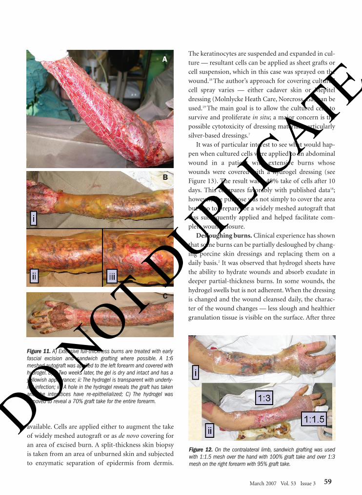
Figure 11. A) Extensive full-thickness burns are treated with earlyfascial excision and sandwich grafting where possible. A 1:6meshed autograft was applied to the left forearm and covered withhydrogel. B) i: Two weeks later, the gel is dry and intact and has ayellowish appearance; ii: The hydrogel is transparent with underly-ing infection; iii: A hole in the hydrogel reveals the graft has takenand the interstices have re-epithelialized; C) The hydrogel wasremoved to reveal a 70% graft take for the entire forearm.
A
B
C
Figure 12. On the contralateral limb, sandwich grafting was usedwith 1:1.5 mesh over the hand with 100% graft take and over 1:3mesh on the right forearm with 95% graft take.
52-62_OWM0307_Burd.qxd 3/5/07 6:21 PM Page 59
DO NOT D
UPLICATE
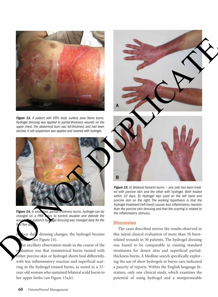
to four daily dressing changes, the hydrogel became
adherent (see Figure 14).
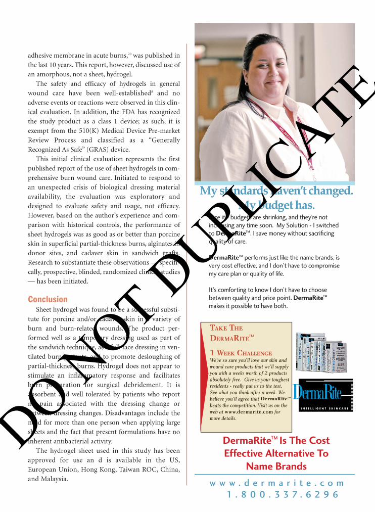
An ancillary observation made in the course of the
evaluation was that symmetrical burns treated with
either porcine skin or hydrogel sheets heal differently,
with less inflammatory reaction and superficial scar-
ring in the hydrogel treated burns, as noted in a 37-
year-old woman who sustained bilateral scald burns to
her upper limbs (see Figure 15a,b).
DiscussionThe cases described mirror the results observed in
this initial clinical evaluation of more than 50 burn-
related wounds in 30 patients. The hydrogel dressing
was found to be comparable to existing standard
treatments for donor sites and superficial partial-
thickness burns. A Medline search specifically explor-
ing the use of sheet hydrogels in burns care indicated
a paucity of reports. Within the English language lit-
erature, only one clinical study, which examines the
potential of using hydrogel and a semipermeable
60 OstomyWound Management
Figure 13. A patient with 65% body surface area flame burns.Hydrogel dressing was applied to partial-thickness wounds on theupper chest. The abdominal burn was full-thickness and had beenexcised. A cell suspension was applied and covered with hydrogel.
Figure 14. In exudative, partial-thickness burns, hydrogel can bechanged on a PRN basis to control exudate and debride thewound. This patient’s hydrogel dressing was changed daily for thefirst few days.
Figure 15. A) Bilateral forearm burns — one side has been treat-ed with porcine skin and the other with hydrogel. Both healedwithin 10 days. B) Hydrogel was used on the left hand andporcine skin on the right. The working hypothesis is that thehydrogel treatment (left hand) causes less inflammatory reactionthan the porcine skin dressing and that the scarring is related tothe inflammatory stimulus.
A
B
52-62_OWM0307_Burd.qxd 3/5/07 6:21 PM Page 60
DO NOT D
UPLICATE
adhesive membrane in acute burns,20 was published in
the last 10 years. This report, however, discussed use of
an amorphous, not a sheet, hydrogel.
The safety and efficacy of hydrogels in general
wound care have been well-established8 and no
adverse events or reactions were observed in this clin-
ical evaluation. In addition, the FDA has recognized
the study product as a class 1 device; as such, it is
exempt from the 510(K) Medical Device Pre-market
Review Process and classified as a “Generally
Recognized As Safe” (GRAS) device.
This initial clinical evaluation represents the first
published report of the use of sheet hydrogels in com-
prehensive burn wound care. Initiated to respond to
an unexpected crisis of biological dressing material
availability, the evaluation was exploratory and
designed to evaluate safety and usage, not efficacy.
However, based on the author’s experience and com-
parison with historical controls, the performance of
sheet hydrogels was as good as or better than porcine
skin in superficial partial-thickness burns, alginates in
donor sites, and cadaver skin in sandwich grafts.
Research to substantiate these observations — specifi-
cally, prospective, blinded, randomized clinical studies
— has been initiated.
ConclusionSheet hydrogel was found to be a successful substi-
tute for porcine and/or cadaver skin in a variety of
burn and burn-related wounds. The product per-
formed well as a temporary dressing used as part of
the sandwich technique, as a full-face dressing in ven-
tilated burn patients, and to promote desloughing of
partial-thickness burns. Hydrogel does not appear to
stimulate an inflammatory response and facilitates
burn preparation for surgical debridement. It is
absorbent and well tolerated by patients who report
no pain associated with the dressing change or
between dressing changes. Disadvantages include the
need for more than one person when applying large
sheets and the fact that present formulations have no
inherent antibacterial activity.
The hydrogel sheet used in this study has been
approved for use an d is available in the US,
European Union, Hong Kong, Taiwan ROC, China,
and Malaysia.
Face it - budgets are shrinking, and they’re not
increasing any time soon. My Solution - I switched
to DermaRite™. I save money without sacrificing
quality of care.
DermaRite™ performs just like the name brands, is
very cost effective, and I don’t have to compromise
my care plan or quality of life.
It’s comforting to know I don’t have to choose
between quality and price point. DermaRite™
makes it possible to have both.
TAKE THE
DERMARITE™
E
1 WEEK CHALLENGE
We’re so sure you’ll love our skin and
wound care products that we’ll supply
you with a weeks worth of 2 products
absolutely free. Give us your toughest
residents - really put us to the test.
See what you think after a week. We
believe you’ll agree that DermaRite™e
beats the competition. Visit us on the
web at www.dermarite.com for
more details.
w w w . d e r m a r i t e . c o m
1 . 8 0 0 . 3 3 7 . 6 2 9 6
My standards haven’t changed. My budget has.
DermaRite™ Is The Cost
Effective Alternative To
Name Brands
nurse challange-1.indd 1 8/12/05 1:27:42 PM
52-62_OWM0307_Burd.qxd 3/5/07 6:21 PM Page 61
DO NOT D
UPLICATE
Overall, hydrogel sheets seem to be a viable alterna-
tive or reserve dressing material for use in a range of
burn-related wounds. In particular, hydrogel sheet
dressings show potential for use in comprehensive
burn wound management, a clinical area that war-
rants further research.
AcknowledgmentThe author would like to acknowledge the invalu-
able help and advice of Michael Tsang who has been a
Project Consultant with the ITF grant and also has
been responsible for the production and development
of the product used in this evaluation. - OWM
References1. Burd A, Chiu T. Allogenic skin in the treatment of
burns. Clin Dermatol. 2005;23(4):376–387.2. Chiu T, Burd A. “Xenograft” dressing in the treat-
ment of burns. Clin Dermatol. 2005;23(4):419–423.3. Burd A. Burn dressings. In: Wnek GE, Bowlin GL
(eds). Encyclopedia of Biomaterials and BiomedicalEngineering. New York, NY: Marcel Dekker, Inc;2004.
4. Shakespeare PG. The role of skin substitutes in thetreatment of burn injuries. Clin Dermatol.2005;23(4):413–418.
5. Lang EM, Eiberg CA, Brandis M, Bjorn Stark GB.Biobrane in the treatment of burn and scald injuriesin children. Ann Plast Surg. 2005;55(5):485–489.
6. Burd A. Glycerolised allogenic skin: transplant ordressing? A Medico-legal question. Burns.2002;28:S34–S39.
7. Burd A, Kwok CH, Hung SC, et al. A comparativestudy of the cytotoxicity of silver-based dressings inmonolayer cells, tissue explant, and animal models.Wound Repair Regen. 2007;15:94–104.
8. Eisenbud D, Hunter H, Kessler L, Zulkowski K.Hydrogel wound dressings: where do we stand in2003? Ostomy Wound Manage. 2003;49(10):52–57.
9. Kiyozumi T, Kanatani Y, Ishihara M, et al. Medium(DMEM/F12)-containing chitosan hydrogel asadhesive and dressing in autologous skin grafts and
accelerator in the healing process. J Biomed MaterRes B Appl Biomater. 2006;79:129–136.
10. Parnell LK, Ciufi B, Gokoo CF. Preliminary use of ahydrogel containing enzymes in the treatment ofStage II and Stage III pressure ulcers. Ostomy WoundManage. 2005;51(8):50–60.
11. Christensen LH, Breiting VB, Aasted A, Jorgensen A,Kebuladze I. Long-term effects of polyacrylamidehydrogel on human breast tissue. Plast Reconstr Surg.2003;111(6):1883–1890.
12. Amin SP, Marmur ES, Goldberg DJ. Complicationsfrom injectable polyacrylamide gel, a new non-biodegradable soft tissue filler. Dermatol Surg.2004;30(12 pt 2):1507–1509.
13. Qiao Q, Wang X, Sun J, et al. Management for post-operative complications of breast augmentation byinjected polyacrylamide hydrogel. Aesth Plast Surg.2005;29(3):156–161.
14. Tengvall OM, Bjornhagen VC, Lindholm C, JonssonCE, Wengstrom Y. Differences in pain patterns forinfected and noninfected patients with burn injuries.Pain Manage Nurs. 2006;7(4):176–182.
15. Wong DL, Hockenberry-Eaton M, Wilson D,Winkelstein ML, Schwartz P. Wong’s Essentials ofPediatric Nursing, 7th ed. Available through:www.mosbysdrugconsult.com/WOW/faces.html.Accessed March 5, 2007.
16. Burd A, Pang PC, Ying SY, Ayyappan T.Microsurgical reconstruction in children’s burns. JPlast Reconstr Aesthet Surg. 2006;59(7):679–692.
17. Ho WS, Ying SY, Choi PC, Wong TW. A prospectivecontrolled clinical study of skin donor sites treatedwith a 1-4,2-acetamide-deoxy-B-D-glucan polymer:a preliminary report. Burns. 2001;27(7):759–761.
18. Wood FM, Kolybaba ML, Allen P. The use of cul-tured epithelial autograft in the treatment of majorburn injuries: a critical review of the literature.Burns. 2006;32(4):395–401.
19. Burd A, Chan E. Keratinocyte-keloid interaction.Plast Reconstr Surg. 2002;110:197–202.
20. Osti E. Cutaneous burns treated with hydrogel(Burnshield) and a semipermeable adhesive film.Arch Surg. 2006;141:39–42.
62 OstomyWound Management
52-62_OWM0307_Burd.qxd 3/5/07 6:22 PM Page 62
DO NOT D
UPLICATE