Embed Size (px)
Citation preview

9/12/2016
1
Pediatric derm stuff: what is it and what to do
Lucia Diaz, MDPediatric and Adolescent Dermatology
Specially for Children/Dell Children’s Hospital
Assistant Professor of Pediatrics
University of Texas Dell Medical School
Disclosures
• Policies and standards of the Texas Medical Association, the Accreditation Council for Continuing Medical Education, and the American Medical Association require that speakers and planners for continuing medical education activities disclose any relevant financial relationships they may have with any entity producing, marketing, re‐selling, or distributing health care goods or services consumed by, or used on, patients whose products, devices or services may be discussed in the content of the CME activity. The planners and speakers have no relevant relationships to disclose.
• I do intent do discuss an unapproved/investigative use of a commercial product/device in my presentation.
Case 1
9/12/2016
2
Hemangioma
• Most common tumor of infancy, more common in females and premature infants
• Glut‐ 1 positive
• Clinical appearance depends on type of hemangioma
• Presents within 1‐3 weeks of age and grows rapidly until 4‐6 months then starts involutingaround 1 year of age
• Larger hemangiomas will not fully go away
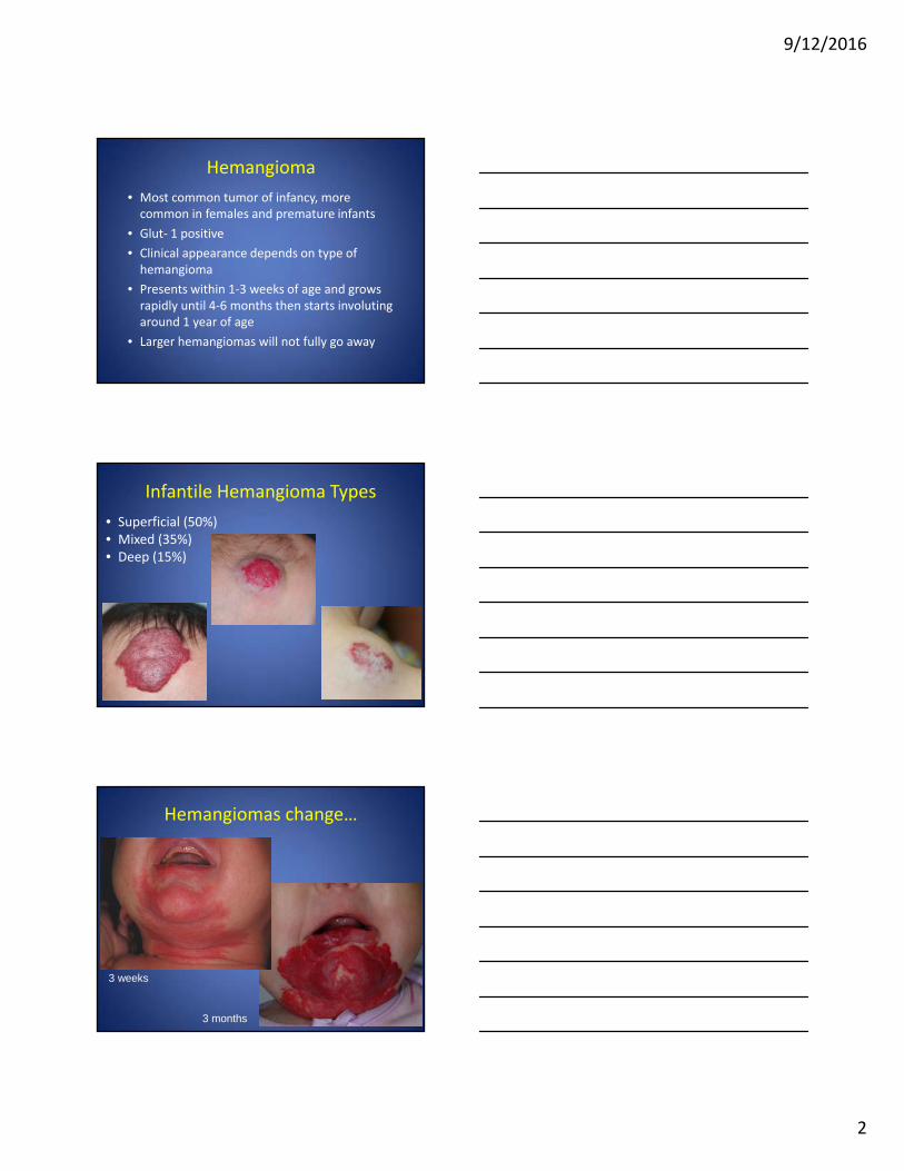
Infantile Hemangioma Types
• Superficial (50%)• Mixed (35%)• Deep (15%)
3 weeks
3 months
Hemangiomas change…

9/12/2016
3
Abortive hemangioma
• GLUT‐1 positive
• Present mostly formed at birth
• Minimal proliferation
High risk hemangiomas
• Location affecting vision, airway, or other vital structure
• Lesions on sites that may cause permanent disfigurement (glabella, lip, nose, ears)
• Size/growth potential
• Ulceration risk
• Segmental.. think about
associated syndromes
Important hemangioma locations
• Periorbital proptosis, amblyopia, visual axis obstruction, chronic conjunctivitis
• Nasal tip distortion of nasal anatomy and residual “Cyrano” deformity
• Lips feeding difficulty, frequent ulceration, and permanent facial scarring
• Ears cosmetically disfiguring, obstruction of auditory canal and conductive hearing loss

9/12/2016
4
Hemangioma treatments
• Active nonintervention
• Beta blockers such as oral propranolol (2‐3 mg/kg/day divided bid or tid) and topical timolol 0.5 % gel
• Pulsed dye laser
• Topical, intralesional, or systemic steroids
• Surgery
• Wound care if ulcerated
• Vincristine, sirolimus (rarely)
Ulcerated hemangioma
• Ulceration is most common complication
– 10% of lesions during proliferative phase
– Most common in intertriginous sites, lips, diaper region
• Treat as appropriate with wound care, topical/oral antibiotics, pain management, pulsed dye laser
Early hemangioma with erosion
9/12/2016
5
Hemangiomas do not always go away
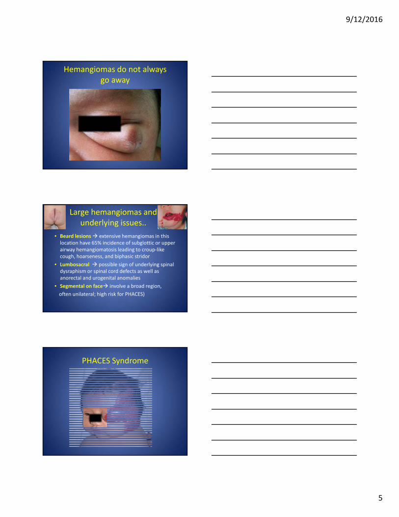
Large hemangiomas and underlying issues..
• Beard lesions extensive hemangiomas in this location have 65% incidence of subglottic or upper airway hemangiomatosis leading to croup‐like cough, hoarseness, and biphasic stridor
• Lumbosacral possible sign of underlying spinal dysraphism or spinal cord defects as well as anorectal and urogenital anomalies
• Segmental on face involve a broad region,
often unilateral; high risk for PHACES)
PHACES Syndrome

9/12/2016
6
PHACES
–Posterior fossa brain malformation (Dandy‐Walker)
–Hemangioma (usually large, facial plaque‐like lesion)
–Arterial anomalies (mainly head and neck)
–Cardiac abnormalities (usually coarctation of the aorta)
–Eye abnormalities
– Sternal clefting and supraumbilical raphe
Port wine stain• Congenital capillary malformation
– May be isolated or as part of various syndromes, most common on face
– Progressively darkens and can develop blebs over years
• Erythematous patches
– Distinguished from hemangioma by congenital presence and static nature
• Treatment: pulsed dye laser
Sturge‐Weber Syndrome
• Facial port wine stain in VI (+/‐ V2, V3) ipsilateral leptomeningeal vascular malformation leading to seizures or brain calcifications
• Choroidal vascular malformation leading to ipsilateral glaucoma
9/12/2016
7
Pyogenic granuloma
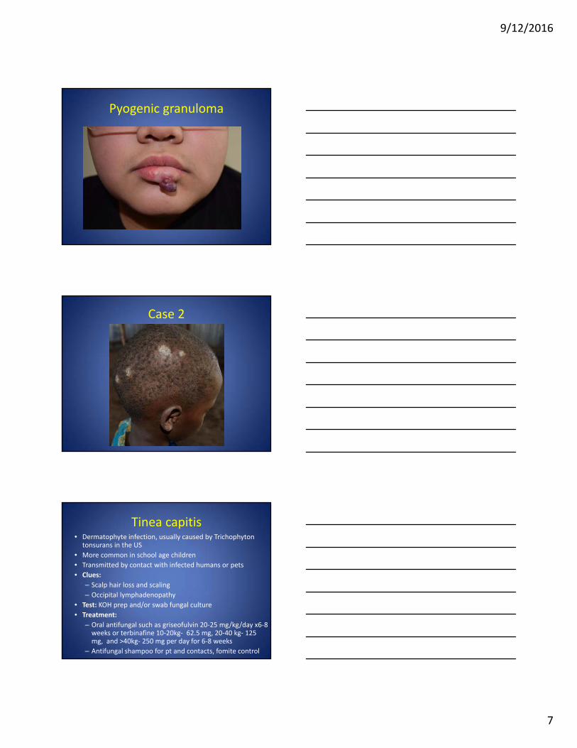
Case 2
Tinea capitis• Dermatophyte infection, usually caused by Trichophyton
tonsurans in the US
• More common in school age children
• Transmitted by contact with infected humans or pets
• Clues:
– Scalp hair loss and scaling
– Occipital lymphadenopathy
• Test: KOH prep and/or swab fungal culture
• Treatment:
– Oral antifungal such as griseofulvin 20‐25 mg/kg/day x6‐8 weeks or terbinafine 10‐20kg‐ 62.5 mg, 20‐40 kg‐ 125 mg, and >40kg‐ 250 mg per day for 6‐8 weeks
– Antifungal shampoo for pt and contacts, fomite control

9/12/2016
8
Kerion and ID reaction
Tinea amiantacea
Alopecia areata

9/12/2016
9
Trichitillomania
Aplasia cutis
Case 3
9/12/2016
10
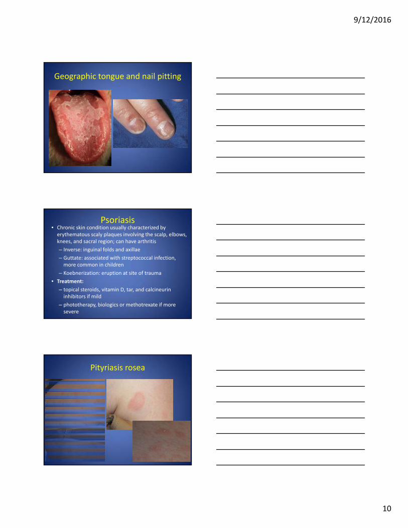
Geographic tongue and nail pitting
Psoriasis• Chronic skin condition usually characterized by erythematous scaly plaques involving the scalp, elbows, knees, and sacral region; can have arthritis
– Inverse: inguinal folds and axillae
– Guttate: associated with streptococcal infection, more common in children
– Koebnerization: eruption at site of trauma
• Treatment:
– topical steroids, vitamin D, tar, and calcineurininhibitors if mild
– phototherapy, biologics or methotrexate if more severe
Pityriasis rosea

9/12/2016
11
Case 4
Scabies burrow and mite!
Scabies• Caused by Sarcoptes scabei
• Transmitted from humans by skin contact or infected objects
• Clues:
– Pruritus, hx of possible exposure
– Crusted or scaly pink papules or burrows in intertrigious areas, genitalia, webspaces, umbilicus or areola.
• Test: Microscopic exam of skin scraping
• Treatment:
– 5% permethrin cream total body in children, 8‐14 hour (overnight); repeat in 7 days
– 2nd line, Ivermectin 200 mcg/kg po
– Wash linens in hot water and treat all close contacts
– Mild to moderate potency topical steroid and/or antihistamine for pruritus

9/12/2016
12
Case 5
Tinea versicolor
• Caused by Malassezia furfur
• More common in adolescents and adults
• Clue: slightly scaly pink or hypopigmented macules coalescing into patches usually on trunk, can be pruritic
• Treatment: Otc selenium sulfide solution/ shampoo or 2% ketoconazole shampoo to affected areas x 10 minutes daily prior to rinsing or topical –azole cream daily; dyspigmentation may last for months
Confluent and reticulated papillomatosis (CARP)

9/12/2016
13
Case 6
Eczema herpeticum• Herpes simplex 1 or 2 infection characterized by punched out erosions and vesicles within eczema lesions
• May have fever, malaise, LAD, pain and pruritus
• Transmission from person, usually from cold sore
• Contagious until crusted over
• Tests: Tzanck smear; DFA, PCR, viral culture
• Treatment: Course of oral or IV antiviral such as acyclovir x 1 week; if young, immunocompromised, extensive infection, or lesions around eyes, consider hospital admission
Excoriated atopic dermatitis

9/12/2016
14
Case 7
Tinea faciei• Dermatophyte infection of the superficial epidermis
• Clues: Annular, scaly pink plaques, ? pustules; tineaincognito: steroid can take away scale
• Test: KOH prep and/or swab fungal culture
• Treatment:
– Topical antifungal cream (ex. terbinafine, ketoconazole) until clear, usually 2x/day for 2 to 4 weeks
– Oral antifungal such as terbinafine daily x 2‐3 weeks if extensive or failing topical therapy
– Look for source to prevent reinfection
Granuloma annulare
9/12/2016
15
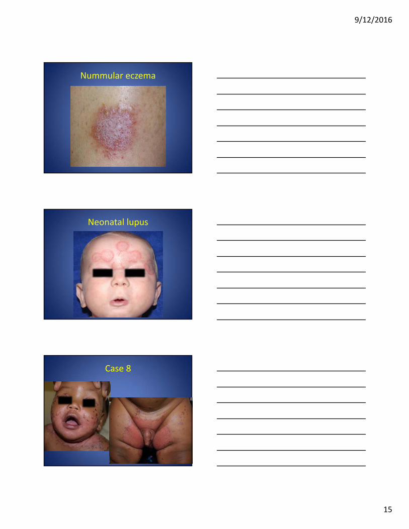
Nummular eczema
Neonatal lupus
Case 8

9/12/2016
16
Acrodermatitis Enteropathica• Nutritional dermatitis due to zinc deficiency that presents as erosive well demarcated periorificialand acral scaly, pink plaques
• Can have alopecia, FTT, irritability
• Can be genetic due to SLC39A4 zinc transporter mutation or acquired from low breast milk zinc, zinc deficient in TPN, or GI disorders with poor zinc absorption
• Tests: low serum zinc and alkaline phosphatase
• Treatment: Zinc supplementation, adequate nutrition
Case 9
Drug rash with eosinophilia an systemic symptoms (DRESS) Syndrome
• Drug hypersensitivity reaction characterized by fever, rash, cervical lymphadenopathy, edema of face and hands in addition to elevated eosinophils, liver enzymes, thrombocytopenia and atypical lymphocytosis
• Onset 3‐6 weeks after starting a drug
• Usual drug offenders: TMP‐SMX, Carbamazepine, phenytoin, phenobarbital, lamotrigine, and allopurinol
• Possible pulmonitis, hepatitis, carditis and thyroiditis Treatment: Stop the offending agent, treat with systemic steroids

9/12/2016
17
Case 10
Bullous impetigo
• Caused by Staph aureus infection
• Production of toxin cleaves desmoglein‐1 which leads to blisters and erosions
• Test: bacterial swab culture for sensitivites
• Treatment: oral antibiotic (ex: cephalexin, clindamycin, tmp/smx) for 1‐2 weeks
Bullous arthropod

9/12/2016
18
Epidermolysis bullosa
Case 11
Molluscum contagiosum
• Poxvirus infection of the epidermis
• May become inflamed, itchy, less likely superinfected
• Lesions usually go away in 18 months on average when the immune system sees them
• May observe or treat depending on lesion number, location, and family preference
• Molluscum dermatitis is an immune reaction against the virus and will resolve when molluscum resolve

9/12/2016
19
Inflamed molluscum
Molluscum treatments
• Office‐based therapy every 3‐6 weeks– curettage– cantharadin green– cryotherapy– candida Antigen intralesional injection
• Home treatments– topical retinoid gel or cream (avoid in patients with eczema)
– imiquimod 5% cream– oral cimetidine x 2‐3 months if many lesions
Folliculitis

9/12/2016
20
Perioral dermatitis
Arthropod bites and papularurticaria
Case 12

9/12/2016
21
Warts
• Human papilloma virus
• Highest incidence in 10‐19 y/o
• 65% disappear in 2 years when immune system sees warts, some may take longer to go away
Warts (verruca vulgaris)
Wart treatments• Office based therapy every 3‐6 weeks:
– cryotherapy– cantharadin red – podophyllin– tricholoracetic acid or squaric acid – candida antigen skin injection
• Home Treatment:– topical salicylic acid with paring– 5‐fluorouracil cream– duct tape – imiquimod 5% cream‐ not good for acral warts– for flat warts: may try topical retinoid gel or cream (avoid in eczema)– oral cimetidine x 2‐3 months if many lesions

9/12/2016
22
Pseudoverrucous papules
Epidermal nevus
Knuckle pads

9/12/2016
23
Dermatomyositis Gottron’s papules
Case 13
Candida diaper dermatitis
• Warmth, moisture, and occlusion lead to overgrowth of candida albicans
• May start as irritant diaper dermatitis
• Treatment: topical antifungal cream (ex. Ketoconazole, nystatin) for 1‐2 weeks

9/12/2016
24
Irritant contact dermatitis
Jacquet’s dermatitis (bad irritation)
Psoriasis

9/12/2016
25
Histiocytosis
Case 14

9/12/2016
26
Staph Scalded Skin Syndrome• Tender erythematous patches on periorifical and flexural regions, can become generalized; superficial bullae
• Primarily neonates and young children but also in adults with renal failure
• Caused by exfoliative toxin by Staph aureus leading to cleavage of desmoglein 1 in upper epidermis
• Clue: positive nikolsky’s sign
• Test: Bacterial cultures from possible source of bacterial infection
• Treatment: pain control, local wound care, oral antibiotic (ex: clindamycin or tmp/smx)
Stevens Johnson Syndrome (SJS)

9/12/2016
27
SJS
• Vesicles or bullae on red base with targetoidappearance on body in addition to at least 2 mucous membranes involved and <10% detached skin BSA
• Caused by drugs or infections such as HSV
• Onset 1‐3 weeks after starting a drug
• Usual drug offenders: TMP‐SMX, carbamazepine, phenytoin, phenobarbital, NSAIDs
• Treatment: stop offending agent and consider treating with intravenous immunoglobulin or immunosuppressive agents if severe
Erythema multiforme
Urticaria multiforme

9/12/2016
28
Case 15
Allergic contact dermatitis to poison ivy
Contact dermatitis
• Can be linear, angulated, or geometric (outside in job)
• Pink papules or plaques, sometimes with blisters or crusting, can be lichenified if chronic
• Allergic: delayed type hypersensitivity reaction, ex. nickel and poison ivy, can have ID
• Irritant: toxic effect of agent, ex. saliva, detergents
• Treatment: avoidance of triggers, topical/oral steroid course and oral antihistamines

9/12/2016
29
Lip licker’s dermatitis(irritant contact dermatitis)
Nickel allergic contact dermatitis with Id reaction
Factitial contact dermatitis

9/12/2016
30
Contact dermatitis cellulitis
Case 16
Vitiligo• Autoimmune skin condition that destroys melanocytes
• Usually symmetric depigmentation not hypopigmentation
• Can be associated with autoimmune conditions such as thyroiditis
• Test: Wood’s lamp can outline involvement
• Treatment: topical steroids, TCIs or excimer laser if mild and NBUVB or systemic immunosuppresants if more severe

9/12/2016
31
Vitiligo regpigmentation
Pityriasis alba
Pigmentary mosaicism

9/12/2016
32
What to refer to dermatology• Recalcitrant mild to moderate skin conditions (inflammatory or infectious) that have failed initial treatments x 2‐3 months or severe skin conditions
• Possible rheumatologic or genetic related skin condition
• Vascular lesions and associated syndromes
• Changing/symptomatic birthmarks/nevi or concerns for skin cancer
• Whenever diagnosis is in question for hair, skin, or nail issues
Thank you!