Embed Size (px)
Citation preview

3,350+OPEN ACCESS BOOKS
108,000+INTERNATIONAL
AUTHORS AND EDITORS115+ MILLION
DOWNLOADS
BOOKSDELIVERED TO
151 COUNTRIES
AUTHORS AMONG
TOP 1%MOST CITED SCIENTIST
12.2%AUTHORS AND EDITORS
FROM TOP 500 UNIVERSITIES
Selection of our books indexed in theBook Citation Index in Web of Science™
Core Collection (BKCI)
Chapter from the book Olive Oil - Constituents , Quality, Health Properties andBioconvers ionsDownloaded from: http://www.intechopen.com/books/olive-oil-constituents-quality-health-properties-and-bioconvers ions
PUBLISHED BY
World's largest Science,Technology & Medicine
Open Access book publisher
Interested in publishing with IntechOpen?Contact us at [email protected]
21
Differential Effect of Fatty Acids in Nervous Control of Energy Balance
Christophe Magnan, Hervé Le Stunff and Stéphanie Migrenne Université Paris Diderot, Sorbonne Paris Cité, Biologie Fonctionnelle et Adaptative, Equipe d'accueil conventionnée Centre National de la Recherche Scientifique, Paris,
France
1. Introduction
Energy homeostasis is kept through a complex interplay of nutritional, neuronal and
hormonal inputs that are integrated at the level of the central nervous system (CNS). A
disruption of this regulation gives rise to life-threatening conditions that include obesity and
type-2 diabetes, pathologies that are strongly linked epidemiologically and experimentally.
The hypothalamus is a key integrator of nutrient-induced signals of hunger and satiety,
crucial for processing information regarding energy stores and food availability. Much effort
has been focused on the identification of hypothalamic pathways that control food intake
but, until now, little attention has been given to a potential role for the hypothalamus in
direct control of glucose homeostasis and nergy balance. Recent studies have cast a new
light on the role of the CNS in regulating peripheral glucose via a hypothalamic fatty acid
(FA)-sensing device that detects nutrient availability and relays, through the autonomic
nervous system, a negative feedback signal on food intake, insulin sensitivity and insulin
secretion. Indeed, accumulating evidences suggest that FA are used in specific areas of CNS
not as nutrients, but as cellular messengers which inform “FA sensitive neurons” about the
energy status of the whole body (Blouet & Schwartz, 2010; Migrenne et al., 2006; Migrenne
et al., 2011). Thus it has been described that up to 70% of hypothalamic arcuate nucleus
(ARC) and ventromedian nucleus (VMN) neurons are either excited or inhibited by long
chain fatty acids such as oleic acid (Jo et al., 2009; Le Foll et al., 2009; Migrenne et al., 2011).
Within the VMN, 90% of the glucosensing neurons also have their activity altered by FA. In
a large percentage of these neurons, glucose and FA have opposing effects on neuronal
activity, much as they do on intracellular metabolism in many other cells (Randle et al.,
1994). Neuronal FA sensing mechanisms include activation of the KATP channel by long
chain fatty acid acyl CoA (Gribble et al., 1998) or inactivation by generation of ATP or
reactive oxygen species during mitochondrial ┚-oxidation (Jo et al., 2009; Le Foll et al., 2009;
Migrenne et al., 2011; Wang et al., 2006). Many fatty acid sensing neurons are activated by
interaction of long chain fatty acids with the fatty acid transporter/receptor, FAT/CD36,
presumably by activation of store-operated calcium channels by a mechanism that is
independent of fatty acid metabolism (Jo et al., 2009). Importantly, most neurons utilize FA
primarily for membrane production rather than as a metabolic substrate (Rapoport et al.,
2001; Smith & Nagura, 2001) and only nanomolar concentrations of fatty acid are required to
www.intechopen.com

Olive Oil – Constituents, Quality, Health Properties and Bioconversions 398
alter the activity of fatty acid sensing neurons in the absence of astrocytes (Jo et al., 2009).
While cerebral lipids are both produced in the brain and transported into it from the
periphery (Rapoport et al., 2001; Smith & Nagura, 2001), the mechanism of this transport
and the actual levels of various FA in the extracellular space in the brain remains largely
unknown. As mentioned above, hypothalamic FA sensing may be involved in the control of
feeding behaviour, hepatic glucose production and insulin secretion. It seems also that
intracellular FA metabolism is important to relay their effects (┚ oxidation has been showed
to be involved in oleate effect in hypothalamus) (Cruciani-Guglielmacci et al., 2004; Obici et
al., 2003). In addition differential effect of FA in regard to feeding behaviour or glucose
production may be related to their chain length and degree of saturation. For exemple, it has
been showed in rodents that oleate both inhibits food intake and hepatic glucose production
whereas octanoate has no effect on these parameters (Obici et al., 2002). In another study we
showed that intracerebroventricular infusion of palmitate induced an hepatic insulin
resistance and an impaired insulin signaling in hypothalamus (Benoit et al., 2009). In contrast
oleate has no deleterious effect in this parameter (Benoit et al., 2009). Poly-unsaturated fatty
acids (PUFA) such as n-3 or n-6 may have also different effects in neuronal activity and
cognitive function such as memorization. The present work was aimed at studying differential
effect of FA or triglycerides emulsion infused in rats in glucose homeostasis. In addition, in
order to identify molecular mechanisms involved in specific effects of FA, mRNA expression
of key genes involved in FA metabolism as well as ceramides and diacylglycerol (DAG)
content have been measured in hypothalamus. Regarding physiopathology aspects it must be
pointed out that dysfunction of central FA sensing could be a contributing factor to the early
development of type 2 diabetes mellitus and/or obesity which leads to further dysfunction in
predisposed subjects. A better understanding of these mechanisms, as well as further
characterization of FA sensitive neurons and their role in physiological and pathological
processes, might lead to identification of novel pharmacological targets for the prevention and
treatment of diabetes and obesity.
2. FA sensing in hypothalamus
There is now growing amount of evidence suggesting, at least in rodents models, that some
neurons located in hypothalamus (and brainstem) are sensitive to FA, ie their electrical
activity is either increased or decreased in presence of variations of FA concentration. This
has been evidenced both in vivo and in vitro. A key point is the transport of FA across the
blood brain barrier (BBB). It cannot be excluded that FA may be produce directly in neurons
from hydrolysis of intracellular troglycerides (TG).
2.1 Transport of FA uptake into the brain and neurons
Cerebral lipids are an essential component of both membranes and intracellular signalling
pathways. They represent 50% of brain dry weight; the highest organ lipid content after
adipose tissue (Edmond, 2001; Watkins et al., 2001). However, the mechanism by which FA
are transported into the brain remains poorly understood. A growing body of evidence
suggests that cerebral lipids are derived both from local synthesis and uptake from the
blood (Rapoport et al., 2001). Several studies show that some poly-unsaturated FA (PUFA)
have the ability to cross the BBB (Rapoport et al., 2001; Smith & Nagura, 2001). The question
www.intechopen.com

Differential Effect of Fatty Acids in Nervous Control of Energy Balance 399
of whether brain FA uptake occurs by passive diffusion or involves a protein which
facilitates the transport is still matter of debate. However, once across the BBB, it is likely
that neurons can take up FA since some neurons do appear to have FA transporters. For
example, dissociated neurons from the VMN of rats express mRNA’s for FA transport
proteins (FATP)-1 and 4 and the FA transporter/receptor FAT/CD36 (Le Foll et al., 2009).
Also, while it is unlikely that neurons derive much of their energy supply from FA, these
same neurons do express mRNA’s for the intracellular metabolism of FA such as long chain
acyl-CoA synthetase, carnitine palmitoyltransferase-1a and 1c and uncoupling protein-2 (Le
Foll et al., 2009). They also express enzymes for de novo FA synthesis such as FA synthetase
(Le Foll et al., 2009). But, it seems likely that much of the reported oxidation of FA such as
palmitate in the brain probably occurs in astrocytes (Escartin et al., 2007) whereas other FA
such as arachidonate are largely incorporated into phospholipids (Rapoport et al., 2001).
2.2 Some hypothalamic neurons are lipid responsive
The presence of neurons sensitive to variations in extracellular glucose levels is clearly
demonstrated in the brain (Gilbert et al., 2003) and in particular in the hypothalamus
(review in (Luquet & Magnan, 2009; Migrenne et al., 2011; Penicaud et al., 2002). Thirty-five
years ago Oomura and colleagues first showed that FA activated lateral hypothalamic neurons
which suggested a role for FA as neuronal signaling molecules (Oomura et al., 1975). As
shown in Figure 1, FA also modify neuronal firing rate in hypothalamic arcuate nucleus (ARC)
(Wang et al., 2006). Both FA “excited” (around 20% of arcuate neurons) and “inhibited”
neurons (about 12%) are detected in arcuate nucleus of rat using this patch clamp technique
(Wang et al., 2006). These FA sensitive neurons are also detected in vivo using multi-unit
recording approaches (Wang et al., 2006). Therefore we demonstrated that single injection of
oleic acid (OA) through carotid artery induced either increased or decreased neuronal activity
depending on location of microelectrode in hypothalamus. It seems that some areas are mainly
composed with FA “excited” neurons whereas others are mainly composed with FA
“inhibited” neurons. Such data also suggest that physiological variations of plasma FA
concentrations (reflecting the metabolic state and energy availability) can be detected and
integrated by FA sensing neurons in critical brain areas involved in the regulation of feeding
behaviour, glucose and lipid metabolism (Clement et al., 2002; Obici et al., 2002). Indeed
increased plasma FA concentration during fasted or starvation may be detected by FA excited
neurons which in turn may have an impact on nervous control of energy balance. On the
contrary decreased plasma FA concentration during a meal could be also detected by these
sensitive neurons which may act a satiety signal like insulin and glucose do during a meal
when acting on hypothalamus sensitive neurons (Gilbert et al., 2003).
The physiological relevance of brain FA sensing is supported by various studies showing
that local increases in brain and hypothalamic FA levels are associated with changes in
insulin secretion and hepatic glucose output with variable effects on food intake (Clement et
al., 2002; Obici et al., 2002; Ross et al., 2010; Schwinkendorf et al., 2010). For example, a 6
hour intracerebroventricular (icv) infusion of the monounsaturated FA, oleic acid (OA),
reduced food intake as well as hepatic glucose production (HGP) (Obici et al., 2002).
Reducing hypothalamic FA oxidation by inhibition of carnitine palmitoyl transferase -1
(CPT1), the enzyme that promotes ┚-oxidation by facilitating transport of medium- and
www.intechopen.com

Olive Oil – Constituents, Quality, Health Properties and Bioconversions 400
long-chain FA into mitochondria, mimicked these effects on food intake and HGP induced
by icv infusion of OA (Obici et al., 2003). In another study a direct bilateral infusion of OA
into the mediobasal hypothalamus decreased hepatic glucose production (Ross et al., 2010).
In addition, it seems that the hypothalamus differentially senses FA. For example, icv
infusions of OA or docosahexanoic acid, but not palmitic acid, reduce food intake and body
weight (Schwinkendorf et al., 2010). However, icv and direct infusions of FA into the brain
are not physiological. Thus, they might produce non-specific effects by evoking an
inflammatory response by irritating ependymocytes and tanycytes lining the ventricles or
by exciting microglia and astrocytes in the brain parenchyma.
Fig. 1. Fragments of whole cell current clamp recordings of oleic acid (OA) excited (A) and inhibited (B) neuron in arcuate nucleus of rat (Adapted from Wang et al, 2006 et Migrenne et al, 2006).The inhibitory effect of OA on neuronal activity is inhibited by tolbutamide (C), suggesting involvement of KATP channels in OA effect.
A
C
B
www.intechopen.com

Differential Effect of Fatty Acids in Nervous Control of Energy Balance 401
More physiological routes include elevating systemic levels of FA or infusing them directly into the carotid arteries, the major route by which FA reach the forebrain. For example, a two-fold increase in plasma triglycerides produced by a two day systemic infusion of triglycerides was associated with decreased sympathetic activity. This reduced sympathetic tone, which is also produced by central FA infusions (Magnan et al., 1999), might contribute to the associated FA-induced exaggeration of glucose-induced insulin secretion (GIIS), a condition which is similar to what occurs in the prediabetic state (Magnan et al., 1999). Also, this exaggerated GIIS and a reduction in HGP were mimicked by infusing triglycerides into the carotid artery (Cruciani-Guglielmacci et al., 2004). These exaggerated responses were reduced by central inhibition CPT1 (Magnan et al., 1999). Similarly, central CPT1 inhibition was associated with an increase in the acyl CoA intracellular pool which was postulated to be the “final” satiety signal rather than FA themselves (review in (Lam et al., 2005; Luquet & Magnan, 2009).
However, there are at least two potential problems involved in the interpretation of such in
vivo data. First, the idea that increases in brain FA levels act as a satiety signal to inhibit
feeding (Obici et al., 2003) is counterintuitive given the fact that plasma FA levels do not rise
substantially after food ingestion, but do rise significantly during fasting (Ruge et al., 2009).
Second, the vast majority of FA oxidation in the brain occurs in astrocytes rather than neurons
(Escartin et al., 2007). While a select group of neurons in the hypothalamus clearly responds
directly to changes in ambient FA levels by altering their activity (Le Foll et al., 2009; Oomura
et al., 1975), only a relatively small percentage of these responses depend upon neuronal FA
metabolism (Le Foll et al., 2009). Furthermore, although ┚-oxidation and formation of malonyl-
CoA and FA metabolites such as acyl-CoA may be mediators of the in vivo effects produced
by FA infusions (Dowell et al., 2005; Migrenne et al., 2011) it is likely that most of these occur at
the level of the astrocyte. If so, then there must be a mechanism by which alterations in
astrocyte FA metabolism can provide a signal to those neurons which regulate HGP and food
intake. We suggest that this communication between astrocyte FA metabolism and neuronal
FA sensing involves the production and export of ketone bodies from astrocytes (Escartin et
al., 2007) and subsequent uptake by neurons. Finally another important issue is the nature of
the FA and its effect on sensitive neurons. As previously mentioned OA and octanoate have
differential effect regarding food intake or hepatic glucose production, suggesting that
medium or long chain fatty acids may have different effects (Obici et al., 2002). Thus, the aim
of the present study was to test whether different FA may have different effect on glucose
homeostasis when infused in rats brains through carotid artery. Triglyceride emulsion either
enriched in ω3, ω6 FA or saturated FA (lard oil) have been also tested.
3. Methods
All animal care and experimental procedures were approved by the animal ethics committee
of the university Paris-Diderot. Four weeks-old male wistar rats were purchased from
Charles Rivers (Lyon, France) and housed at 21°C with normal light/dark cycle and free
access to water and food.
3.1 First serie of experiments
Rats received an intracerebroventricular (icv) infusion of FA during 3 days. Briefly, rats anesthetized with isoflurane were stereotactically implanted with a chronic stainless steel
www.intechopen.com
Olive Oil – Constituents, Quality, Health Properties and Bioconversions 402
cannula in the right lateral cerebral ventricle. The cannula was connected via a polyethylene catheter to a subcutaneously osmotic minipump filled up with FA (oleate, octanoate or linolenate) or saline. Infusions started 6h after surgery. The rate of infusion was 0.5 µl/h. Blood was daily removed (~ 80 µl) from caudal vessels for measurement of plasma substrate (FA and glucose) and insulin. Food intake was daily measured. At day 3 of infusion glucose-induced insulin secretion (GIIS) was measured in response to a single intraperitoneal injection of glucose (0.5g/kg bw). was made in overnight fasted rats. The glycemia was determined by a glucometer (AccuChek, Rabalot, France) from 2 µl collected from the tip of the tail vein at time 0, 5, 10, 15, 20, 30 and 60 min. In addition 20 µl of blood was sampled at the same time for insulin measurement (RIA, Diasorin, France). In another serie of experiments, etomoxir (CPT1 inhibitor) was concomitantly infused with FA. At the end of experiment brain were removed and five hypothalamus nuclei (arcuate, lateral, ventromedian, paraventricular and dorsomedian) were micropunched in order to measure gene expression (acetylCoA carboxylase, ACC, carnitine palmitoyl transferase, CPT1, FA synthase, FAS, G protein related peptide GPR41). Briefly, total RNA was isolated from the hypothalamus using RNeasy Lipid kit (Qiagen). To remove residual DNA contamination, the RNA samples were treated with DNAse RNAse-free (Qiagen). 4 µg of total RNA from each sample was reverse transcribed with 40 U of M-MLV Reverse Transcriptase (Invitrogen, life technologies) using random hexamer primers.
3.2 Second serie of experiments
In a second serie of experiments our goal was to test the effect of different triglyceride
emulsion on glucose tolerance and both diacylglycerol and ceramides content in
hypothalamus. To that end rats received an intracarotid infusion during 24 h of lard oil,
mainly composed of saturated FA (SFA), ω3-enriched (Omegaven, Santec, France) or ω6-
enriched (Ivelip, Rabalot, France) polyunsaturated FA (PUFA) triglyceride emulsion. The
long-term unrestrained infusion technique was used, as previously described (Gilbert et al.,
2003). Briefly, 5 days before the beginning of the infusion, rats were anaesthetized with
isoflurane for catheterization of right carotid artery, towards the brain. Catheter was then
exteriorized at the vertex of the head, and animals were allowed to recover for 5 days. For
infusion, catheter is connected to a swiveling infusion device, allowing the animal free
access to water and food and infused with a triglyceride emulsion. Food intake was
measured after the 24h infusion period. In another set of experiments, oral glucose tolerance
test (3g/kg, OGTT) was also performed.
3.3 Extraction and analysis of ceramids and DAG content in the hypothalamus
Diacylglycerol and ceramide levels in tissues extracts were measured by the diacylglycerol kinase enzymatic method as previously described (Escalante-Alcalde et al., 2003; Le Stunff et al., 2002). Briefly, aliquots of the chloroform phases from cellular lipid extracts were re-suspended in 7.5% (w/v) octyl-┚-D-glucopyranoside/5 mM cardiolipin in 1 mM DETPAC/10 mM imidazole (pH 6.6). The enzymatic reaction was started by the addition of
20 mM DTT, 0.88 U/ml E. coli diacylglycerol kinase, 5 µCi/10 mM [-32P]ATP and the reaction buffer (100 mM imidazole (pH 6.6), 100 mM NaCl, 25 mM MgCl2, and 2 mM EGTA). After incubation for 1 h at room temperature, lipids were extracted with
www.intechopen.com

Differential Effect of Fatty Acids in Nervous Control of Energy Balance 403
chloroform/methanol/HCl (100:100:1, v/v) and 1 M KCl. [-32P]-phosphatidic acid was resolved by TLC with chloroform/acetone/methanol/acetic acid/water (10:4:3:2:1, v/v) and quantified with a Molecular Dynamics Storm PhosphorImager. Known amounts of diacylgycerol and ceramide standards were included with each assay. Ceramide and diacylglycerol levels were expressed as pmol by nmol of phospholipid (PL) levels. Total phospholipids present in cellular lipid extracts used for ceramide analysis were quantified as described previously (Escalante-Alcalde et al., 2003; Le Stunff et al., 2002) with minor modifications. Briefly, a mixture of 10N H2SO4/70% perchloric acid (3:1, v/v) was added to lipid extracts which were incubated for 30 min at 210°C. After cooling, water and 4.2% ammonium molybdate in 4 N HCl/0.045% malachite green (1:3 v/v) was added. Samples were incubated at 37°C for 30 min, and absorbance was measured at 660 nm.
4. Results
In the first serie of experiment, whatever the FA ie oleate or linolenate), there was no change
in food intake during experiment (data not shown). Basal plasma glucose, FA, and insulin
concentrations were also similar in all groups. As displayed in figure 2, in response to
glucose load in linoleate group time course of glycemia was similar to control but was
associated with an increased glucose induced insulin secretion (GIIS), suggesting an insulin
resistance state which was compensated by this increased GIIS. In oleate infused group
there was no change in plasma glucose or GIIS compared to controls.
Fig. 2. Time course of plasma glucose and insulin concentration in response to glucose injection in oleate, linolenate, and NaCl (controls) icv 24h infused rats. **, p < 0.01 vs oleate and controls.
In order to test whether ┚ oxidation was required to relay FA effect, GIIS was measured in
presence or not of etomoxir a specifix inhibitor of CPT1 activity, a rate-limiting enzyme of ┚
oxidation (figure 3). Results are expressed as insulinogenic index (ie ratio of areas under the
curve of insulin to glucose during GIIS). Effect of linolenate on GIIS was reversed by etomoxir.
www.intechopen.com

Olive Oil – Constituents, Quality, Health Properties and Bioconversions 404
Fig. 3. Insulinogenic index in control rats, oleate and linolenate infused rats with or without etomoxir, a specific inhibitor of CPT1.*** p<0.01 vs controls
Hypothalamic gene expression have been measured in five areas (figure 4) involved in nervous control of energy balance: arcuate nucleus (ARC), ventromedian (VMH), lateral (LH), dorsomedian hypothalamus (DMH) and paraventricular nucleus (PVN). Studied genes were CPT1 (carnitine palmitoyl transferase 1), FAS (fatty acid synthase), GPR41 (G protein related receptor 41, GPR40 and 43 have been also tested but were not detected in our models), ACC┚ (Acetyl carboxylase ┚) and AMPK┙2. Results are displayed in figure 5.
Fig. 4. Map of hypothalamic areas involved in nervous control of energy homeostasis. Arcuate nucleus (ARC), ventromedian (VMH), lateral (LH), dorsomedian hypothalamus (DMH) and paraventricular nucleus (PVN).
www.intechopen.com

Differential Effect of Fatty Acids in Nervous Control of Energy Balance 405
Hypothalamic gene expression were also modified in some areas depending on FA (figure 5). For example, FAS expression was inhibited in all nuclei except in VMH of linolenate infused rats. GPR41 was up-regulated in ARC of linolenate infused rats.
Fig. 5. mRNA expressionof target gene in different hypothalamic areas in oleate or linolenate +/- etomoxir. The green line represents gene expression in control rats (infused with NaCl). **, p <0.01 vs controls. §§, p <0.01 vs controls. ARC: arcuate nucleus; VMH: ventromedian hypothalamus; LH: lateral hypothalamus; DMH: dorsomedian hypothalamus; PVN: paraventricular nucleus.
www.intechopen.com
Olive Oil – Constituents, Quality, Health Properties and Bioconversions 406
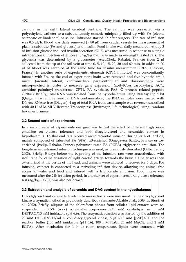
In the second serie of experiments, we first measured food intake (figure 6). As depicted, there was a decreased in food intake with omegaven and ivelip infusion but not with lard oil.
Fig. 6. Measurement of food intake. *, p<0.05 vs controls, **p<0.01 vs controls.
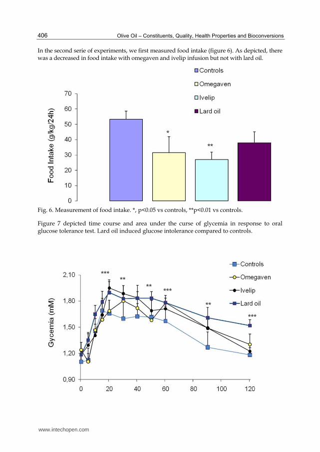
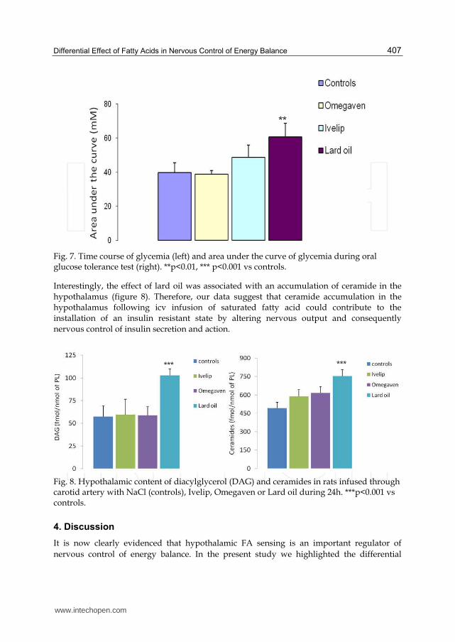
Figure 7 depicted time course and area under the curse of glycemia in response to oral glucose tolerance test. Lard oil induced glucose intolerance compared to controls.
**
*
***
*****
*****
**
www.intechopen.com
Differential Effect of Fatty Acids in Nervous Control of Energy Balance 407
Fig. 7. Time course of glycemia (left) and area under the curve of glycemia during oral glucose tolerance test (right). **p<0.01, *** p<0.001 vs controls.
Interestingly, the effect of lard oil was associated with an accumulation of ceramide in the
hypothalamus (figure 8). Therefore, our data suggest that ceramide accumulation in the
hypothalamus following icv infusion of saturated fatty acid could contribute to the
installation of an insulin resistant state by altering nervous output and consequently
nervous control of insulin secretion and action.
Fig. 8. Hypothalamic content of diacylglycerol (DAG) and ceramides in rats infused through carotid artery with NaCl (controls), Ivelip, Omegaven or Lard oil during 24h. ***p<0.001 vs controls.
4. Discussion
It is now clearly evidenced that hypothalamic FA sensing is an important regulator of
nervous control of energy balance. In the present study we highlighted the differential
*** ***
**
www.intechopen.com

Olive Oil – Constituents, Quality, Health Properties and Bioconversions 408
effects of FA regarding their chain length and degree of saturation. We firstly evidenced
here that oleate and linolenate have differential effects in regard to glucose homeostasis and
GIIS. Especially linolenate induced increased GIIS compared to both oleate and control
group whereas time course of glycemia remained similar. Thus there is a difference between
effect of monounsaturated and polyunsaturated fatty acids when infused toward the brain.
This suggest activation of different pathways. It must be pointed out that we previously
demonstrated that short term infusion of oleate (6h and 24h) induced an increased in insulin
secretion induced by glucose compared to control rats (Migrenne et al., 2006; Wang et al.,
2006). In the present study infusion was made during 3 days which can explain different
effect in short vs long term infusion peridod. Indeed we cannot exclude an adaptation to
oleate effect when infusion stay longer. In the same way of ideas, inhibitory effect of central
infusion of oleate on food intake was also lost after 3 days of infusion as previously
evidenced by obici et al (Obici et al., 2002). In contrast, in the present study effect of
linolenate was still present after 3 days of infusion. Linolenate effect may induce an insulin-
resistance state and increased GIIS could be an adaptation to this insulin resistance state. By
acting on FA sensitive neurons, linolenate may affect nervous output from CNS, especially
autonomic nervous system. This change in autonomic nervous system balance will in turn
modify nervous control of insulin secretion and action. We previously demonstrated that
lipid infusion induced changes in both sympathetic and parasympathetic nervous activity in
both rodents (Magnan et al., 1999) and humans (Magnan et al., 2001). In both studies
decreased sympathetic nervous activity induced an increased insulin secretion in response
to glucose and insulin resistance. In addition, in the present study we showed that linolenate
effect involved its metabolism since it had no more effect in presence of etomoxir an
inhibitor of ┚ oxidation. The involvement of ┚ oxidation to relay FA effect on sensitive
neurons have been also described in different models by us (Cruciani-Guglielmacci et al.,
2004) and others (Obici et al., 2003). Finally that specific effects of linolenate compared to
oleate could be, at least in part, related to differential gene transcription involved in FA
metabolism such as CPT1, FAS or ACC in key areas of hypothalamus. More precisely in
ARC and LH CPT1 expression was increased in linolenate infused rats and etomoxir
induced a decreased in this gene and its return to basal value. However, it is difficult to
further analyze these results since in other nuclei, there is no change of CPT1 expression. In
addition, in some area others genes are differently expressed such as AMPK┙2 or ACC┚,
both key enzymes of glucose and FA metabolism. Altogether these data suggest that oleate
or linolenate may act on different subpopulations of neurons (or astrocytes) thus
highlighting the fact that FAs may have different effect in regard of the area in which they
act. It is also interesting that expression of gene such as GPR41 can be also modified by
linolenate infusion. Indeed it has been recently evidenced that short-chain fatty acids and
ketones directly regulate sympathetic nervous system via GPR41 at the level of sympathetic
ganglion (Kimura et al., 2011). Thus changes in hypothalamic GRP41 gene expression may
have an impact during starvation, a situation in which ketone bodies production is
increased. thereby control body energy expenditure in maintaining metabolic homeostasis.
In the second part of our work we demonstrated a differential role of PUFA vs saturated FA
(SFA) regarding induction of insulin resistance and ceramides production in hypothalamus
by using triglyceride emulsion infusion, in order to mimic a more “physiological approach”.
www.intechopen.com

Differential Effect of Fatty Acids in Nervous Control of Energy Balance 409
Indeed 24h of lard oil infusion in carotid which had no effect on plasma TG or FA
concentrations (data not shown) induced a glucose intolerance suggesting a deregulation of
insulin sensitivity and or secretion. This deleterious effect of lard oil in nervous control of
glucose homeostasis was associated with an increased in DAG and ceramides content in
hypothalamus. An important role for ceramides has emerged from research on the
pathogenesis of metabolic diseases associated with obesity, such as diabetes (Holland &
Summers, 2008). Indeed, ceramides appear to be particularly deleterious components of the
lipid milieu that accrues in obesity, and levels of ceramides are often elevated in skeletal
muscle, liver, and/or serum of obese humans and rodents (Adams et al., 2004; Clement et
al., 2002). DAG and ceramides are known to activate kinase such as PKC, which
phosphorylate insulin receptor substrate and Akt leading to an inhibition of the insulin
signaling (Mullen et al., 2009; Newton et al., 2009). A recent study also evidenced that
sphingolipids such as ceramide might be key components of the signaling networks that
link lipid-induced inflammatory pathways to the antagonism of insulin action that
contributes to diabetes (Holland et al., 2011). We also recently demonstrated that the
atypical protein kinase C, PKCΘ, is expressed in discrete neuronal populations of the ARC
and the dorsal medial hypothalamic nucleus (Benoit et al., 2009). CNS exposure to saturated
palmitic acid via direct infusion or by oral gavage increased the localization of PKCΘ to
hypothalamic cell membranes in association impaired hypothalamic insulin and leptin
signaling (Benoit et al., 2009). This finding was specific for palmitic acid, as the
monounsaturated FA, OA, neither increased membrane localization of PKCΘ nor reduced
insulin signaling. Finally, ARC-specific knockdown of PKCΘ attenuated diet-induced
obesity and improved hypothalamic insulin signaling (Benoit et al., 2009). These results
suggest that many of the deleterious effects of high fat diets, specifically those enriched with
palmitic acid, are CNS mediated via PKCΘ activation, resulting in reduced insulin activity.
Therefore, our data suggest that ceramide accumulation in the hypothalamus following icv
infusion of saturated fatty acid could contribute to the installation of an insulin resistant
state by altering nervous output and consequently nervous control of insulin secretion and
action.
Further studies are needed to clearly identify molecular mechanism relaying ceramides production. However there is now several experiments highlighting some of these mechanisms in FA sensitive neurons as described below.
4.1 Molecular mechanisms involved in neuronal FA sensing
In FA sensitive neurons, exposure to long chain FA can alter the activity of a wide variety of
ion channels including Cl-, GABAA (Tewari et al., 2000), potassium, K+-Ca2+ (Honen et al.,
2003) or calcium channels (Oishi et al., 1990). Additionally, FA inhibit the Na+-K+ ATPase
pump (Oishi et al., 1990). For example, OA activates ARC POMC neurons by inhibiting
ATP-sensitive K+ (KATP) channel activity (Jo et al., 2009) and the effect of OA on HGP is
abolished by icv administration of a KATP channel inhibitor (Jo et al., 2009). However, KATP
channels are ubiquitously expressed on neurons throughout the brain, not only in FA
sensing neurons, making the mechanism and site of such in vivo manipulations difficult to
discern (Dunn-Meynell et al., 1998). Using in vivo and in vitro electrophysiological
approaches, OA sensitive-neurons have been characterized using whole cell patch clamp
www.intechopen.com

Olive Oil – Constituents, Quality, Health Properties and Bioconversions 410
records in ARC slices from 14 to 21 day old rats (Wang et al., 2006). Of these 13 % were
excited by OA and 30% were inhibited by OA (Oomura et al., 1975). The excitatory effects of
OA appeared to be due to closure of chloride channels leading to membrane depolarization
and increased action potential frequency (Migrenne et al., 2006). On the other hand,
inhibitory effect of OA may involve the KATP channels since this inhibition was reversed by
the KATP channel blocker tolbutamide (Migrenne et al., 2006). Using fura-2 Ca2+ imaging in
dissociated neurons from the ventromedial hypothalamic nucleus (VMN) neurons, we
found that OA excited up to 43% and inhibited up to 29% of all VMN neurons
independently of glucose concentrations (Le Foll et al., 2009). However, in these neurons,
inhibition of the KATP channel mediated FA sensing in only a small percentage of FA sensing
neurons. Importantly, although a relatively large percentage of hypothalamic neurons are
FA sensors, a select population that also sense glucose are highly dependent upon ambient
glucose concentrations for the resultant effect of FA on the activity of these neurons (Le Foll
et al., 2009). Such data suggest that the responses of hypothalamic FA sensitive neurons are
dependent upon the metabolic state of the animal and thus might be expected to respond
differently during fasting (when FA levels rise and glucose levels fall) vs. the overfed state
when glucose levels rise while free FA levels remain relatively unchanged (Le Foll et al.,
2009). However, it must be pointed out that FA are naturally complexed to serum albumin
in the blood and the concentration of circulating free FA is less than 1% of total FA levels.
All the studies investigating FA sensing in the hypothalamus either use non-complexed FA
or cyclodextrin-complexed FA in vitro or in vivo. The concentration of free FA in
cyclodextrin-complexed FA preparation is unknown. Whether or not the FA concentration
used mimics FA levels in physiological states needs to be determined.
4.2 Metabolic-dependent FA sensing effects
The effects of FA on activity of some neurons are dependent upon intracellular metabolism
of FA. Enzymes involved in FA metabolism such as FA synthase (FAS), CPT1 and acetyl-
CoA carboxylase (ACC) are expressed in some hypothalamic neurons as well as in glial cells
(reviewed in (Blouet & Schwartz; Le Foll et al., 2009). Malonyl-CoA may be an important
sensor of energy levels in the hypothalamus. It is derived from either glucose or FA
metabolism via the glycolysis or -oxidation, respectively. The steady-state level of malonyl-
CoA is determined by its rate of synthesis catalysed by ACC relative to its rate of turnover
catalysed by FAS. The synthesis of malonyl-CoA is the first committed step of FA synthesis
and ACC is the major site of regulation in that process. Thus, when the supply of glucose is
increased, malonyl CoA levels increase in keeping with a decreased need for FA oxidation.
This increase in both malonyl CoA and acyl CoA levels is associated with reduced food
intake. Central administration of C75, an inhibitor of FAS, also increases malonyl-CoA
concentration in the hypothalamus, suppresses food intake and leads to profound weight
loss (Proulx & Seeley, 2005). It has been proposed that centrally, C75 and cerulenin (another
inhibitor of FAS) alter the expression profiles of feeding-related neuropeptides, often
inhibiting the expression of orexigenic peptides such as neuropeptide Y (Proulx et al., 2008).
Whether through centrally mediated or peripheral mechanisms, C75 also increases energy
expenditure, which contributes to weight loss (Clegg et al., 2002; Tu et al., 2005). In vitro and
in vivo studies demonstrate that at least part of C75's effects are mediated by the
www.intechopen.com

Differential Effect of Fatty Acids in Nervous Control of Energy Balance 411
modulation of AMP-activated kinase, a known energy-sensing kinase (Ronnett et al., 2005).
Indeed, icv administration of 5-aminoimidazole-4-carboxamide ribonucleoside (AICAR), a
5'-AMP kinase activator, rapidly lowers hypothalamic malonyl-CoA concentration and
increases food intake (Tu et al., 2005). These effects correlate closely with the
phosphorylation-induced inactivation of ACC, an established target of AMP kinase.
Collectively, these data suggest a role for FA metabolism in the perception and regulation of
energy balance. However, it must be also pointed out that C75 and AICAR may also have non-
specific or even opposite effects. For example, a major effect of C75 is to activate CPT-1 rather
than lead to its inhibition in vitro (Aja et al., 2008). Finally the route of administration and the
type of FA used are also critical. For example, bolus intracerebroventricular injections of OA,
but not palmitic acid, reduce food intake and body weight, possibly mediated through
POMC/MC4R signaling (Schwinkendorf et al., 2010). Again, such bolus icv injections could
cause non-specific effects related to inflammation of ependymocytes and tanycytes. Also
because so much of FA metabolism takes place in astrocytes, such manipulations done in vivo
and in slice preparations are likely to alter FA metabolism that takes place in astrocytes which
could then indirectly alter neuronal FA sensing (Escartin et al., 2007).
4.3 Non metabolic-dependent neuronal FA sensing
While intracellular FA metabolism may be responsible for altering neuronal activity in some
FA sensitive neurons such as ARC POMC neurons (Jo et al., 2009) it accounts for a relatively
small percent of the effects of OA on dissociated VMN neurons (Le Foll et al., 2009). In those
neurons, inhibition of CPT1, reactive oxygen species formation, long-chain acyl CoA
synthetase and KATP channel activity or activation of uncoupling protein 2 (UCP2) accounts
for no more than 20% of the excitatory or approximately 40% of the inhibitory effects of OA
(Le Foll et al., 2009). On the other hand, pharmacological inhibition of FAT/CD36, a FA
transporter/receptor that can alter cell function independently of intracellular FA
metabolism reduced the excitatory and inhibitory effects of OA by up to 45% (Le Foll et al.,
2009). Thus, in almost half of VMN FA sensing neurons, CD36 may act primarily as
receptor, rather than a transporter, for long chain FA as it does on taste cells on the tongue
where it activates store-operated calcium channels to alter membrane potential and release
of serotonin (Gaillard et al., 2008). These effects all occur in the presence of nanomolar
concentrations of OA, whereas micromolar concentrations are generally required to effect
similar changes in neuronal activity in brain slice preparations (Jo et al., 2009; Migrenne et
al., 2011; Wang et al., 2006). Thus, in the absence of astrocytes, OA can directly affect VMN
neuronal activity through both metabolic and non-metabolic pathways. Alternatively, FA
might act as signaling molecules by covalent attachment to proteins (N-terminal acylation)
to alter the function of membrane and intracellular signaling molecules. For example,
palmitoylation facilitates the targeting and plasma membrane binding of proteins which
otherwise would remain in the cytosolic compartment (Resh, 1999). Some membrane
proteins (TGF, synaptosomal associated protein of 25KDa (required for exocytosis) and
plasma membrane receptors (seven transmembrane receptors such as 2a- and 2-
adrenoceptors) are typically palmitoylated on one or several cysteine residues located
adjacent to or just within the transmembrane domain (Resh, 1999) Such mechanisms might
also modulate neuronal FA sensing.
www.intechopen.com

Olive Oil – Constituents, Quality, Health Properties and Bioconversions 412
4.4 Which neurotransmitters or neuropeptides?
The ultimate consequence of the activation or inactivation of a neuron is the release of neurotransmitters and neuropeptides. Since FA decrease food intake, they might be expected to alter activity neurons specifically involved in the regulation of feeding. In fact, OA activates catabolic POMC neurons directly, apparently via ß-oxidation and inactivation of the KATP channel in hypothalamic slice preparations (Jo et al., 2009). In vivo, Obici et al. (Obici et al., 2003) reported that icv administration of OA markedly inhibits glucose production and food intake, accompanied by a decrease in the hypothalamic expression of the anabolic peptide, neuropeptide Y. This decrease in the expression of such a critical anabolic peptide might contribute to the reduced food intake associated with direct central administration of OA. On the other hand, an n-3 FA enriched diet increases food intake in anorexic tumor-bearing rats, in association with reduced tumor appearance, tumor growth and onset of anorexia (Ramos et al., 2005). In these treated rats, neuropeptide Y immunoreactivity increased 38% in ARC and 50% in paraventricular nucleus, whereas ┙-melanocyte stimulating hormone (a catabolic peptide cleavage product of POMC) decreased 64% in the ARC and 29% in the paraventricular nucleus (Ramos et al., 2005). Finally, in the hippocampus, docosahexaenoic acid (22:6(n-3) increased the spontaneous release of acetylcholine (Aid et al., 2005).
4.5 Pathological implications of excess FA
Besides physiological regulation of energy balance by hypothalamic neuronal FA sensing, impaired regulation of such sensing might contribute to the development of metabolic diseases such as obesity and type 2 diabetes in predisposed subjects exposed to a chronic lipid overload (Luquet & Magnan, 2009; Migrenne et al., 2011). Excessive brain lipid levels may indeed alter control of glucose and lipid homeostasis through changes of autonomic nervous system activity. Increasing brain FA levels reduces sympathetic activity and increases GIIS in rats (Clement et al., 2002; Obici et al., 2003) a condition which would exacerbate the development of type 2 diabetes mellitus. Also, a lipid overload due to high-fat diet intake alters both hypothalamic monoamine turnover (Levin et al., 1983) and peripheral sympathetic activity in rats (Young & Walgren, 1994). In humans, overweight is often associated with an altered sympathetic tone (Peterson et al., 1988) suggesting a relationship between lipids and autonomic control centers in brain.
5. Conclusion
In conclusion, there is now increasing evidence that specialized neurons within
hypothalamus and other areas such as the brainstem or hippocampus can detect changes in
plasma FA levels by having FA directly or indirectly alter the of FA sensitive neurons
involved in the regulation of energy and glucose homeostasis. Central FA effects on insulin
secretion and action are related to their chain length or degree of saturation. Such effects are
also mediated through differential changes in gene expression.
The neuronal networks of these FA sensitive neurons that sense and respond to FA are
likely very complex given the fact that FA can either inhibit or excite specific neurons. In
addition, many of these neurons also utilize glucose as a signaling molecule and there is
often an inverse responsiveness of such “metabolic sensing” neurons to FA vs. glucose.
www.intechopen.com

Differential Effect of Fatty Acids in Nervous Control of Energy Balance 413
Thus, these neurons are ideally suited to respond differentially under a variety of metabolic
conditions such as fasting, feeding, hypo- or hyperglycemia. However, while it is clear that
specific neurons can respond to changes in ambient FA levels, many questions remain. We
still do not know for certain how FA are transported into the brain, astrocytes or neurons
and whether those FA that are transported are derived from circulating free FA or
triglycerides. Since most studies suggest that rising FA levels reduce food intake, then we
must explain why plasma FA levels are most elevated during fasting when the drive to seek
and ingest food should be at its strongest. Another major issue relates to the interaction
between astrocytes and neurons with regard to the metabolism and signaling of FA. Also,
we still know little about the basic mechanisms utilized by neurons to sense FA, where such
FA sensitive neurons reside throughout the brain and what neurotransmitters and peptides
they release when responding to FA.
Finally, it has been postulated that diabetes may be a disorder of the brain (Elmquist &
Marcus, 2003). If so, dysfunction of these FA sensitive neurons could be, at least in part, one
of the early mechanisms underlying impairment of neural control of energy and glucose
homeostasis and the development of obesity and type 2 diabetes in predisposed subjects. A
better understanding of this central nutrient sensing, including both FA and glucose, could
provide clues for the identification of new therapeutic targets for the prevention and
treatment of both diabetes and obesity.
6. Acknowledgements
This work was partially supported by an award from European Foundation for Study of Diabetes (EFSD)/GSK 2007 (Stéphanie Migrenne).
7. References
Adams JM, 2nd; Pratipanawatr T; Berria R; Wang E; DeFronzo RA; Sullards MC &
Mandarino LJ. (2004). Ceramide content is increased in skeletal muscle from obese
insulin-resistant humans. Diabetes, Vol. 53, No. 1, pp 25-31, 0012-1797 (Print) 0012-
1797 (Linking)
Aid S; Vancassel S; Linard A; Lavialle M & Guesnet P. (2005). Dietary docosahexaenoic acid
[22: 6(n-3)] as a phospholipid or a triglyceride enhances the potassium chloride-
evoked release of acetylcholine in rat hippocampus. J Nutr, Vol. 135, No. 5, pp 1008-
1013,
Aja S; Landree LE; Kleman AM; Medghalchi SM; Vadlamudi A; McFadden JM; Aplasca A;
Hyun J; Plummer E; Daniels K; Kemm M; Townsend CA; Thupari JN; Kuhajda FP;
Moran TH & Ronnett GV. (2008). Pharmacological stimulation of brain carnitine
palmitoyl-transferase-1 decreases food intake and body weight. Am J Physiol Regul
Integr Comp Physiol, Vol. 294, No. 2, pp R352-361, 0363-6119 (Print) 0363-6119
(Linking)
Benoit SC; Kemp CJ; Elias CF; Abplanalp W; Herman JP; Migrenne S; Lefevre AL;
Cruciani-Guglielmacci C; Magnan C; Yu F; Niswender K; Irani BG; Holland WL
& Clegg DJ. (2009). Palmitic acid mediates hypothalamic insulin resistance by
www.intechopen.com
Olive Oil – Constituents, Quality, Health Properties and Bioconversions 414
altering PKC-theta subcellular localization in rodents. J Clin Invest, Vol. 119, No.
9, pp 2577-2589,
Blouet C & Schwartz GJ. (2010). Hypothalamic nutrient sensing in the control of energy
homeostasis. Behav Brain Res, Vol. 209, No. 1, pp 1-12,
Clegg DJ; Air EL; Woods SC & Seeley RJ. (2002). Eating elicited by orexin-a, but not
melanin-concentrating hormone, is opioid mediated. Endocrinology, Vol. 143, No. 8,
pp 2995-3000,
Clement L; Cruciani-Guglielmacci C; Magnan C; Vincent M; Douared L; Orosco M;
Assimacopoulos-Jeannet F; Penicaud L & Ktorza A. (2002).
Intracerebroventricular infusion of a triglyceride emulsion leads to both altered
insulin secretion and hepatic glucose production in rats. Pflugers Arch, Vol. 445,
No. 3, pp 375-380,
Cruciani-Guglielmacci C; Hervalet A; Douared L; Sanders NM; Levin BE; Ktorza A &
Magnan C. (2004). Beta oxidation in the brain is required for the effects of non-
esterified fatty acids on glucose-induced insulin secretion in rats. Diabetologia, Vol.
47, No. 11, pp 2032-2038,
Dowell P; Hu Z & Lane MD. (2005). Monitoring energy balance: metabolites of
fatty acid synthesis as hypothalamic sensors. Annu Rev Biochem, Vol. 74, No. pp
515-534,
Dunn-Meynell AA; Rawson NE & Levin BE. (1998). Distribution and phenotype of neurons
containing the ATP-sensitive K+ channel in rat brain. Brain Res, Vol. 814, No. 1-2,
pp 41-54, 0006-8993 (Print) 0006-8993 (Linking)
Edmond J. (2001). Essential polyunsaturated fatty acids and the barrier to the brain: the
components of a model for transport. J Mol Neurosci, Vol. 16, No. 2-3, pp 181-193;
discussion 215-121,
Elmquist JK & Marcus JN. (2003). Rethinking the central causes of diabetes. Nat Med, Vol. 9,
No. 6, pp 645-647,
Escalante-Alcalde D; Hernandez L; Le Stunff H; Maeda R; Lee HS; Jr Gang C; Sciorra VA;
Daar I; Spiegel S; Morris AJ & Stewart CL. (2003). The lipid phosphatase LPP3
regulates extra-embryonic vasculogenesis and axis patterning. Development, Vol.
130, No. 19, pp 4623-4637, 0950-1991 (Print) 0950-1991 (Linking)
Escartin C; Boyer F; Bemelmans AP; Hantraye P & Brouillet E. (2007). IGF-1 exacerbates the
neurotoxicity of the mitochondrial inhibitor 3NP in rats. Neurosci Lett, Vol. 425, No.
3, pp 167-172, 0304-3940 (Print) 0304-3940 (Linking)
Escartin C; Pierre K; Colin A; Brouillet E; Delzescaux T; Guillermier M; Dhenain M; Deglon
N; Hantraye P; Pellerin L & Bonvento G. (2007). Activation of astrocytes by CNTF
induces metabolic plasticity and increases resistance to metabolic insults. J Neurosci,
Vol. 27, No. 27, pp 7094-7104, 1529-2401 (Electronic) 0270-6474 (Linking)
Gaillard D; Laugerette F; Darcel N; El-Yassimi A; Passilly-Degrace P; Hichami A; Khan NA;
Montmayeur JP & Besnard P. (2008). The gustatory pathway is involved in CD36-
mediated orosensory perception of long-chain fatty acids in the mouse. FASEB J,
Vol. 22, No. 5, pp 1458-1468, 1530-6860 (Electronic) 0892-6638 (Linking)
www.intechopen.com

Differential Effect of Fatty Acids in Nervous Control of Energy Balance 415
Gilbert M; Magnan C; Turban S; Andre J & Guerre-Millo M. (2003). Leptin receptor-deficient
obese Zucker rats reduce their food intake in response to a systemic supply of
calories from glucose. Diabetes, Vol. 52, No. 2, pp 277-282,
Gribble FM; Proks P; Corkey BE & Ashcroft FM. (1998). Mechanism of cloned ATP-sensitive
potassium channel activation by oleoyl-CoA. J Biol Chem, Vol. 273, No. 41, pp
26383-26387, 0021-9258 (Print) 0021-9258 (Linking)
Holland WL; Bikman BT; Wang LP; Yuguang G; Sargent KM; Bulchand S; Knotts TA; Shui
G; Clegg DJ; Wenk MR; Pagliassotti MJ; Scherer PE & Summers SA. (2011). Lipid-
induced insulin resistance mediated by the proinflammatory receptor TLR4
requires saturated fatty acid-induced ceramide biosynthesis in mice. J Clin Invest,
Vol. 121, No. 5, pp 1858-1870, 1558-8238 (Electronic) 0021-9738 (Linking)
Holland WL & Summers SA. (2008). Sphingolipids, insulin resistance, and
metabolic disease: new insights from in vivo manipulation of sphingolipid
metabolism. Endocr Rev, Vol. 29, No. 4, pp 381-402, 0163-769X (Print) 0163-769X
(Linking)
Honen BN; Saint DA & Laver DR. (2003). Suppression of calcium sparks in rat ventricular
myocytes and direct inhibition of sheep cardiac RyR channels by EPA, DHA and
oleic acid. J Membr Biol, Vol. 196, No. 2, pp 95-103,
Jo YH; Su Y; Gutierrez-Juarez R & Chua S, Jr. (2009). Oleic acid directly regulates POMC
neuron excitability in the hypothalamus. J Neurophysiol, Vol. 101, No. 5, pp 2305-
2316, 0022-3077 (Print) 0022-3077 (Linking)
Kimura I; Inoue D; Maeda T; Hara T; Ichimura A; Miyauchi S; Kobayashi M; Hirasawa A &
Tsujimoto G. (2011). Short-chain fatty acids and ketones directly regulate
sympathetic nervous system via G protein-coupled receptor 41 (GPR41). Proc Natl
Acad Sci U S A, Vol. 108, No. 19, pp 8030-8035, 1091-6490 (Electronic) 0027-8424
(Linking)
Lam TK; Schwartz GJ & Rossetti L. (2005). Hypothalamic sensing of fatty acids. Nat Neurosci,
Vol. 8, No. 5, pp 579-584,
Le Foll C; Irani BG; Magnan C; Dunn-Meynell AA & Levin BE. (2009). Characteristics and
mechanisms of hypothalamic neuronal fatty acid sensing. Am J Physiol Regul Integr
Comp Physiol, Vol. 297, No. 3, pp R655-664,
Le Stunff H; Galve-Roperh I; Peterson C; Milstien S & Spiegel S. (2002). Sphingosine-1-
phosphate phosphohydrolase in regulation of sphingolipid metabolism and
apoptosis. J Cell Biol, Vol. 158, No. 6, pp 1039-1049, 0021-9525 (Print) 0021-9525
(Linking)
Levin BE; Triscari J & Sullivan AC. (1983). Altered sympathetic activity during development
of diet-induced obesity in rat. Am J Physiol, Vol. 244, No. 3, pp R347-355,
Luquet S & Magnan C. (2009). The central nervous system at the core of the regulation of
energy homeostasis. Front Biosci (Schol Ed), Vol. 1, No. pp 448-465,
Magnan C; Collins S; Berthault MF; Kassis N; Vincent M; Gilbert M; Penicaud L; Ktorza A &
Assimacopoulos-Jeannet F. (1999). Lipid infusion lowers sympathetic nervous
activity and leads to increased beta-cell responsiveness to glucose. J Clin Invest, Vol.
103, No. 3, pp 413-419,
www.intechopen.com

Olive Oil – Constituents, Quality, Health Properties and Bioconversions 416
Magnan C; Cruciani C; Clement L; Adnot P; Vincent M; Kergoat M; Girard A; Elghozi JL;
Velho G; Beressi N; Bresson JL & Ktorza A. (2001). Glucose-induced insulin
hypersecretion in lipid-infused healthy subjects is associated with a decrease in
plasma norepinephrine concentration and urinary excretion. J Clin Endocrinol
Metab, Vol. 86, No. 10, pp 4901-4907,
Migrenne S; Cruciani-Guglielmacci C; Kang L; Wang R; Rouch C; Lefevre AL; Ktorza A;
Routh V; Levin B & Magnan C. (2006). Fatty acid signaling in the hypothalamus
and the neural control of insulin secretion. Diabetes, Vol. 55 S2, No. pp S139-S144,
Migrenne S; Le Foll C; Levin BE & Magnan C. (2011). Brain lipid sensing and nervous
control of energy balance. Diabetes Metab, Vol. 37, No. 2, pp 83-88, 1878-1780
(Electronic) 1262-3636 (Linking)
Migrenne S; Marsollier N; Cruciani-Guglielmacci C & Magnan C. (2006). Importance of the
gut-brain axis in the control of glucose homeostasis. Curr Opin Pharmacol, Vol. 6,
No. 6, pp 592-597,
Mullen KL; Pritchard J; Ritchie I; Snook LA; Chabowski A; Bonen A; Wright D & Dyck DJ.
(2009). Adiponectin resistance precedes the accumulation of skeletal muscle lipids
and insulin resistance in high-fat-fed rats. Am J Physiol Regul Integr Comp Physiol,
Vol. 296, No. 2, pp R243-251, 0363-6119 (Print) 0363-6119 (Linking)
Newton RU; Taaffe DR; Spry N; Gardiner RA; Levin G; Wall B; Joseph D; Chambers SK &
Galvao DA. (2009). A phase III clinical trial of exercise modalities on treatment
side-effects in men receiving therapy for prostate cancer. BMC Cancer, Vol. 9, No.
pp 210, 1471-2407 (Electronic) 1471-2407 (Linking)
Obici S; Feng Z; Arduini A; Conti R & Rossetti L. (2003). Inhibition of hypothalamic
carnitine palmitoyltransferase-1 decreases food intake and glucose production. Nat
Med, Vol. 9, No. 6, pp 756-761,
Obici S; Feng Z; Morgan K; Stein D; Karkanias G & Rossetti L. (2002). Central administration
of oleic acid inhibits glucose production and food intake. Diabetes, Vol. 51, No. 2, pp
271-275,
Oishi K; Zheng B & Kuo JF. (1990). Inhibition of Na,K-ATPase and sodium pump by protein
kinase C regulators sphingosine, lysophosphatidylcholine, and oleic acid. J Biol
Chem, Vol. 265, No. 1, pp 70-75,
Oomura Y; Nakamura T; Sugimori M & Yamada Y. (1975). Effect of free fatty acid on the rat
lateral hypothalamic neurons. Physiol Behav, Vol. 14, No. 04, pp 483-486,
Penicaud L; Leloup C; Lorsignol A; Alquier T & Guillod E. (2002). Brain glucose sensing
mechanism and glucose homeostasis. Curr Opin Clin Nutr Metab Care, Vol. 5, No. 5,
pp 539-543,
Peterson HR; Rothschild M; Weinberg CR; Fell RD; McLeish KR & Pfeifer MA. (1988). Body
fat and the activity of the autonomic nervous system. N Engl J Med, Vol. 318, No.
17, pp 1077-1083,
Proulx K; Cota D; Woods SC & Seeley RJ. (2008). Fatty acid synthase inhibitors modulate
energy balance via mammalian target of rapamycin complex 1 signaling in the
central nervous system. Diabetes, Vol. 57, No. 12, pp 3231-3238,
Proulx K & Seeley RJ. (2005). The regulation of energy balance by the central nervous
system. Psychiatr Clin North Am, Vol. 28, No. 1, pp 25-38, vii,
www.intechopen.com

Differential Effect of Fatty Acids in Nervous Control of Energy Balance 417
Ramos EJ; Romanova IV; Suzuki S; Chen C; Ugrumov MV; Sato T; Goncalves CG &
Meguid MM. (2005). Effects of omega-3 fatty acids on orexigenic and
anorexigenic modulators at the onset of anorexia. Brain Res, Vol. 1046, No. 1-2, pp
157-164,
Randle PJ; Priestman DA; Mistry S & Halsall A. (1994). Mechanisms modifying glucose
oxidation in diabetes mellitus. Diabetologia, Vol. 37 Suppl 2, No. pp S155-161, 0012-
186X (Print) 0012-186X (Linking)
Rapoport SI; Chang MC & Spector AA. (2001). Delivery and turnover of plasma-derived
essential PUFAs in mammalian brain. J Lipid Res, Vol. 42, No. 5, pp 678-685,
Resh MD. (1999). Fatty acylation of proteins: new insights into membrane targeting of
myristoylated and palmitoylated proteins. Biochim Biophys Acta, Vol. 1451, No. 1,
pp 1-16,
Ronnett GV; Kim EK; Landree LE & Tu Y. (2005). Fatty acid metabolism as a target for
obesity treatment. Physiol Behav, Vol. 85, No. 1, pp 25-35,
Ross RA; Rossetti L; Lam TK & Schwartz GJ. (2010). Differential effects of hypothalamic
long-chain fatty acid infusions on suppression of hepatic glucose production. Am J
Physiol Endocrinol Metab, Vol. 299, No. 4, pp E633-639, 1522-1555 (Electronic) 0193-
1849 (Linking)
Ruge T; Hodson L; Cheeseman J; Dennis AL; Fielding BA; Humphreys SM; Frayn
KN & Karpe F. (2009). Fasted to fed trafficking of Fatty acids in human adipose
tissue reveals a novel regulatory step for enhanced fat storage. J Clin
Endocrinol Metab, Vol. 94, No. 5, pp 1781-1788, 1945-7197 (Electronic) 0021-972X
(Linking)
Schwinkendorf DR; Tsatsos NG; Gosnell BA & Mashek DG. (2010). Effects of central
administration of distinct fatty acids on hypothalamic neuropeptide expression and
energy metabolism. Int J Obes (Lond), Vol. No. pp 1476-5497 (Electronic) 0307-0565
(Linking)
Smith QR & Nagura H. (2001). Fatty acid uptake and incorporation in brain: studies with
the perfusion model. J Mol Neurosci, Vol. 16, No. 2-3, pp 167-172; discussion 215-
121,
Tewari KP; Malinowska DH; Sherry AM & Cuppoletti J. (2000). PKA and arachidonic acid
activation of human recombinant ClC-2 chloride channels. Am J Physiol Cell Physiol,
Vol. 279, No. 1, pp C40-50,
Tu Y; Thupari JN; Kim EK; Pinn ML; Moran TH; Ronnett GV & Kuhajda FP. (2005). C75
alters central and peripheral gene expression to reduce food intake and increase
energy expenditure. Endocrinology, Vol. 146, No. 1, pp 486-493,
Wang R; Cruciani-Guglielmacci C; Migrenne S; Magnan C; Cotero VE & Routh VH. (2006).
Effects of oleic acid on distinct populations of neurons in the hypothalamic arcuate
nucleus are dependent on extracellular glucose levels. J Neurophysiol, Vol. 95, No. 3,
pp 1491-1498,
Watkins PA; Hamilton JA; Leaf A; Spector AA; Moore SA; Anderson RE; Moser HW;
Noetzel MJ & Katz R. (2001). Brain uptake and utilization of fatty acids:
applications to peroxisomal biogenesis diseases. J Mol Neurosci, Vol. 16, No. 2-3, pp
87-92; discussion 151-157,
www.intechopen.com

Olive Oil – Constituents, Quality, Health Properties and Bioconversions 418
Young JB & Walgren MC. (1994). Differential effects of dietary fats on sympathetic nervous
system activity in the rat. Metabolism, Vol. 43, No. 1, pp 51-60,
www.intechopen.com

Olive Oil - Constituents, Quality, Health Properties andBioconversionsEdited by Dr. Dimitrios Boskou
ISBN 978-953-307-921-9Hard cover, 510 pagesPublisher InTechPublished online 01, February, 2012Published in print edition February, 2012
InTech EuropeUniversity Campus STeP Ri Slavka Krautzeka 83/A 51000 Rijeka, Croatia Phone: +385 (51) 770 447 Fax: +385 (51) 686 166www.intechopen.com
InTech ChinaUnit 405, Office Block, Hotel Equatorial Shanghai No.65, Yan An Road (West), Shanghai, 200040, China
Phone: +86-21-62489820 Fax: +86-21-62489821
The health-promoting effects attributed to olive oil, and the development of the olive oil industry haveintensified the quest for new information, stimulating wide areas of research. This book is a source of recentlyaccumulated information. It covers a broad range of topics from chemistry, technology, and qualityassessment, to bioavailability and function of important molecules, recovery of bioactive compounds,preparation of olive oil-based functional products, and identification of novel pharmacological targets for theprevention and treatment of certain diseases.
How to referenceIn order to correctly reference this scholarly work, feel free to copy and paste the following:
Christophe Magnan, Hervé Le Stunff and Stéphanie Migrenne (2012). Differential Effect of Fatty Acids inNervous Control of Energy Balance, Olive Oil - Constituents, Quality, Health Properties and Bioconversions,Dr. Dimitrios Boskou (Ed.), ISBN: 978-953-307-921-9, InTech, Available from:http://www.intechopen.com/books/olive-oil-constituents-quality-health-properties-and-bioconversions/differential-effect-of-fatty-acids-in-nervous-control-of-energy-balance
![309 ' # '8& *#3 & 2cdn.intechopen.com/pdfs-wm/36273.pdfInfrared radiation was discovered by Sir William Herschel in 1800 [1]. Herschel was investigating the energy levels associated](https://img.pdfslide.us/doc/110x75/5e91da7a04704626213affb3/309-8-3-2cdn-infrared-radiation-was-discovered-by-sir-william.jpg)