Embed Size (px)
Citation preview
3D OCT Report Template Practical Guide Software ver.8.5x- (for US only)
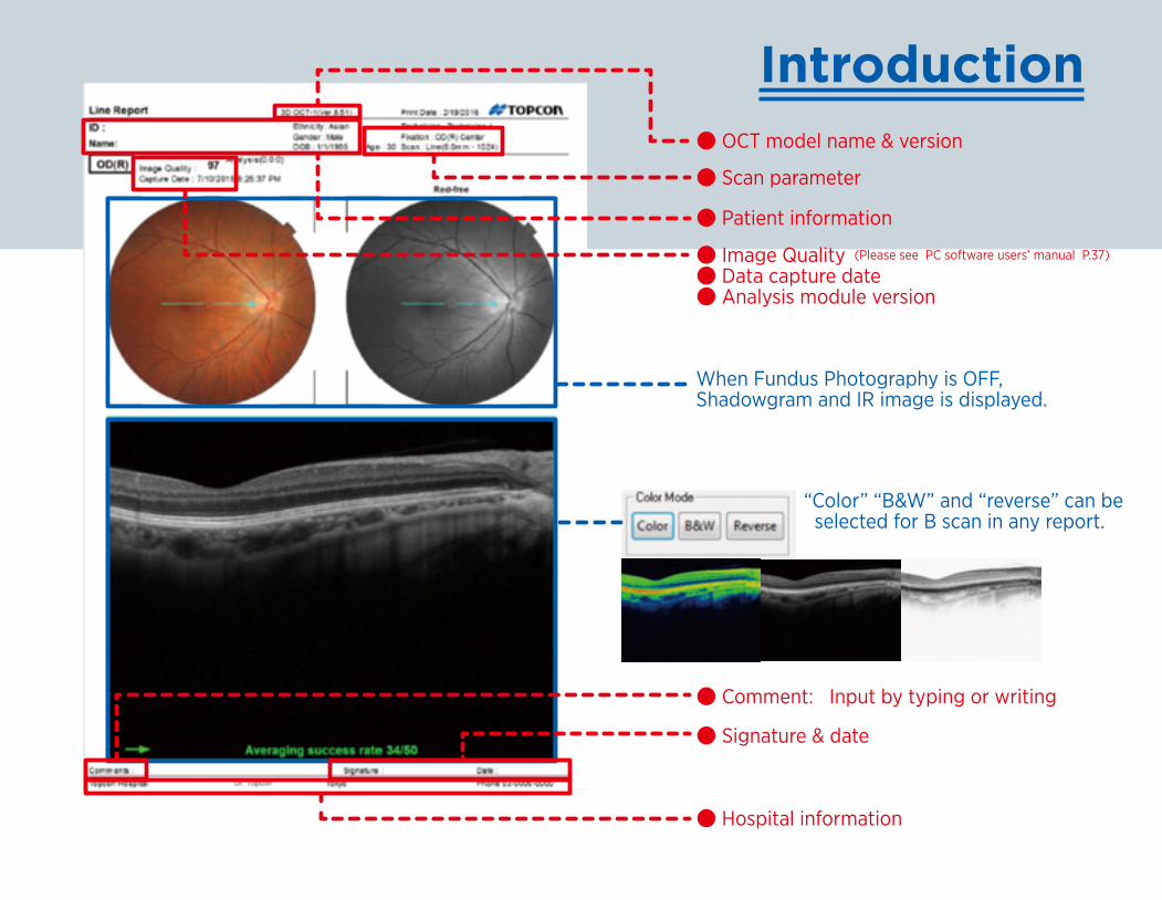
Introduction
● Patient information
● Comment: Input by typing or writing
● Signature & date
● Hospital information
● Image Quality (Please see PC software users’ manual P.37)
● Data capture date● Analysis module version
When Fundus Photography is OFF, Shadowgram and IR image is displayed.
“Color” “B&W” and “reverse” can be selected for B scan in any report.
● OCT model name & version
● Scan parameter
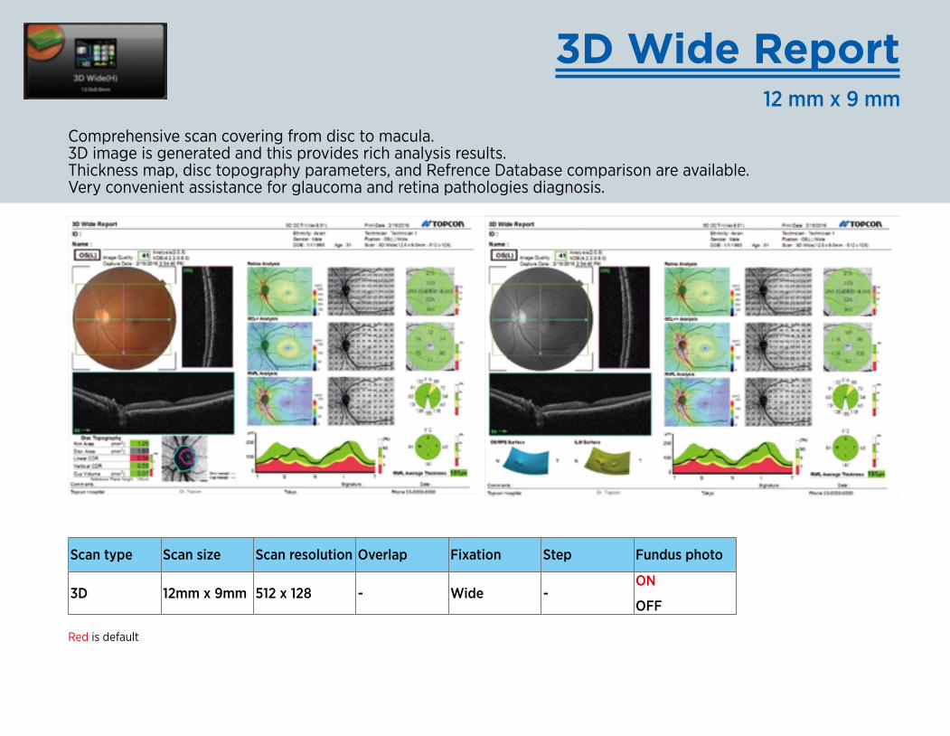
3D Wide Report12 mm x 9 mm
Red is default
Comprehensive scan covering from disc to macula. 3D image is generated and this provides rich analysis results. Thickness map, disc topography parameters, and Refrence Database comparison are available. Very convenient assistance for glaucoma and retina pathologies diagnosis.
Scan type Scan size Scan resolution Overlap Fixation Step Fundus photo
3D 12mm x 9mm 512 x 128 - Wide -ON
OFF
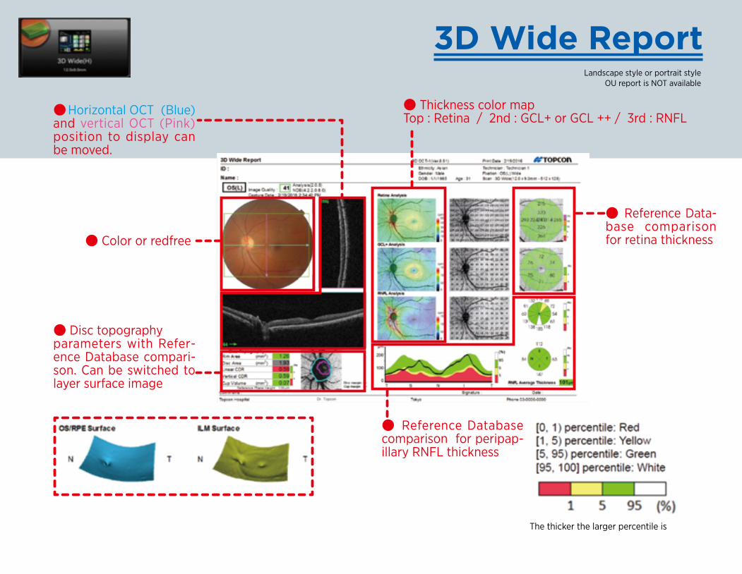
● Color or redfree
● Disc topography parameters with Refer-ence Database compari-son. Can be switched to layer surface image
●Horizontal OCT (Blue) and vertical OCT (Pink) position to display can be moved.
● Thickness color mapTop : Retina / 2nd : GCL+ or GCL ++ / 3rd : RNFL
● Reference Data-base comparison for retina thickness
3D Wide ReportLandscape style or portrait style
OU report is NOT available
● Reference Database comparison for peripap-illary RNFL thickness
The thicker the larger percentile is
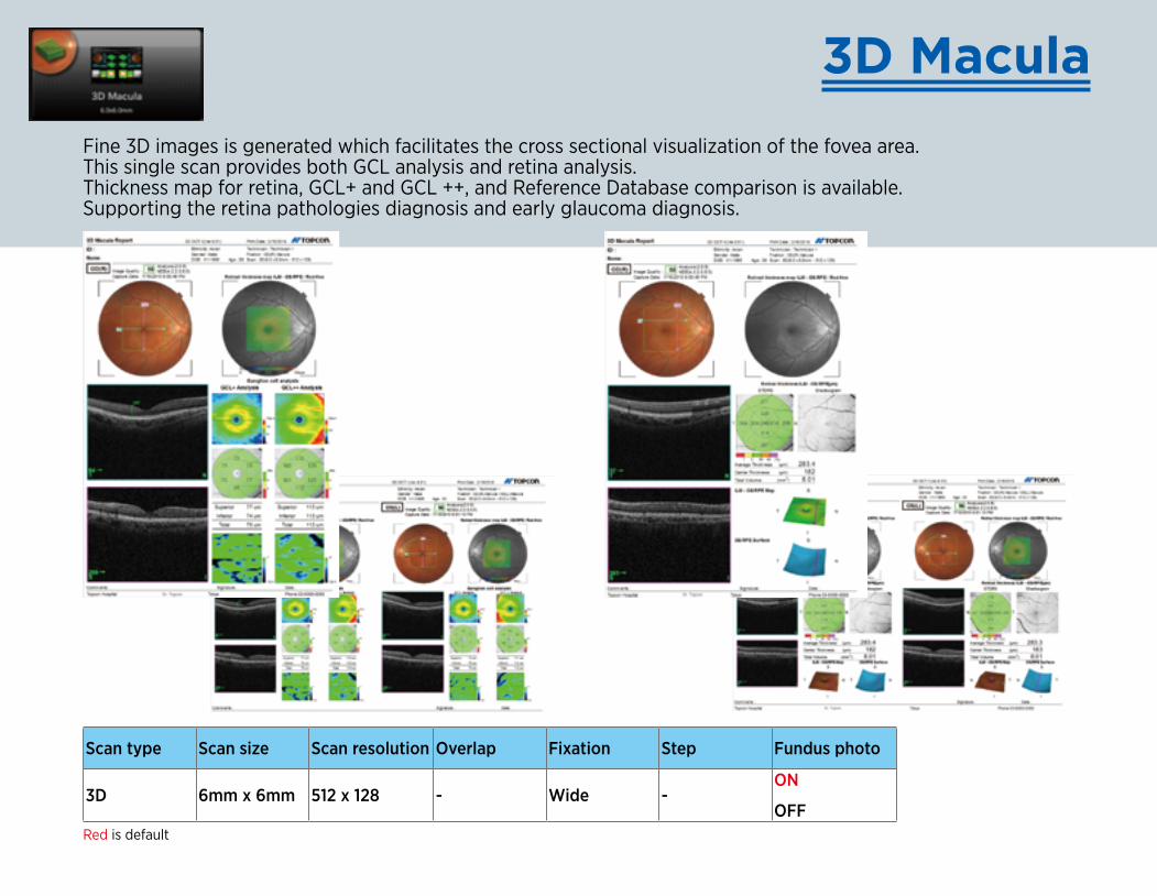
Fine 3D images is generated which facilitates the cross sectional visualization of the fovea area. This single scan provides both GCL analysis and retina analysis. Thickness map for retina, GCL+ and GCL ++, and Reference Database comparison is available. Supporting the retina pathologies diagnosis and early glaucoma diagnosis.
3D Macula
Red is default
Scan type Scan size Scan resolution Overlap Fixation Step Fundus photo
3D 6mm x 6mm 512 x 128 - Wide -ON
OFF
3D Macula Report
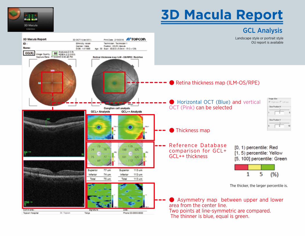
● Horizontal OCT (Blue) and vertical OCT (Pink) can be selected
● Thickness map
The thicker, the larger percentile is.
Reference Database comparison for GCL+ GCL++ thickness
● Asymmetry map between upper and lower area from the center line. Two points at line-symmetric are compared. The thinner is blue, equal is green.
● Retina thickness map (ILM-OS/RPE)
GCL AnalysisLandscape style or portrait style
OU report is available
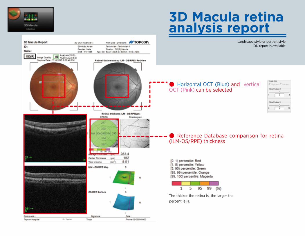
3D Macula retinaanalysis report
● Horizontal OCT (Blue) and vertical OCT (Pink) can be selected
● Reference Database comparison for retina (ILM-OS/RPE) thickness
The thicker the retina is, the larger the percentile is.
Landscape style or portrait style OU report is available
3D Disc
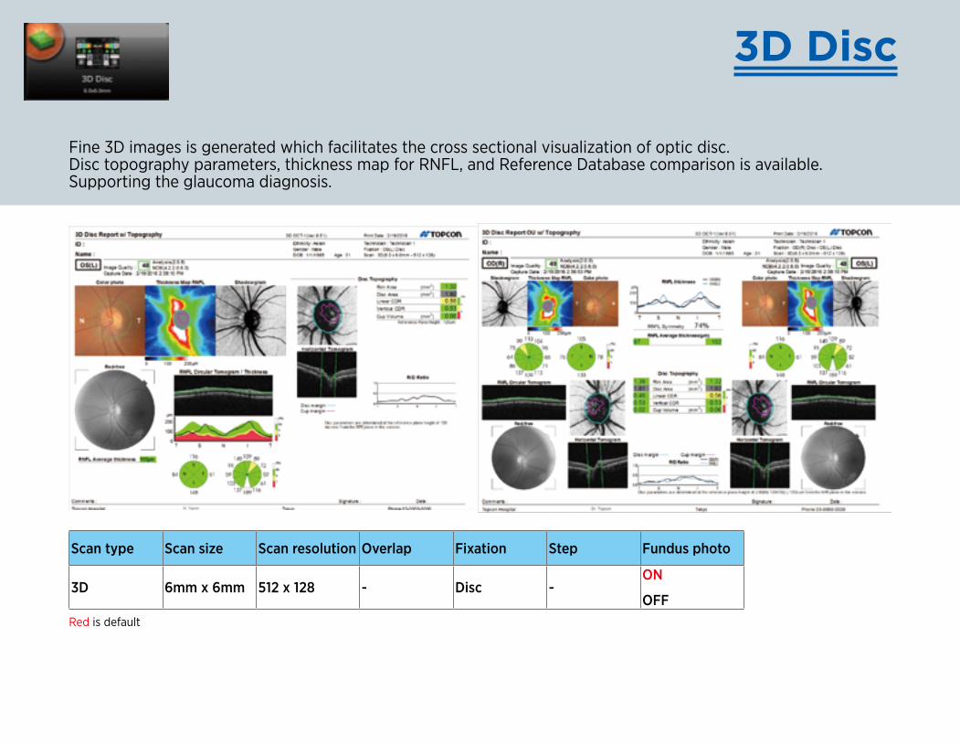
Fine 3D images is generated which facilitates the cross sectional visualization of optic disc. Disc topography parameters, thickness map for RNFL, and Reference Database comparison is available. Supporting the glaucoma diagnosis.
Scan type Scan size Scan resolution Overlap Fixation Step Fundus photo
3D 6mm x 6mm 512 x 128 - Disc -ON
OFFRed is default
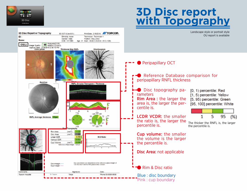
3D Disc report with Topography
● Peripapillary OCT
● Reference Database comparison for peripapillary RNFL thickness
● Rim & Disc ratio
Blue : disc boundaryPink : cup boundary
● Disc topography pa-rametersRim Area : the larger the area is, the larger the per-centile is.
LCDR VCDR: the smaller the ratio is, the larger the percentile is.
Cup volume: the smaller the volume is the larger the percentile is.
Disc Area: not applicable
Landscape style or portrait style OU report is available
The thicker the RNFL is, the larger the percentile is.
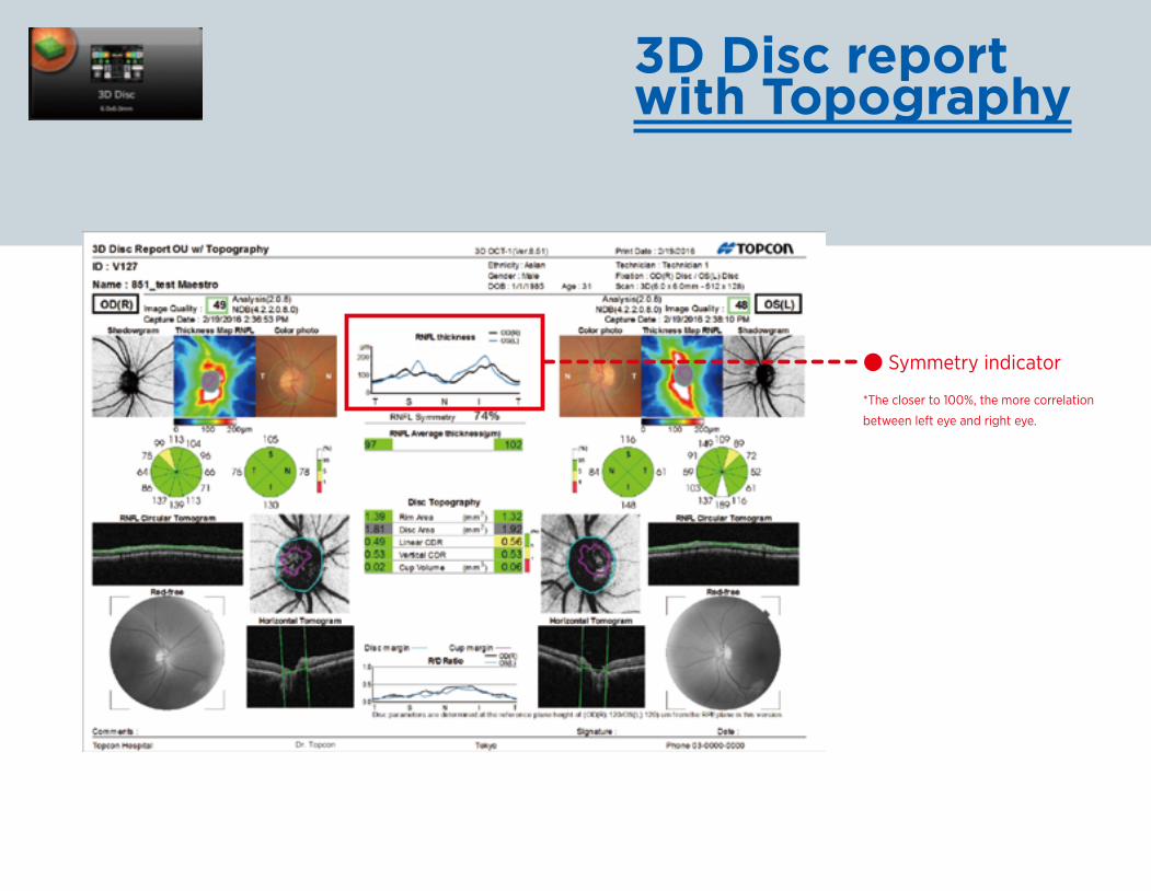
3D Disc report with Topography
● Symmetry indicator
*The closer to 100%, the more correlation
between left eye and right eye.
Landscape style or portrait style OU report is available
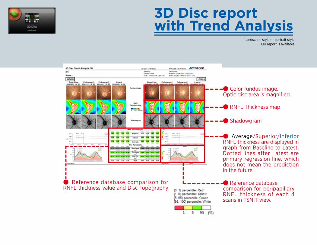
3D Disc report with Trend Analysis
● Color fundus image. Optic disc area is magnified.
● RNFL Thickness map
● Shadowgram
● Reference databasecomparison for peripapillary RNFL thickness of each 4 scans in TSNIT view.
● Average/Superior/Inferior RNFL thickness are displayed in graph from Baseline to Latest. Dotted lines after Latest are primary regression line, which does not mean the prediction in the future.
● Reference database comparison for RNFL thickness value and Disc Topography
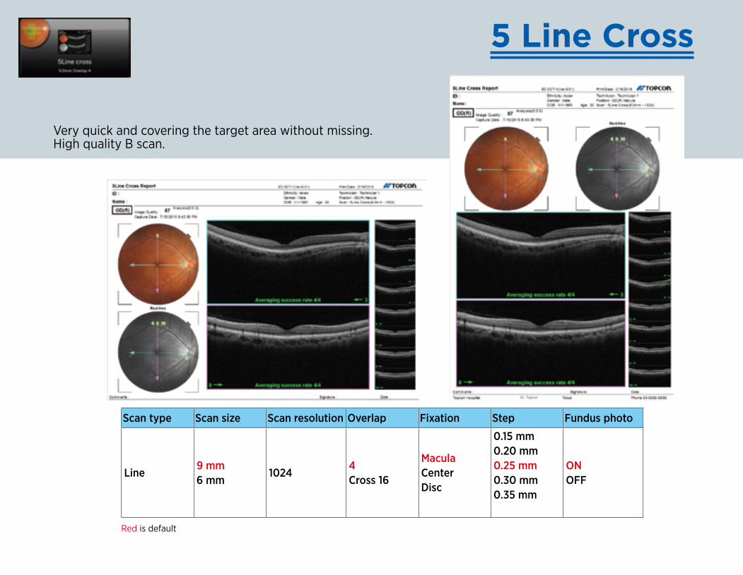
5 Line Cross
Very quick and covering the target area without missing. High quality B scan.
Scan type Scan size Scan resolution Overlap Fixation Step Fundus photo
Line9 mm6 mm
10244Cross 16
MaculaCenterDisc
0.15 mm0.20 mm0.25 mm0.30 mm0.35 mm
ONOFF
Red is default
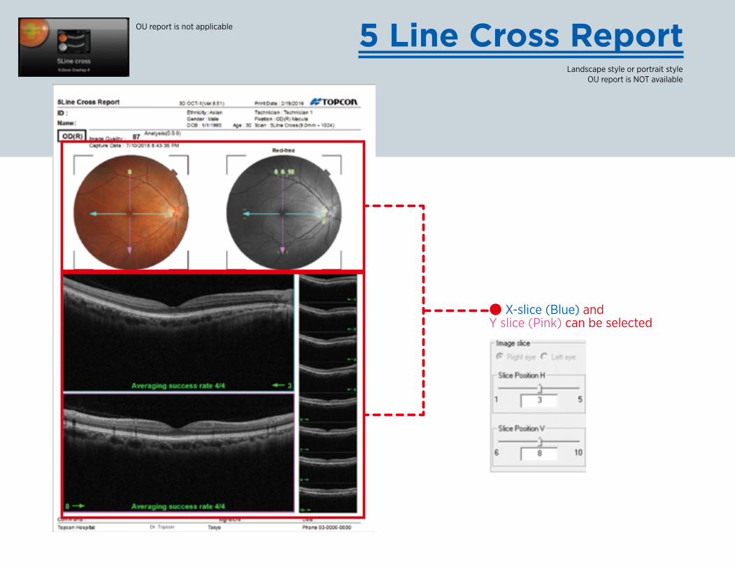
5 Line Cross Report
● X-slice (Blue) and Y slice (Pink) can be selected
Landscape style or portrait style OU report is NOT available
OU report is not applicable
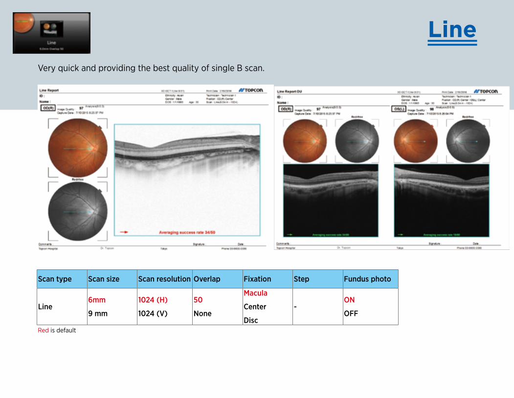
Line
Red is default
Scan type Scan size Scan resolution Overlap Fixation Step Fundus photo
Line6mm
9 mm
1024 (H)
1024 (V)
50
None
Macula
Center
Disc
-ON
OFF
Very quick and providing the best quality of single B scan.
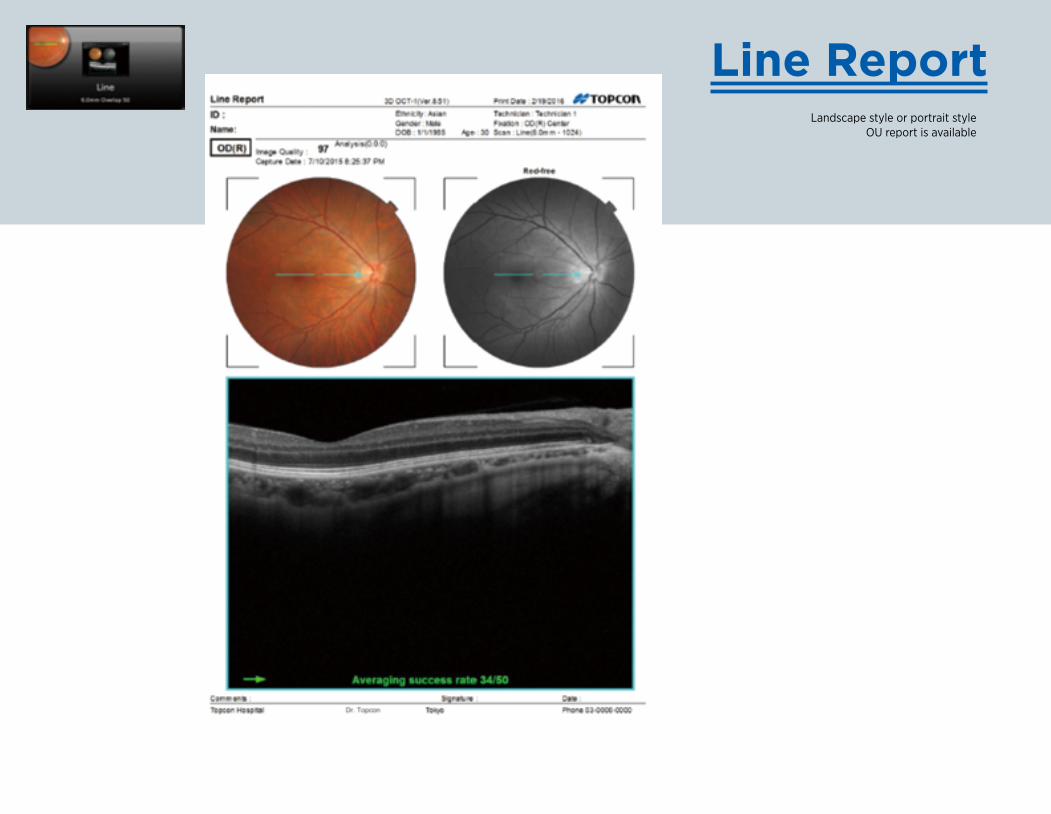
Landscape style or portrait style OU report is available
Line Report


![User Manual - psn-web.net · PDF file[For KX-NCP Series PBX Users Only]](https://img.pdfslide.us/doc/110x75/5ab0db307f8b9ad9788b8c96/user-manual-psn-webnet-for-kx-ncp-series-pbx-users-only.jpg)