Embed Size (px)
Citation preview
330 IEEE TRANSACTIONS ON NEURAL SYSTEMS AND REHABILITATION ENGINEERING, VOL. 17, NO. 4, AUGUST 2009
HermesC: Low-Power Wireless Neural RecordingSystem for Freely Moving Primates
Cynthia A. Chestek*, Student Member, IEEE, Vikash Gilja*, Paul Nuyujukian, Student Member, IEEE,Ryan J. Kier, Student Member, IEEE, Florian Solzbacher, Member, IEEE, Stephen I. Ryu,
Reid R. Harrison, Member, IEEE, and Krishna V. Shenoy, Senior Member, IEEE
Abstract—Neural prosthetic systems have the potential to re-store lost functionality to amputees or patients suffering fromneurological injury or disease. Current systems have primarilybeen designed for immobile patients, such as tetraplegics func-tioning in a rather static, carefully tailored environment. However,an active patient such as amputee in a normal dynamic, everydayenvironment may be quite different in terms of the neural controlof movement. In order to study motor control in a more uncon-strained natural setting, we seek to develop an animal model offreely moving humans. Therefore, we have developed and testedHermesC-INI3, a system for recording and wirelessly transmittingneural data from electrode arrays implanted in rhesus macaqueswho are freely moving. This system is based on the integratedneural interface (INI3) microchip which amplifies, digitizes, andtransmits neural data across a wireless channel.The wireless transmission has a range of in free space. Alltogether this device consumes 15.8 mA and 63.2 mW. On a single
Manuscript received September 04, 2008; revised December 30, 2008; ac-cepted February 23, 2009. First published June 02, 2009; current version pub-lished August 07, 2009. The work of C. A. Chestek was supported in part bythe National Science Foundation (NSF) Graduate Research Fellowships andin part by the William R. Hewlett Stanford Graduate Fellowship. The workof V. Gilja was supported in part by the National Science Foundation (NSF)Graduate Research Fellowship and in part by NDSEG Fellowship. The workof P. Nuyujukian was supported by the Stanford Medical Scholars Program.The work of R. R. Harrison was supported in part by the NSF CAREER awardECS-0134336 and in part by the Johns Hopkins University Applied PhysicsLaboratory under the DARPA Revolutionizing Prosthetics program, contractN66001-06-C-8005. The work of F. Solzbacher was supported in part by thein part by the Johns Hopkins University Applied Physics Laboratory under theDARPA Revolutionizing Prosthetics program, contract N66001-06-C-8005 andin part by the NIH-NINDS N01-NS-4-2362. The work of R. R. Harrison wassupported by the NIH-NINDS N01-NS-4-2362. The work of K. V. Shenoy wassupported by the Burroughs Wellcome Fund Career Award in the BiomedicalSciences, the Christopher Reeve Paralysis Foundation, Stanford Center for Inte-grated Systems, the NSF Center for Neuromorphic Systems Engineering at Cal-tech, the ONR, the Sloan Foundation, the Whitaker Foundation, and the McK-night Endowment Fund for Neuroscience. *These authors contributed equallyto this work.
C. A. Chestek is with the Department of Electrical Engineering, StanfordUniversity, Stanford, CA 94305 USA (email: [email protected]).
V. Gilja is with the Department of Computer Science, Stanford University,Stanford, Stanford, CA 94305 USA.
P. Nuyujukian is with the School of Medicine, Stanford University, Stanford,Stanford, CA 94305 USA.
R. J. Kier is with the Department of Electrical and Computer Engineering,Computer Science, University of Utah, Salt Lake City, UT 84112 USA.
F. Solzbacher and R. R. Harrison are with the Department of Electrical andComputer Engineering and the Department of Bioengineering at the Universityof Utah, Salt Lake City, UT 84112 USA.
S. Ryu is with the Department of Neurosurgery, Stanford University, CA94305 USA.
K. V. Shenoy is with the Department of Electrical Engineering and the Neuro-sciences Program, Stanford University, CA 94305 USA (e-mail: [email protected]).
Color versions of one or more of the figures in this paper are available onlineat http://ieeexplore.ieee.org.
Digital Object Identifier 10.1109/TNSRE.2009.2023293
2 A-hr battery pack, this device runs contiguously for approxi-mately six days. The smaller size and power consumption of thecustom IC allows for a smaller package (51 38 38 mm ) thanprevious primate systems. The HermesC-INI3 system was usedto record and telemeter one channel of broadband neural data at15.7 kSps from a monkey performing routine daily activities inthe home cage.
Index Terms—Brain–machine interface, low power, neural pros-thetics, telemetry, wireless.
I. INTRODUCTION
C ORTICAL neural prostheses extract signals from thebrain in order to control prosthetic devices such as
limbs and computer cursors [1]. This is a rapidly growingfield with the potential to provide treatment for amputees orpatients suffering from neurological injury and disease. Afterseveral proof-of-concept studies [2], [3], subsequent studieshave demonstrated improving performance in monkeys [4]–[6]and even humans [7]. However, several obstacles stand in theway of translating these experiments into a clinical system.Two obstacles are reasonably well recognized while a third,which we focus on in this report, has to date been somewhatunderappreciated. First, multielectrode array lifetime is ap-proximately a year or less, which seriously limits the potentialclinical usefulness of cortical implants. Second, current sys-tems require a percutaneous connector, which is associatedwith infection risk as well as aesthetic concerns. This issuecan be addressed with implantable electronics to record neuralactivity and wirelessly transmit this data through the skin toan external device [8]–[14]. In this study, however, we willfocus on a third major obstacle to clinical adoption: neuralprosthetics experiments to date have occurred in highly con-trolled settings over a short time span with immobile or nearlyimmobile animals or humans. Consequently, it is possible thatposture, body movement, head movement, or brain movementwithin the skull could have a strong effect on performance.Also, many experiments have been done in environments withreduced visual, auditory, and tactile stimulation over limitedperiods of time. In some experiments, animals were trained tofixate their gaze on a specific point, which may significantlyimprove the performance of decoders [15]. In a human clinicalsetting, particularly for active amputees, these are not realisticconstraints.
Therefore, one important next challenge for neural prostheticsystems is to release these constraints, and attempt to replicateprevious high performance results in this more practical, yet
1534-4320/$26.00 © 2009 IEEE
Authorized licensed use limited to: Stanford University. Downloaded on July 09,2010 at 16:40:37 UTC from IEEE Xplore. Restrictions apply.
CHESTEK et al.: HERMESC: LOW-POWER WIRELESS NEURAL RECORDING SYSTEM FOR FREELY MOVING PRIMATES 331
complex setting. To address this challenge, our overarching goalis to establish an animal model of freely moving humans. To doso requires the ability to transmit neural data wirelessly froma subject. Rhesus macaque monkeys are appropriate subjectssince they can make the coordinated arm movements that onewould like to decode. Also, the rhesus brain is substantially ho-mologous to the human brain, and there is a large body of neuro-science and neural prosthetics research in macaques. In additionto the neural prosthetic application, such a system could be usedto study complex voluntary behaviors that have been previouslydifficult to access for researchers, such as aggression, vocaliza-tion, social behavior, and locomotion. Also, the large quantityof neural data that could be obtained using a wireless deviceover many days might be useful to computational neuroscien-tists studying general properties of the cortex.
There has been substantial previous work on systems thatrecord neural data during free movement in many different an-imal models. Several systems have been developed for freelymoving rats. Farshchi et al. has reported a rodent system usingan off the shelf microcontroller and radio transceiver [16]. Othersystems have been built with both custom circuitry, and com-mercial off the shelf (COTS) electronics. Cheney et al. devel-oped the Pico Neural Data Collection system using a custombioamplifier front end, and commercially available processingand wireless circuits [17]. A similar approach was taken byChae et al. [18]. There is also a commercial system availablefor wirelessly recording 31 channels of neural data from freelymoving rodents, which runs for 6 h contiguously before re-quiring a new battery (Triangle Biosystems Inc., Durham, NC).It is notable that packaging of these systems in general can bevery specific to the particular animal model and cage system.For example, Takeuchi et al. have developed a wearable neuralrecording system for insects using a 15- -thickness flexiblepolyimide cable which wraps around the insect’s body betweenthe circuitry and the electrode [19].
While the use of COTS components reduces cost and devel-opment time compared to a fully integrated approach, a majordisadvantage is high power consumption, which makes it verydifficult to run freely behaving experiments for extended periodsof time. The first biotelemetry system integrated onto a singlechip was demonstrated by Song et al. [20]. More recently, sev-eral development efforts have been underway to increase thenumber of channels, decrease the power, and integrate all theelectronics and electrodes into a single implantable package.DeMichele and Troyk developed a 16 channel system that con-sumed 18 mW [21]. Yin et al. have reported a 15 channel systemthat draws only 4.5 mW [11]. Moving closer to freely movingprimates, Mohseni et al. have developed and used a 4 channel,2.2 mW biotelemetry system to record from awake restrainedmarmoset monkeys [12]. In freely moving rats, Sodagar et al.have developed a 64-channel fully integrated wireless systemand successfully recorded in vivo neural data while supplyingpower through a nearby inductive link [13]. However, it ap-pears that none of these systems have yet been adapted for freelymoving primates, possibly due to surgical or packaging difficul-ties.
A small number of systems have been implemented for freelybehaving primate experiments. Prior to wireless systems, teth-
ered recordings have been used to study natural behaviors suchas spatial navigation [22]. However, chronic experiments wouldbe highly challenging using such a system since the animalwould have access to the tethering wire. Therefore, several self-contained wireless systems have been developed. Two systemsrecord 1–2 channels of neural data to onboard memory, whichcan be subsequently downloaded when the device is retrieved[23], [24]. It would be difficult to scale these systems up sub-stantially since memory can fill rather quickly with broadbandneural data. Also, it can be difficult to synchronize neural datawith behavioral measures such as video, and impossible to ac-cess the data in real time to look at task modulation or to do BMIexperiments. Several systems use COTS electronics to transmitneural data wirelessly [25], [26]. However, all of these systemshave relatively high power consumption, running for 1–8 h be-fore requiring a new battery. We would like to record for severaldays without servicing the device. Finally, COTS systems wouldbe difficult to scale up to many channels in the future, since thesize of the electronics for 1–2 channels is already at the limit ofwhat a large animal can carry in an unobtrusive device. We seekto develop a system with a clear development path to 96-channelwireless neural recording.
To that end, we have developed HermesC-INI3, a wirelesssystem for recording neural data from freely moving primates.This system uses the custom Integrated Neural Interface (INI)microchip, which is part of a larger project to develop a fullyimplantable 96-channel system [14] and is described in detailin the companion paper [29]. The INI3 chip digitizes the signalfrom one electrode on a 96-electrode array (Cyberkinetics Inc.,Salt Lake City, UT) and transmits those data wirelessly to areceiver outside of the cage. HermesC refers to the system ofconnecting this device to the chronically-implanted electrodearray, encapsulating the circuitry in a small wearable enclosure,and additional electronics for the specific needs of this primateresearch system, such as acquiring data from receivers placedoutside the cage. HermesC-INI3 has been used to record datafrom a rhesus macaque performing many unconstrained regularactivities. This device differs from previous primate systems byhaving lower power consumption, a smaller form factor, and thecapability to expand to more channels in the future as part ofa planned development path. Portions of this work have beenpreviously presented in conference form [27], [28].
II. METHODS
The system consists of a neural connector and a printed cir-cuit board (PCB) with a custom microchip to record, digitize,and transmit the data, all of which are housed in a protective en-closure. Data is recorded using an external receiver.
A. Physical Design
Fig. 1 shows a diagram of the physical design. Neural dataare obtained through a 96-channel cortical array implanted inmacaque motor and premotor cortex (Cyberkinetics Inc., SaltLake City, UT). Layers of preclude and duragen help protectthe dura and array, and help avoid material adhesions. A siliconeelastomer fills the craniotomy and allows a flexible ribbon cableto connect to a zero-insertion force (ZIF) connector on the skull(Cyberkinetics Inc., Salt Lake City, UT). The entire implant is
Authorized licensed use limited to: Stanford University. Downloaded on July 09,2010 at 16:40:37 UTC from IEEE Xplore. Restrictions apply.
332 IEEE TRANSACTIONS ON NEURAL SYSTEMS AND REHABILITATION ENGINEERING, VOL. 17, NO. 4, AUGUST 2009
Fig. 1. Physical design of HermesC. 100-electrode arrays are implanted inmacaque motor and premotor cortex. Preclude, duragen, a silicone elastomer,and methyl methacrylate protect the brain, skull, and array. ZIF connectorattaches to skull (CKI). Custom connector provides 32 of 96 channels toPCB which includes INI3 microchip. Aluminum housing embedded in methylmethacrylate protects electronics and batteries.
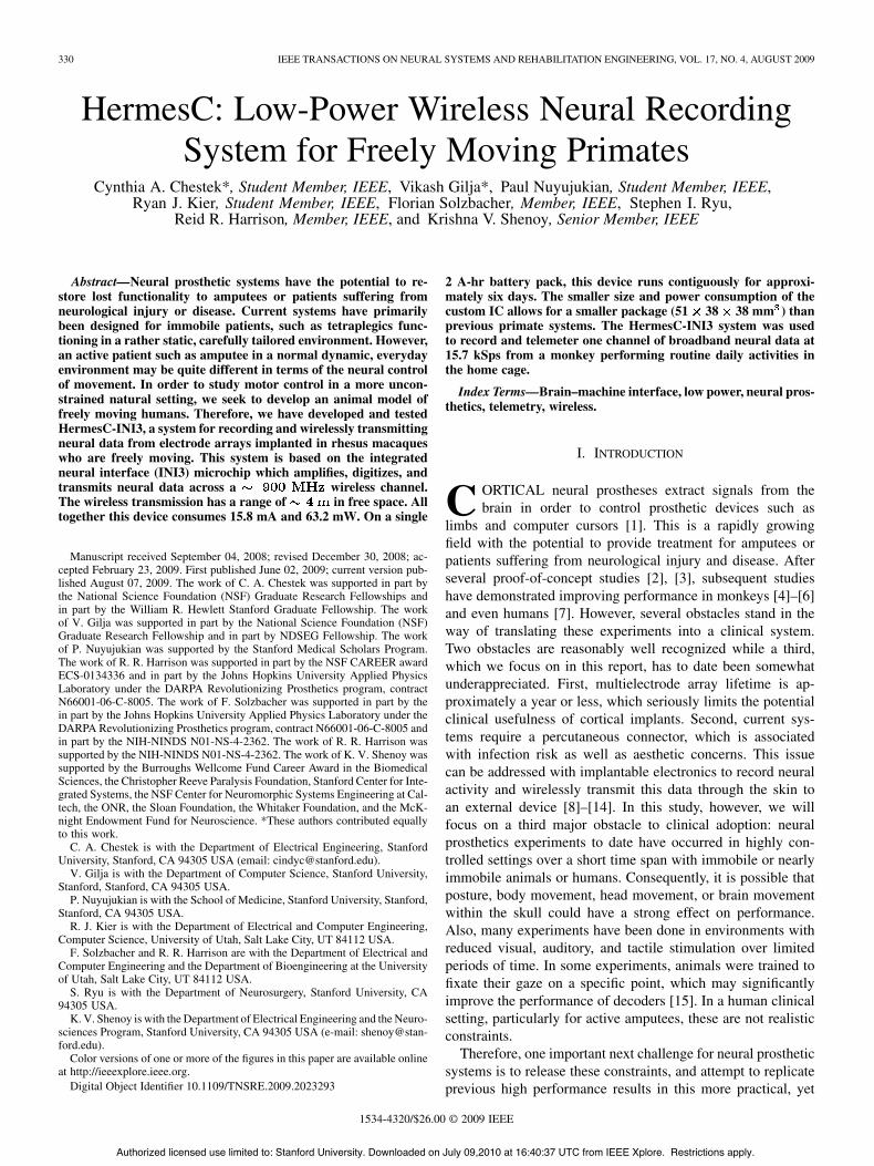
Fig. 2. Aluminum enclosure with stub antenna in lid for (a) larger prototypesystem and (b) final design.
protected with methyl metacrylate (dental cement). Three dif-ferent custom head-stages provide access to three banks of 32channels with connectors that attach directly to the PCB. Theentire system, which includes the percutaneous connector, thePCB, and a lithium battery pack, is housed in an aluminumenclosure attached to the implant with titanium hardware andmethyl methacrylate. Initial testing with a larger prototype PCBwas completed in aluminum enclosure identical to the HermesBsystem [24], which measured 60 70 45 mm . However, thefinal design was verified in a smaller enclosure that measured51 38 38 mm . For each enclosure, a stub antenna protrudes8 mm through a hole in the lid, as shown in Fig. 2. This antennais immobilized and sealed with epoxy. The total weight of thissystem including the batteries is 114 g.
B. Electronics
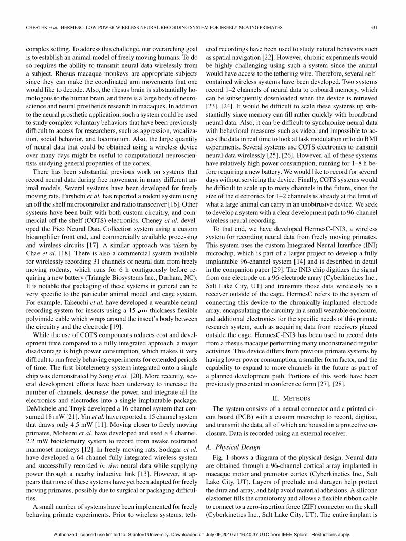
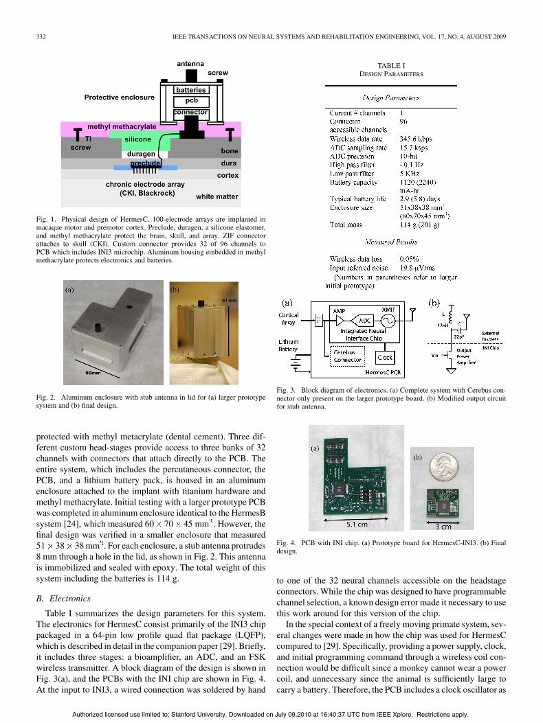
Table I summarizes the design parameters for this system.The electronics for HermesC consist primarily of the INI3 chippackaged in a 64-pin low profile quad flat package (LQFP),which is described in detail in the companion paper [29]. Briefly,it includes three stages: a bioamplifier, an ADC, and an FSKwireless transmitter. A block diagram of the design is shown inFig. 3(a), and the PCBs with the INI chip are shown in Fig. 4.At the input to INI3, a wired connection was soldered by hand
TABLE IDESIGN PARAMETERS
Fig. 3. Block diagram of electronics. (a) Complete system with Cerebus con-nector only present on the larger prototype board. (b) Modified output circuitfor stub antenna.
Fig. 4. PCB with INI chip. (a) Prototype board for HermesC-INI3. (b) Finaldesign.
to one of the 32 neural channels accessible on the headstageconnectors. While the chip was designed to have programmablechannel selection, a known design error made it necessary to usethis work around for this version of the chip.
In the special context of a freely moving primate system, sev-eral changes were made in how the chip was used for HermesCcompared to [29]. Specifically, providing a power supply, clock,and initial programming command through a wireless coil con-nection would be difficult since a monkey cannot wear a powercoil, and unnecessary since the animal is sufficiently large tocarry a battery. Therefore, the PCB includes a clock oscillator as
Authorized licensed use limited to: Stanford University. Downloaded on July 09,2010 at 16:40:37 UTC from IEEE Xplore. Restrictions apply.
CHESTEK et al.: HERMESC: LOW-POWER WIRELESS NEURAL RECORDING SYSTEM FOR FREELY MOVING PRIMATES 333
well as connectors for a battery and a wired connection for initialprogramming. The device can be programmed in ms withan 836 bit command (which includes many parameters not yetfully implemented on this device). After device programming,this 4-wire connection can simply be removed for the rest of theexperiment. Another difference is the wireless range requiredfor freely moving monkeys. While the INI chip can transmit ata range of 5 cm at 8 mW, in the HermesC system, a single dis-crete resistor is added to the final RF amplifier to increase thetransmit range, which results in a total power consumption of63.2 mW. Finally, since this device is using a different antennathan described in [29] several 0603 components are used. Themodified output circuit is shown in Fig. 3(b). The output stageuses a discrete inductor to bias the amplifier, and an addition ca-pacitor to provide a dc block to the antenna. This system, Her-mesC “nano,” is small enough to be used on any large animalwith a Cyberkinetics neuroport connector.
In an earlier iteration, a larger PCB was used for initial de-sign and testing, as shown in Fig. 4(a). In addition to the com-ponents described above, this PCB also provides an alternatedata path to a traditional head-stage connector for a commercialneural recording system, Cerebus (Cyberkinetics Inc., Salt LakeCity, UT). In this way, data can be obtained simultaneously andthen compared. It also includes fuses, various test points for ac-cessing the chip, and larger components for easier removal andtesting. This version represents a general in vivo test platformfor the INI chip, in which signals can be easily accessed, andthe external circuitry can be rapidly reconfigured.
The power consumption of this system is adjustable in twoways. First, a bias resistor can control the gain of the wirelessamplifier, which allows the user to adjust the range. Second, anoptional 6 dB attenuator can be used at the output to the antennato minimize the effect of environmentally-induced changes inantenna impedance on the transmit frequency. Due to powerconstraints, the INI does not include a phase lock loop (PLL)[29], which would better stabilize the transmitting frequency. Inthe current configuration, HermesC consumes 15.8 mA at 4.0 V,for a total of 63.2 mW. Of that amount, is requiredby the RF transmitter on the INI chip, and 21 mW is requiredby the off chip clock. The INI chip itself can run on a voltagesource between 3-4 V. This represents a large improvement overHermesB, which recorded from two broadband channels, andconsumed 71 mA at 4.0 V, for a total of 284 mW [24]. Despitethe smaller enclosure size, HermesC can run for 2.9 days on one1120 mA-hr battery or times longer. Alternatively, using thelarger HermesB enclosure and a second battery pack due to thesmaller electronics, it can run for 5.8 days contiguously. In thetypical usage mode with the smaller enclosure, the device runsfor over three days on two disposable Li-ion 1/2AA batteries.To our knowledge, this is the longest running wireless neuralsystem reported that has been implemented in freely movinganimals.
At the output RF stage, data are transmitted in 16-sampleframes at 345.6 kb/s with one parity bit computed for each10-bit ADC sample. The wireless data were collected with acommercial FSK transceiver, the ADF7025 development board(Analog Devices), receiving in the 902–928 MHz range. Tocompensate for small fluctuations in the transmitting frequency,
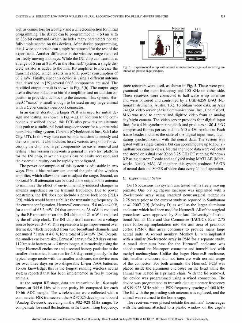
Fig. 5. Experimental setup with animal in metal home cage and receiving an-tennae on plastic cage window.
three receivers were used, as shown in Fig. 5. These were pro-grammed to the main frequency and 100 KHz on either side.These receivers were connected to half-wave whip antennaeand were powered and controlled by a USB-6259 DAQ (Na-tional Instruments, Austin, TX). To obtain video data, an Axis241QA video server (Axis Communications, Inc., Chelmsford,MA) was used to capture and digitize video from an analogday/night camera. The video server provides four digital inputlines for a 4-bit synchronizing clock and producescompressed frames per second at a 640 480 resolution. Eachframe header includes the state of the digital input lines, facil-itating synchronization with the neural data. The system wastested with a single camera, but can accommodate up to four si-multaneous camera views. Neural and video data were collectedand stored on a dual core Xeon 3.25 GHz PC running WindowsXP using custom C code and analyzed using MATLAB (Math-works, Natick, MA). All together, this system produces 3.6 GBof neural data and 80 GB of video data every 24 h of operation.
C. Experimental Setup
On 16 occasions this system was tested with a freely movingprimate. One 6.9 kg rhesus macaque was implanted with a96-electrode array using standard neurosurgical techniques2.75 years prior to the current study as reported in Santhanamet al. 2007 [19] (Monkey D) as well as the larger aluminumenclosure which had been used for HermesB. All of the surgicalprocedures were approved by Stanford University’s Institu-tional Animal Care and Use Committee (IACUC). Even 2.75years following implantation into the arm area of premotorcortex (PMd), this array continues to provide many largeneural units. A second monkey, Monkey L, was implantedwith a similar 96-electrode array in PMd for a separate study.A small aluminum base for the HermesC enclosure wasadded around the Neuroport connector and immobilized withmethyl methacrylate. Unlike the larger HermesB enclosure,this smaller enclosure did not interfere with normal usageof the connector. For both animals, the HermesC PCB wasplaced inside the aluminum enclosure on the head while theanimal was seated in a primate chair. With the lid removed,the device was programmed using a wired connection. Thedevice was programmed to transmit data at a center frequencyof 919–923 MHz with an FSK frequency spacing of 460 kHz.The lid with the protruding stub antenna was replaced, and theanimal was returned to the home cage.
The receivers were placed outside the animals’ home cageswith the antenna attached to a plastic window on the cage’s
Authorized licensed use limited to: Stanford University. Downloaded on July 09,2010 at 16:40:37 UTC from IEEE Xplore. Restrictions apply.
334 IEEE TRANSACTIONS ON NEURAL SYSTEMS AND REHABILITATION ENGINEERING, VOL. 17, NO. 4, AUGUST 2009
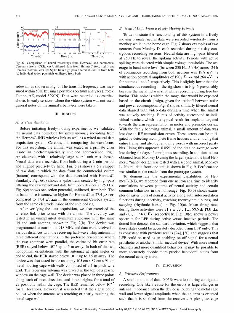
Fig. 6. Comparison of neural recordings from HermesC and commercialCerebus system (CKI). (a) Unfiltered data from HermesC (top, right) andCerebus (bottom, left). (b) Spike train high pass filtered at 250 Hz from both.(c) Individual action potentials unfiltered from both.
sidewall, as shown in Fig. 5. The transmit frequency was mea-sured within 50 kHz using a portable spectrum analyzer (Protek,Tempe, AZ, model 3290N). Data were recorded as describedabove. In early sessions where the video system was not used,general notes on the animal’s behavior were taken.
III. RESULTS
A. System Validation
Before initiating freely-moving experiments, we validatedthe neural data collection by simultaneously recording fromthe HermesC-INI3 wireless link as well as a wired neural dataacquisition system, Cerebus, and comparing the waveforms.For this recording, the animal was seated in a primate chairinside an electromagnetically shielded neuroscience “rig.”An electrode with a relatively large neural unit was chosen.Neural data were recorded from both during a 2 min period,and aligned precisely by hand. Fig. 6(a) shows a 5 s snippetof raw data in which the data from the commercial system(bottom) correspond with the data recorded with HermesC.Similarly, Fig. 6(b) shows a spike train created by high-passfiltering the raw broadband data from both devices at 250 Hz.Fig. 6(c) shows one action potential, unfiltered, from both. Thein-band noise is somewhat higher for HermesC, at 27.4compared to 17.4 in the commercial Cerebus systemfrom the same electrode inside of the shielded rig.
After verifying the data acquisition, we also exercised thewireless link prior to use with the animal. The circuitry wastested in an unimplanted aluminum enclosure with the samelid and stub antenna, shown in Fig. 2(b). The device wasprogrammed to transmit at 918 MHz and data were received atvarious distances with the receiving half-wave whip antenna inthree different orientations. In the preferred orientation wherethe two antennae were parallel, the estimated bit error rate(BER) stayed below up to 5 m away. In both of the twononoptimal orientations with the antennae at right angles orend to end, the BER stayed below up to 3.5 m away. Thedevice was also tested inside an empty 105 cm x 87 cm x 91 cmmetal housing cage with walls composed of a 1-in pitch wiregrid. The receiving antenna was placed at the top of a plasticwindow on the cage wall. The device was placed in three pointsalong each of three directions and three heights, for a total of27 positions within the cage. The BER remained belowfor all locations. However, it was noted that the signal couldbe lost when the antenna was touching or nearly touching themetal cage wall.
B. Neural Data From a Freely Moving Primate
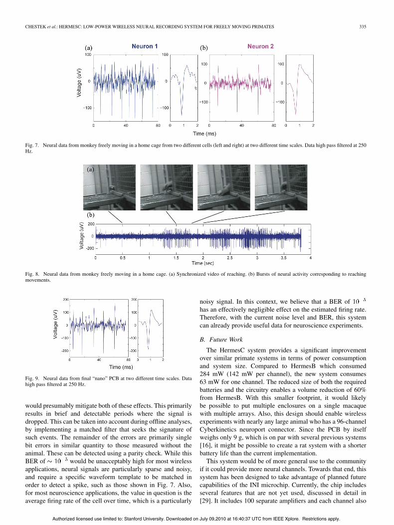
To demonstrate the functionality of this system in a freelymoving primate, neural data were recorded wirelessly from amonkey while in the home cage. Fig. 7 shows examples of twoneurons from Monkey D, each recorded during six day con-tiguous recording sessions. Neural data are high-pass filteredat 250 Hz to reveal the spiking activity. Periods with activespiking were detected with simple voltage thresholds. The av-erage in-band noise level (between 250 Hz–5 kHz) across 24 hof continuous recording from both neurons was 19.8with action potential amplitudes of 190 and 264for neurons 1 and 2, respectively. This is slightly lower than thesimultaneous recording in the rig shown in Fig. 6 presumablybecause the metal lid was shut while recording during free be-havior. This noise is within the range that would be expectedbased on the circuit design, given the tradeoff between noiseand power consumption. Fig. 8 shows similarly filtered neuraldata aligned with video data during a time when the animalwas actively reaching. Bursts of activity correspond to indi-vidual reaches, which is a typical result for implants targetedtowards the arm representation in motor and premotor cortex.With the freely behaving animal, a small amount of data waslost due to RF transmission errors. These errors can be miti-gated by detecting incomplete frame headers and removing thatentire frame, and also by removing words with incorrect paritybits. Using this approach 0.05% of the data on average werelost during six days of contiguous recording. In addition to dataobtained from Monkey D using the larger system, the final Her-mesC “nano” design was tested with a second animal, MonkeyL. Neural data from one unit is shown in Fig. 9. Performancewas similar to the results from the prototype system.
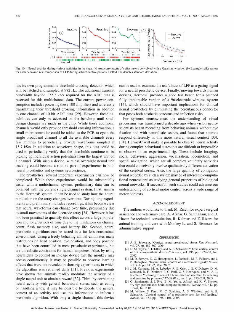
To demonstrate the experimental capabilities of Her-mesC-INI3, we recorded from one neuron at a time to identifycorrelations between patterns of neural activity and certaincommon behaviors in the homecage. Fig. 10(b) shows exam-ples of raster plots of neural activity along with autocorrelationfunctions during inactivity, reaching (nonrhythmic bursts) andswaying (rhythmic bursts) in Fig. 10(a). Mean firing ratesduring those activities were , ,and , respectively. Fig. 10(c) shows a powerspectrum for LFP during active versus inactive periods. Thedotted line denotes the standard deviation, which suggests thatthese states could be accurately decoded using LFP only. Thisis consistent with previous results [24], [30] and suggests thatLFP could be used as an enabling on-off signal for a neuralprosthetic or another similar medical device. With more neuralchannels and more quantified behaviors, it may be possible tomore accurately decode more precise behavioral states fromthe neural activity alone
IV. DISCUSSION
A. Wireless Performance
A small amount of data, 0.05% were lost during contiguousrecording. One likely cause for the errors is large changes inantenna impedance when the device is touching the metal cagewall and lower signal amplitude when the antenna is orientedsuch that it is shielded from the receivers. A plexiglass cage
Authorized licensed use limited to: Stanford University. Downloaded on July 09,2010 at 16:40:37 UTC from IEEE Xplore. Restrictions apply.
CHESTEK et al.: HERMESC: LOW-POWER WIRELESS NEURAL RECORDING SYSTEM FOR FREELY MOVING PRIMATES 335
Fig. 7. Neural data from monkey freely moving in a home cage from two different cells (left and right) at two different time scales. Data high pass filtered at 250Hz.
Fig. 8. Neural data from monkey freely moving in a home cage. (a) Synchronized video of reaching. (b) Bursts of neural activity corresponding to reachingmovements.
Fig. 9. Neural data from final “nano” PCB at two different time scales. Datahigh pass filtered at 250 Hz.
would presumably mitigate both of these effects. This primarilyresults in brief and detectable periods where the signal isdropped. This can be taken into account during offline analyses,by implementing a matched filter that seeks the signature ofsuch events. The remainder of the errors are primarily singlebit errors in similar quantity to those measured without theanimal. These can be detected using a parity check. While thisBER of would be unacceptably high for most wirelessapplications, neural signals are particularly sparse and noisy,and require a specific waveform template to be matched inorder to detect a spike, such as those shown in Fig. 7. Also,for most neuroscience applications, the value in question is theaverage firing rate of the cell over time, which is a particularly
noisy signal. In this context, we believe that a BER ofhas an effectively negligible effect on the estimated firing rate.Therefore, with the current noise level and BER, this systemcan already provide useful data for neuroscience experiments.
B. Future Work
The HermesC system provides a significant improvementover similar primate systems in terms of power consumptionand system size. Compared to HermesB which consumed284 mW (142 mW per channel), the new system consumes63 mW for one channel. The reduced size of both the requiredbatteries and the circuitry enables a volume reduction of 60%from HermesB. With this smaller footprint, it would likelybe possible to put multiple enclosures on a single macaquewith multiple arrays. Also, this design should enable wirelessexperiments with nearly any large animal who has a 96-channelCyberkinetics neuroport connector. Since the PCB by itselfweighs only 9 g, which is on par with several previous systems[16], it might be possible to create a rat system with a shorterbattery life than the current implementation.
This system would be of more general use to the communityif it could provide more neural channels. Towards that end, thissystem has been designed to take advantage of planned futurecapabilities of the INI microchip. Currently, the chip includesseveral features that are not yet used, discussed in detail in[29]. It includes 100 separate amplifiers and each channel also
Authorized licensed use limited to: Stanford University. Downloaded on July 09,2010 at 16:40:37 UTC from IEEE Xplore. Restrictions apply.
336 IEEE TRANSACTIONS ON NEURAL SYSTEMS AND REHABILITATION ENGINEERING, VOL. 17, NO. 4, AUGUST 2009
Fig. 10. Neural activity during various activities in the cage. (a) Autocorrelations of spike rasters convolved with a Gaussian window. (b) Example spike rastersfor each behavior. (c) Comparison of LFP during active/inactive periods. Dotted line denotes standard deviation.
has its own programmable threshold-crossing detector, whichwill be latched and sampled at 982 Hz. The additional transmitbandwidth beyond 172.7 kb/s required for the ADC data isreserved for this multichannel data. The current power con-sumption includes powering these 100 amplifiers and wirelesslytransmitting their threshold crossing information in additionto one channel of 10-bit ADC data [29]. However, these ca-pabilities can only be accessed on the benchtop until smalldesign changes are made in the chip. While these additionalchannels would only provide threshold crossing information, asmall microcontroller could be added to the PCB to cycle thesingle broadband channel to all the available channels everyfew minutes to periodically provide waveforms sampled at15.7 kS/s. In addition to waveform shape, this data could beused to periodically verify that the thresholds continue to bepicking up individual action potentials from the largest unit ona channel. With such a device, wireless overnight neural unittracking could become a routine part of experiments in bothneural prosthetics and systems neuroscience.
For prosthetics, several important experiments can now becompleted. While these experiments would be substantiallyeasier with a multichannel system, preliminary data can beobtained with the current single channel system. First, similarto the HermesB system, it can be used to study how the neuralpopulation on the array changes over time. During long experi-ments and preliminary multiday recordings, it has become clearthat neural waveforms can change over time, presumably dueto small movements of the electrode array [24]. However, it hasnot been practical to quantify this effect across a large popula-tion and long periods of time due to the limitations on channelcount, flash memory size, and battery life. Second, neuralprosthetic algorithms can be tested in a far less constrainedenvironment. Using a freely behaving animal eliminates manyrestrictions on head position, eye position, and body positionthat have been controlled in most prosthetic experiments, butare unrealistic constraints for human amputees. Also, by usingneural data to control an in-cage device that the monkey mayaccess continuously, it may be possible to observe learningeffects that were not revealed in short rig experiments in whichthe algorithm was retrained daily [31]. Previous experimentshave shown that animals readily modulate the activity of asingle neural unit to obtain a reward [32]. Third, by correlatingneural activity with general behavioral states, such as eatingor handling a toy, it may be possible to decode the generalcontext of an activity and use this information to inform aprosthetic algorithm. With only a single channel, this device
can be used to examine the usefulness of LFP as a gating signalfor a neural prosthetic device. Finally, moving towards humandevices, HermesC provides a good test bench for a plannedfully implantable version of a 96-electrode wireless system[14], which should have important implications for clinicalneural prosthetics by eliminating the percutaneous connectorthat poses both aesthetic concerns and infection risks.
For systems neuroscience, the understanding of visualprocessing was transformed a decade ago when vision neuro-scientists began recording from behaving animals without eyefixation and with naturalistic scenes, and found that neuronsrespond differently in this more natural visual context [33],[34]. HermesC will make it possible to observe neural activityduring complex behavioral states that are difficult or impossibleto observe in an experimental rig. These include foraging,social behaviors, aggression, vocalization, locomotion, andspatial navigation, which are all complex voluntary activitiesthat could conceivably involve qualitatively different activationof the cerebral cortex. Also, the large quantity of contiguousneural recorded by such a system may be of interest to computa-tional neuroscientists studying general properties of biologicalneural networks. If successful, such studies could advance ourunderstanding of cortical motor control across a wide range ofmotor contexts.
ACKNOWLEDGMENT
The authors would like to thank M. Risch for expert surgicalassistance and veterinary care, A. Afshar, G. Santhanam, and D.Haven for technical consultation, R. Kalmar and Z. Rivera foranimal training and care with Monkey L, and S. Eisensee foradministrative support.
REFERENCES
[1] A. B. Schwartz, “Cortical neural prosthetics,” Annu. Rev. Neurosci.,vol. 27, pp. 487–507, 2004.
[2] D. M. Taylor, S. I. Tillery, and A. B. Schwartz, “Direct cortical controlof 3D neuroprosthetic devices,” Science, vol. 296, pp. 1829–32, Jun.2002.
[3] M. D. Serruya, N. G. Hatsopoulos, L. Paninski, M. R. Fellows, and J.P. Donoghue, “Instant neural control of a movement signal,” Nature,vol. 416, pp. 141–2, Mar. 2002.
[4] J. M. Carmena, M. A. Lebedev, R. E. Crist, J. E. O’Doherty, D. M.Santucci, D. F. Dimitrov, P. G. Patil, C. S. Henriquez, and M. A. L.Nicolelis, “Learning to control a brain-machine interface for reachingand grasping by primates,” PLOS Biol., vol. 1, pp. 193–208, 2003.
[5] G. Santhanam, S. I. Ryu, B. M. Yu, A. Afshar, and K. V. Shenoy,“A high-performance brain-computer interface,” Nature, vol. 442, pp.195–8, Jul. 2006.
[6] M. Velliste, S. Perel, M. C. Spalding, A. S. Whitford, and A. B.Schwartz, “Cortical control of a prosthetic arm for self-feeding,”Nature, vol. 453, pp. 1098–1101, 2008.
Authorized licensed use limited to: Stanford University. Downloaded on July 09,2010 at 16:40:37 UTC from IEEE Xplore. Restrictions apply.
CHESTEK et al.: HERMESC: LOW-POWER WIRELESS NEURAL RECORDING SYSTEM FOR FREELY MOVING PRIMATES 337
[7] L. R. Hochberg, M. D. Serruya, G. M. Friehs, J. A. Mukand, M. Saleh,A. H. Caplan, A. Branner, D. Chen, R. D. Penn, and J. P. Donoghue,“Neural ensemble control of prosthetic devices by a human withtetraplegia,” Nature, vol. 442, pp. 164–71, Jul. 2006.
[8] R. R. Harrison, “The design of integrated circuits to observe brain ac-tivity,” Proc. IEEE, vol. 97, no. 7, pp. 1203–1216, Jul. 2008.
[9] K. D. Wise, D. J. Anderson, J. F. Hetke, D. R. Kipke, and K. Na-jafi, “Wireless implantable Microsystems: High-density electronic in-terfaces to the nervous system,” Proc. IEEE, vol. 92, no. 1, pp. 76–97,Jan. 2004.
[10] Y. K. Song, W. R. Patterson, C. W. Bull, N. J. Hwang, A. P. Deangelis,C. Lay, J. L. McKay, A. V. Nurmikko, J. D. Donoghue, and B. W. Con-nors, “Development of an integrated microelectrode, microelectronicdevice for brain implantable neuroengineering applications,” in Proc.IEEE Eng. Med. Biol. Soc., 2004, pp. 4053–4056.
[11] M. Yin, R. Field, and M. Ghovanloo, “A 15-channel wireless neuralrecording system based on time division multiplexing of pulse widthmodulated signals,” in Proc. Int. Conf. Microtech. Med. Biol., Okinawa,Japan, 2006, pp. 297–300.
[12] P. Mohseni, K. Najafi, S. J. Eliades, and X. Wang, “Wireless mul-tichannel biopotential recoding using an integrated FM telemetrycircuit,” IEEE Trans. Neural Syst. Rehabil. Eng., vol. 13, no. 3, pp.263–71, Sep. 2005.
[13] A. M. Sodagar, K. D. Wise, and K. Najafi, “A fully integratedmixed-signal neural processor for implantable multichannel corticalrecording,” IEEE Trans. Biomed. Eng., vol. 54, no. 6, pp. 1075–88,Jun. 2007.
[14] R. R. Harrison, P. T. Watkins, R. J. Kier, R. O. Lovejoy, D. J. Black,B. Greger, and F. Solzbacher, “A low-power integrated circuit for awireless 100-electrode neural recording system,” IEEE J. Solid StateCircuits, vol. 42, no. 1, pp. 123–133, Jan. 2007.
[15] A. P. Batista, B. M. Yu, G. Santhanam, S. I. Ryu, A. Afshar, and K. V.Shenoy, “Cortical neural prosthesis performance improves when eyeposition is monitored,” IEEE Trans. Neural Syst. Rehabil. Eng., vol.16, no. 1, pp. 24–31, Feb. 2008.
[16] S. Farshchi, P. H. Nuyujukian, A. Pesterev, I. Mody, and J. W. Judy, “ATinyOS-enabled MICA-2based wireless neural interface,” IEEE Trans.Biomed. Eng., vol. 53, no. 7, pp. 1416–1424, Jul. 2006.
[17] D. Cheney, A. Goh, K. Gugel, J. G. Harris, J. C. Sanchez, and J. C.Principe, “Wireless, in vivo neural recording using a custom integratedbioamplifier and the pico system,” in Proc. 2007 EMBS, Kohala Coast,HI, 2007, pp. 4387–4391.
[18] M. Chae, K. Chen, W. Liu, J. Kim, and M. Sivaprakasam, “4-Channelwearable wireless neural recording system,” in Proc. IEEE Int. Symp.Circuits Syst. (ISCAS), Seattle, WA, May 2008, pp. 1760–1763.
[19] S. Takeuchi and I. Shimoyama, “A radio-telemetry system with a shapememory alloy microelectrode for neural recording of freely movinginsects,” IEEE Trans. Biomed. Eng., vol. 51, no. 1, pp. 133–137, Jan.2004.
[20] H. J. Song, D. R. Allee, and K. T. Speed, “Single chip system for bio-data acquisition, digitization, and telemetry,” in Proc. IEEE Int. Symp.Circuits Syst. (ISCAS), Hong Kong, 1997, vol. 3, pp. 1848–1851.
[21] G. A. DeMichele and P. R. Troyk, “Integrated multi-channel wirelessbiotelemetry system,” in Proc. IEEE Eng. Med. Biol. (EMBS), Cancun,Mexico, 2003, pp. 3372–3375.
[22] N. Ludvig, J. M. Botero, H. M. Tang, B. Gohil, and J. G. Kral, “Single-cell recording from the brain of freely moving monkeys,” J. Neurosci.Meth., vol. 106, pp. 179–187, 2001.
[23] J. Mavoori, A. Jackson, C. Diorio, and E. E. Fetz, “An autonomous im-plantable computer for neural recording and stimulation in unrestrainedprimates,” J. Neurosci. Meth., vol. 148, pp. 71–7, Oct. 2005.
[24] G. Sanathanam, M. D. Linderman, V. Gilja, A. Afshar, S. I. Ryu, T. H.Meng, and K. V. Shenoy, “HermesB: A continuous neural recordingsystem for freely behaving primates,” IEEE Trans. Biomed. Eng., vol.54, no. 11, pp. 2037–50, Nov. 2007.
[25] U. Jurgens and S. R. Hage, “Telemetric recordings of neuronal activity,”Methods, vol. 38, pp. 195–201, 2006.
[26] N. L. Sun, Y. L. Lei, B. H. Kim, J. W. Ryou, Y. Y. Ma, and F. A. W.Wilson, “Neurophysiological recordings in freely moving monkeys,”Methods, vol. 38, pp. 202–209, 2006.
[27] C. A. Chestek, V. Gilja, P. Nuyujukian, R. J. Kier, F. Solzbacher, S. I.Ryu, R. R. Harrison, and K. V. Shenoy, “HermesC: RF wireless low-power neural recording for freely behaving primates,” in Proc. IEEEInt. Symp. Circuits Syst. (ISCAS), Seattle, WA, 2008, pp. 1752–1755.
[28] R. R. Harrison, R. J. Kier, C. A. Chestek, V. Gilja, P. Nuyujukian,S. I. Ryu, B. Greger, F. Solzbacher, and K. V. Shenoy, “Wirelessneural signal acquisition with single low-power integrated circuit,” inProc. IEEE Int. Symp. Circuits Syst. (ISCAS), Seattle, WA, 2008, pp.1748–1751.
[29] R. R. Harrison, R. J. Kier, C. A. Chestek, V. Gilja, P. Nuyujukian, S.I. Ryu, B. Greger, F. Solzbacher, and K. V. Shenoy, “Wireless neuralrecording with single low-power integrated circuit,” IEEE Trans.Neural Syst. Rehabil. Eng., vol. 17, no. 4, Aug. 2009.
[30] J. Donoghue, J. Sanes, N. Hatsopoulos, and G. Gyngyi, “Neural dis-charge and local field potential oscillations in primate motor cortexduring voluntary movements,” J. Neurophysiol., vol. 79, pp. 159–173,1998.
[31] E. E. Fetz, “Volitional control of neural activity: Implications for brain-computer interfaces,” J. Physiol., vol. 579, pp. 571–579.
[32] E. E. Fetz, “Operant conditioning of cortical unit activity,” Science, vol.163, pp. 955–958, 1969.
[33] M. S. Livingstone, D. C. Freeman, and D. H. Hubel, “Visual responsesin V1 of freely viewing monkeys,” in Cold Spring Harbor Symp. Quant.Biol., 1996, vol. 61, pp. 27–37.
[34] J. L. Gallant, C. E. Connor, and D. C. Van Essen, “Neural activity inareas V1, V2 and V4 during free viewing of natural scenes comparedto controlled viewing,” NeuroReport, vol. 9, pp. 2153–2158, 1998.
Cynthia A. Chestek (S’04) received the B.S. andM.S. degrees in electrical engineering from CaseWestern Reserve University, Cleveland, OH, in 2003and 2005, respectively. She is currently workingtoward the Ph.D. degree in electrical engineering atStanford University, Stanford, CA.
Her research is focused on neural prosthetic sys-tems.
Ms. Chestek was awarded the National ScienceFoundation Fellowship and the William R. HewlettStanford Graduate Fellowship.
Vikash Gilja received the S.B. degrees in electricalengineering and computer science and brain and cog-nitive sciences and the M.Eng. degree in electricalengineering and computer science from the Massa-chusetts Institute of Technology, Cambridge, in 2003and 2004, respectively. He is currently working to-ward the Ph.D. degree in computer science at Stan-ford University, Stanford, CA.
At Stanford University, he joined the NeuralProsthetics Laboratory. His research interests centeraround the design of practical and robust neural
prosthetics systems.Mr. Gilja is the recipient of awards and honors including the National De-
fense Science and Engineering Graduate Fellowship and the National ScienceFoundation Graduate Fellowship.
Paul Nuyujukian (S’03) received the B.S. degreein cybernetics from the University of California,Los Angeles, in 2006. He is currently a third yearM.D./Ph.D. candidate at Stanford University, Stan-ford, CA, studying bioengineering.
Ryan J. Kier (S’03) received the B.S. and M.S. de-grees in electrical engineering from the University ofUtah, Salt Lake City, in 2004. His thesis work in-volved the implementation of neurally-inspired cir-cuits in analog VLSI. He is currently working towardthe Ph.D. degree at the University of Utah, wherehis research interests include low-power VCO designand integrated inductor optimization.
Authorized licensed use limited to: Stanford University. Downloaded on July 09,2010 at 16:40:37 UTC from IEEE Xplore. Restrictions apply.
338 IEEE TRANSACTIONS ON NEURAL SYSTEMS AND REHABILITATION ENGINEERING, VOL. 17, NO. 4, AUGUST 2009
Florian Solzbacher (M’04) received the M.Sc.EEdegree from the Technical University Berlin, Berlin,Germany, in 1997 and the Ph.D. degree from theTechnical University Ilmenau, Ilmenau, Germany, in2003.
He is Director of the Microsystems Laboratoryat the University of Utah, Co-Director of the UtahNanotechnology Institute, President of BlackrockMicrosystems and holds faculty appointments inElectrical and Computer Engineering, MaterialsScience and Bioengineering. His research focuses
on harsh environment microsystems and materials, including implantable,wireless microsystems for biomedical and healthcare applications, but alsohigh temperature and harsh environment compatible micro sensors. He isco-founder of several companies such as Blackrock Microsystems, First SensorTechnology and NFocus. He is Chairman of the German Association for SensorTechnology AMA, and serves on a number of company and public privatepartnership advisory boards. He is author of over 100 journal and conferencepublications, five book chapters, and 16 pending patents.
Stephen I. Ryu received the B.S. and M.S. degreein electrical engineering from Stanford University,Stanford, CA, in 1994 and 1995, respectively, andthe M.D. degree from the University of Californiaat San Diego, La Jolla, in 1999. He completedneurosurgical residency and fellowship training atStanford University in 2006.
He was on faculty as an Assistant Professor ofNeurosurgery at Stanford University until 2009. Henow practices at the Palo Alto Medical Foundationin Palo Alto, CA. His research interests include
brain–machine interfaces, neural prosthetics, minimally invasive neurosurgery,and stereotactic radiosurgery.
Reid R. Harrison (S’98–M’00) was born in De-Funiak Springs, FL. He received the B.S. degree inelectrical engineering from the University of Florida,Gainesville, in 1994 and the Ph.D. degree from theCalifornia Institute of Technology, Pasadena, in2000.
He joined the University of Utah, Salt Lake City,in 2000, where he is now an Associate Professor ofElectrical and Computer Engineering and an AdjunctAssociate Professor of Bioengineering. His researchinterests include low-power analog and mixed-signal
CMOS circuit design, integrated electronics for neural interfaces and otherbiomedical devices, and biologically inspired computation.
Dr. Harrison received the National Science Foundation CAREER Award in2002, and in 2006 he received the Jack Raper Award for Outstanding Tech-nology Directions Paper from the International Solid-State Circuits Conference(ISSCC). He has served on the technical program committees of ISSCC and theIEEE International Symposium on Circuits and Systems (ISCAS).
Krishna V. Shenoy (S’87–M’01–SM’06) receivedthe B.S. degree in electrical engineering from theUniversity of California, Irvine, in 1990, and theS.M. and Ph.D. degrees in electrical engineeringfrom the Massachusetts Institute of Technology,Cambridge, in 1992 and 1995, respectively.
He was a neurobiology postdoctoral fellow at Cal-tech from 1995 to 2001 and then joined the Stan-ford University faculty where he is an Associate Pro-fessor in the Departments of Electrical Engineeringand Bioengineering, and in the Neurosciences Pro-
gram. His research interests include computational motor neurophysiology andneural prosthetic system design.
Dr. Shenoy received the 1996 Hertz Foundation Doctoral Thesis Prize, a Bur-roughs Wellcome Fund Career Award in the Biomedical Sciences, an Alfred P.Sloan Research Fellowship, and a McKnight Endowment Fund in NeuroscienceTechnological Innovations in Neurosciences Award.
Authorized licensed use limited to: Stanford University. Downloaded on July 09,2010 at 16:40:37 UTC from IEEE Xplore. Restrictions apply.
![IEEE TRANSACTIONS ON NEURAL SYSTEMS & …IEEE TRANSACTIONS ON NEURAL SYSTEMS & REHABILITATION ENGINEERING, VOL. XX, NO. XX, XXXXXX 2 based technologies [21], [22], have the benefit](https://img.pdfslide.us/doc/110x75/5fb85007e75bf356042dcf7c/ieee-transactions-on-neural-systems-ieee-transactions-on-neural-systems-.jpg)