Embed Size (px)
Citation preview
JULY 2010 | The Surgical Technologist | 305
For patients who are interested in reducing the loose look of sagging skin in the neck area under the jaw line, a platys-maplasty, or neck lift, is an option. It can be performed in conjunction with a face lift, but it is often performed as a
stand-alone procedure. This article examines the surgical options for resolving turkey neck, as well as alternatives to surgery.
E T I O L O G Y A N D A N A T O M YThe platysma muscle is one of a pair of plate-like, wide muscles at the side of the neck. It arises from the fascia covering the supe-rior parts of the pectoralis major and the deltoideus. It crosses the clavicle and rises obliquely and medially along the side of the neck. The platysma covers the external jugular vein as the vein descends from the angle of the mandible to the clavicle. It is innervated by the cervical branch of the facial nerve and serves to draw down the lower lip and the corner of the mouth. When the platysma fully contracts, the skin over the clavicle is drawn toward the mandible, increasing the diameter of the neck.1
The muscle has several distinct points of reference. It is attached to the mentum and the inferior madibular edge and
by Nydia Morales, CST
The term “turkey neck” refers to the lateral ptosis of the frontal neck derma.
This can occur when the platysma muscles separate, a result of natural weak-
ening of the ligaments in the cervical region, as well as excessive lipid build-
up. Other causes of this condition include genetics, bone loss and decline of
skin elasticity—possibly from weight loss. It is most commonly regarded as
a factor of aging, although sun exposure and smoking may also contribute.
by Nydia I Morales, CST
PlatysmaplastyA surgical resolution for the “turkey neck”
L E A R N I N G O B J E C T I V E S
▲ Review the relevant anatomy for this
procedure
▲ Examine the set-up and surgical
positioning for this procedure
▲ Compare and contrast the differences
between an in-office procedure and a
typical OR procedure
▲ Assess the indications for
platysmaplasty
▲ Evaluate the recovery process, as
well as the potential postoperative
complications for platysmaplasty
| The Surgical Technologist | JULY 2010 306
times. There is no circulating nurse or anesthesiologist pres-ent. Prior to the procedure, the surgical technologist con-firms that the consent form has been signed and counter signs it. He or she also reviews all medical entries at this time. Vital signs, including blood pressure and pulse oxim-
eter readings are recorded and close attention is given to any irregularities, such as cardiac dysrhythmia.
The choice of anesthetic will vary depending on several factors, includ-ing the patient’s overall health, medi-cations the patient is currently taking and the number and length of time of the procedures being performed. Patient and surgeon preference are
also considered. A platysmaplasty alone can be performed under general anesthesia, with IV sedation or local anes-thesia. Most cases are done in-office and are performed under local anesthesia.
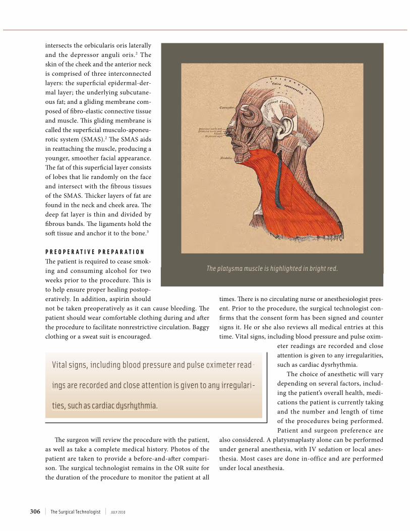
intersects the orbicularis oris laterally and the depressor anguli oris.2 The skin of the cheek and the anterior neck is comprised of three interconnected layers: the superficial epidermal-der-mal layer; the underlying subcutane-ous fat; and a gliding membrane com-posed of fibro-elastic connective tissue and muscle. This gliding membrane is called the superficial musculo-aponeu-rotic system (SMAS).2 The SMAS aids in reattaching the muscle, producing a younger, smoother facial appearance. The fat of this superficial layer consists of lobes that lie randomly on the face and intersect with the fibrous tissues of the SMAS. Thicker layers of fat are found in the neck and cheek area. The deep fat layer is thin and divided by fibrous bands. The ligaments hold the soft tissue and anchor it to the bone.3
P R E O P E R A T I V E P R E P A R A T I O NThe patient is required to cease smok-ing and consuming alcohol for two weeks prior to the procedure. This is to help ensure proper healing postop-eratively. In addition, aspirin should not be taken preoperatively as it can cause bleeding. The patient should wear comfortable clothing during and after the procedure to facilitate nonrestrictive circulation. Baggy clothing or a sweat suit is encouraged.
The surgeon will review the procedure with the patient, as well as take a complete medical history. Photos of the patient are taken to provide a before-and-after compari-son. The surgical technologist remains in the OR suite for the duration of the procedure to monitor the patient at all
VViittaall ssiiggnnss,, iinncclluuddiinngg bblloooodd pprreessssuurree aanndd ppuullssee ooxxiimmeetteerr rreeaadd-
iinngggss aarree rreeccoorrddeedd aanndd cclloossee aatttteennttiioonn iiss gggiivveenn ttoo aannyyy iirrrreeggguullaarrii-
ttiieess,, ssuucchh aass ccaarrddiiaacc ddyyssrrhhyytthhmmiiaa..
The platysma muscle is highlighted in bright red.
JULY 2010 | The Surgical Technologist | 307
The local anesthetic for this procedure is a tumescent solution: a combination of 400 ml normal saline, 90 ml one percent lidocaine without epinephrine, 10 ml 8.4 percent sodium bicarbonate and one ml of epinephrine 1:1000.
Preoperative antibiotics are also administered to prevent bacterial infections. Cephalexin is the primary antibiotic of choice as it can be used in patients with certain heart problems as a means to prevent coronary infection (bac-terial endocarditis). Cleocin may be used to fight bacterial infections in patients who are allergic to penicillin. Azithromycin—known as z-pack—may be used postoperatively. In most cases, a sedative is used. Five to 10 mg of diazepam is administered sub-lingually to treat anxiety in patients who request it. The sedative is used based on patient preference; however, the surgeon will dictate the dose to be administered.
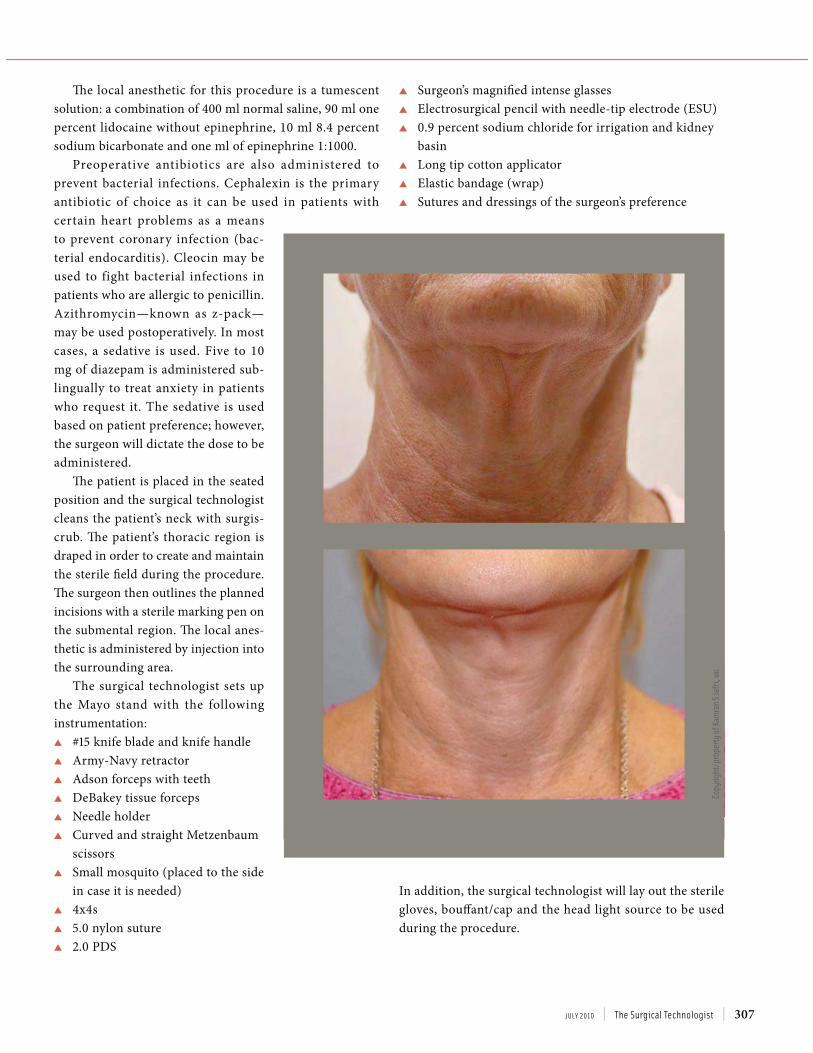
The patient is placed in the seated position and the surgical technologist cleans the patient’s neck with surgis-crub. The patient’s thoracic region is draped in order to create and maintain the sterile field during the procedure. The surgeon then outlines the planned incisions with a sterile marking pen on the submental region. The local anes-thetic is administered by injection into the surrounding area.
The surgical technologist sets up the Mayo stand with the following instrumentation:▲ #15 knife blade and knife handle▲ Army-Navy retractor▲ Adson forceps with teeth▲ DeBakey tissue forceps▲ Needle holder▲ Curved and straight Metzenbaum
scissors▲ Small mosquito (placed to the side
in case it is needed)▲ 4x4s▲ 5.0 nylon suture▲ 2.0 PDS
▲ Surgeon’s magnified intense glasses▲ Electrosurgical pencil with needle-tip electrode (ESU)▲ 0.9 percent sodium chloride for irrigation and kidney
basin▲ Long tip cotton applicator▲ Elastic bandage (wrap)▲ Sutures and dressings of the surgeon’s preference
In addition, the surgical technologist will lay out the sterile gloves, bouffant/cap and the head light source to be used during the procedure.
Cop
yrig
ht/p
rope
rty
of K
amra
n S
Jafr
i, M
D
| The Surgical Technologist | JULY 2010 308
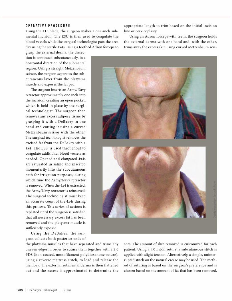
O P E R A T I V E P R O C E D U R EUsing the #15 blade, the surgeon makes a one-inch sub-mental incision. The ESU is then used to coagulate the blood vessels while the surgical technologist pats the area dry using the sterile 4x4s. Using a toothed Adson forceps to grasp the external derma, the dissec-tion is continued subcutaneously, in a horizontal direction of the submental region. Using a straight Metzenbaum scissor, the surgeon separates the sub-cutaneous layer from the platysma muscle and exposes the fat pad.
The surgeon inserts an Army/Navy retractor approximately one inch into the incision, creating an open pocket, which is held in place by the surgi-cal technologist. The surgeon then removes any excess adipose tissue by grasping it with a DeBakey in one hand and cutting it using a curved Metzenbaum scissor with the other. The surgical technologist removes the excised fat from the DeBakey with a 4x4. The ESU is used throughout to coagulate additional blood vessels as needed. Opened and elongated 4x4s are saturated in saline and inserted momentarily into the subcutaneous path for irrigation purposes, during which time the Army/Navy retractor is removed. When the 4x4 is extracted, the Army/Navy retractor is reinserted. The surgical technologist must keep an accurate count of the 4x4s during this process. This series of actions is repeated until the surgeon is satisfied that all necessary excess fat has been removed and the platysma muscle is sufficiently exposed.
Using the DeBakey, the sur-geon collects both posterior ends of the platysma muscles that have separated and trims any uneven edges in order to suture them together with a 2.0 PDS (non-coated, monofilament polydioxanone suture), using a reverse mattress stitch, to load and release the memory. The external submental derma is then flattened out and the excess is approximated to determine the
appropriate length to trim based on the initial incision line or cervicoplasty.
Using an Adson forceps with teeth, the surgeon holds the external derma with one hand and, with the other, trims away the excess skin using curved Metzenbaum scis-
sors. The amount of skin removed is customized for each patient. Using a 5.0 nylon suture, a subcutaneous stitch is applied with slight tension. Alternatively, a simple, uninter-rupted stitch on the natural crease may be used. The meth-od of suturing is based on the surgeon’s preference and is chosen based on the amount of fat that has been removed,
Cop
yrig
ht/p
rope
rty
of K
amra
n S
Jafr
i, M
D
JULY 2010 | The Surgical Technologist | 309
thus it will vary from patient to patient. The suture ends are cut short to prevent them from causing interference. The 4x4 with antibiotic ointment should not adhere to the stitches and snag the skin when wrapped with the elastic bandage wrap.3
Another suturing technique that may be used is the “corset platysmaplasty,” so named for its similarity in appearance to the “X” pattern of an old-fashioned corset when fully laced. In this alternative, the muscle is gathered and sutured with a snug jaw line and a right-angled neck contour. This produces a successful restructuring of the platysma muscle at the midline.4
Once the procedure is completed, the patient’s vital signs are taken and recorded again. Prescriptions for anti-biotics, which are mandatory, and any necessary pain relievers are noted. Many patients’ pain-relieving needs can be met with over-the-counter extra strength acetaminophen. A follow-up appointment is scheduled for the following week, and the patient is provided with written postoperative instructions. The surgeon will call the patient to follow up, but the patient is also instructed to call the surgeon with any concerns or to report any changes in their condition.
P O S T O P E R A T I V E C A R EAn elastic bandage should be wrapped around the head and neck for the first 24 hours postoperatively, and patients should be monitored by a friend or family member during this time. The bandage should be snug and comfortable, but not tightly wrapped. At the patient’s discretion, it may be worn for five days following the surgery for support, how-ever, it should be removed to bathe and eat. Patients are advised to keep ice packs on the surgical area every three to four hours for the first 48-72 hours after surgery. The patent’s head must be elevated while sleeping to minimize edema, and it is suggested that patients sleep with two pil-lows to ensure this is maintained. No strenuous exercise is allowed, although short walks are acceptable. The patent’s diet must also be monitored as a restricted salt intake will reduce facial swelling. In addition, patients are encouraged to eat foods that are enriched with vitamin C, zinc and iron to promote healing.
Mild swelling in the surgical area may be present for up to six months postoperatively. Patients must also use a ther-
mometer to monitor increases in temperature, which may be an early sign of a possible surgical site infection. During this time, the patient should cleanse the surgical site using sterile gauze and a 50-50 mixture of hydrogen peroxide and water. Antibacterial ointment should be applied to the inci-sion site after each cleansing.
All antibiotics must be finished as prescribed, and aspi-rin, ibuprofen, vitamin E and fish oil supplements are not allowed during the first week following surgery to help pre-vent bleeding.
P O S T O P E R A T I V E R I S K S A N D C O M P L I C A T I O N SSwelling, bruising and mild discomfort are common and should be expected in the week following a procedure. Infections, seroma, bleeding and hematoma, while not normally considered serious, may occur and should be monitored carefully. If a hematoma is detected—generally indicated by excessive swelling within 12 hours of the pro-cedure’s completion—the patient must return to the sur-geon’s office to have it drained with a needle in a sterile setting. Left untreated, it may become infected and possi-bly cause necrosis of the derma around the surgical site or enter the blood stream, leading to more complications.6 If the incision site acquires a green or yellow coloration, it could signify a possible infection or that blood circulation to the area is impaired.
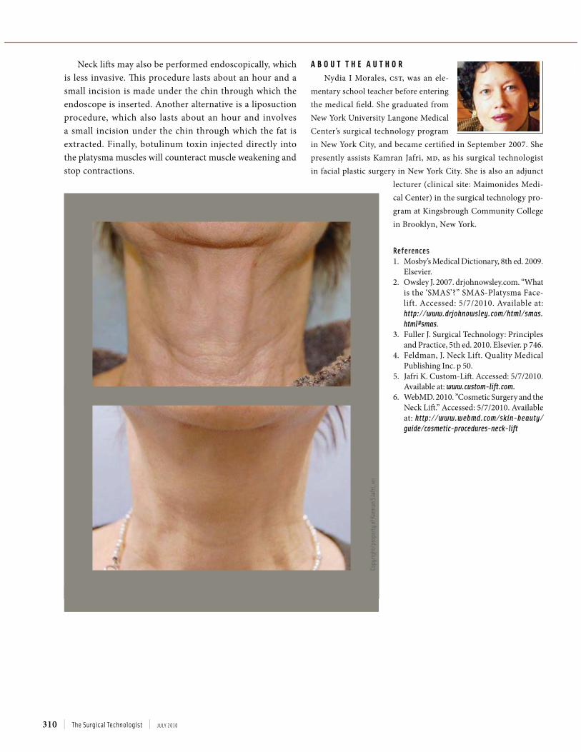
Follow-up visits are scheduled for one week post-pro-cedure, two weeks post-procedure and four weeks post-procedure. At the final visit, another set of photographs are taken to compare to the pre-surgical images.
C O N C L U S I O NThe end result of a successful platysmaplasty is a refined, smooth neck that enhances the patient’s features for a more youthful appearance. It can be combined with a face lift, filler, chemical peel and botulinum toxin injections.
TThhe bbanddage shhoulldd bbe snug andd comffortabblle, bbut not tiighhtlly
wrapped. At the patient’s discretion, it may be worn for five days
following the surgery for support, however, it should be removed
ttoo bbaatthhee aanndd eeaatt.
| The Surgical Technologist | JULY 2010 310
Neck lifts may also be performed endoscopically, which is less invasive. This procedure lasts about an hour and a small incision is made under the chin through which the endoscope is inserted. Another alternative is a liposuction procedure, which also lasts about an hour and involves a small incision under the chin through which the fat is extracted. Finally, botulinum toxin injected directly into the platysma muscles will counteract muscle weakening and stop contractions.
A B O U T T H E A U T H O RNydia I Morales, cst, was an ele-
mentary school teacher before entering the medical field. She graduated from New York University Langone Medical Center’s surgical technology program in New York City, and became certified in September 2007. She presently assists Kamran Jafri, MD, as his surgical technologist in facial plastic surgery in New York City. She is also an adjunct
lecturer (clinical site: Maimonides Medi-cal Center) in the surgical technology pro-gram at Kingsbrough Community College in Brooklyn, New York.
References1. Mosby’s Medical Dictionary, 8th ed. 2009.
Elsevier.2. Owsley J. 2007. drjohnowsley.com. “What
is the ‘SMAS’?” SMAS-Platysma Face-lift. Accessed: 5/7/2010. Available at: http://www.drjohnowsley.com/html/smas.html#smas.
3. Fuller J. Surgical Technology: Principles and Practice, 5th ed. 2010. Elsevier. p 746.
4. Feldman, J. Neck Lift. Quality Medical Publishing Inc. p 50.
5. Jafri K. Custom-Lift. Accessed: 5/7/2010. Available at: www.custom-lift.com.
6. WebMD. 2010. ”Cosmetic Surgery and the Neck Lift.” Accessed: 5/7/2010. Available at: http://www.webmd.com/skin-beauty/guide/cosmetic-procedures-neck-lift
Copy
righ
t/pr
oper
ty o
f Kam
ran
S Ja
fri,
MD