Embed Size (px)
Citation preview

47474747
CHAPTER
3
STUDENT LEARNING OUTCOMES
After studying this chapter, the student will be able to:
1. Give clear verbal instructions to an ambulatory
patient concerning the correct manner of dressing
and undressing for a radiographic procedure.
2. Correctly assess a patient’s need for assistance
to complete a radiographic procedure safely.
3. Demonstrate the correct method of moving and
positioning a patient to prevent injury to the
patient or the radiographer.4. Demonstrate the correct method of assisting a
disabled patient with dressing or undressing for a
radiographic procedure.
5. List the safety measures that must be taken
when transferring a patient from a hospital
room to the radiographic imaging department.
6. Describe steps that must be taken as the
radiographer to protect the patient’s
integumentary system from injury.
7. Explain the criteria to be used when
immobilization of a patient is necessary.
8. List the types of immobilizers available, and demon-
strate the correct method of applying each one.
9. List the precautions to be taken if a patient is in
traction or wearing a cast.
10. Demonstrate the correct manner of assisting a
patient with a bedpan or urinal.
11. Explain the responsibilities of a radiographer
concerning radiation safety.
12. List the departmental safety measures that must
be taken to prevent and control fires, patient
falls, poisoning or injury from hazardous
materials, and burns as well as the measures to
evacuate patients in case of a disaster.
KEY TERMS
Ambulatory: Walking, or able to walk
Atrophy: Decrease in the size of the organ, tissue, or muscle
Decubitus ulcer: A pressure sore or ulcer
Dyspnea: Labored or difficult breathing
Immobilizer: Velcro straps that are used on a patient’slimbs or waist to prevent a patient from injuring him orherself or others
Ischemia: Deficiency of blood in a body part due tofunctional constriction or actual obstruction of a bloodvessel
Tissue necrosis: Localized death of tissue due to injury orlack of oxygen
Ulceration: An area of tissue necrosis that penetrates belowthe epidermis; excavation of the surface of any bodyorgan
Patient Care and Safety
Patient Care and Safety
LWBK129-C03_47-70.qxd 11/6/08 11:22 PM Page 47 Aptara Inc.

48 Patient Care in Imaging Technology
A pproaching the profession and patients in a cour-teous and tactful manner can put the patients at
ease and decrease their level of embarrassment so thatthe procedure can be performed in a smooth and timelymanner. The radiographer sets the tone for the entireexchange when a patient arrives as an outpatient. Theradiographer is responsible for protecting him of her-self and the patient from injury in every way possible.Health care workers are often injured while movingand lifting patients, but almost all of these injuries arepreventable if the correct body mechanics and rules ofsafety are used. Patients are also victims of injuriescaused by being improperly moved or lifted. Most ofthese injuries can also be prevented.
Moving patients from the radiographic table to agurney or wheelchair, or from a hospital bed to a gur-ney or wheelchair, requires some forethought regard-ing the safety of the patient as well as to the bodymechanics used. Special care with the ancillary equip-ment must be taken when moving it with a patient dur-ing transport. A patient’s integumentary system mustbe protected from damage. This is of particular con-cern when the patient is unable to move by his or herown power.
Occasionally, a patient may have to be immobilizedfor his or her own safety during a radiographic proce-dure. Not only must the institution-specific rules con-cerning immobilizers be learned, but also the correctuse of these devices must be carefully learned to pro-tect the patient from harm.
The need for a bedpan or urinal may be a require-ment that a patient may find embarrassing butunavoidable. As a professional, the radiographer willbe able to put the patient at ease and proceed with tactand confidence that will facilitate the procedure to aswift conclusion. The different styles of bedpansrequire some knowledge as to the correct placementunder the patient. An understanding of how a bedpanfeels underneath a patient and the embarrassment thatthe patient experiences will help the radiographerempathize with the patient.
Adhering to rules of radiation safety, preventingand controlling fires, using and disposing of hazardouschemicals correctly, and observing other rules ofpatient and departmental safety are important parts ofthe radiographer’s education.
CARE OF PATIENT’SBELONGINGS
A patient who comes to the radiology department as anoutpatient is frequently required to remove all or someitems of clothing and to put on a patient gown beforean examination procedure or treatment can be per-formed. It is usually the radiographer who receives the
patient and determines which items of clothing are tobe removed. The patient’s discomfort or embarrass-ment can be decreased if the situation is approached ina courteous and professional manner.
The patient should be taken to the specific dressingarea and shown how to close the dressing room door ordraw the curtain of the cubicle while undressing. Clearlyexplain that he or she is to put on the examining gownand point out where to go for the examination once pre-pared. (Remember that not everyone knows that sometypes of examining gowns open at the back rather thanat the front; this information should be part of the expla-nation.) Doing this takes only a few moments, and it willmake the patient feel more comfortable.
The patient should be given hangers for clothing. Ifit is permissible to leave clothing in the dressing room,explain this to the patient. If the patient cannot leavethe clothing, show him or her what to do with it.Purses, jewelry, and other valuables should be treatedwith special care so that they will not be lost or stolen.
Many patients wear jewelry or carry a purse orother valuable items to the radiology department. Thedressing rooms in most departments are not safe placesto leave these items, and the patient may feel justifiablyuneasy about leaving them there. Again, consider thepatient’s concern and explain what must be done withpersonal items to keep them safe.
Metal items such as necklaces, rings, and watchesare not to be worn for many diagnostic procedures andmust be removed before the procedure can begin. Anenvelope or other container large enough to accommo-date all such items should be offered to the patient.Identifying information should be written on a receipt,and all items should be tagged and placed in the desig-nated safety area. This procedure will prevent lossesthat may result in inconvenience and expense to boththe patient and the department.
Do not place value on a patient’s belongings. Anitem that may seem insignificant to others may be thepatient’s most treasured belonging. Every article ofclothing or jewelry and the personal effects that apatient brings to the diagnostic imaging departmentshould be treated with care.
BODY MECHANICS
Constant abuse of the spine from moving and liftingpatients is the leading cause of injury to health carepersonnel in all health care institutions. Following thecorrect rules of body mechanics will reduce the amountof fatigue and chance of injury. Rules of body mechan-ics are based on the laws of gravity.
Gravity is the force that pulls objects toward thecenter of the earth. Any movement requires an expen-diture of energy to overcome the force of gravity. When
LWBK129-C03_47-70.qxd 11/6/08 11:22 PM Page 48 Aptara Inc.

an object is balanced, it is firm and stable. If it is offbalance, it will fall because of the pull of gravity. Thecenter of gravity is the point at which the mass of anybody is centered. When a person is standing, the centerof gravity is at the center of the pelvis.
Safe body mechanics require good posture. Goodposture means that the body is in alignment with all theparts in balance. This permits the musculoskeletal sys-tem (the bones and joints) to work at maximal effi-ciency with minimal amount of strain on joints, ten-dons, ligaments, and muscles. Good posture also aidsother body systems to work efficiently. For instance, ifthe chest is held up and out (the musculoskeletal sys-tem), then the lungs (the respiratory system) can workat maximal efficiency.
Rules for correct upright posture are as follows:
• Hold chest up and slightly forward with thewaist extended. This allows the lungs to expandproperly and fill to capacity.
• Hold head erect with the chin held in. This putsthe spine in proper alignment, and there is nocurve in the neck.
• Stand with the feet parallel and at right anglesto the lower legs. The feet should be 4 to 8inches apart. Keep body weight equally distrib-uted on both feet.
• Keep the knees slightly bent; they act as shockabsorbers for the body.
• Keep the buttocks in and the abdomen up andin. This prevents strain on the back and abdom-inal muscles.
The forces of weight and friction must be overcomewhen moving and lifting objects. Keep the heaviestpart of the object close to the body. If this is not possi-ble, one or more persons should assist with moving orlifting the load.
The force of friction opposes movement. Whenmoving or transferring a patient, reduce friction to theminimum to facilitate movement. This can be done byreducing the surface area to be moved or, in the case ofa patient, by using some of the patient’s own strengthto assist with the move, if possible. If the patient isunable to assist, reduce friction by placing the patient’sarms across the chest to reduce the surface area. Thesurface over which the patient must be moved must bedry and smooth. Pulling rather than pushing alsoreduces friction when moving a heavy object or person.A sliding board or pull sheet placed under an immobilepatient also reduces friction. Directions for the use ofthese items are presented later in this chapter.
To avoid self-injury when moving heavy objects,remember to keep the body’s line of balance closest tothe center of the load. Rules for picking up or liftingheavy objects are:
• When picking up an object from the floor, bendthe knees and lower the body. Do not bend fromthe waist (Fig. 3-1)
• The biceps are the strongest arm muscles andare effective when pulling; therefore, pull heavyitems or patients rather than push them.
• When assisting a patient to move, balance theweight over both feet. Stand close to the patient,flex the gluteal muscles, and bend the knees tosupport the load. Use arm and leg muscles toassist in the move.
• Always protect the spine. Rather than twistingthe body to move a load, change the foot posi-tion instead. Always keep the body balancedover the feet, which should be spread to providea firm base of support.
• Make certain the floor area is clear of all objects.
CHAPTER 3: Patient Care and Safety 49
CALL OUT!
To prevent lower back injury, always keep the center
of gravity, the knees flexed, and the weight over both
feet. Do not bend at the waist or twist with the body.
MOVING AND TRANSFERRINGPATIENTS
The radiographer may be called upon to transfer orassist in transferring a patient from a hospital room tothe diagnostic imaging department. Several precautionsmust be taken when moving a patient from the hospitalroom to the imaging department. They are as follows:
1. Establish the correct identity of the patient.Approach the patient and identify yourself and thereason for being there. Ask to see their identificationwristband. This is extremely important, as manytimes the patient has been transferred to anotherroom since the radiology request was submitted.
2. Request pertinent information concerning thepatient’s ability to comply with the physical demandsof the procedure while at the nurses’ station.
3. Request information concerning the patient’s abil-ity to ambulate and any restriction or precautionsto be taken concerning the patient’s mobility.
4. Move the patient to the imaging department accord-ing to the necessary restrictions after greeting andidentifying him or her and providing an explana-tion of what is to occur.
CALL OUT!
Never move a patient without enough assistance to
prevent injury to yourself and/or the patient.
LWBK129-C03_47-70.qxd 11/6/08 11:22 PM Page 49 Aptara Inc.

When the procedure is completed, return the patient tothe hospital room using the following procedure:
1. Stop at the appropriate nurses’ station, return thechart, and inform the unit personnel that thepatient is being returned to the room. Request helpif it is needed at this time.
2. Return the patient to the room, help the patient getinto bed, and make him or her comfortable andsafe. Place the patient’s bed in the position that isclosest to the floor with the side rails raised and thecall button within reach in case the patient needsassistance.
50 Patient Care in Imaging Technology
Incorrect Correct
FIGURE 3-1 Keep the body balanced over the feet to provide
a broad base of support.
CALL OUT!
Never move a patient without assessing the patient’s
ability to assist.
WARNING!
To prevent possible patient injury, always lower the bed to
the lowest position, and secure the rails in the upright posi-
tion when a patient is returned to bed.
!
Assessing the Patient’s Mobility
Before beginning to move a patient, critical thinking andproblem-solving skills must be used to plan the mosteffective manner of accomplishing the task. The expectedoutcome of this plan will be to accomplish the movewithout causing additional pain or injury to the patient.Use interviewing and assessment skills to complete this.
Look for the following during patient assessment:
1. Deviations from correct body alignment. Deviationsin normal physiologic body alignment may resultfrom the following: poor posture, trauma, muscledamage, dysfunction of the nervous system, malnu-trition, fatigue, or emotional disturbance. Supportblocks or pillows, which are used to assist thepatient during the procedure, must be available.
2. Immobility or limitations in range of joint motion.Any stiffness, instability, swelling, inflammation,pain, limitation of movement, or atrophy of musclemass surrounding each joint must be noted andconsidered in the plan of care.
3. The ability to walk. Gait includes rhythm, speed,cadence, and any characteristic of walking that mayresult in a problem with balance, posture, or inde-pendence of movement. Before beginning themove, the amount of assistance needed to safelycomplete the move and procedure must be planned.
LWBK129-C03_47-70.qxd 11/6/08 11:22 PM Page 50 Aptara Inc.
4. Respiratory, cardiovascular, metabolic, and mus-culoskeletal problems. Obvious respiratory or car-diovascular symptoms that impair circulation andsignal potential problems in positioning must beplanned for. Metabolic problems such as diabetesmellitus or rheumatoid arthritis may be discoveredduring the interview process and planned for asnecessary (symptoms and care of patients withmedical problems are discussed in Chapter 8).
Other assessment considerations are:
1. The patient’s general condition. How well or howpoorly is he or she functioning?
2. Range of motion and weight-bearing ability. Hasthe patient had a surgical procedure that restrictsmotion or limits weight bearing until it is healed?
3. The patient’s strength and endurance. Will thepatient become fatigued and be unable to completethe transfer with only stand-by assistance?
4. The patient’s ability to maintain balance. Can thepatient sit or stand for as long as the procedurerequires?
5. The patient’s ability to understand what is expectedduring the transfer. Is he or she responsive andalert?
6. The patient’s acceptance of the move. Does thepatient fear or resent the transfer? Will the trans-fer increase the pain? Does the patient feel that themove is unnecessary?
7. The patient’s medication history. Has the patientreceived a sedative, hypnotic, or other psychoac-tive drug in the past 2 or 3 hours? Will any med-ication that he or she has taken affect the ability tomove safely?
Before going to the patient, a consultation with thenurse in charge of the patient is recommended so thatthe patient’s condition and limitations can be under-stood. If assistants are needed, they must be on hand.A patient must never be moved without adequate assis-tance; to do so may cause injury to the patient or theradiographer. The radiographer must decide how thepatient can be transferred safely and comfortably,whether by gurney or by wheelchair. Hospital patientsare seldom allowed to walk to and from the diagnosticimaging department for reasons of safety. Someonemust always be at the patient’s side as he or she moves.The following rules should be observed during a move:
1. Give only the assistance that the patient needs forcomfort and safety.
2. Always transfer a patient across the shortest dis-tance.
3. Lock all wheels on beds, gurneys, and wheelchairsbefore the move begins.
4. Generally, it is better to move a patient toward hisor her stronger side while assisting on the patient’sweaker side.
5. The patient should wear shoes for standing trans-fers, not slippery socks.
6. Inform the patient of the plan for moving andencourage him or her to help.
7. Give the patient short, simple commands and helpthe patient to accomplish the move.
Methods of Moving Patients
There are essentially three ways of transferringpatients: by gurney, by wheelchair, and by ambulation.
By Gurney
When a patient is moved from a gurney to a radi-ographic table, or the reverse, great care must betaken to prevent injury. If the patient is unconsciousor unable to cooperate in the move, the patient’sspine, head, and extremities must be well supported.Convenient and safe ways to do this are by using asliding board or a sheet to slide the patient from onesurface to another.
Sheet Transfer
To place a sheet under a patient, use a heavy drawsheet or a full bed sheet that is folded in half. Have oneperson stand on each side of the table or bed at thepatient’s side. Turn the patient onto his or her sidetoward the distal side of the bed or table. Place thesheet on the table or bed with the fold against thepatient’s back (Fig. 3-2A). Roll the top half of thesheet as close to the patient’s back as possible (Fig. 3-2B). Inform the patient that he or she will be turnedonto the side toward the opposite side and will bemoving over the rolled sheet. Then turn the patientacross the sheet roll and have the assistant straightenthe sheet on the distal side (Fig. 3-2C). Return thepatient to a supine position, and the transfer maybegin.
If the patient is an adult, three or four peopleshould participate in the maneuver. One person standsat the patient’s head to guide and support it during themove, with another at the side of the surface to whichthe patient will be moved, and a third person at the sideof the surface on which the patient is lying. If there arefour people, two may stand at each side. The sheet isrolled at the side of the patient so that it can easily begrasped, close to the patient’s body. In unison (usuallyon the count of three), the team transfers the patient tothe other surface. Extra care over the metal parts of theradiographic table’s metal edges should be taken aswell as assuring that the tube housing is positioned outof the way.
CHAPTER 3: Patient Care and Safety 51
LWBK129-C03_47-70.qxd 11/6/08 11:22 PM Page 51 Aptara Inc.

Sliding Board Transfer
The sliding board (also called a smooth mover and a“smoothie”) is a glossy, plasticized board approxi-mately 5 feet 10 inches in length and about 2 feet 6inches wide. This item facilitates moving patients fromone surface to another, usually from a gurney to anexamining table. The sliding board usually requiresfewer personnel to make the move than the sheet trans-fer because it creates a firm bridge between the twosurfaces over which the patient can be easily moved.The sliding board transfer procedure is as follows:
1. Obtain the sliding board and spray it with antista-tic spray if necessary.
2. Obtain the assistance of one other person if thepatient is of average size and weight; if the patientis large, three people may be necessary to move thepatient safely.
3. Move the patient to the edge of the gurney. Oneperson should hold the sheet that the patient is lay-ing on over the top of the patient to keep thepatient from possibly rolling off the gurney.
4. Move the gurney up against the radiographic tableand lock the wheels of the gurney.
5. Assist the patient to turn onto his or her side, awayfrom the radiographic table, and place the slidingboard under the sheet upon which the patient waslying.
6. Create a bridge with the board between the edge ofthe radiographic table and the edge of the gurney(Fig. 3-3A).
7. Place the sheet over the board, and allow thepatient to roll back onto the board.
8. With one person at the side of the radiographictable and the other at the side of the gurney, slidethe patient over the board and onto the radi-ographic table (Fig. 3-3B).
9. Assist the patient to roll toward the distal side ofthe radiographic table, keeping the patient secureby holding onto the sheet on which he or she waslying. The person standing on the side of the gur-ney should remove the sliding board from underthe patient (Fig. 3-3C).
10. Remove the gurney and perform the radiographicprocedure.
11. When the procedure is completed, the patient canbe transferred back to the gurney by repeating thesteps above.
52 Patient Care in Imaging Technology
A B
C
FIGURE 3-2 (A) Place the sheet on the table with the fold
against the patient’s back. (B) Take the top half of the sheet and
roll it against the patient. (C) After the patient is rolled to the
opposite side, the rolled half of the sheet is straightened out.
LWBK129-C03_47-70.qxd 11/6/08 11:22 PM Page 52 Aptara Inc.

CHAPTER 3: Patient Care and Safety 53
A B
C
FIGURE 3-3 (A) Create a bridge with the board between the
table and the gurney. (B) Roll the sheet close to the patient and
slide the patient onto the table. (C) Remove the sliding board
while safely securing the patient.
12. Once the patient is back on the gurney, place a pil-low under the patient’s head, if this is permitted,and put the side rails of the gurney up. Place a softimmobilizer over the patient. The patient may thenbe transferred.
13. When the move is complete, discard the soiledlinen that was used on the radiographic table, andclean the sliding board and the table with a disin-fectant spray.
14. After washing hands, place clean linen on thetable.
physician in charge of the patient’s care and applied incompliance with institutional policy.
The Joint Commission states that immobilizersshould be used only after less restrictive measureshave been attempted and have proved ineffective inprotecting the patient. Remember this and use criticalthinking skills to avoid the use of immobilizers if at allpossible. Immobilizers are defined as any manualmethod or physical or mechanical device, material, orequipment attached or adjacent to the person’s bodythat the person cannot remove easily that restrictsfreedom of movement or normal access to one’s body(Omnibus Reconciliation Act, 1989).
The most effective method of avoiding the need torestrain an adult patient is the use of therapeutic com-munication to explore the patient’s fears. If a patientseems fearful or is striking out or moving in an unsafemanner, assure the patient that the procedure will becarried out quickly and in a manner that keeps him orher as comfortable as possible. If this does not reassurethe patient, other less restrictive devices, such as soft,Velcro straps (Fig. 3-5A), sandbags (Fig. 3-5B and 3-5C),and sponges may be used to remind the patient torefrain from moving. If immobilizers are to be used, be
CALL OUT!
Always obtain enough assistance to move a patient,
even with a smooth mover. This is for the safety of
both the patient and the radiographer.
USE OF IMMOBILIZERS
The ethical and legal restrictions concerning use ofimmobilizers (often called restraints) in patient careare discussed in Chapter 1. The radiographer mustremember that immobilizers must be ordered by the
LWBK129-C03_47-70.qxd 11/6/08 11:22 PM Page 53 Aptara Inc.

54 Patient Care in Imaging Technology
PROCEDURE
Wheelchair Transfer
6. The footrests on the wheelchair should then be put
down and the wheels unlocked. A safety belt should
be put across an unsteady patient.
If a patient must be moved from a bed or radiographic
table to a wheelchair, or the reverse, he or she must be
helped. Never allow a patient to get off a table or onto a
wheelchair without some assistance. The patient is often
not as strong as he or she thinks. The sudden movement
may cause dizziness, and the patient may fall.
If the patient has been in a supine position and is to be
helped to a sitting position, have the patient turn to the side
with knees flexed. Then stand in front of the patient with
one arm under the shoulder and the other across the knees.
1. If the patient can assist, instruct him or her to push up
with the upper arm when told to do so (Fig. 3-4A).
2. On the count of three,move or help the patient to a sit-
ting position at the edge of the table. Before helping the
patient to stand, allow him or her to sit for a moment
and regain a sense of balance.While the patient is “dan-
gling,” place nonskid slippers on the patient’s feet.
3. If the patient needs minimal assistance to get off the
table, stand at the patient’s side and take the patient’s
arm to help.
4. If the radiographic table is high, never allow a patient
to step down without providing a secure stepping
stool.Always stay at the patient’s side to assist.A tele-
scoping radiographic table must be placed in the
lower position before a patient is assisted to move
off of it.
5. The wheelchair must be close enough so that the
patient can be seated in the chair with one pivot (Fig.
3-4B). Have the foot supports of the chair up and the
wheels locked.
A B
FIGURE 3-4 (A) Stand in from of the patient and place an arm under her shoulders and over across her knees. Assist to a
sitting position. (B) Wheel the wheelchair close to the table pivot and help the patient sit in the chair.
CALL OUT!
When moving a patient from hospital bed to wheel-
chair, always place nonskid slippers on the patient’s
feet, provide assistance to prevent falls, and secure
the seatbelt on the wheelchair.
WARNING!
Before allowing a patient to get out of a wheelchair, raise
the foot supports out of the way. Many patients step on
these, causing the wheelchair to flip over, which causes
injury to the patient!
!
Once placed on the radiographic table, cover the patient
with a protective sheet. Do not allow the patient to
become chilled.
A patient who has received a narcotic, hypnotic, or
other type of psychoactive medication; a confused, disori-
ented, unconscious, or head-injured person; or a child
must never be left alone on a radiographic table or gur-
ney. If the patient’s behavior cannot be predicted or if the
patient is in a wheelchair ; observe him or her carefully. A
soft immobilizer belt should be placed over any patient on
a gurney or in a wheelchair. The side rails of the gurney
must always be up.
LWBK129-C03_47-70.qxd 11/6/08 11:22 PM Page 54 Aptara Inc.

CHAPTER 3: Patient Care and Safety 55
certain that they are being used to protect the patient’ssafety and that their use is the only alternative.
There are various types of immobilizing devicesthat may be used for adult patients. Immobilizers foruse with children are discussed in Chapter 10. Reasonsfor application of immobilizers in the care of an adultpatient include the following:
1. To control movement of an extremity when an intra-venous infusion or diagnostic catheter is in place
2. To remind a patient who is sedated and havingdifficulty remembering to remain in a particularposition
3. To prevent a patient who is unconscious, delirious,cognitively impaired, or confused from falling froma radiographic table or a gurney; from removing atube or dressing that may be life sustaining; orfrom injuring him or herself by impact with diag-nostic imaging equipment
When caring for a patient who has been immobilized,explain the reason for using immobilizers to the patientand to anyone who may accompany the patient. Afterimmobilizers are applied, do not leave the patient unat-tended, and inform the patient that he or she is notalone and is not being punished. Also explain that the
immobilizers are only temporary and that as soon as theprocedure is finished, the immobilizer will be removed.
A calm, reassuring manner often soothes an agitatedor confused patient who has been immobilized. A patientin this state needs repeated orientation as well as a quietand quick explanation to complete the procedure. Returnthe patient to the hospital room or wherever he or she isto be taken on completion of the radiographs.
Always apply immobilizers carefully and in themanner prescribed by the manufacturer of the device.The type of immobilizer to be used is dictated by need.As explained in Chapter 1, all radiographers must doc-ument application of immobilizers. The following arerules for application of immobilizers:
1. The patient must be allowed as much mobility as issafely possible.
2. The areas of the body where immobilizers areapplied must be padded to prevent injury to theskin beneath the device.
3. Normal anatomic position must be maintained.4. Knots that will not become tighter with movement
must be used (a half-knot is recommended; Fig. 3-6).5. The immobilizer must be easy to remove quickly, if
this is necessary.
A B
C
FIGURE 3-5 (A) A Velcro strap being placed snuggly but not
tightly. (B) Sandbags will help remind the adult patient to
remain still. (C) Sandbags will also assist in immobilizing the
infant while holding the shield in place.
LWBK129-C03_47-70.qxd 11/6/08 11:22 PM Page 55 Aptara Inc.

6. Neither circulation nor respiration must beimpaired by the immobilizer.
7. If leg immobilizers are necessary, wrist immobiliz-ers must also be applied to prevent the patientfrom either unfastening the device or, in anattempt to leave the radiographic table or gurney,accidentally hanging him or herself.
Although the radiographer is not usually the healthcare worker who monitors the patient in immobilizersfor long periods of time, it should be known thatimmobilizers may need to be removed and the jointsaffected by the immobilizer be put through range ofmotion exercises. Only one immobilizer at a timeshould be released, and then retied prior to releasing asecond immobilizer. Always tie the immobilizer to astationary object such as the gurney frame or side ofthe radiographic table (if possible).
There are various types of immobilizers that maybe used, including the following:
1. Limb holders or four-point restrains (Fig. 3-7A)2. Ankle or wrist immobilizers (Fig. 3-7B)3. Immobilizing vest for keeping a patient in a wheel-
chair4. Waist immobilizer, which keeps the patient safe on
an examining table or in a bed, but allows thepatient to change position (Fig. 3-7C)
56 Patient Care in Imaging Technology
Pull here to untie
Springs
Frame
Pull to tightenMattress
Immobilizer tie
To immobilizer
FIGURE 3-6 Following this sequence will result in tying a half-
bow knot. The knot will remain secure until the free end is
pulled.
FIGURE 3-7 (A) Limb holders (four-point restraints). (B)
Wrist immobilizer. (C) Waist immobilizer.
A
C
B
LWBK129-C03_47-70.qxd 11/6/08 11:22 PM Page 56 Aptara Inc.

CHAPTER 3: Patient Care and Safety 57
At times, a patient who is aggressive and delusional mayneed to have waist as well as four-point immobilizersthat are stronger than those shown in Figures 3-7A andB. Immobilizers of this type also have locks. If this typeof immobilizer is necessary, one or more security officersshould be called to assist with application. A radio-grapher, with only the assistance of one other radiogra-pher, must never attempt to apply immobilizers to anextremely aggressive or combative patient. Persons whohave been trained to deal with this type of patient prob-lem must do this. When this type of immobilization isnecessary, the rules of immobilizer application still apply.
POSITIONING THE PATIENT FORDIAGNOSTIC IMAGINGEXAMINATIONS
When a patient must spend a long period of time in thediagnostic imaging department, it is the radiographer’sduty to assist the patient to maintain his or her body innormal alignment for comfort and to maintain normalphysiologic functioning. There are several protectivepositions that the body may assume or be assisted toassume for comfort. There are also several positions thatthe patient may be requested to assume to facilitate diag-nosis or treatment. These positions are likely to be usedin the radiology department for various procedures:
Supine or dorsal recumbent position: Patient is flaton the back. The feet and the neck will need tobe protected when the patient is lying in thisposition. A pillow may be placed under thehead to tilt it forward. The feet should be sup-ported to prevent plantar flexion or footdrop(Fig. 3-8A).
Lateral recumbent position: Patient is on the rightor the left side with both knees flexed. Thisposition relieves pressure on most bonyprominences. The patient may be supportedwith pillows or sandbags to maintain the posi-tion (Fig. 3-8B).
Prone position: Patient lies face down. A small pillowshould support the head to prevent flexion of thecervical spine. The patient maybe moved downon the table so that the feet drop over the edge,or a pillow may be placed under the lower legsat the ankles to prevent footdrop (Fig. 3-8C).
High Fowler position: Patient semi-sits with headraised at an angle of 45 to 90 degrees off thetable. This position is used for patient in res-piratory distress (Fig. 3-8D).
Semi-Fowler position: Patient’s head is raised at anangle of 15 to 30 degrees off the table. Thearms must be supported to prevent pull on theshoulders, and the feet must be supported to
prevent plantar flexion or footdrop. Pillows orblocks under knees must be removed after abrief time (15 to 20 minutes) to prevent circu-latory impairment (Fig. 3-8E).
Sims position: Patient lies on either left or rightside with the forward arm flexed and the pos-terior arm extended behind the body. The bodyis inclined slightly forward with the top kneebent sharply and the bottom knee slightly bent.This position is frequently used for diagnosticimaging of the lower bowel as an aid in insert-ing the enema tip (Fig. 3-8F).
Trendelenburg position: The table or bed is inclinedwith the patient’s head lower than the rest ofthe body. Patients are occasionally placed inthis position during diagnostic imaging proce-dures and for promotion of venous return inpatients with inadequate peripheral perfusioncaused by disease (Fig. 3-8G).
Patients in respiratory distress or who have COPDmust not be left in a prone, supine, or Sims’ positionfor more than brief periods of time to avoid becomingincreasingly dyspneic.
ASSISTING THE PATIENT TODRESS AND UNDRESS
The patient may arrive in the diagnostic imagingdepartment alone if he or she comes from outside thehospital. The patient may need assistance in removingclothing. This may be necessary if the patient is in acast or a brace, is very young, or is in too weakened acondition to help him or herself. The patient mayhave a contracture of an extremity or poor eyesight.Whatever the problem, if the radiographer sensesthat the patient will have difficulty undressing if leftalone, then assistance should be offered and given asneeded.
If a trauma patient is brought to the diagnosticimaging department from the emergency unit, remov-ing the clothing in the conventional manner may causefurther injury or pain. It may be necessary to cut awaygarments that interfere with acceptable radiographs;however, clothing must not be cut without the patient’sconsent except in extreme emergencies. If the patient isunable to give consent, a family member should do soin writing for protection.
If clothing must be cut off, try to cut into a seam ifat all possible. The clothes should not be automaticallythrown in the trash. They should be offered to thepatient and placed with the patient’s other belongs.
If the patient is very young and is accompanied bya familiar adult, he or she will be more relaxed andcooperative if the adult helps him to dress and undress.Explain to the adult how the child should be dressed
LWBK129-C03_47-70.qxd 11/6/08 11:22 PM Page 57 Aptara Inc.

A
E F
B
G
C D
FIGURE 3-8 (A) Supine position. (B) Lateral position. (C)
Prone position. (D) High Fowler position. (E) Semi-Fowler
position. (F) Sims position. (G) Trendelenburg position.
58
LWBK129-C03_47-70.qxd 11/6/08 11:22 PM Page 58 Aptara Inc.

for the procedure, arrange a meeting place, and leavethem alone.
If a patient with a disability of the lower extremi-ties must have assistance, the clothing should beremoved from the top part of the body first.
1. Place a long examining gown on the patient.Instruct him to loosen belt buckles, buttons, orhooks around the waist and slip the trousers overthe hips. If the patient cannot do this, reach underthe gown and pull the trousers down over the hips.
2. Have the patient sit down. Squat down in front ofthe patient and gently pull the clothing over thelegs and feet to remove it. If the patient is not ableto help, call for an assistant.
Some dresses may be removed in the same way. If thismethod is not practical, however, and the dress must bepulled over the woman’s head, proceed as follows:
1. Place a draw sheet over the patient and then helpher to remove her slip and brassiere.
2. Help her to put on an examining gown and thenremove the draw sheet.
The following are steps to re-dress a patient with a par-alyzed leg, a leg injury, a cast, or a brace:
1. Slide the clothing (pants or skirt) over the feet orlegs as far as the hips while the patient is sittingand still wearing an examining gown.
2. Have the patient stand and pull the clothing overthe hips if he or she can tolerate it.
3. If the patient is not able to pull the clothing overthe hips alone, have an assistant raise the patientoff the chair so that you may slip the clothing overthe hips and waist.
4. Remove the patient’s arms from the sleeves of thegown. Have the patient hold the gown over his orher chest, and carefully pull the shirt over the head,or put it on one sleeve at a time.
5. When the outside items of clothing are on thepatient, remove the gown from under the clothes.
THE DISABLED PATIENT
If the patient is on a gurney or the radiographic tableand the patient’s clothing must be changed, this canmost easily be accomplished with the patient in thesupine position.
1. Cover the patient with a draw sheet and have anexamining gown ready. Explain what is to be doneand ask the patient to help if he or she is able. Ifthe patient is paralyzed or unconscious, summonhelp before beginning the procedure.
2. Remove the clothing from the less affected side firstand then remove the clothing from the moreaffected side and place the clean gown on that side,making sure to keep the patient covered with thedraw sheet.
3. Next place the clean gown on the unaffected sideand tie the gown at the back, if practical.
4. If the patient is wearing an article of clothing thatmust be pulled over the head, roll the garment upabove the waist. Then remove the garment upabove the waist. Next, remove the patient’s armsfrom the clothing, first from the unaffected sideand then from the affected side.
5. Neatly, gently lift the clothing over the patient’shead. One person alone should not attempt toundress a disabled patient; to do so may cause fur-ther injury or discomfort.
6. To remove trousers, loosen buckles and buttonsand have the patient raise his buttocks as thetrousers are slipped over his hips. If the patient isunable to help, have an assistant stand at theopposite side of the table. After the trousers havebeen loosened, have the assistant pull the patienttoward him or her, and then slide the trousers offone side of the hip. Next, draw the patient towardthe opposite side and have the assistant slide thetrousers off the other hip.
7. Slip the trousers below the knees and off.8. Fold the clothing and place it in a paper bag on
which the patient’s name has been printed. If a rel-ative or a friend accompanies the patient, ask thatperson to keep the patient’s clothing. If the patientis alone, the radiographer is responsible for caringfor the clothing.
When a patient’s gown becomes wet or soiled in theradiology department, it is the duty of the radiographerto change it. If a patient is allowed to remain in a wetor soiled gown, the skin may become damaged, or heor she may become chilled.
When changing the gown of a patient who has aninjury or is paralyzed on one side, remove the gownfrom the unaffected side first. Then, with the patientcovered by the soiled gown, place the clean gown firston the affected side and then on the unaffected side.Pull the soiled gown from under the clean one.
Always make sure that the patient is covered dur-ing the process.
CHAPTER 3: Patient Care and Safety 59
CALL OUT!
When changing a disabled patient’s gown, allow enough
material to work with by removing the unaffected side
first or by placing the gown on the affected side first.
LWBK129-C03_47-70.qxd 11/6/08 11:22 PM Page 59 Aptara Inc.
THE PATIENT WITH ANINTRAVENOUS INFUSION
Frequently, patients are taken to the diagnostic imagingdepartment with an IV infusion in place.
1. If the patient’s gown must be changed, slip theclothing off the unaffected side first.
2. Carefully slide the sleeve of the unaffected sideover the IV tubing and catheter, then over the con-tainer of fluid. For this step, the container must beremoved from the stand.
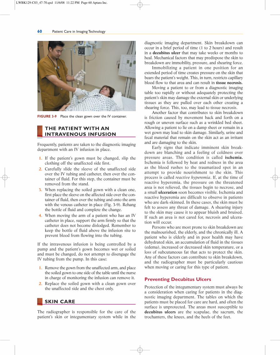
3. When replacing the soiled gown with a clean one,first place the sleeve on the affected side over the con-tainer of fluid, then over the tubing and onto the armwith the venous catheter in place (Fig. 3-9). Rehangthe bottle of fluid and complete the change.
4. When moving the arm of a patient who has an IVcatheter in place, support the arm firmly so that thecatheter does not become dislodged. Remember tokeep the bottle of fluid above the infusion site toprevent blood from flowing into the tubing.
If the intravenous infusion is being controlled by apump and the patient’s gown becomes wet or soiledand must be changed, do not attempt to disengage theIV tubing from the pump. In this case:
1. Remove the gown from the unaffected arm, and placethe soiled gown to one side of the table until the nursein charge of monitoring the infusion can remove it.
2. Replace the soiled gown with a clean gown overthe unaffected side and the chest only.
SKIN CARE
The radiographer is responsible for the care of thepatient’s skin or integumentary system while in the
diagnostic imaging department. Skin breakdown canoccur in a brief period of time (1 to 2 hours) and resultin a decubitus ulcer that may take weeks or months toheal. Mechanical factors that may predispose the skin tobreakdown are immobility, pressure, and shearing force.
Immobilizing a patient in one position for anextended period of time creates pressure on the skin thatbears the patient’s weight. This, in turn, restricts capillaryblood flow to that area and can result in tissue necrosis.
Moving a patient to or from a diagnostic imagingtable too rapidly or without adequately protecting thepatient’s skin may damage the external skin or underlyingtissues as they are pulled over each other creating ashearing force. This, too, may lead to tissue necrosis.
Another factor that contributes to skin breakdownis friction caused by movement back and forth on arough or uneven surface such as a wrinkled bed sheet.Allowing a patient to lie on a damp sheet or remain in awet gown may lead to skin damage. Similarly, urine andfecal material that remain on the skin act as an irritantand are damaging to the skin.
Early signs that indicate imminent skin break-down are blanching and a feeling of coldness overpressure areas. This condition is called ischemia.Ischemia is followed by heat and redness in the areaas the blood rushes to the traumatized spot in anattempt to provide nourishment to the skin. Thisprocess is called reactive hyperemia. If, at the time ofreactive hyperemia, the pressure on the threatenedarea is not relieved, the tissues begin to necrose, anda small ulceration soon becomes visible. Ischemia andreactive hyperemia are difficult to observe in patientswho are dark-skinned. In these cases, the skin must befelt to assess any threat of damage. A shearing injuryto the skin may cause it to appear bluish and bruised.If such an area is not cared for, necrosis and ulcera-tion will occur.
Persons who are most prone to skin breakdown arethe malnourished, the elderly, and the chronically ill. Apatient who is elderly and in poor health may havedehydrated skin, an accumulation of fluid in the tissues(edema), increased or decreased skin temperature, or aloss of subcutaneous fat that acts to protect the skin.Any of these factors can contribute to skin breakdown,and the radiographer must be particularly cautiouswhen moving or caring for this type of patient.
Preventing Decubitus Ulcers
Protection of the integumentary system must always bea consideration when caring for patients in the diag-nostic imaging department. The tables on which thepatients must be placed for care are hard, and often thesurface is unprotected. The areas most susceptible todecubitus ulcers are the scapulae, the sacrum, thetrochanters, the knees, and the heels of the feet.
60 Patient Care in Imaging Technology
FIGURE 3-9 Place the clean gown over the IV container.
LWBK129-C03_47-70.qxd 11/6/08 11:22 PM Page 60 Aptara Inc.

The patient who is on the imaging table for a longperiod of time should be allowed to change positionoccasionally to keep pressure off the hips, knees, andheels. This can be done by placing a pillow or softblanket under the patient or by turning him or her to adifferent position whenever possible. This is done inthe usual hospital situation every 2 hours. If the patientis lying on a hard surface, such as the radiographictable, it should be done every 30 minutes. If a patientis perspiring profusely or is incontinent of urine orfeces, make certain that he or she is kept clean and dry,and take precautions when moving the patient to pre-vent skin abrasions.
Special precautions should be taken to protect thepatient’s feet and lower legs during a position changeor transfer. Shoes should protect the feet, and careshould be taken to prevent bruising while the move ismade. Circulatory impairment in the lower extremitiesis common, and the slightest bump may be the begin-ning of ulceration.
CAST CARE AND TRACTION
Radiographic exposures of fractures that have beencasted are often needed to determine correct position-ing of musculoskeletal tissues. Casts may be made ofplaster, fiberglass, plastic, or cast-tape materials. Thematerial used depends on the type of injury, the lengthof time needed for immobilization, and the physician’spreference.
The radiographer will often care for the patient whohas a newly applied cast. Some of the materials used, par-ticularly plaster, contain water and can accidentally becompressed. Compression of a cast may produce pres-sure on the patient’s skin under the cast, and this, in turn,may lead to the formation of a decubitus ulcer at the siteof cast compression. A cast that becomes too tight maycause circulatory impairment or nerve compression. Toprevent these complications, the radiographer must beable to assess the patient for circulatory or neurologicimpairment and must learn to move a cast with care.
When moving a patient who is wearing a cast, slidean opened, flattened hand under the cast. Avoid grasp-ing the cast with fingers, since this may cause indenta-tions if the cast material is sill damp. A cast must besupported at the joints when it is moved. A castedextremity must be moved as a unit with flat hands sup-porting it at the joints (Fig. 3-10). When moving apatient who has an abduction bar placed between thelegs of a spica cast, it is imperative that the abductionbar not be used as a moving or turning device.
To position a patient who is in a cast, positioningsponges or sandbags must be on hand so that the castcan be well supported. A recently casted limb usuallyshould be kept elevated. If a cast is allowed to put pres-
sure on the skin in any area, it may impede circulationor damage underlying nerves.
A patient with a cast who is in the diagnostic imag-ing department for any length of time should beassessed for signs of impaired circulation or nerve com-pression every 15 minutes. A cast applied to an armmay cause a circulatory disturbance in the hand; a legor body cast may affect circulation in the feet, toes, orlower leg. Signs of impaired circulation or nerve com-pression that may be easily detected are as follows:
• Pain: Sudden pain or pain that increases withpassive motion may indicate nerve damage.
• Coldness: Fingers or toes distal to a cast shouldfeel warm.
• Numbness: A cast that is too tight may causenumbness, another sign of nerve damage.
• Burning or tingling of fingers or toes: Thesesymptoms may indicate circulatory impairment.
• Swelling: Indicative of edema, swelling may resultin circulatory impairment or nerve compression.
• Skin color changes (to a pale or bluish color):Skin should remain pink and warm. In dark-skinned persons, temperature and comparisonwith the normal extremity are evaluated.
• Inability to move fingers or toes: All fingers andtoes should be able to be moved and fullyextended and flexed.
CHAPTER 3: Patient Care and Safety 61
FIGURE 3-10 Support a casted or wrapped limb at both joints
when moving it.
LWBK129-C03_47-70.qxd 11/6/08 11:23 PM Page 61 Aptara Inc.

• Decrease in or absence of pulses: These changesmay indicate circulatory impairment.
If the last three changes are observed, the physicianshould be notified and an attempt to relieve the pres-sure must be made.
If the patient in a body cast or a spica cast reportsdifficult respirations or nausea or is vomiting, notifythe physician because this may indicate abdominal dis-tress that requires immediate treatment.
Radiographic images of patients who are in tractionwill require the use of the portable unit. When workingwith a patient in a traction device, the traction appara-tus must never be removed or pulled on. To do so maycause a reduced fracture to become misaligned. Enlistthe help of another radiographer or a nurse to obtain theimages without endangering the patient’s well-being.
3. After the patient has finished using the lavatory,help him or her to wash hands if unable to do so.
4. Accompany the patient back to the examinationarea and cover the patient to make him or her com-fortable.
5. Return to the lavatory and make certain that it isclean.
6. The radiographer must wash his or her hands.
The Bedpan
The patient who is unable to get to the lavatory must beoffered a bedpan or urinal. In the diagnostic imagingdepartment, clean bedpans and urinals are usually storedin a specific place. Most departments stock disposableunits.
There are two types of bedpans. The standardbedpan is made of metal or plastic and is approxi-mately 4 inches high. Most patients can use this type.However, a patient may have a fracture or anotherdisability that makes it impossible to use a pan of thisheight. For these patients, the fracture pan is used.All diagnostic departments should have these pansavailable (Fig. 3-11).
1. Before assisting the patient, obtain tissue and abedpan with a towel to cover it. Close the exam-ining room door, or screen the patient to ensureprivacy. Always place a sheet over the patientwhile helping her onto the bedpan. Put on clean,disposable gloves.
2. Approach the patient and place the bedpan at theend of the table. If the patient is able to move,place one hand under the lower back and ask thepatient to raise the hips.
3. Place the pan under the hips (Fig. 3-12). Be surethe patient is covered with a sheet. If the patientis unable to sit up, assist the patient to a sitting
62 Patient Care in Imaging Technology
WARNING!
Never remove or move a traction bar from a patient while
performing radiographic procedures.
!
ASSISTING THE PATIENT WITHA BEDPAN OR URINAL
A patient may spend several hours in the diagnosticimaging department; often the patient is not able topostpone urination or defecation. He or she may beembarrassed about making the request and will waituntil the last possible moment to do so. When a patientmakes such a request, the radiographer should respondquickly yet treat it in a matter-of-fact manner.
1. If possible, help the patient to reach the lavatorynear the examining room; this is the most desirableway to handle the situation. However, do not allowthe patient to go to the toilet without assistance.Help the patient put on slippers or shoes, and wrapthe draw sheet around him or her if no robe isavailable. Help the patient off the radiographictable or out of the wheelchair and lead the patientto the lavatory. The patient may have been fastingor may have been given drugs that make him or hervery unsteady; therefore, it is not safe to leave thepatient unattended.
2. If the patient can help him or herself in the lava-tory, close the door and tell the patient that assis-tance is just outside the door waiting if help isneeded. Each lavatory should be equipped with anemergency call button, and its use should beexplained to the patient. If there is no emergencycall button, check on the patient at frequent inter-vals to be certain that his or her condition is stable. FIGURE 3-11 A fracture bedpan and a male urinal.
LWBK129-C03_47-70.qxd 11/6/08 11:23 PM Page 62 Aptara Inc.

position. Do not leave a patient sitting on a bedpan—he or she is poorly balanced and may fall.
4. Place the toilet tissue where the patient can reachit. Let the patient be alone as much as possible byturning around and facing away from the patient,or if the patient is able to sit by him or herself, stepaway from the patient to provide privacy.
5. When the patient has finished using the bedpan,put on clean, disposable gloves and help thepatient off the pan. Have the patient lie back, placeone hand under the lumbar spine and have thepatient raise the hips.
6. Remove the pan, cover it, and empty it in the lava-tory. Rinse it with clean cold water (dump the waterinto the toilet, not the sink) and then discard thedisposable bedpan in the trash receptacle.
7. Offer the patient a wet paper towel or washcloth towash the hands and a dry towel to dry them.
8. The radiographer must then remove his or hergloves as described in Chapter 4 and wash thehands thoroughly.
If a patient is unable to assist in getting onto and off abedpan, do not attempt to help him or her alone. Enlistthe aid of another team member. Have that personstand at the opposite side of the table. With the assis-tance of the second radiographer, turn the patient to aside-lying position. Place the pan against the patient’ships, then turn the patient back to a supine positionwhile holding the pan in place. Be certain that the hipsare in good alignment on the pan. Place pillows underthe patient’s shoulders and head and stay nearby. Whenthe patient has finished using the pan, put on cleangloves and reverse the procedure to remove the pan. Besure to secure the pan before rolling the patient as it willtip and spill the contents as the patient rolls to the side.
If the patient is not able to clean the perineal area,the radiographer will have to do this. Wear clean dis-posable gloves.
1. Take several thicknesses of tissue and fold theminto a pad. Wipe the patient’s perineum from frontto back and drop the tissue into the pan. If neces-sary, repeat the procedure until the perineum isclean and dry.
2. Cover the pan to take it to the bathroom and emptyit. If the bedpan is disposable, and the patient willnot be staying in the department long enough touse it a second time, the pan may be discarded.
3. Remove the gloves and wash hands correctly.
If a patient has difficulty in moving or adjusting to theheight of a regular bedpan, follow the procedure usingthe fracture pan. The end with the lip is the back of thepan and goes under the patient’s buttocks.
CHAPTER 3: Patient Care and Safety 63
FIGURE 3-12 Place one hand under the patient’s lower back,
and ask the patient to raise the hips so that the bedpan may be
placed under the patient.
CALL OUT!
Wipe the perineal area from front to back to prevent
a possible urinary tract infection.
The Male Urinal
The male urinal is made of plastic and is shaped so itcan be used by a patient who is supine, lying on theright or left side, or in Fowler position. The urinalmay be offered to the male patient who is unable toget off of the gurney or examining table to go to thelavatory.
1. If the patient is able to help himself, simply handhim an aseptic urinal and allow him to use it, pro-viding privacy whenever possible.
2. When he has finished, put on clean, disposablegloves, remove the urinal, empty it, and rinse itwith cold water. If the urinal is disposable and thepatient will not be staying in the diagnostic imag-ing department long enough to use the urinal a sec-ond time, the urinal may be discarded.
3. Offer the patient a washcloth with which tocleanse his hands.
4. Remove the clean glove and wash hands.
If a patient is unable to assist himself in using the uri-nal, the radiographer must position the urinal for him.
1. Put on clean, disposable gloves; raise the coversheet sufficiently to permit adequate visibility, butdo not expose the patient unduly.
2. Spread the patient’s legs and put the urinalbetween them.
LWBK129-C03_47-70.qxd 11/6/08 11:23 PM Page 63 Aptara Inc.

3. Put the penis into the urinal far enough so that itdoes not slip out, and hold the urinal in place bythe handle until the patient finishes voiding.
4. Remove the urinal, empty it, discard it, remove thegloves, and wash the hands.
DEPARTMENTAL SAFETY
Prevention of patient and personnel injury is the respon-sibility of all health care workers. It is the responsibilityof the radiographer to practice safety in all aspects ofwork. This includes fire and electrical safety, preventionof patient or staff falls, prevention of poisoning, and safedisposal of hazardous waste and toxic chemicals.
Institutional, local, state, and federal agencies regu-late safety in health care institutions, and there are safetycommittees in all JCAHO-accredited health care agen-cies. Fire departments in all cities routinely evaluate thefire safety of health care and community institutions. Poi-son control centers advise health care institutions if poi-soning is possible. The Nuclear Regulatory Commissionenforces radiation safety and nuclear medicine standards,and the Environmental Protection Agency establishesguidelines for the disposal of radioactive waste.
Fire Safety
The radiographer has an obligation to learn the firecontainment guidelines in any institution in which heor she is employed. The following are essential:
1. The telephone number of the institution forreporting a fire; the number must be posted in aclearly visible location next to the telephone
2. The agency’s fire drill and fire evacuation plan3. The location of the fire alarms4. The routes of evacuation in case of fire5. The locations of fire extinguishers and the correct
type of extinguisher for each type of fire
Carbon dioxide extinguisher: grease or electrical fireSoda and acid water extinguisher: paper and wood
fireDry chemical extinguisher: rubbish or wood fireAntifreeze or water: rubbish, wood, grease, or
anesthetic fire
6. A fire must be reported before an attempt is madeto extinguish it, regardless of the size.
7. Hallways must be kept free of unnecessary equip-ment and furniture.
8. Fire hoses must be kept clear at all times.9. Fire extinguishers must be inspected at regular
intervals. Fire drills must be regularly scheduledfor agency personnel.
10. Warning signs must be posted stating that, in caseof fire, elevators are not to be used and stairwaysmust be used instead.
If fire occurs, the correct procedure for patient safetymust be followed:
1. Persons in imminent danger are to be moved out ofthe area first.
2. Windows and doors are to be closed.3. If oxygen is in use, it must be turned off.4. Patient and staff evacuation procedures must be
followed.
General rules for the prevention of accidents involvingelectrical equipment should include the following:
1. Use only grounded electrical plugs (three pronged)inserted into a ground outlet.
2. Do not use electrical equipment when hands orfeet are wet or when standing in water becausewater conducts electricity.
3. When removing an electrical plug from an outlet,grasp the plug at its base. Do not pull on the elec-trical cord.
4. Electrical cords must be unkinked and unfrayed; ifthey are kinked or frayed, don’t use them.
5. Any electrical equipment must be in sound work-ing order to be used for patient care. If it is not, theequipment must be returned to the manufactureror to the area designated for repair service.
6. All electrical equipment must be tested before it isused for patient care.
7. Report any shock experienced; do not use equip-ment if a patient reports that it gives a tingling feel-ing or a shock.
8. Do not use a piece of electrical equipment that hasnot been explained.
9. To prevent falls, do not use extension cords thatare not rounded and secured to the floor with elec-tric tape.
Prevention of Falls
Patient falls are one of the most common hospital acci-dents. The radiographer must always be on guard toprevent falls. No patients should be allowed to get outof a wheelchair or off a gurney or radiographic tablewithout assistance from the radiographer or designee.
The patients most prone to falls are the frail elderly,persons with neurologic deficits, persons who are weakand debilitated due to prolonged illness or lengthypreparations for procedures, persons with head trauma,persons with sensory deprivations, persons who havebeen medicated with sedating or psychoactive drugs,
64 Patient Care in Imaging Technology
LWBK129-C03_47-70.qxd 11/6/08 11:23 PM Page 64 Aptara Inc.

and confused patients. Adhere to the following rules toprevent falls:
1. Learn the condition of the patient and determinewhether he or she is safely able to enter, remain in,or leave the diagnostic imaging department withoutassistance.
2. Keep floors clear of objects that may obstructpathways.
3. Keep equipment such as gurneys, portable radi-ographic machines, and wheelchairs in areas wherethey do not obstruct passageways.
4. Side rails must always be up when a patient is on agurney.
5. A wheelchair must be locked if a patient is in it; asoft restraint may be needed if the patient is notreliable and may try to get up without assistance.
Poisoning and Disposition of Hazardous Waste Materials
The number of the nearest Poison Control Center mustbe posted near department telephones. As the radiog-rapher, the following must be adhered to:
1. Any toxic chemical or agent that may poisonpatients or staff must be clearly labeled as such.
2. These substances must be stored in a safe area asdesignated.
3. Emergency instructions to be followed in case ofpoisoning must be conspicuously posted in thediagnostic imaging department.
4. Chemicals must remain in their own containersand marked as toxic substances.
5. Chemical and toxic substances must be disposedof according to federal mandates and institutionalpolicy.
6. Restrictions for disposal of hazardous materialsmust be posted in a conspicuous area and followedby all in the department.
7. Contrast agents and other drugs must be kept in asafe storage area where access to them is not avail-able to anyone not designated to use them.
8. All containers of hazardous substances must beclearly marked with the name of the substance, ahazard warning, and the name and address of themanufacturer.
9. Hazardous substances may be labeled with a colorcode that designates the hazard category, for instancehealth, flammability, or reactivity.
The radiographer must read and fully understand all haz-ard warnings before using any product, and follow theguidelines as stated on the label. If there is no label, orif the label is unclear, the product should not be used.
If an accidental spill of a hazardous substanceoccurs, first aid guidelines are as follows:
Eye contact. Flush eyes with water for 15 minutesor until irritation subsides. Consult a physicianimmediately.
Skin contact. Remove any affected clothing; washskin thoroughly with gentle soap and water.
Inhalation. Remove from exposure; if breathinghas stopped, begin CPR; call emergency num-ber and a physician.
Ingestion. Do not induce vomiting; call emergencynumber and Poison Control Center.
Diagnostic imaging personnel must understand the poten-tial hazards of scalds or burns that may occur in theirdepartment. Although the radiographer does not com-monly deal with heating pads and hydrotherapy, they dopresent potential hazards and must be handled safely.
Hot beverages must be kept away from children.Coffee and tea equipment used in staff lounges must bekept in safe working order and deactivated when emptyor not in use. If a patient is offered a hot beverage, itshould be at a temperature that will not scald him orher if it is accidentally spilled.
Radiation Safety
It is the responsibility of a radiographer to protectpatients and personnel from radiation exposure. Whilethe benefit of rapid medical diagnosis by exposure of thepatient to radiation outweighs the associated risks, radi-ation exposure must be kept to a consistently low level.
Ionizing radiation in excessive amounts or inamounts higher than the accepted level in a brief timeperiod can result in either illness to the recipient or apotential genetic disturbance to the descendants of therecipient. Other factors that can increase the risk ofsuffering the adverse effects of ionizing radiation arethe patient’s age at exposure, sensitivity of exposedcells, and the size and area of the body exposed. Thevery young, the very old, and pregnant women are themost vulnerable to adverse effects of radiation.
The goal of the radiographer must be to limit theamount of ionizing radiation acceptable limits in thepatient, others in the vicinity, and personnel. To do this,the following precautions must be taken:
1. Maintain exposure to a level as low as reasonablyachievable (ALARA).
2. Minimize the length of time the patient or others inthe vicinity are placed in the path of the x-ray beam.
3. Maximize the distance between the source of theionizing radiation and the person exposed to it.
4. Maximize the shielding from exposure of thepatient and others in the vicinity of the radiation.
CHAPTER 3: Patient Care and Safety 65
LWBK129-C03_47-70.qxd 11/6/08 11:23 PM Page 65 Aptara Inc.
Time. Use the shortest exposure time possible.Remember that radiation dosage increaseswith fluoroscopic imaging.
Distance. The closer a person is to the radiationbeam, the greater the exposure. The larger thefield of radiation, the greater the risks of scat-tering the ionizing radiation and the greaterthe exposure risk. Increasing distance from thesource greatly reduces the exposure risk of theradiographer and others in the vicinity.
Shielding. Shielding persons who are unable toreduce their exposure either by limiting time orincreasing distance is the third alternative forprotection from ionizing radiation. Shielding isdone by setting up a protective barrier, usuallylead or an equivalent, between the source ofthe ionizing radiation and the subject involved,whether the patient or others in the vicinity.There are primary and secondary barriers. Pri-mary barriers are usually made of lead or similarmaterial; they are designed to withstand beingstruck by the beam exiting the x-ray tube with-out allowing passage of ionizing radiation.Secondary barriers are designed to prevent pas-sage of scatter and leakage, rather than direct,radiation.
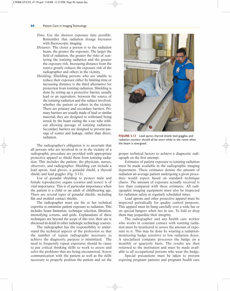
The radiographer’s obligation is to ascertain thatall persons who are involved in or in the vicinity of aradiographic procedure are provided with appropriateprotective apparel to shield them from ionizing radia-tion. This includes the patient, the physician, nurses,observers, and radiographer. Shielding can include alead apron, lead gloves, a gonadal shield, a thyroidshield, and lead goggles (Fig. 3-13).
Use of gonadal shielding to protect male andfemale reproductive organs (ovaries and testes) is ofvital importance. This is of particular importance whenthe patient is a child or an adult of childbearing age.There are several types of gonadal shields, includingflat and molded contact shields.
The radiographer must use his or her technicalexpertise to minimize patient exposure to radiation. Thisincludes beam limitation, technique selection, filtration,intensifying screens, and grids. Explanations of thesetechniques are beyond the scope of this text; their use isdiscussed in detail in other radiologic technology courses.
The radiographer has the responsibility to under-stand the technical aspects of the profession so thatthe number of repeat radiographs necessary toachieve the diagnostic purpose is minimized. Theneed to frequently repeat exposures should be causeto put critical thinking skills to work to assess andsolve the problems that are being encountered. Assesscommunication with the patient as well as the skillsnecessary to properly position the patient and set the
proper technical factors to achieve a diagnostic radi-ograph on the first attempt.
Estimates of patient exposure to ionizing radiationmust be made available in the radiographic imagingdepartment. These estimates denote the amount ofradiation an average patient undergoing a given proce-dure would expect based on standard techniquecharts. The amount of exposure actually received isless than compared with these estimates. All radi-ographic imaging equipment must also be inspectedfor radiation safety at regularly scheduled times.
Lead aprons and other protective apparel must beinspected periodically for quality control purposes.This apparel must be hung carefully over a wide bar oron special hangers when not in use. To fold or dropthem may jeopardize their integrity.
The radiographer and any health care workerwho works in constant contact with ionizing radia-tion must be monitored to assess the amount of expo-sure to it. This may be done by wearing a radiation-monitoring badge sensitive to low radiation doses. A specialized company processes the badge on amonthly or quarterly basis. The results are thenreturned to the institution and must be made avail-able to all occupational persons who wear the badge.
Special precautions must be taken to preventexposing pregnant patients and pregnant health care
66 Patient Care in Imaging Technology
FIGURE 3-13 Lead apron, thyroid shield, lead goggles, and
radiation monitor should all be worn while in the room when
the beam is energized.
LWBK129-C03_47-70.qxd 11/6/08 11:23 PM Page 66 Aptara Inc.

workers to ionizing radiation. This is particularly trueduring the early weeks of pregnancy, when particularfetal tissues are especially sensitive to radiation. This iswhy it is critical to ask the female patient if there is anypossibility of her being pregnant and also when her lastmenstrual period was. Pregnant workers who “declare”themselves to be pregnant are double badged, and rota-tions in the department are varied so as to limit theamount of exposure to radiation. The occupational
dose limit for a fetus must not exceed 0.5 rem duringthe entire gestation. The exposure must be limited tono more than 0.05 rems in any month.
To minimize radiation exposure, the radiographershould not hold the patient during a procedure on aroutine basis. Sand bags and positioning spongesshould be used if possible. If this is not feasible, then arelative or a person who is not working regularly inradiography should be requested to assist.
CHAPTER 3: Patient Care and Safety 67
her at all times and release the immobilizers at leastevery 2 hours. Follow the correct manner of docu-menting immobilization use.
Take care to prevent the patient’s skin from beingdamaged while being cared for in the diagnostic imagingdepartment. This can be done by preventing injury thatmay come from immobility, pressure, shearing force, orfriction. Patients most susceptible to skin breakdown arethe malnourished, the elderly, and the chronically ill.Take special care to protect these patients from injuriesto their integumentary system, because they may resultin a decubitus ulcer that can take months to heal. Also,take extra precautions when caring for a patient who iswearing a cast or who is in traction. Observe thepatient’s extremities for evidence of neurocirculatoryimpairment, which may result from the pressure of acast on the skin. Some symptoms of neurocirculatoryimpairment that are easily detected are pain, coldness,numbness, burning or tingling of fingers or toes, swelling,color changes of the skin, and an inability to move fingersor toes. If these symptoms are noted, change the patient’sposition and report the problem to the physician imme-diately. Do not release a traction apparatus while takinga radiographic image. If the procedure cannot be com-pleted because of the traction bar, request assistancefrom the nurse in charge of the patient.
If a patient is unable to undress alone, offer assis-tance. Give assistance in a matter-of-fact manner thatdoes not violate the patient’s privacy. Patients must bekept clean and dry while in the diagnostic imagingdepartment. It is the radiographer’s duty to change thedisabled patient’s gown and covering if they becomewet or soiled. Do this in a prescribed manner to ensureprivacy, safety, and comfort.
Some examinations in the imaging department arelong and tedious. They often stimulate peristalsis and aneed to defecate or urinate. Meeting these needs can-not be postponed. Be prepared to assist with either thebedpan or urinal if necessary and do it in a way thatensures the patient as much privacy as possible. Infec-tion control measures must be taken when assisting apatient with a bedpan or urinal. These items must be
SUMMARY
When an outpatient arrives in the diagnostic imagingdepartment, it is often necessary for that patient toundress entirely or partially for the diagnostic exami-nation or treatment. Always show the patient whereand how to do so in a sensitive manner to spare thepatient embarrassment.
It is the responsibility of the radiographer to pro-vide the patient with a safe place for personal belong-ings. Remember that the patient may treasure an arti-cle of clothing or jewelry that may not seem valuable.Everything that belongs to the patient must be treatedas if it were of value.
Correct body mechanics must always be used.When moving or lifting in the workplace, keep theweight close to the body and maintain a firm base ofbody support. This is accomplished by having the feetslightly spread out and knees flexed. Twist or bend thebody at the waist when lifting a heavy load. Weightshould be pulled, not pushed. Use arm and leg muscles,not the spine for lifting.
The three ways of moving patients are by gurney, bywheelchair, or by ambulation. When moving and liftingpatients, assess the patient and resolve potential prob-lems before beginning the transfer. The plan for movingthe patient should be explained, and the patient’s helpshould be enlisted before beginning. Always notify theward personnel when taking a patient to or from his orher hospital room. The use of enough assistants andequipment such as a smooth mover facilitates the moveand protects personnel and the patient from possibleinjury.
When a patient is on the radiographic table or on agurney in the diagnostic imaging department, his or herbody must be in good alignment. If the patient ismoved to a particular position for an examination,restore correct body alignment as soon as possible.
There are times when immobilizers must be usedfor the safety of the adult patient. When immobilizersare required, apply them according to the manufac-ture’s directions and the policy of the institution. Donot immobilize a patient without an order by a physi-cian. When a patient is immobilized, attend to him or
LWBK129-C03_47-70.qxd 11/6/08 11:23 PM Page 67 Aptara Inc.

used for one patient only and then disposed of in theproper waste receptacle. Put on clean, disposable gloveswhen assisting with the patient’s elimination needs andwash thoroughly after removing the gloves. The patientmust not be left unattended while on a bedpan, gurney,or diagnostic imaging table. Patients must never beallowed to get on or off an examining table or out of awheelchair without assistance. They must also be care-fully attended on trips to the lavatory and in a dressingarea after an examination or treatment.
The radiographer must be constantly on guard toprotect patients and other staff members from acci-dents in hospitals. Most falls and injuries to patientscan be prevented if the radiographer is knowledgeableabout the patient’s condition and comfort. The radiog-
rapher must understand the precautions to take rou-tinely to prevent fire and the correct procedures to fol-low if a fire occurs. Prevention of accidents due tofaulty electrical equipment or poisoning and the correctuse and disposal of hazardous materials are also theradiographer’s professional obligation.
Unnecessary exposure to radiation to the patientand personnel is the responsibility of the radiographer.Excessive amounts of radiation from improper techni-cal factors or from repeat exposures have an adverseaffect on living tissue. The very young, the very old,and pregnant women are particularly susceptible toadverse affects of ionizing radiation. Precautions toprevent excessive exposure involve knowledge, techni-cal expertise, and constant vigilance.
68 Patient Care in Imaging Technology
a. Turning the patient every 1 to 2 hoursb. Friction and pressurec. Frequent diagnostic imaging proceduresd. A wet environment
6. If a patient who has a cast in place complains ofpain that is sudden in onset and increases in inten-sity when the affected limb is moved, what shouldbe done? (Circle all that apply.)a. Complete the procedure and discharge the
patient.b. Elevate the affected limb.c. Notify a physician immediately.d. Find a nurse to administer pain medication.
7. When caring for a patient who has a new castapplied to an extremity, what must be remem-bered? (Circle all that apply.)a. Hold the cast firmly at a position between the
joints when moving it.b. Observe for signs of impaired circulation.c. Support the cast with bolsters and sandbags
where needed.d. The extremity is now almost impervious to pain
and can be twisted as needed for the image.8. When caring for a patient who is disabled and is
difficult to move, it is best to:a. Keep the patient as quiet as possible.b. Work quickly.c. Obtain as much help as necessary to avoid
injury to the patient and to the radiographer.d. Move the patient by gurney.
9. When moving a heavy object, you should___________ the weight, not ______________ it.
10. Patients most prone to falls are (circle all that apply):a. The frail elderlyb. The person who is confused
CHAPTER 3 TEST
1. When admitting a patient to the diagnostic imag-ing department, what should be done? (Circle allthat apply)a. Take the patient to the dressing area and
explain in some detail how he or she shoulddress for the procedure.
b. Give the patient directions concerning how tocare for valuables brought to the department.
c. Assist any patient who appears to need assis-tance with preparation for an examination.
2. The most effective means of reducing frictionwhen moving a patient is by:a. Placing the patient’s arms across the chest and
using a pull sheetb. Pushing rather than pulling the patientc. Rolling the patient to a prone positiond. Asking the patient to cooperate
3. When transporting a patient back to the hospitalroom, some safety measures to be used are (circleall that apply):a. Place the side rails up, the bed in “low” position,
and the call bell at hand.b. Inform the nurse in charge of the patient that
the patient has been returned to the room.c. Give the patient something to eat or drink.d. Be sure that the TV is in place for the patient’s
viewing.4. Which procedures must be observed when assist-
ing a patient with a bedpan (circle all that apply):a. Respect the patient’s privacy.b. Seek assistance for an immobile patient.c. Wear clean gloves to remove the bedpan.d. Make sure to offer tissue to the patient and a
towel to clean his or her hands.5. Contributing factors to skin breakdown are (circle
all that apply):
LWBK129-C03_47-70.qxd 11/6/08 11:23 PM Page 68 Aptara Inc.
CHAPTER 3: Patient Care and Safety 69
c. Persons who have been given a psychoactivedrug
d. Persons with sensory deficits11. When moving a patient into an unnatural position
for a radiographic examination, the patient shouldmaintain that position:a. Until he or she asks to be movedb. Until the radiograph has been processed and
approved by the radiologistc. Only for the time it takes to make the
exposure12. Match the following:
a. Fowler position
b. Supine position c. Semi-Fowler
positiond. Trendelenburg
positione. Sims position
13. Name the two convenient and safe methods of mov-ing a patient from a radiographic table to a gurney.
14. Describe three legitimate reasons for applicationof immobilizers to an adult patient.
15. List four signs of circulatory impairment if apatient is wearing a cast.
16. What are three methods of reducing a patient’sexposure to ionizing radiation?
17. The leading cause of work-related injuries in thefield of health care is:a. Bumping into misplaced equipmentb. Overexposure to radiationc. Infection owing to poor hand-washing
techniquesd. Abuse of the spine when moving and lifting
patients
i. Patient on side with for-ward arm flexed and topknee flexed
ii. Semi-sitting position withhead raised 45 to 60degrees
iii. Patient laying flat onback
iv. Patient on back with headlower than extremities
v. Patient on back with headraised 15 to 30 degrees
LWBK129-C03_47-70.qxd 11/6/08 11:23 PM Page 69 Aptara Inc.

LWBK129-C03_47-70.qxd 11/6/08 11:23 PM Page 70 Aptara Inc.


![SYN - Lippincott Williams & Wilkinsdownloads.lww.com/wolterskluwer_vitalstream_com/sample-content/... · SYN musculus zy-gomaticus major [TA], greater zygomatic mus-cle, musculus](https://img.pdfslide.us/doc/110x75/5e21415630172f658d026ddc/syn-lippincott-williams-syn-musculus-zy-gomaticus-major-ta-greater-zygomatic.jpg)