Embed Size (px)
Citation preview

2D Digital Radiography 3D Computed Tomography Inspection Services Laboratories
4nsi.com xrayinspectionservice.com

Celebrating over 28 years of InnovationNorth Star Imaging was founded in 1986 by Ken Ness as a Non-destructive
Testing (NDT) equipment representative and accessory supplier based in the
St. Paul, MN area. The firm specialized in industrial X-ray testing. NSI served
the upper Midwestern states of Minnesota, North Dakota, South Dakota,
Wisconsin and Iowa.
In 1991, North Star Imaging manufactured their first Digital Radiography
system. Throughout the 1990’s, NSI continued to expand their equipment
representative and accessory supplier business across the USA. In 2002, NSI
manufactured their first Computed Tomography system.
In 2004, NSI outgrew their St. Paul, MN site and moved into a brand new facility
in Rogers, MN. In 2006, the company formed their Inspection Services Group
(ISG), which provides need based consulting services for anyone needing X-ray
and/or Computed Tomography scanning. The company’s X-ray and Computed Tomography systems and services business continued to flourish and in 2009
they expanded their Rogers complex by 33% to keep up with demand.
In late 2010, North Star Imaging, Inc. was acquired by Illinois Tool Works (ITW) and became part of ITW’s Test and Measurement segment. ITW is a leading
diversified manufacturing company with over 100 years of history and over 800 individual business units.
In 2012, with demand continuing to increase both nationally and internationally, North Star Imaging nearly doubled the square footage of their facility in
Rogers, MN. The same year, NSI added a physical location in Paris, France.
In 2013, North Star Imaging, Inc. was awarded with ISO 9001:2008 certification as a result of significant changes and improvements to their Quality System
to increase efficiency, reduce errors and enrich customer satisfaction.
2

Truly a Global CompanyToday, North Star Imaging is one of the leading manufacturers of 2D Digital Radiography and 3D Computed Tomography systems in the
World. Additionally, each worldwide location houses state of the art equipment for demonstration and need based X-ray/CT Inspection
Services. No matter your location, NSI has local employees ready to help evaluate your needs, explain the technology and provide
thorough training upon installation. Furthermore, each NSI location employs dedicated service personnel, so local help is never more
than a phone call away.
3

What is Digital Radiography?Digital Radiography, or DR for short, is a 2D X-ray inspection method using a digital X-ray detector in place of X-ray
film. DR allows for real-time X-ray inspection of your part or object - no more waiting for film to process! You can
make scan adjustments on the fly and also apply digital image enhancements quickly and easily – saving you time.
Digital Radiography detectors are designed to be used time after time, helping to eliminate the cost of consumables
– saving you money.
North Star Imaging’s Digital Radiography systems are designed to make your business and your team as efficient as
possible. Programmed and repeatable inspection sequences, easy to use software and superior image quality lets
you focus more monitoring your product quality while also increasing throughput.
Capacitor
Flashlight
We offerDR Training
4nsi.com/training
4

What is Digital Radiography?
Battery Cylinder Head5

3D Computed Tomography (CT) is a nondestructive scanning technology that allows you to view and inspect the external and internal
structures of an object in 3D space. Computed Tomography works by taking hundreds or thousands of 2D Digital Radiography projections
around a 360 degree rotation of an object. Proprietary algorithms are then used to reconstruct the 2D projections into a 3D CT volume, which
will allow you to view and slice the part at any angle.
3D CT virtually eliminates interpretation errors and opens the door to many capabilities that are not available with any other technology.
CT capabilities include:• Internal and external measurements
• 3D CAD comparisons
• Void analysis
• Surface reconstructions for reverse engineering
• Finite Element Analysis
• And much more
North Star Imaging Computed Tomography systems are the easiest to use in the industry. NSI’s efX-CT software
uses five simple steps to guide you through the CT scanning process and have you inspecting your product in no
time – increasing your quality and efficiency.
What is Computed Tomography
Composites
We offerCT Training
4nsi.com/training
6

What is Computed Tomography
7
Munitions Plastics IC Chips7

Applications & Markets
Castings
Museum
Electronics
Aerospace
8

The uses of Digital Radiography and
Computed Tomography are very diverse.
There are few limits on what we can scan
and what we can see; we’re working every
day to push those limits.
Automotive
Plastics
MedicalDevices
Research
9

Systems
SYSTEM CAPABILITIES• Advanced 2D X-ray inspection
• 2D CT Slice reconstruction
• CT volume reconstruction for 3D inspection
• 3D internal and external surface scanning
• 5” (12cm) diameter x 5” (12cm) tall nominal part envelope
CT SOFTWARE• Comprehensive acquisition, processing and archival program with
user-friendly interface
• Automated CT calibration software
• Very fast reconstruction software with option for Graphic Card based algorithm
• Local CT and Limited angle CT reconstruction capabilities
• 3D visualization module with real-time volume rendering, density segmentation, & high performance measurements
• 3D surface model export for CAD compatibility
• Multiple image formats input/output
X-RAY SOURCE• Micro-focus X-ray tube
• Voltage Range: 10kV – 150 kV
• Focal Spot Size : < 5 microns
• Maximum system resolution: better than 5 microns
X-RAY DETECTOR• Digital X-ray detector types: Flat Panel or Image Intensifier
• Detector Size: up to 8’’ x 10’’ (20cm x 25cm)
MANIPULATOR• Maximum Sample Weight: 10 lbs (4.5kg)
• 4 Axis of Scan Travel (customization available) : Focal Distance : 24’’ (61cm) Vertical : 7.5’’ (19cm) (*Exact measurements vary depending
on tube, detector and optional configurations)
• Rotational stage with high precision
• Optional motion controlled 3 axis
CABINET• External Dimension: 57’’ Wide, 30’’ Deep, 58’’ Tall (144cm Wide,
76cm Deep, 147cm Tall)
• Cabinet Features: interior lighting, sliding access door, leaded glass viewing window
• Meets or exceeds 21 CFR 1020.40 and EN 61010-2-091 2012
• System includes one ergonomic desk and chair
The ImagiX is North Star Imaging’s most compact system. The system can
be configured as a desktop unit or a freestanding system. The generous
scanning envelope can handle products up to 5” (12cm) in size making it a
great choice for laboratories, small electronics and R&D applications.
Affordability and Simplicity
10
Systems4nsi.com25
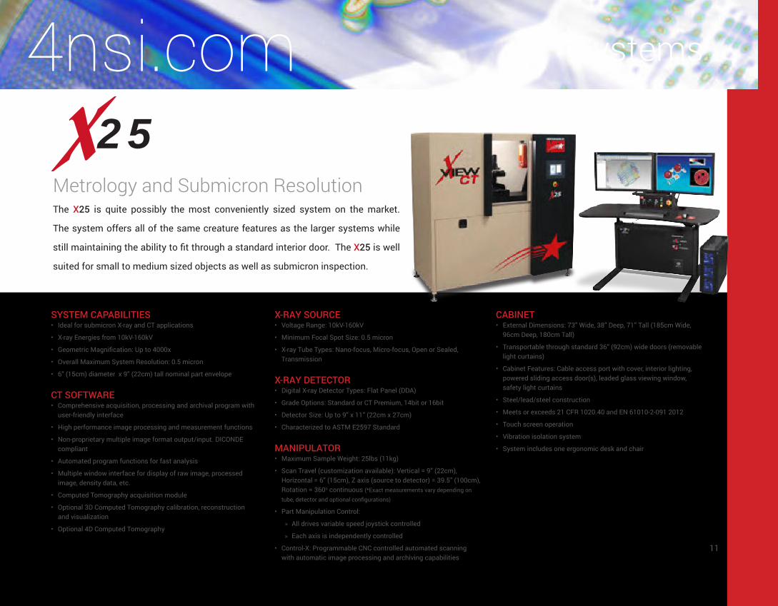
SYSTEM CAPABILITIES• Ideal for submicron X-ray and CT applications
• X-ray Energies from 10kV-160kV
• Geometric Magnification: Up to 4000x
• Overall Maximum System Resolution: 0.5 micron
• 6” (15cm) diameter x 9” (22cm) tall nominal part envelope
CT SOFTWARE• Comprehensive acquisition, processing and archival program with
user-friendly interface
• High performance image processing and measurement functions
• Non-proprietary multiple image format output/input. DICONDE compliant
• Automated program functions for fast analysis
• Multiple window interface for display of raw image, processed image, density data, etc.
• Computed Tomography acquisition module
• Optional 3D Computed Tomography calibration, reconstruction and visualization
• Optional 4D Computed Tomography
X-RAY SOURCE• Voltage Range: 10kV-160kV
• Minimum Focal Spot Size: 0.5 micron
• X-ray Tube Types: Nano-focus, Micro-focus, Open or Sealed, Transmission
X-RAY DETECTOR• Digital X-ray Detector Types: Flat Panel (DDA)
• Grade Options: Standard or CT Premium, 14bit or 16bit
• Detector Size: Up to 9” x 11” (22cm x 27cm)
• Characterized to ASTM E2597 Standard
MANIPULATOR• Maximum Sample Weight: 25lbs (11kg)
• Scan Travel (customization available): Vertical = 9” (22cm), Horizontal = 6” (15cm), Z axis (source to detector) = 39.5” (100cm), Rotation = 360o continuous (*Exact measurements vary depending on
tube, detector and optional configurations)
• Part Manipulation Control:
» All drives variable speed joystick controlled ◦
» Each axis is independently controlled
• Control-X: Programmable CNC controlled automated scanning with automatic image processing and archiving capabilities
CABINET• External Dimensions: 73” Wide, 38” Deep, 71” Tall (185cm Wide,
96cm Deep, 180cm Tall)
• Transportable through standard 36” (92cm) wide doors (removable light curtains)
• Cabinet Features: Cable access port with cover, interior lighting, powered sliding access door(s), leaded glass viewing window, safety light curtains
• Steel/lead/steel construction
• Meets or exceeds 21 CFR 1020.40 and EN 61010-2-091 2012
• Touch screen operation
• Vibration isolation system
• System includes one ergonomic desk and chair
The X25 is quite possibly the most conveniently sized system on the market.
The system offers all of the same creature features as the larger systems while
still maintaining the ability to fit through a standard interior door. The X25 is well
suited for small to medium sized objects as well as submicron inspection.
Metrology and Submicron Resolution
11

Systems
50
SYSTEM CAPABILITIES• X-ray Energies from 10kV-240kV
• Geometric Magnification: Up to 4000x
• Overall Maximum System Resolution: better than 1μm
• Meets ASTM E2597 Standard
• 8” (20cm) diameter x 12” (30cm) tall nominal part envelope
CT SOFTWARE• Comprehensive acquisition, processing and archival program with
user-friendly interface
• High performance image processing and measurement functions
• Non-proprietary multiple image format output/input. DICONDE compliant
• Automated program functions for fast analysis
• Multiple window interface for display of raw image, processed image, density data, etc.
• Computed Tomography acquisition module
• Optional 3D Computed Tomography calibration, reconstruction and visualization
• Optional 4D Computed Tomography
• Available with vorteX
X-RAY SOURCE• Voltage Range: 10kV-240kV
• Minimum Focal Spot Size: <1μm
• X-ray Tube Types: Nano-focus, Micro-focus, Mini-focus, Open or Sealed, Transmission or Directional or Dual Head
X-RAY DETECTOR• Digital X-ray Detector Types: Flat Panel (DDA), Linear Diode Array
(LDA), Image Intensifier
• Grade Options: Standard or CT Premium, 14bit or 16bit
• Detector Size: Up to 16” x 16” (40cm x 40cm)
MANIPULATOR• Maximum Sample Weight: 25lbs (11kg)
• Scan Travel (customization available): Vertical = 12” (30cm), Horizontal = 12” (30cm), Z axis (source to detector) = 53” (134cm), Tilt = +20°/-10°, Rotation = 360° continuous (*Exact measurements vary
depending on tube, detector and optional configurations)
• Part Manipulation Control:
» All drives variable speed joystick controlled
» Each axis is independently controlled
• Control-X: Programmable CNC controlled automated scanning with automatic image processing and archiving capabilities
CABINET• External Dimensions: 87” Wide, 52” Deep, 79” Tall (221cm Wide,
132cm Deep, 201cm Tall)
• Cabinet Features: Cable access port with cover, interior lighting, powered sliding access door(s), leaded glass viewing window, safety light curtains
• Steel/lead/steel construction
• Meets or exceeds 21 CFR 1020.40 and EN 61010-2-091 2012
• Touch screen operation
• System includes one ergonomic desk and chairs
The X50 is one of NSI’s most popular models for electronics, aerospace
components and medical devices. It offers an excellent balance of power
and space sensitivity. The system can handle products up to 12” (30cm) in
size while seated nicely in your failure analysis lab or busy production line.
Powerful and Ergonomic
12

Systems4nsi.com5000
SYSTEM CAPABILITIES• X-ray Energies from 10kV-450kV
• Geometric Magnification: Greater than 2000x
• Overall Maximum System Resolution: better than 500nm
• Meets ASTM E2597 Standard
• 32” (81cm) diameter x 48” (121cm) tall nominal part envelope
CT SOFTWARE• Comprehensive acquisition, processing and archival program with
user-friendly interface
• High performance image processing and measurement functions
• Non-proprietary multiple image format output/input. DICONDE compliant
• Automated program functions for fast analysis
• Multiple window interface for display of raw image, processed image, density data, etc.
• Computed Tomography acquisition module
• Optional 3D Computed Tomography calibration, reconstruction and visualization
• Optional 4D Computed Tomography
• Available with vorteX, subpiX, and mosaiX
X-RAY SOURCE• Voltage Range: 10kV-450kV
• Minimum Focal Spot Size: < 500nm
• X-ray Tube Types: Nano-focus, Micro-focus, Mini-focus, Open or Sealed, Transmission or Directional or Dual Head
• Optional Dual Tube configuration
X-RAY DETECTOR• Digital X-ray Detector Types: Flat Panel (DDA), Linear Diode Array
(LDA), Image Intensifier
• Grade Options: Standard or CT Premium, 14bit or 16bit
• Detector Size: Up to 16” x 16” (40cm x 40cm)
MANIPULATOR• Maximum Sample Weight: 250lbs (113kg) (400lbs (181kg)
optional)
• Scan Travel (customization available): Vertical = 48” (121cm), Horizontal = 33” (83cm), Z axis (source to detector) = 48” (121cm), Tilt = +20°/-15°, Rotation (optional +/-30°) = 360° continuous (*Exact measurements vary depending on tube, detector and optional
configurations)
• Motorized detector travel for variable focal distance adjustment
• Part Manipulation Control:
» All drives variable speed joystick controlled
» Each axis is independently controlled
• NSI CNC motion control software for automated scanning with automatic image processing and archiving capabilities
• Option: Rotational stage indexes outside of the cabinet for ergonomic part loading/unloading
CABINET• External Dimensions:
» 240kV model: 107” Wide x 80” Deep x 92” Tall (271cm Wide, 203cm Deep, 233cm Tall)
» 450kV model: 126” Wide x 91” Deep x 102” Tall (320cm Wide, 231cm Deep, 259cm Tall)
• Cabinet Features: Cable access port with cover, interior lighting, powered sliding access door(s), leaded glass viewing window (240kV model only), internal camera monitoring system, safety light curtains
• Steel/lead/steel construction
• Meets or exceeds 21 CFR 1020.40 and EN 61010-2-091 2012
• Touch screen operation
• Includes one ergonomic desk and chair
The X5000 is the most versatile system offered by North Star Imaging.
The system boasts a large scanning envelop and excellent ergonomics for
loading sizable objects while still maintaining the sensitivity to inspect even
the smallest of items.
Universal and Flexible
13

Systems
The X6000 is specifically designed for castings and other large and heavy
products. The system features a programmable C-arm manipulator for
automated and repeatable inspection sequences. The massive access door
and external indexing rotational stage make loading quick and easy.
SYSTEM CAPABILITIES• X-ray Energies from 10kV-225kV
• Shielded to 160kV or 225kV
• Geometric Magnification: Greater than 2000x
• Capable of scanning large components
• 48” (121cm) diameter x 60” (152cm) all nominal part envelope
CT SOFTWARE• Comprehensive acquisition, processing and archival program with
user-friendly interface
• High performance image processing and measurement functions • Non-proprietary multiple image format output/input. DICONDE compliant
• Automated program functions for fast analysis
• Multiple window interfaces for display of raw image, processed image, density data, etc.
• Optional 2D, 3D & 4D Computed Tomography acquisition module
X-RAY SOURCE• Voltage Range: 10kV-225kV
• X-ray Tube Types: Transmission or Directional Micro-focus, Mini-focus
• Optional Microfocus Rod Anode or Center Tube Design
X-RAY DETECTOR• Digital X-ray Detector Types: Flat Panel (DDA)
• Flat Panel Detector Size: Up to 16” x 16” (40cm x 40cm)
MANIPULATOR• Maximum Sample Weight: 400lbs (181kg)
• Scan Travel: Vertical (Y-axis) = 70” (177cm), Horizontal (X-axis) = 48” (121cm), Lateral (Z-axis) = 48” (121cm), Source to Detector = Max 48” (121cm), C-arm Rotation = +/-120°, Stage Rotation = 360° continuous (*Exact measurements vary depending on tube, detector and
optional configurations)
• Motorized detector travel for variable focal distance adjustment
• Part Manipulation Control:
» All drives variable speed joystick controlled
» Each axis is independently controlled
• Rotational stage indexes outside of the cabinet for easy part loading/unloading
• Options:
» efX-CNC programmable CNC controlled automated scanning with automatic image processing and archiving capabilities
» Lateral detector motion
» Additional X-axis for long component scanning
CABINET• External Dimensions: 139” Wide x 120” Deep x 132” Tall (353cm
Wide, 304cm Deep, 335cm Tall) (varies depending on shielding)
• Cabinet Features: Cable access ports with cover, interior lighting, 52” x 90” (132cm x 228cm) powered bi-parting sliding access door, two 15” x 24” (38cm x 60cm) leaded glass viewing windows, internal camera monitoring system, safety light curtains
• Steel/lead/steel construction
• Meets or exceeds 21 CFR 1020.40 and EN 61010-2-091 2012
• Touch screen operation
• Includes one ergonomic desk and chair
6000Automated and Versatile
14

Systems4nsi.com
The X7000 is North Star Imaging’s largest standard system. The optional
independent horizontal (x-axis) travel of the tube and detector allow for
unparalleled inspection capabilities of an elongated object. The system is
great for composites, castings, pipes, tubes, welds and similar parts.
SYSTEM CAPABILITIES• X-ray Energies from 10kV-450kV
• Geometric Magnification: Greater than 2000x
• Capable of scanning large components
• 60” (152cm) diameter x 60” (152cm) tall nominal part envelope
CT SOFTWARE• Comprehensive acquisition, processing and archival program with
user-friendly interface
• High performance image processing and measurement functions • Non-proprietary multiple image format output/input. DICONDE compliant
• Automated program functions for fast analysis
• Multiple window interface for display of raw image, processed image, density data, etc.
• Computed Tomography acquisition module
• Optional 3D & 4D Computed Tomography acquisition module
• Available with vorteX, subpiX and mosaiX
X-RAY SOURCE• Voltage Range: 10kV-450kV
• X-ray Tube Types: Transmission or Directional Micro-focus, Mini-focus
• Optional dual tube configuration
X-RAY DETECTOR• Digital X-ray Detector Types: Flat Panel (DDA), Linear Diode Array
(LDA)
• Flat Panel Detector Size: Up to 16” x 16” (40cm x 40cm)
• LDA size up to 36” (91cm)
• Optional dual detector configuration
MANIPULATOR• Maximum Sample Weight: 800lbs (362kg)
• Scan Travel: Vertical = 60” (152cm), Horizontal = 120” (305cm), Z axis (source to detector) = 80” (203cm), Tilt = +30°/-30°, Rotation = 360° continuous (*Exact measurements vary depending on tube,
detector and optional configurations)
• Motorized detector travel for variable focal distance adjustment
• Rotational stage indexes outside of the cabinet for easy part loading/unloading
• Part Manipulation Control:
» All drives variable speed joystick controlled
» Each axis is independently controlled
• Options:
» Control-X programmable CNC controlled automated scanning with automatic image processing and archiving capabilities
» Lateral detector motion
» Dual tubes and/or dual detectors
» Additional X-axis for long component scanning
CABINET• External Dimensions: 156” Wide x 156” Deep x 125” Tall (396cm
Wide, 396cm Deep, 317cm Tall) (varies depending on shielding)
• Cabinet Features: Cable access ports with cover, interior lighting, powered bi-parting sliding access door (60” x 90” (172cm x 228cm) door opening), internal camera monitoring system, safety light curtains
• Steel/lead/steel construction
• Meets or exceeds 21 CFR 1020.40 and EN 61010-2-091 2012
• Touch screen operation
• Includes one ergonomic desk and chair
7000Modular and Robust
15
Upgrades
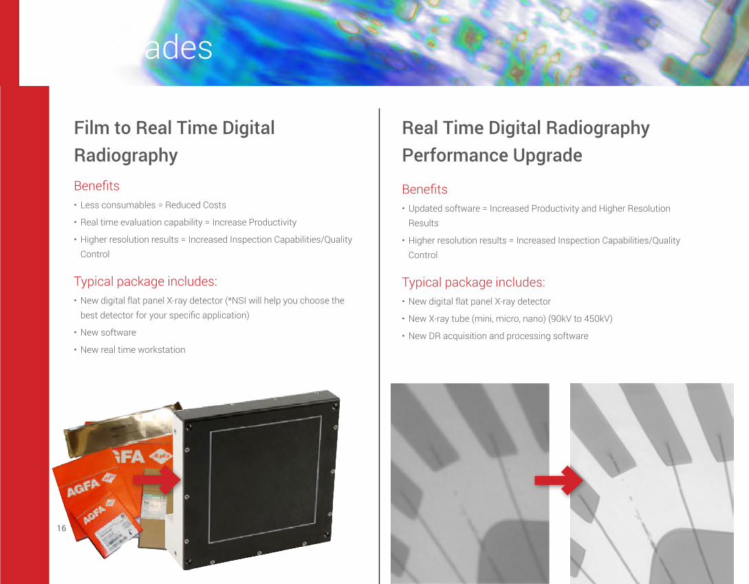
Film to Real Time Digital Radiography
Real Time Digital Radiography Performance Upgrade
Benefits• Less consumables = Reduced Costs
• Real time evaluation capability = Increase Productivity
• Higher resolution results = Increased Inspection Capabilities/Quality Control
Typical package includes:• New digital flat panel X-ray detector (*NSI will help you choose the
best detector for your specific application)
• New software
• New real time workstation
Benefits• Updated software = Increased Productivity and Higher Resolution
Results
• Higher resolution results = Increased Inspection Capabilities/Quality Control
Typical package includes:• New digital flat panel X-ray detector
• New X-ray tube (mini, micro, nano) (90kV to 450kV)
• New DR acquisition and processing software
16
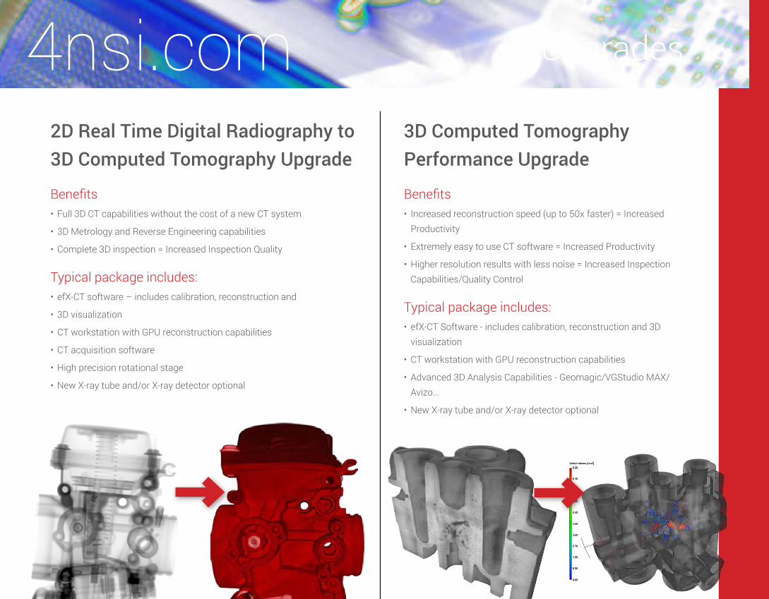
Upgrades4nsi.com2D Real Time Digital Radiography to 3D Computed Tomography Upgrade
3D Computed Tomography Performance Upgrade
Benefits• Full 3D CT capabilities without the cost of a new CT system
• 3D Metrology and Reverse Engineering capabilities
• Complete 3D inspection = Increased Inspection Quality
Typical package includes:• efX-CT software – includes calibration, reconstruction and
• 3D visualization
• CT workstation with GPU reconstruction capabilities
• CT acquisition software
• High precision rotational stage
• New X-ray tube and/or X-ray detector optional
Benefits• Increased reconstruction speed (up to 50x faster) = Increased
Productivity
• Extremely easy to use CT software = Increased Productivity
• Higher resolution results with less noise = Increased Inspection Capabilities/Quality Control
Typical package includes:• efX-CT Software - includes calibration, reconstruction and 3D
visualization
• CT workstation with GPU reconstruction capabilities
• Advanced 3D Analysis Capabilities - Geomagic/VGStudio MAX/Avizo...
• New X-ray tube and/or X-ray detector optional
17

Software
is a new generation DR software developed entirely by North Star Imaging. Exclusively featuring:
• High performance image processing and measurement functions using GPU
• Automatic creation of customizable Technique sheet for operator records
• Easy CT acquisitions: continuous or step, Fan Beam, Cone Beam, vorteX
• Enhanced detector capabilities: larger size (mosaiX) or improved resolution (subpiX)
• Seamless integration with efX-CT software
• DICONDE compliant
• Motion programming and Automated barcode triggered program execution
efX-DR IMAGE PROCESSING SOFTWARE • Windows 7 Based (XP supported)
• Non-proprietary image storage format (TIFF)
• High performance image processing and measurement functions using GPU
• Live Averaging
• Live Histogram with multiple color tables
• Live line Profile
• Live Rotation between portrait and landscape modes
• Live Measurements
• Live image offset and multiple gain calibration, defective pixel correction
• Live signal to noise and live contrast to noise measurement
• Filters to improve image quality
• Automatic creation of customizable Technique sheet for operator records
• Capture video into AVI files
• Supports digital flat panel detectors, LDA’s and digital/analog cameras at 8, 10, 12 and 16 bits
• Supports X-ray sources
• Read and store images in TIFF 32 bit / 16 bit / 8bit, BMP, JPEG, DICONDE
• Seamless integration with efX-CT software
• Optional CNC motion control and teach-based programming
• Support for production mode with barcoded input and automated system operation
OPTIONAL DETECTOR QUALIFICATION MODULE:• Designed to meet ASTM 2597, 2737 and BSS 7044 Rev B.
specifications
• Simplifies reporting process to meet above guidelines
• Simple SRb calculation
efX-DR ACQUISITION WORKSTATION::• Windows®7 x64 Based (XP supported)
• Quad Core Xeon Processor
• 8 GB RAM
• 1 TB SATA High Speed Hard Drive
• DVD+/-RW Drive
• 10/100/1000 network interface card
• 30” high resolution flat panel monitor
18

is the Easiest, Fastest and Most Complete Industrial CT Software on the Market. Exclusively featuring:
• GPU accelerated CT reconstruction module
• Automatic Parallelization for systems with multiple CPU’s and GPU’s
• 5-step guided wizard for easy CT reconstruction
• Intuitive interface and OpenGL based 3D volume rendering
• Unique geometry and dimensional calibration, exceeds system/mechanical precision
• Non-proprietary data formats, handles broad range of input formats
efX-CT PACKAGE INCLUDES: • Full software license
• High-end, multi-processor CT reconstruction and 3D visualization workstation
• Complete user guide, documentation and calibration tools
efX-CT SOFTWARE INCLUDES:• User friendly volume viewer
• 2D Viewer: efX-view for X-ray images and CT slices
• CT slices stack import
• Compatible 2D formats include BMP, TIFF, DICOM, DICONDE and most standard formats
• Automated focal spot drift compensation
• Volume format conversion capabilities
• Advanced CT mode for full access to all CT reconstruction parameters
• Filters on projections for noise and artefact correction
• Unique ultra-fast 3D preview of CT reconstructions
• Region of Interest CT reconstruction
• Job list – process all CT reconstructions in a queue
• Interactive density segmentation
• Real time multi-slicing (up to six planes) with measurements
• Volume resizing, cropping and reorienting
• Imperial and Metric measurement systems
• Beam hardening correction
• Surface extraction with export to STL, OBJ, DXF, WRL, PLY, etc.
• No limitation in reconstruction size and resolution
• Easy screen capture, video recording and exporting of x/y/z slices
• Production mode with automated reconstruction
efX-CT IS WINDOWS 8.1 BASED (7 AND XP SUPPORTED)
CT RECONSTRUCTION ALGORITHMS AVAILABLE IN efX-CT:• Cone-Beam (FDK) conventional and vorteX
• Fan-Beam
OPTIONS INCLUDE:• GPU acceleration package with NVIDIA supercomputer hardware
• High capacity high speed storage with hardware RAID support
• Geomagic, VGStudioMAX and/or Avizo software packages for more advanced CT processing
Software4nsi.com
19
Software Innovations
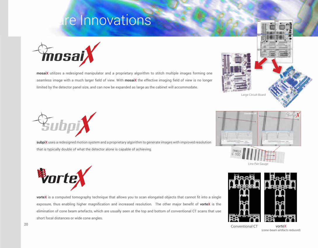
mosaiX utilizes a redesigned manipulator and a proprietary algorithm to stitch multiple images forming one
seamless image with a much larger field of view. With mosaiX the effective imaging field of view is no longer
limited by the detector panel size, and can now be expanded as large as the cabinet will accommodate.
subpiX uses a redesigned motion system and a proprietary algorithm to generate images with improved resolution
that is typically double of what the detector alone is capable of achieving.
vorteX is a computed tomography technique that allows you to scan elongated objects that cannot fit into a single
exposure, thus enabling higher magnification and increased resolution. The other major benefit of vorteX is the
elimination of cone beam artefacts, which are usually seen at the top and bottom of conventional CT scans that use
short focal distances or wide cone angles.
Conventional CT vorteX(cone-beam artifacts reduced)
Large Circuit Board
Line Pair Gauge
20
Software Innovations
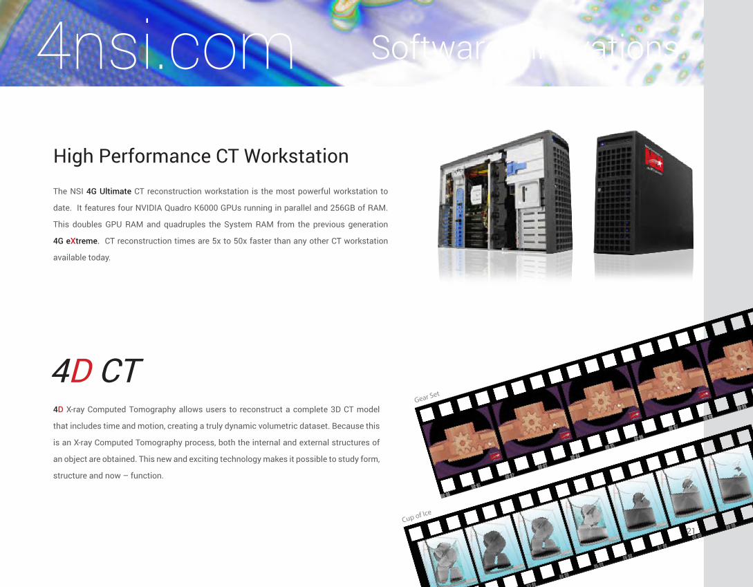
4D CT4D X-ray Computed Tomography allows users to reconstruct a complete 3D CT model
that includes time and motion, creating a truly dynamic volumetric dataset. Because this
is an X-ray Computed Tomography process, both the internal and external structures of
an object are obtained. This new and exciting technology makes it possible to study form,
structure and now – function.
High Performance CT WorkstationThe NSI 4G Ultimate CT reconstruction workstation is the most powerful workstation to
date. It features four NVIDIA Quadro K6000 GPUs running in parallel and 256GB of RAM.
This doubles GPU RAM and quadruples the System RAM from the previous generation
4G eXtreme. CT reconstruction times are 5x to 50x faster than any other CT workstation
available today.
Gear Set
Cup of Ice
4nsi.com
21

North Star Imaging’s Inspection Services Group provides real-time X-ray
inspection and CT scanning services to virtually anyone needing to verify the
integrity of internal components. The “inside view” that our team produces is
unparalleled in the industry and is the foundation for all of the services that we
provide. When you need high accuracy examination of internal components or
wish to inspect the dimensions of any assembly, call on NSI’s Inspection Services
Group. No other company offers a broader range of services or the depth of
nondestructive testing expertise.
The Most Advanced X-ray Inspection Laboratory in North America
Electronics Castings Automotive Aerospace Medical Devices
xrayinspectionservice.com
22

• Failure Analysis
• Research and Development (R&D)
• Product Quality Compliance/Screening
• Internal and External Measurements
• Reverse Engineering
• Density Analysis
• Product Contamination
• 3D Metrology
• Museum Artifact Digitization
• Weld Quality Analysis
• Assembly Verification
Applications Include:
Inspection Services Groupxrayinspectionservice.com
23
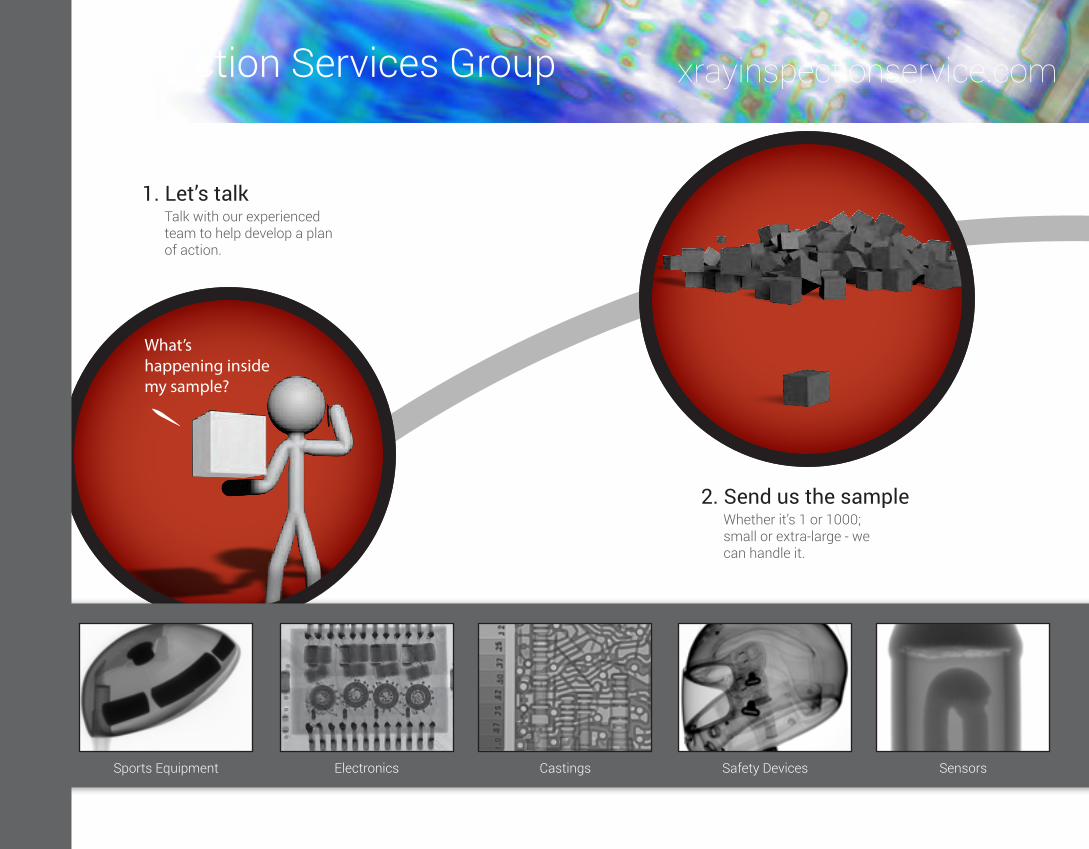
2. Send us the sampleWhether it’s 1 or 1000; small or extra-large - we can handle it.
What’s happening inside my sample?
1. Let’s talkTalk with our experienced team to help develop a plan of action.
3. We scan itWe have the Largest Lab in North America and facilities throughout the world - We’ll get it done!
4. Deliver the dataVisit our facility, join us for a web meeting or request a USB drive.
24
Inspection Services Group
Sports Equipment Electronics Castings Safety Devices Sensors
xrayinspectionservice.com

2. Send us the sampleWhether it’s 1 or 1000; small or extra-large - we can handle it.
What’s happening inside my sample?
1. Let’s talkTalk with our experienced team to help develop a plan of action.
3. We scan itWe have the Largest Lab in North America and facilities throughout the world - We’ll get it done!
4. Deliver the dataVisit our facility, join us for a web meeting or request a USB drive.
5000225kV & 450kV
50
25
25
Inspection Services Group
Plastics Circuit Boards Assemblies Rubber Power Equipment
xrayinspectionservice.com
25

Our goal is to help you avoid interruptions by keeping your system state-of-the-art and running smoothly.
Unlimited phone and remote access support
List of recommendations generated upon evaluation of system
Discounts on service labor and parts.
All service is performed by factory trained and authorized system specialist.
Maintenance Services 4nsi.com
26

1
2
3 4
5
6
7
8
9
10
11
12
1. Clean and adjust X-ray Tubes, replace
o-rings and adjust Controllers to
manufacturers specs
2. Clean, inspect, set, compression and
reapply dielectric grease
3. Vacuum system check and change oil if
applicable
4. Clean cooler and test safety switches
5. Clean and verify adjustments on the HT
generators to preserve tube filament life
6. Clean , inspect and lubricate manipulator
7. Test and Adjust shutter
8. Test and adjust Safety Interlocks and Safety
Lamps
9. Test power and supplies and adjust to
factory specifications
10. Inspect for proper cable drape
11. Install software updates
12. Perform a Radiation Safety Survey with
documentation
Thorough 12 Point Inspection..
Maintenance Services
We offer replacement X-ray tubes,
detectors and additional
components
4nsi.com
27

Corporate Office:
North Star Imaging, Inc.
19875 S. Diamond Lake Road
Rogers, Minnesota 55374 USA
Phone: (763) 463-5650
Toll Free: 1-800-635-8392
Fax: (763) 463-5651
4nsi.com
North Star Imaging Europe
Les Fregates Paris Nord 2
13 rue de la Perdrix
BP66151 Tremblay en France
95978 Roissy Charles de Gaulle Cedex France
Phone: +33 (0) 1 48 17 02 00
Fax: +33 (0) 1 48 17 02 09
4nsi.eu
North Star Imaging Worldwide
NSI has representative professionals
located throughout Europe, Asia
and other countries throughout the world.
Contact us for details.
ISO 9001:2008
NSI Quality Policy:The people of ITW North Star Imaging are committed to understanding and achieving our customer’s expectations
and providing world class imaging products and services driven by a culture of continual improvement.
What can NSI do for you?
4nsi.comxrayinspectionservice.com