Embed Size (px)
Citation preview

453
may not be “cured.” The optimal postoperative managementof patients with congenital heart disease (CHD) requires a multidisciplinary approach, combining the disciplines of cardiology, cardiac surgery, anesthesia, critical care, andnursing. A thorough understanding of the precise anatomicdiagnosis, pathophysiology, and details of the surgical tech-nique, including the potential for residual defects, is neces-sary when managing pediatric cardiac patients in the ICU.
For most patients, postoperative recovery is uncompli-cated, reflecting the improvements in preoperative diagnosisand stabilization, surgical techniques and, in particular, CPBmanagement. In general, when the patient’s clinical pro-gress or postoperative cardiorespiratory function does notfollow the expected course, the accuracy of the preoperativediagnosis should be questioned and the adequacy of the surgical repair investigated, either with echocardiographyand/or cardiac catheterization.
Pathophysiology of congenital cardiacdefects
A thorough understanding of the pathophysiology of
Introduction
The primary aims of treatment strategies for managing chil-dren with congenital cardiac defects are to promote normalgrowth and development and to limit the pathophysiologicconsequences of congenital cardiac defects such as volumeoverload, pressure overload, and chronic hypoxemia. As aresult, there has been a distinct change in management philosophy over the past 10–20 years towards performingreparative operations on neonates and infants, rather thaninitial palliation and later repair.1 However, because of a limited physiologic reserve and the complications associatedwith cardiopulmonary bypass (CPB) and open heart surgery, the risk for cardiorespiratory dysfunction in neonates andyoung infants in the immediate postoperative period may be increased.
The successful management of congenital heart defectsrequires detailed knowledge, experience, and technicalexpertise because of the significant heterogeneity in patientage, structural disease, and cardiorespiratory physiology.The range of operative procedures performed at Children’sHospital Boston during calendar year 2001 is shown inChapter 1, Table 1.1. Many of the postoperative managementproblems are therefore quite different from those experi-enced in adults in the intensive care unit (ICU) followingsurgery for acquired heart disease. The wide age range ofpatients undergoing congenital cardiac surgery is anotherfactor that has had a substantial bearing on postoperativemanagement. The age ranges for patients undergoing gen-eral anesthesia for cardiac surgery at Children’s HospitalBoston in the calendar year 2002 are shown in Fig. 27.1.Patients at the extremes of this age spectrumathe low birthweight and premature newborns at one end and adults withcongenital cardiac disease at the other endaare providingnew challenges for postoperative management and resourcemanagement in the ICU.
Virtually all congenital cardiac defects are now amenableto either an anatomic or functional repair, but “corrected”
Cardiac intensive care
Peter C. LaussenStephen J. Roth
27
0
5
10
15
20
25
30
<1m 1m-12m
Perc
ent (
%)
12m-5y
Age
5y-18y >18y
Fig. 27.1 Cardiac surgery procedures performed according to age at the
Children’s Hospital Boston in 2002.
AFCC27 01/14/2005 03:46PM Page 453

Simple shuntThe amount of flow across a “simple” left-to-right shuntdepends on the size of the defect and balance between pul-monary and systemic vascular resistance. It is important tounderstand that this is a physiologic term and has no directrelationship to specific diagnoses (Table 27.1). Therefore,patients who have a simple shunt may have:1 A normal SaO2 with two ventricles, such as in a large VSD,
complete atrioventricular canal (CAVC) defect, and largePDA.
2 A normal SaO2 with single ventricular outflow trunk andtwo ventricles, such as in truncus arteriosus.
3 A low SaO2 and two ventricles, such as in patients with D-TGA and VSD.
4 A low SaO2 and a single ventricle, such as in atrioventricu-lar valve atresia (tricuspid or mitral) and following place-ment of a systemic-to-pulmonary artery shunt as in theNorwood-type procedure.
If the simple shunt is “unrestrictive,” the physiologic con-sequence for all the above diagnoses will be the same, i.e.excessive pulmonary blood flow and volume overload to thesystemic ventricle. The clinical manifestation will also be thesame, i.e. congestive cardiac failure and pulmonary hyper-tension, although some patients will be cyanotic and othersacyanotic depending on the amount of intracardiac mixing.
On the other hand, for a simple “restrictive” shunt, theorifice or size of the defect is small, and the pressure gradientacross this now determines the magnitude of shunting rather
congenital cardiac defects is essential when managing thesepatients in the ICU. Not only will this influence preoperativemanagement strategies for stabilization and/or resuscitationprior to surgery, but the effects of pre-existing cyanosis andpressure and volume overload may have a substantial impacton myocardial performance and recovery after surgery. Fur-ther, if there are hemodynamically significant residual intra-cardiac lesions or defects after surgery, the accompanyingalterations in pulmonary blood flow, systemic perfusion andventricular compliance may significantly affect recovery inthe ICU.
Mixing
Intra-atrial mixing of pulmonary and systemic venous returnis essential for maintenance of cardiac output (CO) in defectswith severe right or left atrioventricular valve stenosis oratresia, e.g. hypoplastic left heart syndrome (HLHS) or tricus-pid atresia, those with an anatomically parallel pulmonaryand systemic circulation, such as d-transposition of the great vessels (D-TGA), and postoperative patients who haveundergone a Norwood-type procedure. If complete mixingoccurs, the systemic arterial oxygen saturation (SaO2) shouldbe approximately 85% in room air, although this can behighly variable depending on the amount of pulmonaryblood flow. Inadequate mixing across a restrictive atrial septal defect (ASD) can cause significant systemic desatura-tion secondary to reduced pulmonary blood flow and/orpulmonary edema from pulmonary venous hypertension.The septal defect can be enlarged either by catheter balloonseptostomy, balloon dilation or atrial stent; or surgically byatrial septectomy.
Shunts
Shunting between the pulmonary and systemic circulationscan be intracardiac occurring between the atria or ventricles(e.g. across an ASD or ventricular septal defect [VSD]), orextracardiac occurring between the pulmonary arteries andaorta (e.g. across a patent ductus arteriosus [PDA], aortopul-monary window or an aortopulmonary artery collateral ves-sel in patients with tetralogy of Fallot [TOF] and pulmonaryatresia). Depending on the size of the communication and thepressure and resistance differences between the systemic andpulmonary circulations, patients may have an increased ordecreased amount of pulmonary blood flow and be eitheracyanotic or cyanotic.
Increased pulmonary blood flow
Shunts that increase pulmonary blood flow may occur eitherbetween the ventricles, atria, or great arteries, and can bedescribed as “simple” (either unrestricted or restricted) or“complex.”
PART 6 Anesthesia outside the cardiac operating room
454
Table 27.1 Simple shunts: defects or surgical procedures contributing to
an increased Qp : Qs.
Acyanotic Cyanotic
Two ventricles ASD D-TGA/VSD
VSD PA/VSD
CAVC
DORV
Single ventricle TA ± TGA
HLHS
DORV/MA
Norwood procedure
BT shunt
Aortopulmonary connection PDA PA/MAPCA
Truncus arteriosus
AP window
AP, aortopulmonary; ASD, atrial septal defect; BT, Blalock–Taussig; CAVC,
complete atrioventricular canal; DORV, double outlet right ventricle; D-TGA,
d-transposition of the great arteries; HLHS, hypoplastic left heart syndrome;
MA, mitral atresia; MAPCA, multiple aortopulmonary collateral arteries; PA,
pulmonary atresia; PDA, patent ductus arteriosus; Qp, pulmonary blood
flow; Qs, systemic blood flow; TA, tricuspid atresia; VSD, ventricular septal
defect.
AFCC27 01/14/2005 03:46PM Page 454
CHAPTER 27 Cardiac intensive care
455
ventilation.10 Minute ventilation is therefore increased, pri-marily by an increase in respiratory rate. Pulmonary arteryand left atrial enlargement may compress mainstem bronchicausing lobar collapse.
It is important to appreciate that such clinical scenarios canbe present after surgery in patients who have significantresidual intracardiac shunts that cause an increase in Qp : Qs.It may be manifest during the early postoperative course as alow CO state (see below) or become apparent some days aftersurgery with an inability to wean from mechanical ventila-tion or persistent requirement for vasoactive support.
Decreased pulmonary blood flow
Pulmonary blood flow may be reduced either from pul-monary outflow obstruction or a right-to-left intracardiacshunt. While elevated PVR is the primary cause of an intra-cardiac right-to-left shunt at the atrial level via a patent fora-men ovale (PFO) in neonates with non-cardiac diseases, suchas persistent pulmonary hypertension of the newborn or con-genital diaphragmatic hernia, the shunt in newborns withcongenital heart defects usually results from right ventricle(RV) outflow obstruction, such as in TOF and pulmonary
than relative vascular resistances.2 In this circumstance, there is less systemic ventricle volume overload and the pulmonary circulation is protected to some extent fromexcessive pressure and flow; as a result patients may be relatively asymptomatic and continue to thrive or presentlater for management.
Complex shuntIn the presence of additional pulmonary or systemic outflowobstruction, the ratio of pulmonary (Qp) to systemic (Qs)blood flow (Qp : Qs) is determined by the size of the orifice,the outflow gradient as well as the resistance across the pul-monary or systemic vascular bed. The obstruction may befixed as with valvular stenosis, or dynamic as in subvalvarstenosis (some forms of TOF).
Clinical consequence of increased Qp : Qs
If the increase in pulmonary blood flow and pressure persistsover months to years, structural changes occur within thepulmonary vasculature, until eventually pulmonary vascularresistance (PVR) becomes irreversibly elevated.2–4 The timecourse for developing this pathology, termed pulmonaryvascular occlusive disease (PVOD), depends on the amountof shunting, but changes may be evident by 4–6 months ofage in some lesions. The progression is more rapid when boththe volume and pressure load to the pulmonary circulation is increased, such as with a large VSD or CAVC defect. Whenpulmonary flow is increased in the absence of elevated pulmonary artery pressure, as with an ASD, persistently elevated PVR develops much more slowly, if at all.
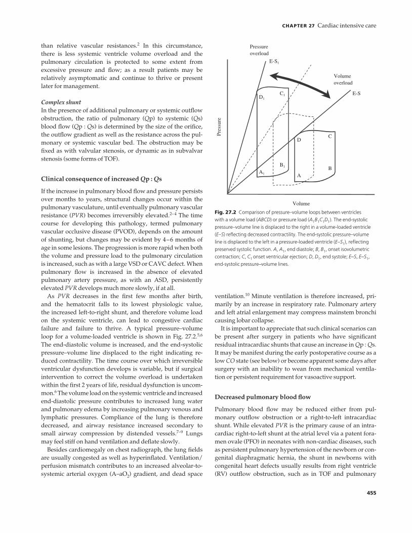
As PVR decreases in the first few months after birth, and the hematocrit falls to its lowest physiologic value, the increased left-to-right shunt, and therefore volume loadon the systemic ventricle, can lead to congestive cardiac failure and failure to thrive. A typical pressure–volume loop for a volume-loaded ventricle is shown in Fig. 27.2.5,6
The end-diastolic volume is increased, and the end-systolicpressure–volume line displaced to the right indicating re-duced contractility. The time course over which irreversibleventricular dysfunction develops is variable, but if surgicalintervention to correct the volume overload is undertakenwithin the first 2 years of life, residual dysfunction is uncom-mon.6 The volume load on the systemic ventricle and increasedend-diastolic pressure contributes to increased lung waterand pulmonary edema by increasing pulmonary venous andlymphatic pressures. Compliance of the lung is thereforedecreased, and airway resistance increased secondary tosmall airway compression by distended vessels.7–9 Lungsmay feel stiff on hand ventilation and deflate slowly.
Besides cardiomegaly on chest radiograph, the lung fieldsare usually congested as well as hyperinflated. Ventilation/perfusion mismatch contributes to an increased alveolar-to-systemic arterial oxygen (A–aO2) gradient, and dead space
Pres
sure
Volume
Pressureoverload
Volumeoverload
E-S1
E-S
AB
CD
A1
B1
C1D1
Fig. 27.2 Comparison of pressure–volume loops between ventricles
with a volume load (ABCD) or pressure load (A1B1C1D1). The end-systolic
pressure–volume line is displaced to the right in a volume-loaded ventricle
(E–S) reflecting decreased contractility. The end-systolic pressure–volume
line is displaced to the left in a pressure-loaded ventricle (E–S1), reflecting
preserved systolic function. A, A1, end diastole; B, B1, onset isovolumetric
contraction; C, C1 onset ventricular ejection; D, D1, end systole; E–S, E–S1,
end-systolic pressure–volume lines.
AFCC27 01/14/2005 03:46PM Page 455

shunt across an ASD or VSD, or a decompressing vesselfrom the pulmonary artery to pulmonary vein.
3 A reduction in mixed venous oxygen saturation, such asfrom reduced oxygen delivery secondary to a low CO stateor low hematocrit, or increased oxygen extraction in afebrile or hyper-metabolic state following surgery.
Outflow obstruction
Severe left or right ventricular outflow obstruction in thenewborn may be associated with ventricular hypertrophyand vessel hypoplasia distal to the level of obstruction. Theincreased pressure load may cause ventricular failure, mix-ing or shunting at the atrial and/or ventricular level occurs tomaintain CO.
A typical pressure–volume loop from a chronic pressureload on the ventricle is shown in Fig. 27.2. The end-diastolicpressure is elevated, and the end-systolic pressure–volumeline displaced to the left reflecting increased contractility.Maintenance of preload, afterload, and normal sinus rhythmis important to prevent a fall in CO or coronary hypoperfu-
atresia. Pulmonary blood flow is reduced and PVR is usu-ally low in these patients. The decreased flow during fetaldevelopment can lead to diminished arborization of the pulmonary vessels and a decrease in total surface area of thepulmonary vascular bed, resulting in a relatively increasedand fixed PVR.
Pulmonary mechanics and lung volumes are generallynormal in patients with reduced pulmonary blood flow.Dead space ventilation is increased although minute ventila-tion is only slightly increased to maintain normocapnia. Thelung fields appear oligemic on chest radiograph.
It is very important to know what the target SaO2 should be in the immediate postoperative period. If the SaO2 is lowerthan anticipated, there are a number of important causeswhich must be evaluated (Table 27.2). These include:1 A reduction in pulmonary venous oxygen saturation indic-
ating an intrapulmonary shunt such as from pulmonaryedema, lung collapse or pleural effusion.
2 A reduction in effective pulmonary blood flow, such as frompulmonary ventricle outflow tract obstruction or increasedpulmonary artery resistance, an intracardiac right-to-left
PART 6 Anesthesia outside the cardiac operating room
456
Etiology Considerations
Low FIO2 Inappropriately low dialed oxygen concentration
Failure of oxygen delivery device
Pulmonary vein desaturation Impaired diffusion:
Alveolar process: Edema
Infectious
Restrictive process: Effusion
Atelectasis
Intrapulmonary shunt:
RDS
Pulmonary AVM
PA-to-PV collateral vessel(s)
Reduced pulmonary blood flow Anatomic RV outflow obstruction
Anatomic pulmonary artery stenosis
Increased PVR
Atrial level right-to-left shunt:
RV hypertension
Restrictive RV physiology (low compliance)
Severe tricuspid regurgitation
Large fenestration (modified Fontan operation)
Intra-atrial baffle leak
Ventricular level right-to-left shunt:
RV hypertension and residual VSD
Low dissolved O2 content Low mixed venous oxygen level:
Increased O2 extraction: Hypermetabolic state
Decreased O2 delivery: Low cardiac output state
Anemia
AVM, arteriovenous malformation; FIO2, fractional inspired concentration of oxygen; PA, pulmonary artery;
PV, pulmonary vein; PVR, pulmonary vascular resistance; RDS, respiratory distress syndrome; RV, right
ventricle; VSD, ventricular septal defect.
Table 27.2 Factors to consider in a post-
cardiac surgery patient who has an arterial
oxygen saturation lower than the anticipated
range.
AFCC27 01/14/2005 03:46PM Page 456

CHAPTER 27 Cardiac intensive care
457
Airway and ventilation management
Altered respiratory mechanics and positive pressure ventila-tion may have a significant influence on hemodynamics fol-lowing congenital heart surgery. While changes in alveolaroxygen (PaO2), PaCO2 and pH significantly affect PVR, themean airway pressure and changes in lung volume duringpositive pressure ventilation will also affect PVR, preloadand ventricular afterload. Therefore, the approach to mech-anical ventilation should not only be directed at achieving adesired gas exchange, but is also influenced by the potentialcardiorespiratory interactions of positive pressure ventilationand method of weaning.
Airway management
Intubation of the trachea in an awake neonate or young infantwith CHD may illicit major undesirable hemodynamic andmetabolic responses, and therefore appropriate anestheticand muscle relaxant techniques are necessary to secure theairway under most circumstances.
The narrowest part of the airway before puberty is belowthe vocal cords at the level of the cricoid cartilage, and the useof uncuffed endotracheal tubes has been generally recom-mended. While a leak around the endotracheal tube at aninflation pressure of approximately 20 cmH2O is desirable, a significant air leak may have a detrimental effect on mech-anical ventilation and delivery of a consistent ventilation pattern. Examples include patients with extensive chest andabdominal wall edema following CPB and patients withlabile PVR and increased Qp : Qs. If a significant air leakexists around the endotracheal tube, lung volume, and in par-ticular functional residual capacity (FRC), will not be main-tained and fluctuations in gas exchange can occur. During theweaning process, a significant leak will also increase the workof breathing for some neonates and infants. In these situa-tions, it is therefore preferable to change the endotrachealtube to a larger size or to use a cuffed endotracheal tube.
In certain circumstances, a smaller than expected endotra-cheal tube may be necessary. This is particularly the case inpatients with other congenital defects such as Down’s syn-drome (trisomy 21). Small airway compression is commonamong patients with TOF with absent pulmonary valve.Tracheal stenosis may also occur in association with somecongenital cardiac defects such as a pulmonary artery sling.Extrinsic compression of the bronchi may occur secondary to pulmonary artery and left atrial dilation. This may be suspected by persistent hyperinflation or lobar atelectasis.
Mechanical ventilation
Altered lung mechanics and ventilation/perfusion abnorma-lities are common problems in the immediate postoperative
sion. As the time course to develop significant ventriculardysfunction is longer in patients with a chronic pressure loadcompared to a chronic volume load, symptoms of congestiveheart failure are uncommon unless the obstruction is severeand prolonged.
In the immediate postoperative period it is important toevaluate both systolic and diastolic ventricular function in apreviously obstructed but still hypertrophied ventricle.1 A hyperdynamic state may be present particularly follow-
ing left ventricle (LV) outflow reconstruction. This will bemanifest as systemic hypertension and should be treatedpromptly with β-blockers to reduce myocardial work andprotect surgical suture lines.
2 Systolic dysfunction of a hypertrophied ventricle may beapparent early after cardiac surgery secondary to myocar-dial ischemia and ventricular dysrhythmias. Ischemia mayoccur particularly if there has been a long aortic cross-clamp time, or if there has been inadequate protection ofthe subendocardium with cardioplegia solution or byhypothermia. In the case of the RV, dysfunction may alsobe present after surgery if an extended ventriculotomy hasbeen performed (e.g. TOF or truncus arteriosus repair) orthere has been direct injury to a coronary artery across theright ventricular outflow tract.
3 Diastolic dysfunction is usually manifest as a poorly com-pliant or stiff ventricle that often contracts well but isunable to relax and fill effectively during diastole. On theleft side of the heart, this is usually manifest as left atrialhypertension with either pulmonary edema, atrial dys-rhythmias or pulmonary hypertension. On the right side of the heart, an increase in RV end-diastolic pressure isdemonstrated by right atrial hypertension along with clinical signs such as a lower SaO2 from a right-to-left atrial shunt (in the presence of a PFO or residual ASD),hepatomegaly, ascites and pleural effusions.
In all the above examples of mixing, shunting and outflowobstruction, the mode and method of mechanical ventilationmay have a substantial impact on hemodynamics and sys-temic perfusion. Particularly for neonates and infants, cardio-respiratory interactions are essential to recognize duringpostoperative management. In addition to evaluating theadequacy of mechanical ventilation settings by arterial bloodgases and chest radiography, it is very important that ventila-tor settings be continually evaluated and adjusted accordingto hemodynamic response. This is completely different fromthe general concepts of mechanical ventilation used in gen-eral pediatric and neonatal ICUs. The application of standardor accepted practices for mechanical ventilation as applied topediatric patients with respiratory disease, or as applied tothe premature newborn or newborn with hyaline membranedisease, will often result in an ineffective matching of ventila-tion with perfusion in patients with congenital cardiac dis-ease, and contribute to delayed postoperative recovery andpossible adverse outcomes.
AFCC27 01/14/2005 03:46PM Page 457

result in a significant increase in PVR.13 At low tidal volumes,alveolar collapse occurs because of reduced interstitial trac-tion on alveolar septae. In addition, radial traction on extra-alveolar vessels such as the branch pulmonary arteries isreduced, therefore reducing the cross-sectional diameter.Conversely, hyperinflation of the lung may cause stretch-ing of the alveolar septae and compression of extra-alveolarvessels.
An increase in PVR increases the afterload or wall stress on the RV, compromising RV function and contributing todecreased LV compliance secondary to interventricular septal shift. In addition to low CO, signs of RV dysfunctionincluding tricuspid regurgitation, hepatomegaly, ascites, andpleural effusions may be observed.
Influence of intrathoracic pressure
An increase in mean intrathoracic pressure during positivepressure ventilation decreases preload to both pulmonaryand systemic ventricles, but has opposite effects on afterloadto each ventricle.14,15
Right ventricle
The increase in pressure in the right atrium and reduction inRV preload that occurs with positive pressure ventilationmay reduce CO. Normally the RV diastolic compliance isextremely high and the pulmonary circulation is able toaccommodate changes in flow without a large change in pres-sure. An increase in mean intrathoracic pressure increases theafterload on the RV from direct compression of extra-alveolarand alveolar pulmonary vessels. This has a number of clinicalconsequences (Table 27.3). An increase in afterload causes an increase in RV end-diastolic pressure and myocardialwork, which may lead to ischemia in a patient with limitedcoronary perfusion. An example of the increase in RV pres-sure during a positive pressure breath is demonstrated in Fig. 27.3. The increase in afterload on the RV will also reduceantegrade pulmonary blood flow and therefore preload tothe systemic ventricle. If there is pulmonary or tricuspidvalve incompetence, the amount of regurgitant flow acrossthese valves will also increase during positive pressure venti-lation from the increase in RV afterload.
Patients with normal RV compliance and without residualvolume load or pressure load on the ventricle followingsurgery usually show little change in RV function from thealteration in preload and afterload that occurs with positivepressure ventilation. However, these effects can be magnifiedin patients with RV hypertrophy and those with restrictiveRV physiology following congenital heart surgery, in partic-ular neonates who have required a right ventriculotomy forrepair of TOF, pulmonary atresia or truncus arteriosus, andpatients with concentric RV hypertrophy. While systolic RV function may be preserved, the ventricles have diastolic
period.11,12 Patients who have an increased Qp : Qs greaterthan 2 : 1 may have cardiomegaly and congested lung fieldson radiograph. Patients who have an elevated left atrial pres-sure from some form of outflow tract obstruction to the LVmay demonstrate signs of pulmonary venous hypertensionand pulmonary edema. Additional considerations includethe surgical incision and lung retraction, increased lungwater following CPB, possible pulmonary reperfusion injury,surfactant depletion in neonates, and restrictive defects fromatelectasis and pleural effusions.
In general, patients with known limited physiologic reserveshould not be weaned from mechanical ventilation untilhemodynamically stable, and problems contributing to anincrease in intrapulmonary shunt and altered respiratorymechanics have improved.
Cardiorespiratory interactions
Cardiorespiratory interactions vary significantly betweenpatients, and it is not possible to provide specific ventilationstrategies or protocols that are appropriate for all patients.Rather, the mode of ventilation must be matched to thehemodynamic status of each patient to achieve adequate CO and gas exchange. The influence of positive pressure ven-tilation on preload and afterload are shown in Table 27.3.Frequent modifications to the mode and pattern of ventila-tion may be necessary during recovery after surgery, withattention to changes in lung volume and airway pressure.
Influence of lung volume
Changes in lung volume have a major effect on PVR, which is lowest at FRC, while both hypo- or hyperinflation may
PART 6 Anesthesia outside the cardiac operating room
458
Table 27.3 The effect of a positive pressure mechanical breath on
afterload and preload to the pulmonary and systemic ventricles.
Afterload Preload
Pulmonary ventricle Elevated ReducedEffect: ↑ RVEDP Effect: ↓ RVEDV
↑ RVP ↓ RAP
↓ Antegrade PBF
↑ PR and/or TR
Systemic ventricle Reduced ReducedEffect: ↓ LVEDP Effect: ↓ LVEDV
↓ LAP ↓ LAP
↓ Pulmonary edema Hypotension
LAP, left atrial pressure; LVEDP, left ventricle end-diastolic pressure;
LVEDV, left ventricle end-diastolic volume; PBF, pulmonary blood flow;
PR, pulmonary regurgitation; RAP, right atrial pressure; RVEDP, right
ventricle end-diastolic pressure; RVEDV, right ventricle end-diastolic volume;
RVP, right ventricle pressure; TR, tricuspid regurgitation.
AFCC27 01/14/2005 03:46PM Page 458

CHAPTER 27 Cardiac intensive care
459
law, wall stress is directly proportional to the transmural LVpressure and the radius of curvature of the LV. The trans-mural pressure across the LV is the difference between theintracavity LV pressure and surrounding intrathoracic pres-sure. Assuming a constant arterial pressure and ventriculardimension, an increase in intrathoracic pressure, as occursduring positive pressure ventilation, will reduce the trans-mural gradient and therefore wall stress on the LV. Therefore,positive pressure ventilation and PEEP can have significantbeneficial effects in patients with left ventricular failure (seeTable 27.3).
Patients with LV dysfunction and increased end-diastolicvolume and pressure can have impaired pulmonary mech-anics secondary to increased lung water, decreased lung compliance and increased airway resistance. The work ofbreathing is increased and neonates can fatigue early becauseof limited respiratory reserve. A significant proportion oftotal body oxygen consumption is directed at the increasedwork of breathing in neonates and infants with LV dysfunc-tion, contributing to poor feeding and failure to thrive.Therefore, positive pressure ventilation has an additionalbenefit in patients with significant volume overload and systemic ventricular dysfunction by reducing the work ofbreathing and oxygen demand.
Weaning from positive pressure ventilation may bedifficult in patients with persistent systemic ventricular dys-function. As spontaneous ventilation increases during theweaning process, swings in mean intrathoracic pressure maysubstantially alter afterload on the systemic ventricle. Onceextubated, the subatmospheric intrapleural pressure meansthat the transmural pressure across the systemic ventricle isincreased. This sudden increase in wall stress may contributeto an increase in end-diastolic pressure and volume, leadingto pulmonary edema and a low output state. It may be diffi-cult to determine which patients are likely to fail extubationbecause of ventricular failure; even a small amount of positive pressure as used during continuous positive airwaypressure (CPAP) or pressure support modes of ventilationmay be sufficient to reduce afterload and myocardial work. Inotropic agents, vasodilators, and diuretics should becontinued throughout the weaning process and followingextubation to maintain stable ventricular function in thesepatients.
Positive end-expiratory pressure
The use of PEEP in patients with CHD has been controver-sial. It was initially perceived not to have a significant posi-tive impact on gas exchange, and there was concern that the increased airway pressure could have a detrimental effect on hemodynamics and contribute to lung injury and air leak.
Nevertheless, PEEP increases FRC enabling lung recruit-ment and redistributes lung water from alveolar septal regions
dysfunction with increased RV end-diastolic pressure andimpaired RV filling.
The potential deleterious effects of mechanical ventilationon RV function are important to emphasize. The aim shouldbe to ventilate with a mode that enables the lowest possiblemean airway pressure, yet maintaining a tidal volume of12–15 cm3/kg. While ventilating with a low peak inspirat-ory pressure, short inspiratory time, increased intermittentmandatory ventilation (IMV) rate, and low levels of positiveend-expiratory pressure (PEEP) has been recommended asone ventilation strategy in patients with restrictive RV phys-iology, the smaller tidal volumes, e.g. 6–8 cm3/kg, with thispattern of ventilation may reduce lung volume and FRC,thereby increasing PVR and afterload on the RV.
An alternative strategy in a pressure-limited mode of ven-tilation is to use larger tidal volumes of 12–15 cm3/kg, with alonger inspiratory time of 0.9–1.0 seconds, increased peakinspiratory pressure of around 30 cmH2O and low PEEP (i.e.wide P), and slow IMV rate of 12–15 breaths/minute. For thesame mean airway pressure, RV filling is maintained and RVoutput augmented by maintaining lung volume and reducedRV afterload.
Left ventricle
Left ventricular preload is also affected by changes in lungvolume. Pulmonary blood flow, and therefore preload to thesystemic ventricle, may be reduced by an increase or decreasein lung volume secondary to alteration in radial traction onalveoli and extra-alveolar vessels.
The systemic arteries are under higher pressure and notexposed to radial traction effects during inflation or deflationof the lungs. Therefore, changes in lung volume will affect LV preload, but the effect on afterload is dependent uponchanges in intrathoracic pressure alone rather than changesin lung volume.
In contrast to the RV, a major effect of positive pressureventilation on the LV is a reduction in afterload. Using Laplace’s
Fig. 27.3 Simultaneous tracings of aortic and right ventricle (RV) pressure
waveforms during positive pressure ventilation in a child with pulmonary
artery stenosis. Note the increase in RV pressure to approximately systemic
(aortic) level during inspiration when the afterload on the RV is increased.
AFCC27 01/14/2005 03:46PM Page 459

Numerous factors contribute to the inability to wean frommechanical ventilation following congenital heart surgery(Table 27.4). As a general rule, however, residual defects following surgery causing either a volume or pressure loadmust be excluded first by echocardiography or cardiaccatheterization.
Restrictive defects
Pulmonary edema, pleural effusions and persistent atelecta-sis may delay weaning from mechanical ventilation. Residualchest and abdominal wall edema, ascites, and hepatomegalylimit chest wall compliance and diaphragmatic excursion.Chest tubes and peritoneal catheters may be necessary todrain pleural effusions and ascites, respectively.
If atelectasis persists, bronchoscopy is often useful toremove secretions and to diagnose extrinsic compressionfrom enlarged and hypersensitive pulmonary arteries, adilated left atrium, or conduits. Upper airway obstructionfrom vocal cord injury (e.g. recurrent laryngeal nerve damage during aortic arch reconstruction), edema or bron-chomalacia can also be evaluated.
Phrenic nerve injury can occur during cardiac surgery,either secondary to traction, thermal injury from electro-cautery or direct transection as a complication of extensive
to the more compliant perihilar regions. Both of these actionswill improve gas exchange and reduce PVR. Positive end-expiratory pressure should, therefore, be used in all mechani-cally ventilated patients following congenital heart surgery.However, excessive levels of PEEP can be detrimental byincreasing afterload on the RV. Usually 3–5 cmH2O of PEEPwill help maintain FRC and redistribute lung water withoutcausing hemodynamic compromise.
The use of PEEP in patients who have undergone a Fontanprocedure or cavopulmonary anastomosis has also beendebated. In this group of patients, pulmonary blood flow isnon-pulsatile and depends on the pressure gradient betweenthe superior vena cava (SVC) and pulmonary venous atrium.During positive pressure ventilation, pulmonary blood flowcan be diminished, and during a Valsava maneuver and athigh levels of PEEP, retrograde pulmonary blood flow maybe demonstrated by Doppler. Nevertheless, the beneficialeffects of PEEP to 5 cmH2O as outlined above, can be demon-strated following the Fontan procedure and rarely contri-bute to a significant clinical decrease in effective pulmonaryblood flow.
Weaning from mechanical ventilation
Weaning from mechanical ventilation is a dynamic processthat requires continued re-evaluation. While most patientsfollowing congenital cardiac surgery who have had no com-plications with repair or CPB will wean without difficulty,some patients with borderline cardiac function and residualdefects may require prolonged mechanical ventilation and a slow weaning process.
The method of weaning varies between patients. Mostpatients can be weaned using either a volume- or pressure-limited mode by simply decreasing the IMV rate. Guided by physical examination, hemodynamic criteria, respiratorypattern, and arterial blood gas measurements, the mechanicalventilator rate is gradually reduced. Patients with limitedhemodynamic and respiratory reserve may demonstratetachypnea, diaphoresis and shallow tidal volumes as theystruggle to breathe spontaneously against the resistance ofthe endotracheal tube. The addition of pressure- or flow-triggered pressure support 10–15 cmH2O above PEEP is oftenbeneficial in reducing the work of breathing.
A flow-triggered mode of pressure or volume support,with a back-up ventilator rate if the patient becomes apneic,such as synchronized intermittent mandatory ventilationwith pressure or volume support, is particularly useful forneonates and infants who have either required prolongedventilation following surgery or who have a residual volumeor pressure load after surgery compromising ventricularfunction. Patients are often more comfortable weaning in thismode and have reduced work of breathing, and the level ofpressure support is adjusted according to their gas exchange,respiratory rate and tidal volume.
PART 6 Anesthesia outside the cardiac operating room
460
Table 27.4 Factors contributing to the inability to wean from mechanical
ventilation after congenital heart surgery.
Residual cardiac defects:
Volume and/or pressure overload
Myocardial dysfunction
Arrhythmias
Restrictive pulmonary defects:
Pulmonary edema
Pleural effusion
Atelectasis
Chest wall edema
Phrenic nerve injury
Ascites
Hepatomegaly
Airway:
Subglottic edema and/or stenosis
Retained secretions
Vocal cord injury
Extrinsic bronchial compression
Tracheobronchomalacia
Metabolic:
Inadequate nutrition
Diuretic therapy
Sepsis
Stress response
AFCC27 01/14/2005 03:46PM Page 460

CHAPTER 27 Cardiac intensive care
461
started before culture results are known. Signs to note inneonates and infants include temperature instability (hyper-or hypothermia), hypoglycemia, unexplained metabolic acidosis, hypotension and tachycardia with poor extremityperfusion and oliguria, increased respiratory effort and ventilation requirements, altered conscious state, and leuko-cytosis with left shift on blood count.
Colonization of the airway occurs frequently in patientsmechanically ventilated for an extended period, but may notrequire intravenous antibiotic therapy unless there is evid-ence of either increased secretions with fever, leukocytosis,new chest radiograph abnormalities or detection of an organ-ism on Gram stain together with abundant neutrophils.Urinary tract infection and both superficial and deep surgicalsite infections must also be excluded in patients with clinicalsuspicion of sepsis (i.e. sternotomy or thoracotomy wounds).
Airway
Bronchospasm can complicate mechanical ventilation andthe weaning process. While this may reflect intrinsic airwaydisease, bronchospasm can also result from increased airwaysecretions and extrinsic airway compression. Treatment with inhaled or systemic bronchodilators may be beneficial,although they should be used with caution because of their chronotropic and tachyarrhythmic potential. Levoal-buterol has less chronotropic potential and is the preferredbronchodilator among children who may be harmed bytachycardia.
The sudden onset of bronchospasm with increased peakinspiratory pressure and difficult hand ventilation shouldraise immediate concern for acute endotracheal tube obstruc-tion or pneumothorax. Bronchospasm in patients with labilePVR may reflect acute pulmonary hypertension, and treat-ment is directed at maneuvers to lower pulmonary arterypressure and improve CO.
Post-extubation stridor may be due to mucosal swelling ofthe large airway, and treatment with dexamethasone beforeextubation can be beneficial to reduce edema in patients who have required prolonged ventilation. Stridor follow-ing extubation is initially treated with nebulized racemicepinephrine, which promotes vasoconstriction and decreasesairway hyperemia and edema. If reintubation is necessary, asmaller endotracheal tube should be used. Vocal cord dys-function should also be considered, particularly as surgeryaround the ductus arteriosus and left pulmonary artery mayinjure the recurrent laryngeal nerve.
The ability to clear secretions and potential for nosocomialinfection are additional concerns in patients who have beenventilated for an extended period of time. Inability to clear secretions because of sedation, bulbar and vocal corddysfunction, ineffective cough following prolonged intuba-tion and poor nutritional state with muscle fatigue will
aortic arch and pulmonary hilar disection, particularly forrepeat operations. Diaphragmatic paresis (no motion) orparalysis (paradoxical motion), should be investigated in any patient who fails to wean.16 Increased work of breathingon low ventilator settings, increased PaCO2 and an elevatedhemidiaphragm on chest radiograph are suggestive of dia-phragmatic dysfunction. Ultrasonography or fluoroscopy isuseful for identifying abnormal diaphragmatic movement.Surgical plication of the diaphragm may be necessary as a last resort when the patient fails to wean repeatedly frommechanical ventilation.
Fluid and nutrition
Fluid restriction and aggressive diuretic therapy can result in metabolic disturbances and limit nutritional intake. Ahypochloremic, hypokalemic metabolic alkalosis with sec-ondary respiratory acidosis is a common complication fromhigh-dose diuretic use and can delay the ventilator weaningprocess. Diuretic therapy should be continually re-evaluatedbased on fluid balance, daily weight (if possible), clinicalexamination and measurement of electrolyte levels andblood, urea, nitrogen (BUN). Chloride and potassium supple-mentation is essential to correct the metabolic acidosis.
It is essential to maintain adequate nutrition, particularlyas patients will be catabolic early following cardiac surgeryand may have a limited reserve secondary to preoperativefailure to thrive. Fluid restriction may limit parenteral nutri-tion, and enteral nutrition may be poorly tolerated fromsplanchnic hypoperfusion secondary to low CO or diastolicpressure.
Sedation
Sedation is often necessary to improve synchronization with the ventilator and maintain hemodynamic stability.However, excessive sedation and/or withdrawal symptomsfrom opioids and benzodiazepines will impair the weaningprocess.
Sepsis
Sepsis is a frequent cause for failure to wean from mechanicalventilation in the ICU. Invasive monitoring catheters are acommon source for blood infections. Beside blood culturesurveillance and antibiotics, removing or replacing centralvenous and arterial catheters should be considered as soon aspossible during an episode of suspected or culture-provensepsis.
The signs of sepsis may be subtle and non-specific, andoften broad spectrum intravenous antibiotic coverage is
AFCC27 01/14/2005 03:46PM Page 461

Surgical factors
Residual or unrecognized defects
A thorough understanding of the underlying cardiac anatomy,surgical findings and surgical procedures is essential becausethis will direct the initial postoperative evaluation and exam-ination. Residual lesions may be evident by auscultation,intracardiac pressures and waveforms, and oxygen saturationdata. For example, a large v-wave on the left atrial waveformmay indicate significant residual mitral valve regurgitation.A step-up of the right atrial to pulmonary artery oxygen satu-ration of more than 10% may indicate a significant intracardiacshunt across a residual VSD.
However, if there are significant concerns for importantresidual lesions that are compromising CO and ventricularfunction, further evaluation with echocardiography and/orcardiac catheterization should be considered. Imaging of theheart may be difficult immediately after surgery because oflimited transthoracic access and acoustic windows. Duringtransthoracic echocardiography, it is important that hemody-namics be closely observed, because inadvertent pressureapplied with the transducer may adversely affect filling pressures and mechanical ventilation. Similarly, vigorousantegrade flexion of a transesophageal echocardiographyprobe may alter left atrial filling or compromise ventilationby partial obstruction of a main stem bronchus.
Surgical procedure and technique
While surgery may be routine for many uncomplicateddefects, such as ASD closure, the approach for more complexintracardiac repairs may cause specific postoperative prob-lems. For example, if a ventriculotomy is performed to closethe VSD in a patient with TOF, RV dyskinesia and poor con-traction may be apparent. On the other hand, if a transatrialapproach had been used to close the VSD in the same patient,the risk for atrioventricular valve injury or dysrhythmiassuch as junctional ectopic tachycardia and heart block isincreased. Often unexpected findings or technical difficult-ies at the time of surgery means that modifications to theapproach or procedure are necessary. A difficult proceduremay lead to a longer time on CPB or additional traction oncardiac structures.
Complications related to surgery
Failure to secure adequate hemostasis may expose the patientto significant volumes of tranfused blood products, and ifthere is inadequate drainage via chest drains placed at thetime of surgery, the risk for cardiac tamponade is significant.This may be an acute event, but more commonly it is evidentby progressive hypotension with a narrow pulse width, tachy-cardia, an increase in filling pressures and reduced peripheral
result in atelectasis and respiratory failure. Frequent chestphysiotherapy, mask CPAP and nasopharyngeal suction arebeneficial, provided patients are hemodynamically stablewith adequate gas exchange. In tachypneic patients, the useof nasopharyngeal CPAP can be beneficial by reducing thework of breathing; however, these patients have limitedreserve and frequent reassessment is essential.
Myocardial dysfunction and monitoring
Assessment of cardiac output
The accurate assessment of the postoperative patient’s COshould be a focus of management in the ICU. Establishing an adequate CO is important, because low CO is associatedwith longer duration of mechanical ventilatory support, ICUstay, and hospital stay, all of which can increase the risk ofmorbidity and/or mortality. Data from physical examina-tion, routine laboratory testing, bedside hemodynamic monitoring, echocardiography, and occasionally bedside COdetermination typically are sufficient to manage patientsoptimally. If patients are not progressing as expected and lowCO persists, a cardiac catheterization should be performed to investigate and exclude the possibility of residual or undiagnosed structural defects.
The systemic CO is defined as the product of ventricularstroke volume (in liters/beat) multiplied by heart rate (inbeats/minute).17 The ventricular stroke volume is deter-mined chiefly by three factors: afterload (the resistance toventricular emptying), preload (the atrial filling pressure),and myocardial contractility. Cardiac output is usuallyindexed to body surface area (BSA, in meters squared)because it is a function of body mass. Thus, CO/BSA is thedesignated cardiac index (CI) (in L/minute/m2). The CIvaries inversely with age, so that normal values in children at rest are 4.0–5.0 L/minute/m2, whereas the normal restingCI at age 70 is 2.5 L/minute/m2.18
Postoperative patients with low CO can present with a variety of abnormalities on physical exam or in bedsidemonitoring and laboratory values. These manifestations oflow CO are listed in Table 27.5. Clinical signs on examinationinclude cool extremities and diminished peripheral perfu-sion, tachycardia, hypotension, oliguria, and hepatomegaly.An increase in the arterial to mixed venous oxygen satura-tion difference (a–vO2) of greater than 30% and a metabolicacidosis provide biochemical evidence for a low CO state.The atrial pressure is a useful measure to follow, and both anincrease and decrease could be observed in a low CO state.Factors that should be considered when evaluating the atrialpressure following surgery are shown in Table 27.6.
The mechanism(s) underlying low CO in a specific patientcan be related to one or a combination of factors followingsurgery.
PART 6 Anesthesia outside the cardiac operating room
462
AFCC27 01/14/2005 03:46PM Page 462

CHAPTER 27 Cardiac intensive care
463
vasospasm. Examples include extrinsic compression of acoronary artery by an outflow tract conduit or annulus of aprosthetic valve, and kinking or distortion of a transferredcoronary artery button. While ECG changes may indicateischemia (ST segment abnormalities), a sudden increase inleft atrial pressure or sudden onset of a dysrhythmia such as ventricular fibrillation or complete heart block may be anearlier warning sign.
Cardiopulmonary bypass and the systemicinflammatory response
The effects of prolonged CPB relate in part to the interactionsof blood components with the extracorporeal circuit. This is
perfusion with possible evolving metabolic acidosis. This isprimarily a clinical diagnosis and treatment (i.e. opening ofthe sternum) should not be delayed while waiting for possibleechocardiographic confirmation.
Myocardial ischemia from inadequate coronary perfusionis often an under-appreciated event in the postoperativepediatric patient. Nevertheless, there are a number of cir-cumstances in which ischemia may occur, compromisingventricular function and CO. Myocardial ischemia may occurintraoperatively because of problems with cardioplegiadelivery or insufficient hypothermic myocardial protection,and from intracoronary air embolism. In the ICU setting,mechanical obstruction of the coronary circulation is usuallythe cause of myocardial ischemia rather than coronary
Physical examination
Mental status: Lethargy or irritability
Vital signs: Core hyperthermia (often associated with peripheral vasoconstriction)
Tachycardia or bradycardia
Tachypnea
Hypotension (for age and weight)
Narrow pulse pressure
Peripheral perfusion: Pale or mottled skin color and cool skin temperature
Prolonged (> 3 s) distal extremity capillary refill
Poorly palpable pulses
Signs of congestive heart failure: Failure to thrive, poor feeding and diaphoresis
Increased respiratory work, chest wall retraction
Tachypnea, grunting
Gallop rhythm
Hepatomegaly
Bedside monitoring data
ECG tracing: Rhythm other than normal sinus
Arterial waveform: Blunted upstroke and narrow pulse pressure
Atrial pressure change: See Table 27.6
Urine output: < 1.0 mL/kg/h in neonates, infants and children
< 25 mL/h in older patients
Laboratory and radiographic data
SvO2: Decreased (< 65–70%) with an increased (> 25–30%)
AV O2 difference
Acid-base balance: Metabolic acidosis with increased anion gap
Increased arterial lactate (> 2.2 mM/L)
Electrolytes: Hyperkalemia
Elevated BUN and Cr
Increased liver transaminases
Chest radiography: Cardiac enlargement
Abnormal (increased or decreased) pulmonary blood flow
Pulmonary edema
AV, arteriovenous; BUN, blood, urea, nitrogen; Cr, creatinine; ECG, electrocardiogram; SvO2, systemic
venous oxygen saturation.
Table 27.5 Manifestations of low cardiac
output.
AFCC27 01/14/2005 03:46PM Page 463

oliguria.21 Sternal closure may need to be delayed due tomediastinal edema and associated cardiorespiratory compro-mise when closure is attempted. Ascites, hepatic ingestionand bowel edema may affect mechanical ventilation, causinga prolonged ileus and delay in enteral feeding. A coagulopa-thy post-CPB may contribute to delayed hemostasis.
Dysrhythmias
The ECG is an essential component of the initial postoperat-ive evaluation because the ICU team must identify whetherthe patient is in sinus rhythm early in the recovery period. If the rhythm cannot be determined with certainty from a sur-face 12- or 15-lead ECG, temporary epicardial atrial pacingwires, if present, can be used with the limb leads to generatean atrial ECG.22 Also right and left atrial waveforms are use-ful in diagnosing atrioventicular synchrony (see Chapter 7).Temporary epicardial atrial and/or ventricular pacing wiresare routinely placed in most patients to allow mechanicalpacing should sinus node dysfunction or heart block occur in the early postoperative period. Because atrial wires areapplied directly to the atrial epicardium, the electrical signalgenerated by atrial depolarization is significantly larger andthus easy to distinguish compared to the P wave on a surfaceECG. Sinus tachycardia, which is common and often second-ary to medications (e.g. sympathomimetics), pain and anxiety,or diminished ventricular function, must be distinguishedfrom a supraventricular, ventricular, or junctional tachycar-dia. Any of these tachyarrhythmias can lower CO by eithercompromising diastolic filling of the ventricles or depressingtheir systolic function.23,24 High-grade second-degree heartblock and third-degree (or complete) heart block can dimin-ish CO by producing either bradycardia or loss of atrioven-tricular synchrony or both. Third-degree block is transient inapproximately one third of cases. If it persists beyond post-operative day 9–10, it is unlikely to resolve, and a permanentpacemaker is indicated.25
Low preload
The diagnosis of insufficient preload is usually made by mon-itoring the mean atrial pressure or central venous pressure(CVP). The most common cause in the ICU is hypovolemiasecondary to blood loss from postoperative bleeding. Initiallyafter surgery and CPB, the filling pressures may be in the normal range or slightly elevated, but this often reflects a centralized blood volume secondary to peripheral vaso- andvenoconstriction following hypothermic CPB. As the patientcontinues to rewarm and vasodilate in the ICU, considerableintravenous volume may be necessary to maintain the circu-lating blood volume. There may also be considerable third-space fluid loss in neonates and small infants who manifestthe most significant systemic inflammatory response follow-ing CPB. The “leaking” of fluid into serous cavities (e.g.
magnified in children due to the large bypass circuit surfacearea and priming volume relative to patient blood volume.Humoral responses include activation of complement,kallikrein, eicosinoid, and fibrinolytic cascades; cellularresponses include platelet activation and an inflammatoryresponse with an adhesion molecule cascade stimulatingneutrophil activation and release of proteolytic and vasoact-ive substances.19,20
The clinical consequences include increased interstitialfluid and generalized capillary leak, and potential multior-gan dysfunction. Total lung water is increased with an associ-ated decrease in lung compliance and increase in A–aO2gradient. Myocardial edema results in impaired ventricularsystolic and diastolic function. A secondary fall in CO by20–30% is common in neonates in the first 6–12 hours follow-ing surgery, contributing to decreased renal function and
PART 6 Anesthesia outside the cardiac operating room
464
Table 27.6 Factors that should be considered when there is a change in
the measured atrial pressure outside of the anticipated range for a particular
postoperative patient.
Increased
Increased ventricular end-diastolic pressure:
Decreased ventricular systolic or diastolic function
Myocardial ischemia
Ventricular hypertrophy
Ventricular outflow obstruction
Semilunar valve disease
Mitral or tricuspid valve disease
Large left-to-right anatomic shunt:
Residual ventricular septal defect
Systemic-to-pulmonary artery connection
Chamber hypoplasia
Intravascular or ventricular volume overload
Cardiac tamponade
Dysrhythmia:
Tachyarrhythmia
Complete heart block
Artifactual:
Catheter tip not in the atrium (e.g. in a ventricle or wedged in a
pulmonary vein)
Pressure transducer below level of heart or improperly calibrated or
zeroed
Concomitant drug infusions through the atrial line
Decreased
Inadequate preload
Artifactual:
Catheter malfunction (e.g. cracked or clotted)
Pressure transducer above level of heart, or improperly calibrated or
zeroed
AFCC27 01/14/2005 03:46PM Page 464

CHAPTER 27 Cardiac intensive care
465
especially older children and adults, may develop an unde-sirable dose-dependent tachycardia.
If a patient does not respond adequately to dopamine at10–15 µg/kg/minute or has severe hypotension (more than30% decrease in mean arterial blood pressure for age), treat-ment with epinephrine should be considered. Epinephrineshould be given exclusively via a central venous catheter andcan be added to dopamine at a starting dose of 0.05–0.10 µg/kg/minute, with subsequent titration of the infusion to achievethe target systemic blood pressure. At high doses (i.e. 0.5 µg/kg/minute), epinephrine can produce significant renaland peripheral vasoconstriction, tachycardia, and increasedmyocardial oxygen demand. Patients with severe ventriculardysfunction who require persistent or escalating doses ofepinephrine greater than 0.3–0.5 µg/kg/minute may benefitfrom opening of the sternum and/or should be evaluated forthe possibility of mechanical circulatory support with a ven-tricular assist device (VAD) or extracorporeal membraneoxygenation (ECMO) (see below).
A combination of epinephrine at low doses (e.g. < 0.1 µg/kg/minute) or dopamine with an intravenous afterloadreducing agent such as nitroprusside or milrinone is frequentlybeneficial to support patients with significant ventriculardysfunction accompanied by elevated afterload. Epinephrineis preferred to the equally potent inotrope norepinephrinebecause it generally is well tolerated in pediatric patients andcauses less dramatic vasoconstriction. Norepinephrine is adirect-acting α-agonist, primarily causing intense arteriolarvasoconstriction, but it also has positive inotropic actions. Atdoses of 0.01–0.2 µg/kg/minute, it can be considered inpatients with severe hypotension and low SVR (e.g. “warm”or “distributive” shock), inadequate coronary artery perfu-sion or inadequate pulmonary blood flow with a systemic-to-pulmonary artery shunt.
Delayed sternal closure
Pericardial and sternal closure following cardiac surgerycauses a restriction to cardiac function and can interfere withefficient mechanical ventilation. This is particularly impor-tant for neonates and infants in whom considerable capillaryleak and edema develops following CPB, and in whom car-diopulmonary interactions impair CO. In the operating room,mediastinal edema, unstable hemodynamic conditions andbleeding are indications for delayed sternal closure. Patientswho commonly develop hemodynamic or respiratory insta-bility in the immediate postoperative period (e.g. following a Norwood procedure for HLHS) should also be consideredfor delayed sternal closure. Urgent reopening of the sternumin the ICU following surgery is associated with higher mor-tality compared to leaving the sternum open in the operat-ing room, and successful sternal closure can usually beachieved by postoperative day 4 with a low risk for surgicalsite infection.33,34
ascites) and the extracellular space (progressive anasarca)requires that these patients receive close monitoring and vol-ume replacement to maintain the circulating blood volume.Patients with a hypertrophied or poorly compliant ventricle,and those with lesions dependent on complete mixing at theatrial level, also often require additional preload in the earlypostoperative period.
High afterload
Elevated afterload in both the pulmonary and systemic circu-lations frequently follows surgery with CPB.26 Excessiveafterload in the systemic circulation is caused by elevatedsystemic vascular resistance (SVR) and typically producesboth diminished peripheral perfusion and low urine output.Treatment of elevated SVR includes recognizing and im-proving conditions that exacerbate vasoconstriction (e.g. painand hypothermia) and administering a vasodilating agent. A vasodilator, which can be either a phosphodiesteraseinhibitor (e.g. milrinone) or a nitric oxide donor (e.g. nitro-prusside), is frequently added to an inotropic agent such asdopamine to augment CO.27–30 Neonates are intolerant ofincreased afterload, and appear to derive particular benefitfrom afterload reduction therapy.
Decreased myocardial contractility
Because decreased myocardial contractility occurs frequentlyafter reparative or palliative surgery with CPB, pharmaco-logic enhancement of contractility is used routinely in theICU. Before initiating treatment with an inotrope, however,the patient’s intravascular volume status, serum ionized Ca2+
level, and cardiac rhythm and acid-base status should be considered. Inotropic agents enhance CO more effectively ifpreload is adequate, so intravenous colloid or crystalloidadministration should be given if preload is low. If hypocal-cemia (normal serum ionized Ca2+ levels are 1.14–1.30 mmol/L)31 is detected, supplementation with intravenous calciumgluconate or calcium chloride is appropriate, because Ca2+ isa potent positive inotrope itself, particularly in neonates andinfants.32
Dopamine is usually the first-line agent to treat of eithermild (10–20% decrease in normal mean arterial blood pres-sure for age) or moderate (20–30% decrease in normal meanarterial blood pressure for age) hypotension. This sympath-omimetic agent promotes myocardial contractility by elevat-ing intracellular Ca2+, both via direct binding to myocyteβ1-adrenoceptors and by increasing norepinephrine levels.Dopamine is administered by a constant infusion because ofits short half-life, and usual starting doses for inotropy are5–10 µg/kg/minute. Dopamine should be infused through acentral venous catheter to avoid superficial tissue damageshould extravasation occur. The dose is titrated to achieve the desired systemic blood pressure, although some patients,
AFCC27 01/14/2005 03:46PM Page 465

survival in this group of patients has been reported to be ashigh as 70%.39 The outcome of patients who require ECMObecause of inability to wean from CPB in the operating roomis generally poor, with a reported survival between 10% and33%.36 –39 Other than factors such as primary myocardial dys-function, pulmonary hypertension, severe hypoxemia, andcardiac dysrhythmia after surgical repair, lack of a significantresidual defect(s) is the major factor determining successfuloutcome.
Patients with a systemic-to-pulmonary artery shunt alsohave worse outcomes from ECMO. In a shunt-dependentpulmonary circulation, runoff through the shunt will limit
Mechanical support of the circulation
Mechanical assist devices have an important role providingshort-term circulatory support to enable myocardial recoveryafter cardiac surgery or myocarditis, and can also providelonger-term support while awaiting cardiac transplantation.Although a variety of assist devices are available for adult-sizedpatients, other than ECMO, the development of alternatepediatric VADs has lagged behind adult devices. A problemfor pediatric patients is determining which method of supportis optimal. While venoarterial ECMO provides biventricularsupport with oxygenation, a number of patients may benefitfrom univentricular support with a left or right VAD.
Extracorporeal membrane oxygenation
Extracorporeal membrane oxygenation has become the mostwidely used mode of mechanical cardiopulmonary supportfor children with CHD. Venoarterial ECMO (venous cannulain the right atrium or SVC and arterial cannula in the aorta or innominate artery) is the mode of support necessary forpatients with cardiac failure. Extracorporeal membrane oxygenation fully supports the heart and lungs, similar toconventional bypass, and requires significant systemic anti-coagulation. Figure 27.4 depicts a standard ECMO circuit.Over 300 children per year receive ECMO for cardiac supportaccording to the Extracorporeal Life Support (ELSO) Registry,with the majority of patients placed on ECMO following car-diotomy.35 The percentage of patients who are successfullydecannulated from ECMO (approximately 40–45%), and thepercentage of patients subsequently discharged home aftercardiac ECMO (approximately 37%), has remained largelyunchanged over the last 5 years.36 –38 Therefore, criticalappraisal of indications and techniques is required if the success rate for cardiac ECMO is to increase to levels currently achieved in neonates with respiratory distress syndrome (approximately 90%).
Centers with an efficient and well-established ECMO ser-vice are more likely to utilize this form of support, and indica-tions for the use of ECMO vary among centers. Therefore,comparisons of the use and outcomes from ECMO betweeninstitutions are difficult to interpret. Nevertheless, this formof mechanical support clearly is life-saving and must beavailable for selected patients following congenital heartsurgery. Extracorporeal membrane oxygenation may be used to stabilize critically ill patients prior to cardiac surgery,thereby limiting end-organ dysfunction prior to surgery.Indications include severe low output state, pulmonaryhypertension and severe hypoxemia.
The best outcomes from ECMO are in postoperativepatients who have a period of relative stability after repara-tive surgery, but then develop progressive myocardial or respiratory failure, or have a sudden cardiac arrest. Hospital
PART 6 Anesthesia outside the cardiac operating room
466
Fig. 27.4 Extracorporeal membrane oxygenation (ECMO) circuit. The
dashed lines represent flow through a venoarterial ECMO circuit. Blood is
drained from the right atrium by direct atrial cannulation, or from a superior
vena cava catheter advanced into the right atrium. When using a roller
pump, the blood is first drained to a small venous reservoir, with a
centrifugal pump, and a reservoir is not used. Blood then passes through
a membrane oxygenator and heat exchanger before returning to the
ascending aorta, which is cannulated directly or via the right carotid artery.
IVC, inferior vena cava; LA, left atrium; LV, left ventricle; PA, pulmonary
artery; PV, pulmonary vein; RA, right atrium; RV, right ventricle; SVC,
superior vena cava.
AFCC27 01/14/2005 03:46PM Page 466

CHAPTER 27 Cardiac intensive care
467
bleeding and coagulopathy is common, and ample bloodproducts must be available for infusion into the circuit. Theneed for surgical exploration is common in these patients dueto accumulation of blood or clot in the mediastinum or undera patch covering an open sternum. The platelet count is main-tained above 100 000, the hematocrit above 30, and the pro-thrombin time within normal ranges. Aminocaproic acid hasbeen shown to be effective at reducing blood loss and transfu-sion in cardiac ECMO.43 Central nervous system bleeding is a catastrophic complication and should be suspected whenthere is sudden volume loss without other explanation.Cranial ultrasound in infants is adequate for screening ordiagnostic purposes. Infectious complications are preventedby meticulous sterile technique, and in many cases, broadspectrum antibiotics are required. Inadequate ECMO circuitvolume is indicated by excessive negative pressure readingson the venous side of the circuit, and an inability to maintaintarget flows and mean arterial pressures. Renal insufficiencycan be managed by placing a hemofilter in the circuit for con-tinuous dialysis and removal of inflammatory mediators.Sedation, analgesia, and paralysis are required for thesepatients, and tolerance develops rapidly. Extracorporealmembrane oxygenation support is weaned when the com-promised myocardium has recovered, which may vary fromas little as 24–48 hours to 3–4 weeks. Transthoracic echocar-diography may give an estimate of contractility if volume isadded to the heart. Before weaning inotropic agents must berestarted or increased, hematocrit and electrolytes optimized,and ventilation increased. The ECMO flow is graduallyreduced, additional cardiovascular support with intravascu-lar volume and intropic support may be required, and CO isassessed at lower levels of flow. After repeated unsuccessfulattempts to wean, the caregivers and parents or guardiansmust decide if support should be withdrawn or the patientlisted for transplantation.
Ventricular assist devices
The experience with VAD in children remains relativelysmall primarily due to technical considerations and concernsregarding suitability of children with CHD for univentricularsupport.44 A major limitation of the application of adult sys-tems for pediatric patients is the risk of thromboembolismwhen lower flows are used.
The majority of pediatric patients reported have receivedleft ventricular support, which has been particularly bene-ficial in patients with an ischemic myocardium secondary to an anomalous left coronary artery from the pulmonaryartery, and in patients who require retraining of an “unpre-pared” LV in D-TGA and intact ventricular septum or a smallVSD after an arterial switch procedure.39 Survival resultsreported to date are similar to those achieved with cardiacECMO. A typical left ventricular assist device (LVAD) circuitused for failure to wean from bypass is shown in Fig. 27.5.
systemic flow and contribute to both pulmonary overcircula-tion and ventricular volume overload. Therefore, temporaryocclusion of the shunt is necessary while on ECMO. Whenattempting to discontinue ECMO, shunt flow is re-established,potentially causing a reperfusion injury in the pulmonaryvasculature and severe pulmonary hypertension. Reducingthe shunt size to allow a limited amount of pulmonary flowwhile on ECMO will reduce the incidence of this complication.
The survival of pediatric patients who require in-hospitalresuscitation following a cardiorespiratory arrest is extremelypoor.40 Nevertheless, intact survival following pediatric car-diac surgery has been reported after prolonged cardiac arrestunresponsive to conventional closed or open chest cardiacmassage with the use of ECMO.38,41 Prolonged resuscitation,particularly when combined with the obligatory time neces-sary to set up the ECMO circuit, increases the likelihood ofsignificant neurologic and other end-organ injury. To addressthis issue, we now have a mobile ECMO circuit set up andprimed for immediate use in the ICU at all times.41 Patientswho do not demonstrate recovery of cardiac function within10–15 minutes of the initiation of advanced life support arecannulated either through the chest, neck or groin and ECMOis initiated.
Extracorporeal membrane oxygenation may also be usedas a bridge to cardiac transplantation. However, prolongedsupport from ECMO is associated with increasing potentialcomplications such as bleeding, renal failure, and sepsis. Inour experience, if there is no significant recovery of myocar-dial function after 48–72 hours on ECMO support, the trans-plant evaluation is completed.42
A brief review of ECMO management principles follows.In the emergency situation an asanguinous prime is used,with blood added later after initiation of ECMO. Initially the patient is supported with ECMO flows approaching full bypass flow, i.e. 100–150 mL/kg/minute for patients lessthan 10 kg. This provides opportunity for a completelyunloaded myocardium to recover from ischemic insult, andfully supports other organ systems. Inotropic agents are dis-continued or minimized, in order to prevent downregulationof adrenergic receptors in the myocardium and peripheralvasculature. Vasodilating agents such as milrinone, nitro-prusside, or phentolamine are often required to maintainmean arterial pressure within desired limits. The left atriummay need to be decompressed with a small cannula inpatients who have blood return to the left heart, i.e. thosewith systemic to pulmonary collaterals. Left atrial pressuremay also be elevated if ECMO flow is inadequate. The lungsare ventilated with “lung rest” settings to prevent atelectasis,but also to avoid oxygen toxicity and barotrauma, e.g. a slowrate of 10–20 breaths/minute, FIO2 less than 0.50, and PEEPof 5 cm or less with long inspiratory times and low peakinflating pressures. Anticoagulation is maintained with aheparin infusion, usually 10–20 U/kg/hour, titrated to keepactivated clotting time (ACT) within 200–250 seconds. Ongoing
AFCC27 01/14/2005 03:46PM Page 467

lungs. Inotropic support and RV afterload reduction toreduce PVR (i.e. milrinone, nitric oxide) may be necessary. Ifthe RV fails, either biventricular assist device or ECMO needsto be instituted. Since the patient’s lungs provide oxygena-tion, ventilation and pulmonary toilet must be optimized.Patients with intracardiac shunts (i.e. a PFO) may not be suit-able candidates for VAD because arterial desaturation maydevelop from right-to-left shunting, and there is increased riskof paradoxical emboli. The principles for weaning attemptsare similar to those discussed above for ECMO.
Application of adult VAD technology to larger pediatricpatients and the miniaturization of existing technology haveincreased the availability of VAD in children and the poten-tial to provide longer periods of mechanical support of thecirculation with fewer complications. The patients can poten-tially be extubated, and ambulate. Two such devices are theThoratec,46 available for patients down to 20 kg, and theBerlin Heart,47 which has been miniaturized for patients evendown to infant sizes. Survival rate for infants and childrenwith application of these technologies is 50–70%.
A VAD has a number of advantages. The circuit is simple indesign, takes less time to prime, and requires little technicalassistance once established. It may more effectively decom-press the LV, and pulsatile flow is possible with somedevices. Bleeding complications and platelet consumptionare often less problematic in patients on VAD compared toECMO. Because of the lower complications associated withVAD, patients may be supported for a longer period as abridge to transplantation.45
Management of the patient on VAD support is similar to ECMO in some respects, but the following important differences exist. An oxygenator is not used, requiring fullventilatory support. Decreased intravascular volume causesa reduction in flow; therefore, left and right atrial pressuresare monitored to establish a Starling curve for each patient.The circuit volume is smaller, and because there is no oxy-genator in the circuit, a lower level of anticoagulation isrequired (ACT of 180–200 seconds). Consequently, bleedingcomplications are fewer with VAD. A patient receiving LVADsupport must have a RV capable of pumping blood into the
PART 6 Anesthesia outside the cardiac operating room
468
Fig. 27.5 Ventricular assist device (VAD)
circuit. The VAD flow is shown in the solid lines.
With a left ventricular assist device (LVAD),
blood is drained from the left atrium or left
ventricle and pumped to the aorta. With a
right ventricular assist device (RVAD), blood is
drained from the right atrium or right ventricle
and pumped to the pulmonary artery. With a
biventricular assist device (BIVAD), both LVAD
and RVAD circuits are utilized. An ECMO circuit
(dashed lines) is shown for comparison. IVC,
inferior vena cava; LA, left atrium; LV, left
ventricle; PA, pulmonary artery; PV, pulmonary
vein; RA, right atrium; RV, right ventricle; SVC,
superior vena cava.
AFCC27 01/14/2005 03:46PM Page 468

CHAPTER 27 Cardiac intensive care
469
premature and full term newborns, causing hearing loss. Acontinuous infusion of 0.2 to 0.3 mg/kg/hour after initialbolus of 1 mg/kg i.v. often provides a consistent and sus-tained diuresis without sudden fluid shifts. Chlorothiazide10 mg/kg i.v. or p.o. every 12 hours is also an effectivediuretic, particularly when used in conjunction with loopdiuretics.
Peritoneal dialysis, hemodialysis and continuous venove-nous hemofiltration (CVVH) provide alternate renal replace-ment therapy in patients with persistent oliguria and renalfailure.52,53 In addition to enabling water and solute clearance,fluids with increased nutritional content can be increased involume to ensure adequate nutrition. The indications forrenal support vary, but include BUN greater than 100 mg/dL, life-threatening electrolyte imbalance such as severehyperkalemia, ongoing metabolic acidosis, fluid restrictionslimiting adequate nutrition, and increased mechanical ven-tilation requirements secondary to persistent pulmonaryedema, reduced chest wall compliance, or ascites.
A peritoneal dialysis catheter can be placed into the peri-toneal cavity at the completion of surgery or later in the ICU.Indications in the ICU include the need for renal support or to reduce intra-abdominal pressure from ascites that may be compromising mechanical ventilation. Drainage may besignificant in the immediate postoperative period as thirdspace fluid losses continue, and replacement with albuminand/or fresh frozen plasma may be necessary to treat hypo-volemia and hypoproteinemia.
To enhance fluid excretion if oliguria persists, “mini-volume dialysis” may be effective using 10 cm3/kg of 1.5% or 2.25% dialysate over a 30–40 minute cycle. A persistentcommunication between the peritoneum, mediastinum and/or pleural cavities following surgery will limit the effective-ness of peritoneal dialysis and is a relative contraindication.
Arteriovenous hemofiltration or hemodialysis throughdouble lumen femoral or subclavian vein catheters can beused effectively in neonates. Complications related to ven-ous access, catheter-related thrombosis and hemodynamicinstability are potential complications that require close monitoring.
Hemostasis
Hemostasis after surgery may be difficult to obtain, particu-larly if CPB has been prolonged, if there are extensive suturelines, and following repeat cardiac surgery. Prompt manage-ment and meticulous control of surgical bleeding is essentialto prevent the complications associated with a massive trans-fusion. Generally, if postoperative bleeding greater than 10cm3/kg/hour persists in the immediate postoperative periodwith a normal coagulation profile and platelet count, surgicalbleeding should be suspected, and re-exploration considered.
Factors contributing to a coagulopathy after CPB include
Intra-aortic balloon pump
Intra-aortic balloon pumps have been used with success in infants and children, although experience is limited.48
Problems in pediatric patients include size constraints of the device and balloon, difficulty synchronizing the ballooninflation–deflation cycle with faster heart rates, and theincreased elasticity of the aorta, which makes effective coun-terpulsation ineffective.
Fluid management and renal function
Because the inflammatory response to CPB often leads to an increase in total body water, fluid management in theimmediate postoperative period is critical. Capillary leak and interstitial fluid accumulation continue for the first 24–48 hours following surgery, necessitating ongoing volumereplacement with colloid or blood products. A fall in CO andincreased antidiuretic hormone secretion contribute to delayedwater clearance and potential pre-renal dysfunction, whichcould progress to acute tubular necrosis and renal failure if alow CO state persists.
During bypass, optimizing the circuit prime hematocritand oncotic pressure, attenuating the inflammatory responsewith steroids and protease inhibitors such as aprotinin, andthe use of modified ultrafiltration techniques have all beenrecommended to limit interstitial fluid accumulation.49,50
During the first 24 hours following surgery, maintenancefluids should be restricted to 50% of full maintenance, andvolume replacement titrated to appropriate filling pressuresand hemodynamic response.
Oliguria in the first 24 hours after complex surgery andCPB is common in neonates and infants until CO recovers.While diuretics are commonly prescribed in the immediatepostoperative period, CO must also be enhanced with vol-ume replacement and vasoactive drug infusions for these tobe most effective. In addition to supporting CO, low dosedopamine (3 µg/kg/minute) has the advantage of increasingrenal blood flow and promoting diuresis. Fenoldopam mesy-late, a selective dopamine (DA1) receptor agonist, leads toboth renal and splanchnic vasodilation, and has been used to provide renal protection during periods of ischemia andhypoxia such as during hypothermic CPB.51 At a dose of0.1–0.5 µg/kg/minute it may also have a role in postopera-tive ICU management to enhance renal perfusion by decreas-ing renal vascular resistance.
Furosemide 1 mg/kg i.v. every 8–12 hours is a commonlyprescribed loop diuretic that must be excreted into the renaltubular system before producing diuresis. Low CO thereforereduces its efficacy. Bolus dosing may result in a significantdiuresis over a short period, thereby causing changes inintravascular volume and possibly hypotension. Rapidboluses may also damage the hair cells of the inner ear in
AFCC27 01/14/2005 03:46PM Page 469

aortic and venous cannula placement, the duration of bypass,the rate of cooling, perfusion pressure and flow rate, airembolism, pH and PCO2 management during deep hypo-thermia, degree of hypothermia, type of oxygenator, circuithematocrit, and duration of circulatory arrest.56,57 For a moredetailed discussion see Chapters 5 and 8.
Seizures are the most frequently observed neurologic com-plication following cardiac surgery using deep hypothermiccirculatory arrest (DHCA) with a reported incidence of4–25%.58,59 Both focal and generalized seizures have beendescribed, usually occurring on the first or second postopera-tive day, and can usually be controlled with anticonvulsants;status epilepticus is uncommon. A brain CT scan is usuallynon-diagnostic, although indicated in patients with persis-tent focal seizures, because a larger area of damage, or a focallesion such as an intracerebral hemorrhage, may be seen.Postoperative seizures do not increase the risk of seizureslater in life, and in the past were thought to be benign.60
However, an ongoing, prospective, randomized trial ofDHCA versus low-flow bypass during the arterial switchoperation for D-TGA has demonstrated that seizures are amarker of neurologic damage and a prognostic indicator ofworse neurodevelopmental outcome.61–63
Detection of seizures in the postoperative period is a diagnostic challenge in the ICU. Clonic, tonic, or myoclonicmanifestations of seizures may be difficult to detect becauseof sedation or paralysis. Autonomic manifestations such asthe sudden onset of tachycardia, hypertension, and pupillarydilation are suggestive of seizure activity, although the causeof these signs is difficult to distinguish from other hemody-namic causes. Nevertheless, the early detection and man-agement of seizures post-deep hypothermic low-flow CPB(DHCPB) is important, and they should not be considered abenign event, rather both a marker for and manifestation ofongoing neurologic injury. In a prospective study followingthe arterial switch procedure (the Boston Circulatory ArrestStudy),64 clinical seizures were detected in 11% of infants, butcontinuous EEG monitoring detected seizure activity in 25%of these infants. Over recent years, postoperative clinicalseizures have become a rare event, with an incidence lessthan 4%,65 which is most likely secondary to improved tech-niques for neurological protection. Deep hypothermic circu-latory arrest is now performed less frequently, and whenused, the duration of cooling on CPB is longer, pH-stat bloodgas management strategy is used, and circulatory arrest timekept to less than 40 minutes, when possible.
Follow up of neonates and infants enrolled in the BostonCirculatory Arrest Study at 1, 4, and 8 years of age indicatesthat seizure activity following DHCA is associated withlonger-term neurodevelopmental abnormalities. At 1 year ofage, scores on the Psychomotor Development Index of theBayley Scales (a measure of fine and gross motor ability) weresignificantly lower in children randomized to circulatoryarrest versus low-flow CPB and in those who developed
hemodilution of coagulation factors and platelets from thebypass circuit prime, stimulation of the intrinsic coagula-tion pathway, and both platelet activation and aggregation. In addition, several preoperative factors may contribute topostoperative coagulopathy: chronic cyanosis, low CO withtissue hypoperfusion and disseminated intravascular coagu-lopathy, hepatic immaturity, and the use of platelet inhibitorssuch as prostaglandin E1 (PGE1) infusion and aspirin.
Transfusion of platelets and fresh frozen plasma or cryo-precipitate is often necessary. Fresh whole blood less than 24 hours old has been demonstrated to be beneficial inneonates after bypass, but not older patients. Antifibrinolyticdrugs such as ε-aminocaproic acid, tranexamic acid andaprotinin are used intraoperatively to reduce postoperativebleeding, but may also be effective in the ICU if coagulopathyand fibrinolysis persists.
Hypothermia and hypocalcemia following bypass will alsocontribute to delayed hemostasis and should be corrected inconjunction with the above-mentioned maneuvers.
Neurologic injury
While patients with CHD may have coexisting abnormalitiesof the central nervous system, they are also at risk for acquiredneurologic injury throughout the perioperative period, whichinclude paradoxical air and thrombotic emboli, cerebralabscess, and venous or arterial thrombosis from erythrocyto-sis caused by chronic cyanosis.54 Premature newborns are atrisk for intraventricular hemorrhage (IVH); cerebral ultra-sonography is recommended before and after CPB. Althoughthere are no data to support an increased incidence of IVHrelated to CPB, this should be viewed with caution based onthe ECMO experience with IVH.
Patients with CHD may develop circulatory collapseand/or severe hypoxemia which may contribute to globalhypoxic–ischemic neurologic damage. Encephalopathy andseizures are manifestations of neurologic injury; however,preoperative assessment is often difficult because of the needfor mechanical ventilation, sedation and paralysis. An elec-troencephalogram (EEG) and a computed tomography (CT)scan may help localize specific sites of neurologic injury, butthere is poor correlation with longer-term neurologic out-come. The potential for neurologic recovery of the immaturebrain is greater compared to older children and adults.55
Because of the risk (transport to radiology) and lack ofproven benefit, these patients often proceed to surgery withan uncertain neurologic status.
The application of deep hypothermia provides protectionof the central nervous system during periods of ischemia.However, prolonged periods (> 45 minutes) of hypothermicarrest may cause an ischemic reperfusion injury to the brain.In addition, there are other variables that increase the risk forbrain injury during CPB. These include problems related to
PART 6 Anesthesia outside the cardiac operating room
470
AFCC27 01/14/2005 03:46PM Page 470

CHAPTER 27 Cardiac intensive care
471
As in adult ICUs, stress ulceration and gastritis occur inpediatric patients. Prophylaxis with H2 receptor blockingdrugs and/or antacids should be used in any patient requir-ing protracted hemodynamic and respiratory support. Earlyresumption of enteral nutrition is encouraged to reduce therisk of nosocomial pulmonary and blood infections by pre-venting bacterial overgrowth.
The onset of abdominal distension, ileus, blood withinstool, and pneumatosis intestinalis on abdominal radiographsuggests necrotizing enterocolitis. Severe cases manifest addi-tional signs of abdominal wall cellulitis, sepsis, hemodynamicinstability, and gut perforation. Initial treatment includesstopping enteral feeds, and initiating intravenous maintenancefluids and broad-spectrum intravenous antibiotics. Hemody-namic support may be necessary, and occasionally laparotomyif perforation occurs or hemodynamic instability persists.
Factors contributing to postoperative liver dysfunctioninclude complications during CPB secondary to low perfu-sion pressure or inadequate venous drainage, and persistentlow CO causing ischemic hepatitis. Following a Fontan pro-cedure, patients may be at particular risk because of hepaticvenous congestion from elevated central venous pressure.Marked elevations in liver transaminases may begin withinhours of surgery and remain elevated for 2–3 days beforegradually returning to normal. These patients also typicallyhave significantly elevated prothrombin and partial throm-boplastin times, and thus are at increased risk of bleedingcomplications. Fulminant hepatic failure is uncommon.
Chylothorax
Chylothorax may occur from injury to the thoracic duct anywhere along its course from the lower mediastinum to its drainage into the systemic venous system near the leftinnominate vein. Inadvertent surgical trauma is the mostcommon cause of thoracic duct injury, particularly in repeatoperations where identification of anatomic structures isdifficult. Chylotorax is heralded by pleural effusions, oftenleft sided, that do not clear with diuresis, and worsen withenteral feeding. First line treatment involves eliminating or restricting enteral intake to non-fat-containing feedings,such as portagen. Persistent chylothorax can be treated withocreotide, tube drainage, or in severe cases, pleurodesis, or ligation of the thoracic duct. Superior vena cava thrombosismay also cause chylothorax. Most cases can be managed conservatively without surgery.
Sedation and analgesia
Maintenance of adequate analgesia and sedation is an essen-tial component of patient management following congenitalheart surgery. Besides relieving pain and anxiety after surgery
seizures in the early postoperative period. At 4 years of agelower verbal and performance intelligence quotient was alsodetected in this group of patients.
Postoperative factors contributing to neurologic injuryinclude cerebral embolism, persistent low output state withlow cerebral perfusion pressure, and hyperventilation withlow PaCO2 reducing cerebral blood flow. Post-bypass hyper-thermia may develop from low CO and peripheral vasocon-striction, or from aggressive rewarming practices, and maycontribute to neurologic injury. Post-ischemia brain tempera-ture has been shown to be important in influencing delayedneuronal death and subsequent neurologic recovery followingtraumatic brain injury. Recent data from a neonatal porcinemodel of circulatory arrest also demonstrated the detrimentaleffect of hyperthermia on neurologic outcome followingDHCA.66 Mild hypothermia may help attenuate neurologicinjury following DHCA, and hyperthermia in the immediatepostoperative period should be aggressively treated. A recentstudy of children after CPB documented that cerebral hyper-thermia (measured with jugular venous bulb thermistors)develops in a majority of patients. The mean cerebral temper-ature was 39.6°C (one patient with 41.4°C) and was under-estimated by the use of rectal temperature monitoring (mean37.7°C).67
Choreoathetosis is a rare, yet sometimes devastating, complication following cardiac surgery in children with orwithout the use of DHCA. It is reported in the neonate, but ismore common in older infants. Factors that contribute to thedevelopment of choreoathetosis include inadequate braincooling, particularly to deeper structures such as the basalganglia and midbrain, a rapid rate of cooling, an alkaline (i.e.α-stat) blood gas strategy during cooling, and the presence of systemic-to-pulmonary artery collateral vessels that mayresult in a “cerebral steal.”68,69
Gastrointestinal problems
Splanchnic hypoperfusion may be secondary to low CO fromventricular dysfunction, or from low diastolic pressure inpatients with systemic-to-pulmonary artery runoff. It oftenmanifests as a persistent ileus or feeding intolerance; gutischemia or necrotizing enterocolitis may also develop.
Besides splanchnic hypoperfusion, other causes of feedingintolerance include bowel edema following CPB, delayedgastric emptying secondary to opioids, gastroesophagealreflux, and small bowel obstruction secondary to malrotation,which occurs in some patients with heterotaxy syndrome.Patients with decreased ventricular function may be unableto increase their CO sufficiently to meet the metabolic demandassociated with oral feeding. Coexisting problems such astachypnea also restrict oral intake. To ensure adequate nutri-tion in these situations, placement of a transpyloric feedingtube should be considered.
AFCC27 01/14/2005 03:46PM Page 471
Stress response
Newborns and infants can generate a significant stressresponse following cardiac surgery and CPB.71,72 High-doseopioid anesthesia continuing into the initial postoperativerecovery has been demonstrated to significantly attenuatethis stress response, leading to a reduction in morbidity andpossibly mortality. The patients who would most benefitfrom this approach include those with limited myocardialreserve, labile pulmonary hypertension, myocardial ischemia,and those undergoing complex repairs that have requiredprolonged CPB and aortic cross-clamp times.
Improvements in preoperative assessment, earlier inter-ventions, and modifications to surgical techniques and CPBhave all contributed to improved patient outcome andreduced total ICU stay. Therefore, the notion of extendinganesthesia into the ICU is not necessary for all patients, butrather should be considered on a case-by-case basis accord-ing to hemodynamic stability after surgery.
Prolonged ventilation may be necessary for some patientsbecause of residual hemodynamic or respiratory complica-tions. Examples include persistent cardiac failure, pulmonarydisease, recurrent pleural effusions, sepsis, phrenic nerveinjury with diaphragm paresis, and muscle weakness frominadequate nutrition and prolonged paralysis. A slow weanfrom mechanical ventilation is necessary and appropriateanalgesia and sedation is important. Commonly, a combina-tion of benzodiazepines and opioids are effective, althoughthis should be frequently re-evaluated to avoid the complica-tions of tolerance and dependence.
Assessment
The assessment of adequate analgesia in children can bedifficult, particularly when paralyzed and ventilated. Prim-arily, autonomic signs such as hypertension, tachycardia,pupillary size and diaphoresis are used. If unparalyzed, chil-dren will grimace and withdraw from a painful stimulus, andif breathing spontaneously, changes in respiratory patternsuch as tachypnea, grunting and splinting of the chest wallmay be evident.
However, changes in autonomic signs do not only reflectpain. Other causes include awareness, fever, hypoxemia,hypercapnea, changes in vasoactive drug infusions, andseizures. If not diagnosed correctly, patients may receiveadditional opioid or benzodiazepine doses when hyperten-sive and tachycardic, which will only contribute to toleranceand possible withdrawal symptoms later.
Sedatives
Chloral hydrate is commonly used to sedate children prior to medical procedures and imaging studies.73 It can be admin-istered orally or rectally in a dose ranging from 50 to 100
and during procedures, attenuation of the stress responseand promoting synchrony with mechanical ventilation areimportant considerations.
A number of patients, particularly newborns, requireresuscitation and stabilization prior to surgery. Sedation andanalgesia, with or without neuromuscular blockade, is oftennecessary to minimize cardiorespiratory work, assist withcoordinated mechanical ventilation, and limit patient move-ment during painful procedures such as catheter placementand balloon septostomy. Drug doses should be titrated to thedesired effect; however, children will develop tolerance aftera few days of exposure to opioids and have increased doserequirements following surgery.
Early extubation
Recovery following cardiac surgery and duration of mechan-ical ventilation depend upon numerous factors, including thepatient’s preoperative clinical condition, type and durationof surgical procedure, hemodynamic stability, and complica-tions such as postoperative bleeding and dysrhythmias. Forthe majority of patients with stable preoperative hemody-namics undergoing uncomplicated prolonged surgical repair,postoperative ventilation is not necessary. Once hemody-namically stable, normothermic, and no longer bleeding,patients can be rapidly weaned and extubated. Examplesinclude infants and older children undergoing uncompli-cated ASD and VSD repair, RV outflow tract reconstructionand conduit replacement, and following LV outflow tractreconstruction such as aortic valve replacement. Low-doseopioid techniques are used to maintain anesthesia, and fol-lowing repairs such as an ASD, extubation in the operatingroom or soon after transfer to the ICU is usually possible.70 Asimilar approach for early extubation is applicable to patientsfollowing a cavopulmonary anastomosis when early resump-tion of spontaneous ventilation is preferable because of thepotential deleterious effects of positive pressure ventilationon preload and pulmonary blood flow.
Patients undergoing early extubation can be managed withintermittent doses of opioids to ensure adequate analgesiawithout respiratory depression, and may also benefit fromsedation with benzodiazepines to treat restlessness or agita-tion to prevent dislodgment of transthoracic catheters andchest drains.
Several problems should be anticipated following extuba-tion in the operating room or soon after transfer to the ICU. A mild respiratory acidosis is common and usually resolveswithin the first 6 hours following surgery.70 Hypertensionand tachycardia may develop during emergence from anes-thesia and sedation, increasing the risk for bleeding fromoperative suture lines; therefore antihypertensive agents maybe necessary. Warming blankets should be used to preventhypothermia and shivering, and the patient closely observedfor possible airway obstruction.
PART 6 Anesthesia outside the cardiac operating room
472
AFCC27 01/14/2005 03:46PM Page 472

CHAPTER 27 Cardiac intensive care
473
Intermittent morphine doses of 0.05–0.1 mg/kg i.v. or as acontinuous infusion at 50–100 µg/kg/hour provides excel-lent postoperative analgesia for most patients. The sedativeproperty of morphine is an advantage over the synthetic opioids; however, histamine release can cause systemicvasodilation and an increase in pulmonary artery pressure. Itshould therefore be used with caution in patients with lim-ited myocardial reserve and labile pulmonary hypertension.
The synthetic opioids, fentanyl, sufentanil, and alfentanil,have a shorter duration of action than morphine and do notcause histamine release, therefore producing less vasodila-tion and hypotension. Fentanyl is commonly prescribed following cardiac surgery. It blocks the stress response in a dose-related fashion while maintaining both systemic and pulmonary hemodynamic stability.77,78 A bolus dose of10–15 µg/kg i.v. effectively ameliorates the hemodynamicresponse to intubation in neonates.79 Patients with highendogenous catecholamine levels, e.g. severe cardiac failureor critical aortic stenosis in the neonate, can become hypo-tensive after a bolus induction dose, and fentanyl must beused with caution in these conditions. Chest wall rigidity isan idiosyncratic and dose-related reaction that can occur witha rapid bolus in newborns as well as older children.
A continuous infusion of fentanyl 3–10 µg/kg/hour pro-vides analgesia following surgery, although it often needs to be combined with a benzodiazepine to maintain sedation.Large variability between children in fentanyl clearanceexists, making titration of an infusion difficult. The experi-ence with ECMO indicates tolerance and dependence to afentanyl infusion develops rapidly and significant increasesin infusion rate may be required.
Sufentanil is more potent than fentanyl, although it hassimilar effects and offers no specific advantage. A continuousinfusion of the ultra-short acting synthetic opioid, remifen-tanil, may be useful in the ICU for patients with limitedhemodynamic reserve undergoing short procedures.
The development of tolerance is dose- and time-related,and is a particular problem following cardiac surgery in pati-ents who received a high-dose opioid technique to maintainanesthesia. Physical dependence with withdrawal symptomssuch as dysphoria, fussiness, crying, agitation, piloerection,tachypnea, tachycardia, and diaphoresis may be seen in chil-dren and can be managed by gradually tapering the opioiddose or administering a longer-acting opioid such as meth-adone. Methadone has a similar potency to morphine withthe advantage of a prolonged elimination half-life between 18 and 24 hours. It can be administered intravenously and is absorbed well orally. It is particularly useful, therefore, to treat patients with opioid withdrawal.
Alternate methods of opioid delivery which are often effective following cardiac surgery include patient-controlledanalgesia and epidural opioids, either as a bolus or continu-ous infusion. Patients receiving epidural opioids must beclosely monitored for potential respiratory depression, and
mg/kg (maximum dose 1 g). Onset of action is within 15–30minutes with a duration of action between 2 and 4 hours.Between 10% and 20% of children may have a dysphoric reaction following chloral hydrate, causing them to becomeexcitable and uncooperative. On the other hand, some chil-dren may become excessively sedated with associated respiratory depression and inability to protect their airway.
The regular administration of chloral hydrate to providesedation in the ICU is controversial. Administered intermit-tently, it can be used to supplement benzodiazepines andopioids, may assist sedation during drug withdrawal, and isuseful as a nocturnal hypnotic when trying to establish nor-mal sleep cycles. Repetitive dosing to maintain prolongedsedation is not recommended by the American Academy ofPediatrics and should be avoided in the ICU.74
Benzodiazepines are the most commonly used sedatives inthe ICU because of their anxiolytic, anticonvulsant, hypnotic,and amnestic properties. While providing excellent conscioussedation, they may cause dose-dependent respiratory depres-sion and result in significant hypotension in patients withlimited hemodynamic reserve. Following chronic adminis-tration, tolerance and withdrawal symptoms are common.
Midazolam, when administered as a continuous infusionat 0.05–0.10 mg/kg/hour is useful in children following con-genital heart surgery.75 It is short-acting and water soluble,although if CO and splanchnic perfusion are diminished,hepatic metabolism is reduced and drug accumulation canoccur. Tachyphylaxis can occur within days of commencing acontinuous infusion, and withdrawal symptoms of restless-ness, agitation and visual hallucinations can occur followingprolonged administration. A reversible encephalopathy hasbeen reported following the abrupt discontinuation of mida-zolam and fentanyl infusions, characterized by movementdisorders, dystonic posturing and poor social interaction.76
Both diazepam and lorazepam can be effectively used withinthe ICU, and they possess the advantage of longer duration ofaction. Prescribed on a regular basis, lorazepam can provideuseful longer-term sedation, supplementing an existing seda-tion regimen and assisting with withdrawal from opioids.
Opioid analgesics
Opioid analgesics are the mainstay of pain management inthe ICU, and in high doses can provide anesthesia. They alsoprovide sedation for patients while mechanically ventilatedand blunt hemodynamic responses to procedures such asendotracheal tube suctioning. Hypercyanotic episodes asso-ciated with TOF and air hunger associated with congestiveheart failure are also effectively treated with opioids.
Intermittent dosing of opioids can provide effective ana-lgesia and sedation following surgery, although periods ofover-sedation and under-medication can occur because ofpeaks and troughs in drug levels. A continuous infusion istherefore advantageous.
AFCC27 01/14/2005 03:46PM Page 473

short-term (i.e. 4–6 hours) continuous infusion of propofol at50–100 µg/kg/minute will keep the patient comfortable,allow for initial recovery after surgery (i.e. to achievehemostasis and normothermia), and permit rapid weaningand extubation after the infusion is discontinued.
Ketamine
Ketamine is a “dissociative” anesthetic agent with a rapidonset and short duration of action. It can be effectivelyadministered intravenously or intramuscularly and providesadequate anesthesia for most ICU procedures including intu-bation, draining of pleural and pericardial effusions, andsternal wound exploration and closure. It produces a type of catalepsy whereby the eyes remain open, usually with nystagmus and intact corneal reflexes. Occasionally non-purposeful myoclonic movements occur. It causes cerebralvasodilation and should be avoided in patients with intra-cranial hypertension.
Because hemodynamic stability is generally maintained, itis commonly used in ICUs. Heart rate and blood pressure areusually increased through sympathomimetic actions second-ary to central stimulation and reduced post-ganglionic cate-cholamine uptake. However, it is important to remember thatthis drug does have direct myocardial depressant effects andshould be used with caution in patients with limited myocar-dial reserve, e.g. neonates with critical aortic stenosis andpoor LV function.
Dose-related respiratory depression may occur; how-ever, most patients continue to breathe spontaneously afteran induction dose of 2–3 mg/kg i.v. Airway secretions areincreased, and even though airway reflexes seem intact, aspi-ration may occur. It is essential that patients be fasted prior toadministration of ketamine and complete airway managementequipment must be available. An increase in airway secre-tions may cause laryngospasm during airway manipulation,and an antisialagogue such as atropine or glycopyrrolateshould be administered concurrently. Side effects of emer-gence delirium and hallucinations may be ameliorated withthe concurrent use of benzodiazepines.
There are conflicting reports about the effect of ketamineon PVR. One small study in children undergoing cardiaccatheterization concluded that PVR was increased followingketamine in patients predisposed to pulmonary hyperten-sion.81 However, another demonstrated minimal effects inyoung children either breathing spontaneously or duringcontrolled ventilation.82 On balance, ketamine has minimaleffects on PVR and can be used safely in patients with pul-monary hypertension, provided secondary events such asairway obstruction and hypoventilation are avoided.
Etomidate
Etomidate is an anesthetic induction agent with the advantage
side effects include pruritis, nausea, vomiting, and urinaryretention.
Non-steroidal analgesics
Non-steroidal anti-inflammatory drugs (NSAIDs) can pro-vide effective analgesia following cardiac surgery, either as asole analgesic agent or in combination with opioids or localanesthetics. Ketorolac 0.5 mg/kg i.v. every 8 hours is particu-larly useful as an adjunct to opioids for patients who areweaned and extubated in the early postoperative period.However, there are significant concerns regarding nephro-toxicity and inhibition of platelet aggregation. The incidenceof acute renal failure is increased if ketorolac administrationis continued for more than 3 days postoperatively, and ingeneral it should be avoided in patients potentially predis-posed to renal failure, such as those with hypovolemia, pre-existing renal disease, and low CO, and those receivingmedications such as angiotensin-converting enzyme (ACE)inhibitors. Acute renal failure is more commonly seen afterinitiation of treatment, or after an increase in dose, and isreversible in most cases.80
Inhibition of platelet aggregation and increased bleedingtime may occur following a single intravenous dose ofketorolac, although it has not been demonstrated to increasethe risk of surgical site bleeding following cardiac surgery.
Actetaminophen can be given rectally, in an initial dose of30–40 mg/kg, followed by 15–20 mg/kg every 6 hours for 24–48 hours following surgery. This regimen is desirable becauseof its adjunctive ability to treat pain and lower temperaturewithout platelet inhibition effects or narcotic side effects.
Anesthetic agents
Barbiturates
Thiopental and methohexital are rarely used in the ICUbecause of direct myocardial depression and venodilationthat may cause severe hypotension in patients with limitedcardiac reserve.
Propofol
Propofol is an anesthesia induction agent that can be suitable for use in the ICU for short procedures such as transesophageal echocardiography, pericardiocentesis andcardioversion. Its use, however, is limited because of thepotential for hypotension from a decrease in SVR and directmyocardial depression. Although it has short duration ofaction and rapid clearance, propofol is currently not approvedfor longer-term continuous infusion for sedation in pediatricpatients. Nevertheless, it has a useful role in facilitating earlyextubation. Rather than relying on frequent intermittentdoses of opioids and benzodiazepines in small children, a
PART 6 Anesthesia outside the cardiac operating room
474
AFCC27 01/14/2005 03:46PM Page 474

CHAPTER 27 Cardiac intensive care
475
at a dose of 0.1 mg/kg i.v. It can cause a mild tachycardia andincrease in blood pressure, but is also safe to administer topatients with limited hemodynamic reserve.
Criteria for intensive care unit discharge
As patients improve after surgery and require less intensivemonitoring and therapy, the timing of discharge from theICU becomes an important management decision. For themajority of patients who have stable hemodynamics withoutsignificant residual defects, and who have been weaned andextubated uneventfully after surgery, the decision to transferout of the ICU is not difficult. The function of all organ sys-tems should be assessed and considered in this decision,although the focus will be on cardiovascular and respiratoryfunction. The cost of intensive care medicine is high, andearly rather than delayed discharge is recommended. Indeed,as the mortality and morbidity associated with congenitalcardiac surgery have declined, length of ICU stay, total hospital stay, and cost effectiveness have become importantoutcome variables.
In addition to poor CO and residual anatomic lesions, thereare a variety of non-cardiac problems that can complicaterecovery and prolong ICU stay. Many of these problemsaffect respiratory function and cause inability to wean frommechanical ventilation (see the Mechanical ventilation sec-tion above). Table 27.7 provides a list of cardiovascular and
of minimal cardiovascular and respiratory depression.83 Anintravenous dose of 0.3 mg/kg induces a rapid loss of con-sciousness with a duration of 3–5 minutes. It can cause painon injection and is associated with spontaneous movements,hiccoughing and myoclonus. Etomidate may be used as analternative to the synthetic opioids for induction of patientswith limited myocardial reserve. It is not approved for con-tinuous infusion because adrenal steroidogenesis can beinhibited.
Muscle relaxants
Muscle relaxants are more commonly used in pediatric ICUs compared to adult units. In addition to their ability tofacilitate intubation and controlled mechanical ventilation,patients with limited cardiorespiratory reserve also benefitfrom paralysis because of reduced myocardial work and oxy-gen demand. However, prolonged paralysis carries the con-comitant risks of prolonged ventilatory support and delayedestablishment of entral nutrition, and can result in toleranceand prolonged muscle weakness after discontinuance.
Succinylcholine is a depolarizing muscle relaxant withrapid onset and short duration of action. While frequentlyused in the pediatric ICU to facilitate intubation, the potentialfor bradycardia and hyperkalemia side effects following cardiac surgery may be disastrous. Its use should therefore be restricted to patients requiring a rapid sequence inductionbecause of the risk for aspiration of gastric contents. Theusual intravenous dose of 1 mg/kg should be increased in newborns and infants to 2 mg/kg because of the greatersurface area-to-weight ratio in these patients. It can also beadministered intramuscularly in an urgent situation whereno vascular access is available, at a dose usually double theintravenous dose (i.e. 3–4 mg/kg). The risk for bradycardia isexaggerated in children, especially after multiple doses, andatropine 20 µg/kg i.v. should be administered concurrently.
Rocuronium is an aminosteroid, non-depolarizing musclerelaxant with fast onset and intermediate duration of action.Time to complete neuromuscular blockade for an intubatingdose of 0.6 mg/kg i.v. ranges from 30 to 180 seconds, althoughadequate intubating conditions are usually achieved within60 seconds. It is therefore a suitable alternative to succinyl-choline during rapid sequence induction. The duration ofaction averages 25 minutes, although recovery is slower ininfants. It is a safe drug to administer to patients with limitedhemodynamic reserve and does not cause histamine release.
Vecuronium and cis-atracurium are non-depolarizingmuscle relaxants with intermediate durations of actions.They can be administered as an intravenous bolus or con-tinuous infusion within the ICU. Both of these agents haveminimal effect on the circulation and can be administeredsafely to patients with limited hemodynamic reserve.
Pancuronium is a commonly used, longer-acting, non-depolarizing relaxant that can be administered intermittently
Table 27.7 Criteria for intensive care unit discharge.
Cardiovascular
• Stable and desired blood pressure without requiring intravenous
vasoactive support
• Invasive intravascular monitoring no longer required for monitoring or
blood sampling
• No requirement for mechanical pacing using temporary wires and an
external pacemaker
• Stable cardiac rhythm (preferably sinus) with a stable blood pressure and
cardiac output
Respiratory
• Not dependent on mechanical ventilatory support
• Stable and adequate ventilation rate and pattern, and no signs of airway
obstruction
• Stable and adequate oxygenation (PO2 depends on lesion and physiology
after repair or palliation) ± supplemental O2 via nasal cannula, mask, or
blow-by
• Neurologic status adequate to protect airway from aspiration
• Appropriate nursing intensity:
Chest physical therapy or bronchodilator treatments at least 3 h apart
in frequency
Established nutrition plan (enteral or parenteral)
Controlled analgesic or sedation requirements
AFCC27 01/14/2005 03:46PM Page 475

17 Grossman W. Blood flow measurement: The cardiac output. In:Grossman W, Baim D, eds. Cardiac Catheterization, Angiography,and Intervention. Baltimore, MD: Williams & Wilkins, 1996: 109–24.
18 Guyton AC, Jones CE, Coleman TG. Normal cardiac output. In:Guyton AC, Jones CE, Coleman TG, eds. Cardiac Output and ItsRegulation, 2nd edn. Philadelphia, PA: Saunders, 1973: 4–80.
19 Verrier ED, Boyle EM, Jr. Endothelial cell injury in cardiovascularsurgery. Ann Thorac Surg 1997; 62: 915–22.
20 Hall RI, Smith MS, Rocker G. The systemic inflammatoryresponse to cardiopulmonary bypass: Pathophysiological, thera-peutic, and pharmacological considerations. Anesth Analg 1997;85: 766–82.
21 Wernovsky G, Wypij D, Jonas RA et al. Postoperative course andhemodynamic profile after the arterial switch operation inneonates and infants. A comparison of low-flow cardiopulmonarybypass and circulatory arrest. Circulation 1995; 92: 2226–35.
22 Perry JC, Walsh EP. Diagnosis and management of cardiacarrhythmias. In: Chang AC, Hanley FL, Wernovsky G, WesselDL, eds. Pediatric Cardiac Intensive Care. Baltimore, MD: Williams& Wilkins, 1998: 461–81.
23 Mukharji J, Rehr RB, Hastillo A et al. Comparison of atrial contri-bution to cardiac hemodynamics in patients with normal andseverely compromised cardiac function. Clin Cardiol 1990; 13:639–43.
24 Leinbach RC, Chamberlain DA, Kastor JA, Harthorne JW,Sanders CA. A comparison of the hemodynamic effects of ven-tricular and sequential A-V pacing in patients with heart block.Am Heart J 1969; 78: 502–8.
25 Weindling SN, Saul JP, Gamble WJ et al. Duration of completeatrioventricular block after congenital heart disease surgery. Am J Cardiol 1998; 82: 525–7.
26 Wessel DL. Hemodynamic responses to perioperative pain andstress in infants. Crit Care Med 1993; 21: S361–2.
27 Lawless ST, Zaritsky A, Miles M. The acute pharmacokinetics and pharmacodynamics of amrinone in pediatric patients. J ClinPharmacol 1991; 31: 800–3.
28 Benzing G, IIIrd, Helmsworth JA, Schreiber JT, Kaplan S.Nitroprusside and epinephrine for treatment of low output inchildren after open-heart surgery. Ann Thorac Surg 1979; 27: 523–8.
29 Chang AC, Atz AM, Wernovsky G, Burke RP, Wessel DL.Milrinone: Systemic and pulmonary hemodynamic effects inneonates after cardiac surgery. Crit Care Med 1995; 23: 1907–14.
30 Hoffman TM, Wernovsky G, Atz AM et al. Prophylactic intra-venous use of milrinone after cardiac operation in pediatrics (PRIMACORP) study. Am Heart J 2002; 143: 15–21.
31 Loughead JL, Mimouni F, Tsang RC. Serum ionized calcium con-centrations in normal neonates. Am J Dis Child 1988; 142: 516–18.
32 Opie LH. Regulation of myocardial contractility. J CardiovascPharmacol 1995; 26 (Suppl. 1): S1–9.
33 Tabutt S, Duncan BW, McLaughlin D et al. Delayed sternal clo-sure after cardiac operations in a pediatric population. J ThoracCardiovasc Surg 1997; 113: 886–93.
34 Alexi-Meskishvili V, Weng Y, Uhlemann F, Lange PE, Hetzer R.Prolonged open sternotomy after pediatric open heart operation:Experience with 113 patients. Ann Thorac Surg 1995; 59: 379–83.
35 Zwischenberger JB, Bartlett RH. ECMO in critical care. In:Zwischenberger JB, Bartlett RH, eds. Extracorporeal Life Support. AnnArbor, MI: Extracorporeal Life Support Registry, 1995: 445–68.
respiratory criteria for consideration prior to patient dischargefrom the ICU. It is important to emphasize that this decisionshould be multidisciplinary, with particular attention paid to nursing availability and experience, and availability ofadequate monitoring.
References
1 Castaneda AR, Mayer JE, Jr, Jonas RA et al. The neonate with criti-cal congenital heart disease: Repairaa surgical challenge. J ThoracCardiovasc Surg 1989; 98: 869–75.
2 Hoffman JI, Rudolph AM, Heymann MA. Pulmonary vasculardisease with congenital heart lesions: Pathologic features andcauses. Circulation 1981; 64: 873–7.
3 Rabinovitch M, Haworth SG, Castaneda AR, Nadas AS, Reid LM.Lung biopsy in congenital heart disease: A morphometricapproach to pulmonary vascular disease. Circulation 1978; 58:1107–22
4 Heath D, Edeards JE. The pathology of hypersensitive pulmonaryvascular disease: A description of six grades of structural changesin the ulmonary arteries with special reference to congenital cardiac septal defects. Circulation 1958; 18: 533–47.
5 Sagawa K. The end-systolic pressure–volume relation of the ventricle: Definition, modifications and clinical use. Circulation1981; 63: 1223–7.
6 Graham TP, Jr. Ventricular performance in congenital heart dis-ease. Circulation 1991; 84: 2259–74.
7 Howlett G. Lung mechanics in normal infants and infants withcongenital heart disease. Arch Dis Child 1972; 47: 707–15.
8 Bancalari E, Jesse MJ, Gelband H, Garcia O. Lung mechanics in congenital heart disease with increased and decreased pul-monary blood flow. J Pediatr 1977; 90: 192–5.
9 Lees MH, Way RC, Ross BB. Ventilation and respiratory gastransfer of infants with increased pulmonary blood flow.Pediatrics 1967; 40: 259–71.
10 Levin AR, Ho E, Auld PA. Alveolar–arterial oxygen gradients ininfants and children with left-to-right shunts. J Pediatr 1973; 83:979–87.
11 Lister G, Talner N. Management of respiratory failure of cardiacorigin. In: Gregory GA, ed. Respiratory Failure in the Child. NewYork: Churchill Livingstone, 1981: 67–87.
12 Jenkins J, Lynn A, Edmonds J, Barker G. Effects of mechanicalventilation on cardiopulmonary function in children after open-heart surgery. Crit Care Med 1985; 13: 77–80.
13 West JB. Blood flow and metabolism. In: West JB, ed. RespiratoryPhysiology: The Essentials. Baltimore, MD: Williams & Wilkins,1985: 31–48.
14 Robotham JL, Lixfeld W, Holland L et al. The effects of positiveend-expiratory pressure on right and left ventricular perform-ance. Am Rev Respir Dis 1980; 121: 677–83.
15 Pinsky MR, Summer WR, Wise RA, Permutt S, Bromberger-Barnea B. Augmentation of cardiac function by elevation ofintrathoracic pressure. J Appl Physiol 1983; 54: 950–5.
16 Watanabe T, Trusler GA, Williams WG et al. Phrenic nerve par-alysis after pediatric cardiac surgery. Retrospective study of 125cases. J Thorac Cardiovasc Surg 1987; 94: 383–8.
PART 6 Anesthesia outside the cardiac operating room
476
AFCC27 01/14/2005 03:46PM Page 476

CHAPTER 27 Cardiac intensive care
477
56 Hickey PR. Pro: Deep hypothermic circulatory arrest is preferableto low-flow bypass. J Cardiothorac Vasc Anesth 1991; 5: 635–7.
57 Mault JR, Ohtake S, Klingensmith ME et al. Cerebral metabolismand circulatory arrest: Effects of duration and strategies for pro-tection. Ann Thorac Surg 1993; 55: 57–63.
58 Jonas RA, Wernovsky G, Ware JH et al. The Boston circulatoryarrest study: Perioperataive neurologic outcome after the arterialswitch operation. Circulation 1992; 86: 360A (Abstract).
59 Newburger JW, Jonas RA, Wernovsky G et al. A comparison ofthe perioperative neurologic effects of hypothermic circulatoryarrest versus low-flow cardiopulmonary bypass in infant heartsurgery. N Engl J Med 1993; 329: 1057–64.
60 Ehyai A, Fenichel GM, Bender HW, Jr. Incidence and prognosis ofseizures in infants after cardiac surgery with profound hypother-mia and circulatory arrest. JAMA 1984; 252: 3165–7.
61 Bellinger DC, Jonas RA, Rappaport LA et al. Developmental andneurologic status of children after heart surgery with hypother-mic circulatory arrest or low-flow cardiopulmonary bypass. NEngl J Med 1995; 332: 549–55.
62 Bellinger DC, Wypij D, Kuban KC et al. Developmental and neu-rological status of children at 4 years of age after heart surgerywith hypothermic circulatory arrest or low-flow cardiopulmonarybypass. Circulation 1999; 100: 526–32.
63 Bellinger DC, Rappaport LA, Wypij D, Wernovsky G, NewburgerJW. Patterns of developmental dysfunction after surgery duringinfancy to correct transposition of the great arteries. J Dev BehavPediatr 1997; 18: 75–83.
64 du Plessis AJ, Jonas RA, Wypij D et al. Perioperative effects of α-stat versus pH-stat strategies for deep hypothermic cardiopulmon-ary bypass in infants. J Thorac Cardiovasc Surg 1997; 114: 991–1000.
65 Menache C, du Plessis AJ, Wessel DL et al. Current incidence ofacute neurologic complications after open-heart operations inchildren. Ann Thorac Surg 2002; 73: 1752–8.
66 Shum-Tim D, Nagashima M, Shinoka T et al. Postischemic hyper-thermia exacerbates neurologic injury after deep hypothermiccirculatory arrest. J Thorac Cardiovasc Surg 1998; 116: 780–92.
67 Bissonnette B, Holtby HM, Davis AJ et al. Cerebral hyperthermiain children after cardiopulmonary bypass. Anesthesiology 2000; 93: 611–18.
68 DeLeon S, Ilbawi M, Arcilla R et al. Choreoathetosis after deephypothermia without circulatory arrest. Ann Thorac Surg 1990; 50: 714–19.
69 Wong PC, Barlow CF, Hickey PR et al. Factors associated withchoreoathetosis after cardiopulmonary bypass in children withcongenital heart disease. Circulation 1992; 86: II118–26.
70 Laussen PC, Reid RW, Stene RA et al. Tracheal extubation of chil-dren in the operating room after atrial septal defect repair as partof a clinical practice guideline. Anesth Analg 1996; 82: 988–93.
71 Anand KJ, Hansen DD, Hickey PR. Hormonal–metabolic stressresponses in neonates undergoing cardiac surgery. Anesthesiology1990; 73: 661–70.
72 Anand KJ, Hickey PR. Halothane–morphine compared withhigh-dose sufentanil for anesthesia and postoperative analgesiain neonatal cardiac surgery. N Engl J Med 1992; 326: 1–9.
73 Cote CJ. Sedation for the pediatric patient. A review. Pediatr ClinNorth Am 1994; 41: 31–58.
74 American Academy of Pediatrics Committee on Drugs. Guidelinesfor monitoring and management of pediatric patients during and
36 Meliones JN, Custer JR, Snedecor S et al. Extracorporeal life support for cardiac assist in pediatric patients. Review of ELSORegistry data. Circulation 1991; 84: III168–72.
37 Pennington DG, Swartz MT. Circulatory support in infants andchildren. Ann Thorac Surg 1993; 55: 233–7.
38 del Nido PJ, Dalton HJ, Thompson AE, Siewers RD. Extra-corporeal membrane oxygenator rescue in children during cardiacarrest after cardiac surgery. Circulation 1992; 86: II300–4.
39 Duncan BW. Mechanical circulatory support for infants and chil-dren with cardiac disease. Ann Thorac Surg 2002; 73: 1670–7.
40 Schindler MB, Bohn D, Cox PN et al. Outcome of out-of-hospitalcardiac or respiratory arrest in children. N Engl J Med 1996; 335:1473–9.
41 Duncan BW, Ibrahim AE, Hraska V et al. Use of rapid-deploymentextracorporeal membrane oxygenation for the resuscitation ofpediatric patients with heart disease after cardiac arrest. J ThoracCardiovasc Surg 1998; 116: 305–11.
42 Duncan BW, Hraska V, Jonas RA et al. Mechanical circulatorysupport for pediatric cardiac patients. Circulation 1995; 94: 173.
43 Downard CD, Betit P, Chang RW et al. Impact of AMICAR ofhemorrhagic complications of ECMO: A 10-year review. J PediatrSurg 2003; 38: 1212–6.
44 Karl TR. Extracorporeal circulatory support in infants and chil-dren. Semin Thorac Cardiovasc Surg 1994; 6: 154–60.
45 Warnecke H, Berdjis F, Hennig E et al. Mechanical left ventricularsupport as a bridge to cardiac transplantation in childhood. Eur J Cardiothorac Surg 1991; 5: 330–3.
46 Reinhartz O, Keith FM, El Banayosy A et al. Multicenter experi-ence with the thoratec ventricular assist device in children andadolescents. J Heart Lung Transplant 2001; 20: 439–48.
47 Reinhartz O, Stiller B, Eilers R, Farrar DJ. Current clinical status of pulsatile pediatric circulatory support. ASAIO J 2002; 48:455–9.
48 Booker PD. Intra-aortic balloon pumping in young children.Paediatr Anaesth 1997; 7: 501–7.
49 Davies MJ, Nguyen K, Gaynor JW, Elliott MJ. Modified ultra-filtration improves left ventricular systolic function in infantsafter cardiopulmonary bypass. J Thorac Cardiovasc Surg 1998; 115:361–9.
50 Elliott MJ. Ultrafiltration and modified ultrafiltration in pediatricopen heart operations. Ann Thorac Surg 1993; 56: 1518–22.
51 Halpenny M, Lakshmi S, O’Donnell A, O’Callaghan-Enright S,Shorten GD. Fenoldopam: Renal and splanchnic effects in patientsundergoing coronary artery bypass grafting. Anaesthesia 2001; 56:953–60.
52 Giuffre RM, Tam KH, Williams WW, Freedom RM. Acute renalfailure complicating pediatric cardiac surgery: A comparison ofsurvivors and nonsurvivors following acute peritoneal dialysis.Pediatr Cardiol 1992; 13: 208–13.
53 Paret G, Cohen AJ, Bohn DJ et al. Continuous arteriovenoushemofiltration after cardiac operations in infants and children. J Thorac Cardiovasc Surg 1992; 104: 1225–30.
54 Jonas RA. Neurological protection during cardiopulmonarybypass/deep hypothermia. Pediatr Cardiol 1998; 19: 321–30.
55 Johnston MV. Brain development and its relationship to patternof injury. In: Jonas RA, Newburger JW, Volpe JJ, eds. Cardiopul-monary Bypass in Neonates, Infants and Young Children. Oxford:Butterworth–Heinemann, 1996; 237–300.
AFCC27 01/14/2005 03:46PM Page 477

79 Yaster M. The dose response of fentanyl in neonatal anesthesia.Anesthesiology 1987; 66: 433–5.
80 Tarkkila P, Rosenberg PH. Perioperative analgesia with non-steroidal analgesics. Curr Opin Anaesthesiol 1998; 11: 407–10.
81 Morray JP, Lynn AM, Stamm SJ et al. Hemodynamic effects ofketamine in children with congenital heart disease. Anesth Analg1984; 63: 895–9.
82 Hickey PR, Hansen DD, Cramolini GM, Vincent RN, Lang P.Pulmonary and systemic hemodynamic responses to ketamine ininfants with normal and elevated pulmonary vascular resistance.Anesthesiology 1985; 62: 287–93.
83 Ostwald P, Doenicke AW. Etomidate revisited. Curr OpinAnaesthesiol 1998; 11: 391–8.
after sedation for diagnostic and therapeutic procedures. Pediatrics1992; 89: 1110–15.
75 Lloyd-Thomas AR, Booker PD. Infusion of midazolam in paedi-atric patients after cardiac surgery. Br J Anaesth 1986; 58: 1109–15.
76 Bergman I, Steeves M, Burckart G, Thompson A. Reversible neu-rologic abnormalities associated with prolonged intravenousmidazolam and fentanyl administration. J Pediatr 1991; 119: 644–9.
77 Hickey PR, Hansen DD, Wessel DL et al. Blunting of stressresponses in the pulmonary circulation of infants by fentanyl.Anesth Analg 1985; 64: 1137–42.
78 Hickey PR, Hansen DD, Wessel DL, Lang P, Jonas RA. Pulmonaryand systemic hemodynamic responses to fentanyl in infants.Anesth Analg 1985; 64: 483–6.
PART 6 Anesthesia outside the cardiac operating room
478
AFCC27 01/14/2005 03:46PM Page 478