Embed Size (px)
Citation preview
26 January 2015 – JB
Identifying Bacteria with DNA Sequences
Bacteria appear in three common shapes, Spirillum, Coccus, and Bacillus. Bacteria can
be either gram-positive, meaning it has a thick layer of peptidoglycan in its cell well, or gram-
negative, meaning it lacks the thick layer of peptidoglycan. Bacteria are most visible when they
are in a colony, a group of a bunch of bacteria together. To determine if a bacterium is gram-
positive or gram-negative, a gram stain procedure is done, and if the bacteria are purple after the
procedure, then it is gram-positive. The purpose of this lab was to become more familiar with
bacterial identification and to identify DNA sequences of bacteria.
In this lab we first observed both the hay infusion and the gar plates. We then created
two wet mounts with two colonies from separate plates. We observed those colonies under a
microscope and determined the shape and type of bacteria that they were. We determined this by
looking at shape, color, motility, and size. Next we carried out the gram staining procedure with
four separate colonies from four separate plates (2 from the same plates used for the wet
mounts). Lastly, we ran PCR on the four colonies from the plates that we used for gram staining.
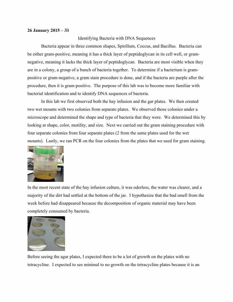
In the most recent state of the hay infusion culture, it was odorless, the water was clearer, and a
majority of the dirt had settled at the bottom of the jar. I hypothesize that the bad smell from the
week before had disappeared because the decomposition of organic material may have been
completely consumed by bacteria.
Before seeing the agar plates, I expected there to be a lot of growth on the plates with no
tetracycline. I expected to see minimal to no growth on the tetracycline plates because it is an

antibiotic and would normally act to prevent growth of bacteria. Some growth may still occur
because tetracycline only fights certain type of bacteria, and others that are resistant to the drug
may persist and grow.
Dilution Agar Type Colonies
Counted
Conversion
Factor
Colonies/mL
10^-3 Nutrient 50 x10^3 50 x 10^3
10^-5 Nutrient 61 x10^5 61 x 10^61
10^-7 Nutrient 1 x10^7 10^7
10^-9 Nutrient 1 x10^9 10^9
10^-3 Nutrient + tet 1 x10^3 10^3
10^-5 Nutrient + tet 56 x10^5 5.6 x 10^8
10^-7 Nutrient + tet 0 x10^7 0
10^-9 Nutrient + tet 0 x10^9 0
Plate 10^-5 Nutrient plus tetracycline may have been high in bacteria population because it was a
bacteria that was resistant to the drug or antibiotic. Also, contamination may have also been
possible, causing the unusual number to occur. Contamination may have also occurred on plates
10^-7 nutrient and 10^-9 because the bacteria population is unusually low. Overall the
tetracycline seemed to reduce bacteria growth indicating that it was a mostly effective drug in
eliminating bacteria. It seems that only one species of bacteria were unaffected by the drug. I
think that the bacteria that survived must have had a very gram-positive peptidoglycan wall,
defending against the bacteria well. The others may have had weaker walls, succumbing the
drug.
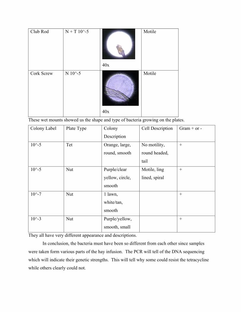
Club Rod N + T 10^-5
40x
Motile
Cork Screw N 10^-5
40x
Motile
These wet mounts showed us the shape and type of bacteria growing on the plates.
Colony Label Plate Type Colony
Description
Cell Description Gram + or -
10^-5 Tet Orange, large,
round, smooth
No motility,
round headed,
tail
+
10^-5 Nut Purple/clear
yellow, circle,
smooth
Motile, ling
lined, spiral
+
10^-7 Nut 1 lawn,
white/tan,
smooth
+
10^-3 Nut Purple/yellow,
smooth, small
+
They all have very different appearance and descriptions.
In conclusion, the bacteria must have been so different from each other since samples
were taken form various parts of the hay infusion. The PCR will tell of the DNA sequencing
which will indicate their genetic strengths. This will tell why some could resist the tetracycline
while others clearly could not.