Embed Size (px)
Citation preview
HAL Id: hal-01808094https://hal-univ-rennes1.archives-ouvertes.fr/hal-01808094
Submitted on 20 Jun 2018
HAL is a multi-disciplinary open accessarchive for the deposit and dissemination of sci-entific research documents, whether they are pub-lished or not. The documents may come fromteaching and research institutions in France orabroad, or from public or private research centers.
L’archive ouverte pluridisciplinaire HAL, estdestinée au dépôt et à la diffusion de documentsscientifiques de niveau recherche, publiés ou non,émanant des établissements d’enseignement et derecherche français ou étrangers, des laboratoirespublics ou privés.
2018 Update of French Recommendations on theManagement of Postmenopausal Osteoporosis
Karine Briot, Christian Roux, Thierry Thomas, Hubert Blain, Daniel Buchon,Roland Chapurlat, Françoise Debiais, Jean Marc Feron, Jean Bernard
Gauvain, Pascal Guggenbuhl, et al.
To cite this version:Karine Briot, Christian Roux, Thierry Thomas, Hubert Blain, Daniel Buchon, et al.. 2018 Updateof French Recommendations on the Management of Postmenopausal Osteoporosis. Joint Bone Spine,Elsevier Masson, 2018, 85 (5), pp.519-530. �10.1016/j.jbspin.2018.02.009�. �hal-01808094�
Page 1 of 48
Accep
ted
Man
uscr
ipt
1
2018 Update of French Recommendations on the Management of Postmenopausal
Osteoporosis
Karine Briot ¹, Christian Roux ¹, Thierry Thomas ², Hubert Blain ³, Daniel Buchon 4, Roland
Chapurlat 5, Françoise Debiais 6, Jean Marc Feron 7, Jean Bernard Gauvain 8, Pascal
Guggenbuhl 9,10,11, Eric Legrand 12, Anne Marie Lehr-Drylewicz 13, Eric Lespessailles 14,
Françoise Tremollieres 15, Georges Weryha 16, Bernard Cortet 17
¹ Service de rhumatologie, Hôpital Cochin, 75014 Paris, France
² INSERM U1059, Service de Rhumatologie, CHU de St Etienne, 42100 St Etienne, France
³ CHU de Montpellier, université Montpellier 1, centre Antonin-Balmes, unité de soins aigus
gériatriques, 34090 Montpellier, France
4 Médecine générale, 87032 Limoges, France
5 Service de rhumatologie, Inserm U1033, université de Lyon, hôpital Edouard-Herriot, 5,
place d'Arsonval, 69437 Lyon, France
6 Service de rhumatologie, CHU Poitiers, 86021 Poitiers, France
7 Service de Chirurgie Orthopédique de l’Hôpital Saint Antoine, Hôpitaux Universitaires Est
Parisiens, 75012 Paris, France
8 Centre de Médecine Gériatrique, 45000 CHR d'Orléans, Orléans, France
9 Institut NUMECAN, INSERM U 1241, INRA U 1341, 35000 Rennes, France
10Service de Rhumatologie, Hôpital Sud, CHU, 35000 Rennes, France
11Université de Rennes 1, 35000 Rennes, France
12 Service de Rhumatologie, CHU Angers, 49000 Angers, France
13 Médecine générale, 37000 Tours, France
14 Service de rhumatologie, CHR Orléans, 45000 Orléans, France

Page 2 of 48
Accep
ted
Man
uscr
ipt
2
15 Centre de ménopause, hôpital Paule-de-Viguier, 31300 Toulouse, France
16 Service d’endocrinologie, CHU Nancy, 54000 Nancy, France
15 EA 4490, Service de Rhumatologie, CHU Lille, 59000 Lille, France
Corresponding author: Karine Briot, Hôpital Cochin, Service de rhumatologie, 27, rue du
Faubourg Saint-Jacques, 75014 Paris, FRANCE
E-mail: [email protected]
Page 3 of 48
Accep
ted
Man
uscr
ipt
3
ABSTRACT
Objectives: To update the 2012 recommendations on pharmacotherapy for postmenopausal
osteoporosis, under the aegis of the Bone Task Force of the French Society for Rheumatology
(SFR) and of the Osteoporosis Research and Information Group (GRIO), in collaboration
with scientific societies (Collège National des Généralistes Enseignants, Collège National
des Gynécologues et Obstétriciens Français, Fédération Nationale des Collèges de
Gynécologie Médicale, Groupe d’Étude de la Ménopause et du Vieillissement hormonal,
Société Française de Chirurgie Orthopédique, Société Française d’Endocrinologie, and
Société Française de Gériatrie et de Gérontologie)
Methods: Updated recommendations were developed by a task force whose members
represented the medical specialties involved in the management of postmenopausal
osteoporosis. The update was based on a literature review and developed using the method
advocated by the French National Authority for Health (HAS).
Discussion and conclusion: The updated recommendations place strong emphasis on the
treatment of women with severe fractures, in whom the use of osteoporosis medications is
recommended. All the available osteoporosis medications are suitable in patients with severe
fractures; zoledronic acid deserves preference as the fist-line drug after a hip fracture. In
patients with or without non-severe fractures, the decision to use osteoporosis medications is
based on bone mineral density values and, in challenging cases, on probabilities supplied by
prediction tools such as FRAX®. All osteoporosis medications are suitable; raloxifene should
be reserved for patients at low risk for peripheral fractures. The fracture risk should be
reevaluated every 2 to 3 years to decide on the best follow-up treatment. These updated
Page 4 of 48
Accep
ted
Man
uscr
ipt
4
recommendations discuss the selection of first-line osteoporosis medications and treatment
sequences.
Keywords: Osteoporosis. Menopause. Fracture. Recommendations. Bone absorptiometry.
FRAX.
Page 5 of 48
Accep
ted
Man
uscr
ipt
5
1. Objectives and methods
These updated recommendations are intended for all physicians involved in managing
women who have, or are at risk for, postmenopausal osteoporosis. The objectives of the
update are to review current epidemiological data on postmenopausal osteoporosis; identify
the key criteria for evaluating patients at high risk for fractures; and clarify the principles of
drug therapy for postmenopausal osteoporosis in the light of recent evidence about
indications, efficacy, and safety. The content of the recommendations was discussed and
elaborated according to the method advocated by the French National Authority for Health
(HAS) then validated by a multidisciplinary study group. When published data were
inadequate or incomplete, expert consensus formed the basis for the recommendations, to give
due weight to current practice and expert opinion.
No recommendations can encompass every specific situation, the full spectrum of
comorbidities, or all hospital care protocols. Therefore, the current update does not claim to
cover all possible management strategies and should not serve as a substitute for individual
physician responsibility regarding treatment decisions. The indications of the drugs and the
information on reimbursement by the statutory health insurance system of drugs, bone
absorptiometry, and serum 25-OH-vitamin D assays are valid for France.
These updated recommendations were developed by a project manager and a scientific
committee then discussed and revised by a multidisciplinary panel of reviewers. As part of the
process of revising and validating the recommendations, advice was obtained from the
following scientific societies: Collège National des Généralistes Enseignants, Collège
National des Gynécologues et Obstétriciens Français, Fédération Nationale des Collèges de
Page 6 of 48
Accep
ted
Man
uscr
ipt
6
Gynécologie Médicale, Groupe d’Étude de la Ménopause et du Vieillissement hormonal,
Groupe de Recherche et d’Information sur les Ostéoporoses (GRIO), Société Française de
Chirurgie Orthopédique, Société Française d’Endocrinologie, Société Française de Gériatrie
et de Gérontologie, and Société Française de Rhumatologie (SFR).
2. Epidemiology of osteoporosis and fractures
2.1. Epidemiology
Osteoporosis is a generalized bone disease in which bone strength is diminished,
resulting in a risk of fractures (1). As a disease that increases the risk and frequency of several
severe fractures associated with devastating consequences, osteoporosis is a major public
health issue. The fracture risk increases substantially with age, and the burden placed by
osteoporosis on public health is therefore heaviest in countries with long life expectancies.
The recommendations presented here apply to patients in whom causes of decreased bone
strength other than postmenopausal osteoporosis have been ruled out.
Osteoporotic or fragility fractures are induced by low-energy trauma such as a fall
from standing height while walking. The only bones where osteoporotic fractures do not
occur are the skull, facial bones, cervical spine, first three thoracic vertebras, hand bones, and
toes; fractures at these sites are due to either injuries or tumors. Falls from standing height are
the leading cause of non-vertebral fragility fractures. Among these falls, 5% are responsible
for fractures at any site (2) and 2% for hip fractures in individuals older than 65 years of age
(3).
2.1.1. Epidemiological data from France

Page 7 of 48
Accep
ted
Man
uscr
ipt
7
Annual estimates for 2001 in France blame osteoporosis for about 70 000 vertebral
fractures, 60 000 hip fractures, and 35 000 wrist fractures (4). The number of patients
requiring surgery for hip fractures increased between 2002 and 2013 by 5% in females (from
49 287[AW1] to 50 215) and by 22% in males (from 12 716 to 15 482) (5). According to a
report issued in January 2016 by a French national research agency (Direction de la
Recherche, des Etudes, de l'Evaluation et des Statistiques), among patients in France older
than 55 years of age who sustain a hip fracture due to any cause, 23.5% die within the
following year (www.data.drees.sante.gouv.fr).
A study by the French statutory health insurance system for salaried workers (Caisse
Nationale d’Assurance Maladie) assessed hospital admissions in France of patients older than
50 years with fractures in 2013 (6). The number of patients admitted with osteoporotic
fractures at any site was 177 000. Among these patients, three-quarters were female and two-
thirds were older than 70 years. The number of admissions for fractures of any type increased
by 9% between 2011 and 2013. Direct annual costs totaled 771 million Euros. During the year
following the admission, 6325 (7%) patients died, 12% experienced another fracture, and
40% were readmitted. The mortality rate was twice as high in males than in females. During
the first year after the fracture, only 10% of patients underwent bone absorptiometry and only
15% were started on osteoporosis medication. Thus, over 80% of patients did not receive
appropriate management after sustaining a fracture that required hospital admission. These
data from France (6) are consistent with those reported worldwide (7).
2.1.2. Consequences of severe fractures
Severe fractures are associated with an increase in mortality. Fractures are severe at
the following sites: hip, proximal humerus, spine, pelvis, sacrum, femoral shaft, distal femur,
ribcage involving at least three ribs, and proximal tibia (8-10). The excess mortality compared
Page 8 of 48
Accep
ted
Man
uscr
ipt
8
to the general population occurs chiefly among patients younger than 70 years of age (8).
Epidemiological studies have confirmed that pelvic and humeral fractures are associated with
increased mortality (11). Furthermore, severe fractures are associated with a risk of further
vertebral and non-vertebral fractures, which account for 25% of the excess mortality (12).
Other common fractures, such as forearm fractures, are classified as non-severe
because they are not associated with an increase in mortality, although their impact may be
substantial. Non-severe fractures can be the first manifestation of osteoporosis and are
associated with a risk of further fractures, which may be severe (13).
2.2. Risk factors for fractures and short-term fracture risk
A recent fracture is a major risk factor for a further fracture in the short term. More
specifically, after a vertebral fracture there is a 25% risk of sustaining another fracture within
the following year (14). The risk of fracture is also increased during the 2 to 3 years after a
non-vertebral fracture (15, 16). In addition to a recent fracture, risk factors for a repeat
fracture in the short term include risk factors for falls (17, 18). Patients who have these risk
factors should be treated promptly to prevent further fractures.
2.3. Comorbidities and fracture risk
Patients with osteoporosis have an increased prevalence of chronic comorbidities such
as dementia, Parkinson’s disease, other neurological disorders, diabetes, and cardiovascular
disease, and many are on multiple medications (19, 20). These comorbidities must receive
appropriate attention, as they increase the fracture risk and the adverse impact of fractures.
Examples of situations in which fractures are particularly deleterious include hip fractures in
patients with dementia and vertebral fractures in those with chronic obstructive pulmonary
Page 9 of 48
Accep
ted
Man
uscr
ipt
9
disease. The risk/benefit ratio should be evaluated carefully in the event of comorbidities and
the treatment options and administration modalities should be discussed with the patient.
3. Evaluation of the fracture risk and treatment decisions
Decisions about offering osteoporosis treatment are guided by the existence, type, and
date of previous fractures; patient age; risk factors for falls; bone mineral density (BMD)
values; and comorbidity profile.
3.1. Fracture risk prediction tools
3.1.1. History of fractures in the patient
A history of fracture is the strongest predictor of further fractures (14, 15, 18, 20,21),
regardless of fracture location at the spine or at a peripheral site. The period of greatest risk
increase is 2 to 3 years after the first fracture. However, the risk increase remains significant
for 10 to 15 years (particularly after vertebral and humeral fractures) (22, 23). The time since
the fracture is important to note, as only recent fractures are associated with an increased
short-term risk of fractures.
Vertebral fractures are common, but their frequency is underestimated, as they are
missed in two-thirds of cases due to the paucity of the symptoms or to a mistaken diagnosis of
disk disease as the source of pain, in the absence of a radiographic assessment. Vertebral
fractures are a key risk factor for vertebral and non-vertebral fractures. The risk of further
fractures increases with the number and severity of the existing vertebral fractures. Even in
the absence of symptoms, a radiographic vertebral fracture is associated with an increased
relative risk of incident fractures during the first year and for up to 15 years after the
diagnosis, after adjustment for age and BMD[AW2] (21).
Page 10 of 48
Accep
ted
Man
uscr
ipt
10
Vertebral fracture assessment (VFA) is a dual-energy X-ray densitometry technique for
detecting vertebral fractures at the thoracic and lumbar spine. VFA is not reimbursed by the
health insurance system in France. VFA is indicated in postmenopausal women with spinal
pain or any of the following criteria: loss of height 4 cm compared to historical height (at 20
years of age), loss of height 2 cm as established prospectively during follow-up, previous
vertebral fracture, chronic comorbidities, and treatments associated with a high risk of
vertebral fracture (glucocorticoids and aromatase inhibitors) (ISCD 2015) (www.iscd.org)
(expert consensus).
3.1.2. Bone mineral density (BMD) measurement
Dual-energy X-ray absorptiometry (DXA) is the reference method for measuring
BMD at the lumbar spine and hip. Bone strength correlates strongly with BMD. In
postmenopausal women, BMD results are reported as T-scores. The T-score is the number of
standard deviations (SDs) of the measured BMD value above or below the same-site mean
BMD in young women. The World Health Organization defines osteoporosis as a T-score ≤-
2.5 at the femoral neck (24). Since July 1, 2006, the French health insurance system
reimburses DXA in women meeting the criteria listed in Box 1.
3.1.2.1. Selecting the BMD measurement site and reference curve
BMD should be measured at two sites, the lumbar spine and proximal femur (femoral
neck and total hip). BMD measurement at the radius is not indicated for the evaluation of
postmenopausal osteoporosis (25). According to the International Osteoporosis Foundation, if
DXA is performed at a single site, the femoral neck or total hip should be selected and the
NHANES III reference curve used (26, 27). BMD at the femoral neck correlates more
Page 11 of 48
Accep
ted
Man
uscr
ipt
11
strongly with the fracture risk in cohort studies overall, is used in the FRAX tool, and can
serve to monitor treatment effects.
3.1.2.2. Relation between a low T-score and the fracture risk
The fracture risk increases as BMD decreases: for each BMD decrease by 1 SD, the
fracture risk increases 2-fold (28-30). As the T-score value declines, the risk of osteoporotic
hip fracture increases (31, 32). BMD decrease at any site is associated with a higher risk of
fracture at any site. Nevertheless, a decline in BMD measured at the femur strongly predicts
the risk of fracture at any site and at the femur (32, 33).
3.1.2.3. Limitations of using a T-score cutoff
Defining osteoporosis based on BMD criteria fails to identify all women at risk for
fractures. Thus, over 50% of non-vertebral fractures occur in women whose T-score is above
-2.5 (30, 34-36). Among patients with osteoporosis diagnosed based on a fracture after a
trivial trauma although they do not meet BMD criteria for the disease, bone tissue
assessments show specific alterations responsible for decreased bone strength. For instance,
obese individuals may have BMD values that are too low for their body weight, diabetic
patients exhibit bone matrix abnormalities related to protein glycation, and women starting
aromatase inhibitor therapy experience excessive bone resorption (37-41).
3.1.3. Evaluating the fall risk
Risk factors for falls play a central role in the occurrence of non-vertebral fractures in
very elderly and/or frail patients (42). Recommendations about identifying individuals at high
risk for falls were issued by the HAS in 2005 (http://www.has-
sante.fr/portail/upload/docs/application/pdf/prevention_des_chutes-argumentaire.pdf).
Page 12 of 48
Accep
ted
Man
uscr
ipt
12
Numerous factors increase the risk of falls (Table 1). Routinely evaluating and
managing risk factors for falls in elderly patients would raise major challenges. Therefore,
HAS guidelines, together with guidelines issued in the UK and US in 2010[AW3] and
recommendations by other scientific societies, support the use of simple tests and questions as
screening tools for elderly patients. In practice, a history of falling, particularly within the last
3-6 months and regardless of the circumstances, or a fear of falling that restricts self-
sufficiency should prompt an evaluation for causes of balance impairment, if needed during a
specialized geriatric visit. In doubtful cases, dynamic balance impairments responsible for an
up-and-go time above 14 seconds or static balance impairments with a single-leg stance time
below[AW4] 5 seconds and/or instability during the sternal push test indicate a need for an
etiological workup and an appropriate management strategy (43, 44).
3.2. Fracture risk prediction tools for specialists
These tools can be helpful to all physicians trained in interpreting their results and
experienced in managing bone diseases.
3.2.1. Absolute fracture risk estimation using the FRAX
The identification of individuals at risk for fractures requires a multifactorial assessment
including BMD measurement and an evaluation of the clinical risk factors associated with the
fracture risk (Box 2). The roles played by the various risk factors varies with age. The
Fracture Risk Assessment Tool FRAX was developed to quantify the fracture risk (45)
(www.sheffield.ac.uk/FRAX). The tool estimates the 10-year probability of a hip fracture and
of major fracture defined as a fracture of the hip, humerus, wrist or a clinical vertebral
fracture. The FRAX tool has been tested in several cohorts in France (46-50). The
Page 13 of 48
Accep
ted
Man
uscr
ipt
13
recommendations set forth below (consensus of experts) are based on national validation and
calibration studies (46-50) and on international recommendations (NOF, NOS, NOGG).
- The FRAX tool is not useful when there is a clear indication to start osteoporosis
therapy, for instance a history of severe fracture or a T-score -3 at the lumbar spine and at
the total hip and/or femoral neck.
- The FRAX cutoff above which osteoporosis treatment is appropriate varies with age. For
a given age, the FRAX cutoff for treatment is the value in same-age women with a history of
fracture (risk of repeat fracture) (45). Figure 1 shows the cutoffs according to age (consensus
of experts).
3.2.2. The trabecular bone score (TBS)
The trabecular bone score (TBS) is a measure of bone texture that is automatically
computed from DXA data at the lumbar spine. TBS values are lower in patients with than
without fragility fractures and have been found effective in discriminating between these two
groups. Combining the BMD and TBS values predicts the fracture risk more effectively than
does the lumbar BMD value alone. Nevertheless, in prospective studies, femoral BMD was
the strongest predictor. Routine TBS measurement for fracture risk prediction and treatment
monitoring is not recommended, as the ability of the TBS to reclassify patients has not been
firmly established (51, 52). The TBS is associated with the risk of osteoporotic hip fractures
after adjustment on the FRAX® probability (53, 54). A metaanalysis of 14 prospective studies
showed that adjustment on the TBS did not substantially improve the predictive performance
of the FRAX® tool (55, 56). In situations where the appropriateness of osteoporosis therapy is
not obvious, the TBS[AW5] can be used in the same way as the FRAX and with the same
treatment-initiation cutoffs.
Page 14 of 48
Accep
ted
Man
uscr
ipt
14
3.2.3. Bone turnover markers
Several laboratory markers are available for noninvasively assessing bone turnover.
Among them, some reflect bone formation (osteocalcin, bone alkaline phosphatase,
procollagen I extension peptides) and others bone resorption (degradation peptides).
However, the clinical meaning of bone marker levels must be interpreted in the light of
potential confounders such as renal function or a recent fracture. No markers are available for
predicting DXA results. In contrast, bone turnover markers can predict bone loss. Combining
several markers and/or combining markers and risk factors might improve the prediction of
bone fragility. No marker associated with an increased fracture risk was reproducibly
identified in published studies. However, combining markers and BMD measurement may
improve predictive performance. According to recommendations recently issued by experts,
there is insufficient evidence that bone turnover markers help to predict the fracture risk in
clinical practice (57). thus, routine bone turnover marker assays are not recommended for this
purpose but may help specialists decide whether osteoporosis therapy is in order in difficult
cases. Furthermore, marker assays are useful for monitoring the effects of anti-resorptive
treatments.
4. Strategies for preventing and treating postmenopausal
osteoporosis
The diagnosis of osteoporosis requires the elimination of other causes of bone fragility,
which include metabolic diseases, malignancies, and genetic disorders. This diagnostic step
must be completed before starting osteoporosis therapy. Fracture prevention is the treatment
goal. Therefore, osteoporosis therapy should aim both to increase bone strength and to
decrease the risk of falls. The management strategy thus combines pharmacological and
Page 15 of 48
Accep
ted
Man
uscr
ipt
15
nonpharmacological components. The efficacy of available osteoporosis medications in
preventing fractures was proven in populations with osteoporosis diagnosed based on BMD
criteria or on a history of fracture (Tables 2 and 3) (58-75).
No head-to-head comparisons of the antifracture efficacy of osteoporosis medications
are available. Neither BMD values nor biochemical parameters can serve to compare efficacy.
Important considerations when selecting the drug include beneficial or undesirable extra-
skeletal effects, specific contraindications of each drug, constraints for patients, and decision-
sharing with patients. Taking the drugs exactly as ordered may contribute to minimize the risk
of certain adverse events. Factors relevant to drug selection include age, the risk of vertebral
and/or non-vertebral fractures, and fracture severity (consensus of experts). Finally, the
conditions under which each drug is reimbursed by the health insurance system must be
respected.
In every case, the patient should be informed about the disease and its treatments.
Emphasis should be placed on treatment adherence as part of the process of shared decision
making. Adherence should be monitored throughout follow-up.
If osteoporosis drug therapy fails or raises challenges, advice should be sought from a
bone disease specialist (consensus of experts). Management by a multidisciplinary network
for fracture patients has been shown to improve the quality of care (76, 77). The
recommendations set forth here consider both the first treatment course and subsequent
treatments. Box 3 gives examples of treatment recommendations for various clinical
situations.
4.1. Osteoporosis medications
4.1.1. Recommendations for fracture patients
Page 16 of 48
Accep
ted
Man
uscr
ipt
16
4.1.1.1. Severe fracture
DXA should be performed before making treatment decisions if allowed by the medical
situation (Grade A). Osteoporosis drug therapy is recommended in patients of all ages after a
severe fragility fracture (hip, vertebra, distal femur, proximal humerus, pelvis, proximal tibia)
if the T-score is ≤-1 (consensus of experts) (Figure 2). DXA provides a quantitative
assessment of bone fragility, confirms that the T-score is ≤-1, and serves as a reference for
monitoring the treatment (Figure 2). If the T-score is >-1, it may be best to seek advice from a
bone disease specialist and to use fracture prediction tools (FRAX®, TBS, and bone turnover
markers).
In patients with severe non-vertebral fractures, the following drugs are reimbursed in
France: alendronate, 70 mg/week or 10 mg/day; risedronate, 35 mg/week or 75 mg on 2
consecutive days once a month or 5 mg/day; zoledronic acid, 5 mg as a single intravenous
infusion per year; and denosumab, 60 mg subcutaneously every 6 months (reimbursed only
when used after a bisphosphonate). Zoledronic acid is the only osteoporosis drug that has
been proven effective in postmenopausal patients with hip fractures (69).
The following treatments are reimbursed in patients with vertebral fractures:
alendronate, 70 mg/week or 10 mg/day; risedronate, 35 mg/week or 75 mg on 2 consecutive
days once a month or 5 mg/day; zoledronic acid, 5 mg as a single intravenous infusion per
year; denosumab, 60 mg subcutaneously every 6 months (reimbursed only when used after a
bisphosphonate); raloxifene, 60 mg/day (reimbursed in patients younger than 70 years);
teriparatide (reimbursed in patients with at least two vertebral fractures); and menopausal
hormone replacement therapy in women aged 50 to 60 years of age who have menopausal
symptoms.
Parenterally administered drugs (zoledronic acid and denosumab) can be given
preference in patients with any of the following: hip fracture; very low BMD values;

Page 17 of 48
Accep
ted
Man
uscr
ipt
17
comorbidities and, more specifically, memory impairments; poor adherence; and
polypharmacy (consensus of experts).
4.1.1.2. Non-severe fractures (wrist and other sites)
DXA should be performed before making treatment decisions (Grade A). Given the
correlation between declining BMD values and rising fracture risk, the treatment indications
depend on the T-score (consensus of experts). These indications are reported in Fig. 2 (29-
31).
Treatment is recommended if the T-score is ≤-2 at the lumbar spine and/or hip. If the T-
score is >-2 and <-1, it may be best to seek advice from a bone disease specialist and to use
fracture prediction tools (FRAX®, TBS, and bone turnover markers). If the T-score is >-1,
treatment is not recommended (consensus of experts).
When osteoporosis drug therapy is indicated, the following drugs may be used (in
alphabetical order): alendronate, 70 mg/week or 10 mg/day; risedronate, 35 mg/week or 75
mg on 2 consecutive days once a month or 5 mg/day; zoledronate, 5 mg as a single
intravenous infusion per year; denosumab, 60 mg subcutaneously every 6 months (reimbursed
only when used after a bisphosphonate); raloxifene, 60 mg/day; and menopausal hormone
replacement therapy in women aged 50 to 60 years of age who have menopausal symptoms.
Raloxifene should be reserved for patients at only moderate risk for non-vertebral fractures
(Grade A), i.e., who are younger than 70 years of age and have none of the following risk
factors: femoral T-score -3, high risk of falls, and history of non-vertebral fracture.
Menopausal hormone replacement therapy is indicated in postmenopausal women who have
menopausal symptoms and are younger than 60 years of age, as proof of efficacy exists only
for the early postmenopausal period. The treatment duration should be determined based on
the menopausal symptoms and on a discussion of the risk/benefit ratio with the patient.

Page 18 of 48
Accep
ted
Man
uscr
ipt
18
Patients without menopausal symptoms may be given hormone replacement therapy if they
fail to tolerate or to respond to other osteoporosis drugs. In patients receiving dosages lower
than those recommended for bone protection, repeat DXA should be performed 2 to 3 years
after treatment initiation (Grade A). If the BMD values are still low, an osteoporosis drug can
be added to the hormone replacement regimen.
4.1.2. Recommendations for patients without fractures
Osteoporosis screening using DXA is recommended in postmenopausal women with
risk factors for osteoporosis (Grade A). The test is reimbursed in this situation. Falls and
osteoporosis are independent risk factors for non-vertebral fractures, and osteoporosis is
common in patients who fall. Therefore, screening DXA should be performed in elderly
patients at risk for falls (consensus of experts) (78).
Given the correlation between declining BMD values and rising fracture risk, the
treatment indications depend on the T-score (consensus of experts). These indications are
reported in fig. 2 (29-31). Treatment is recommended if the T-score is ≤-3 at the lumbar spine
and/or hip. If the T-score is >-3 et ≤-2, it may be best to seek advice from a bone disease
specialist and to use fracture prediction tools (FRAX®, TBS, and bone turnover markers). If
the T-score is >-2, treatment is not recommended (consensus of experts).
When osteoporosis drug therapy is indicated, the treatment options are those listed for
non-severe fractures.
The criteria for using raloxifene and menopausal hormone replacement therapy are
described in the previous section.
4.2. Treatment measures to be used in combination with osteoporosis drug therapy
4.2.1. Fall prevention
Page 19 of 48
Accep
ted
Man
uscr
ipt
19
Preventing falls and their consequences is crucial in elderly and/or frail patients. The
implementation of appropriate fall prevention measures has been reported to decrease falls in
elderly patients who are at high risk for falls and who live at home (79). Fall prevention
measures include exercises to improve balance, vitamin D supplementation if serum 25-OH-
vitamin D levels are low, decreasing the use of medications that impair alertness or induce
postural hypotension, eliminating environmental hazards, improving vision, and providing
appropriate treatment for lower limb pain.
The assessment of risk factors for falls in individual patients and the provision of
appropriate management requires collaboration among the networks involved in fracture care
and in geriatric care, rehabilitation departments, and geriatric teams.
Participation in physical activity programs that include specific balance exercises is key
to successful fall prevention. Also needed are muscle strengthening exercises; work on
coordination and stamina; and activities to increase joint motion range, particularly at the
ankle (79). These exercises have been proven effective in decreasing the risk of falls and of
complicated falls (Grade A).
Patients older than 65 years of age should be advised to engage in moderate-to-high
intensity exercises at least twice a week, preferably on nonconsecutive days, with 8 to 12
repeats of 8 to 10 exercises (updated recommendations by the French national health and
nutrition program [PNNS, Programme National Nutrition Santé-Révisions des repères relatifs
à l’activité physique et à la sédentarité 2016] (www.anses.fr).
Many barriers to engaging in physical activities have been reported. Patients often feel
that their age or health problems make exercising difficult. However, these two factors
indicate a strong need for exercise. Patients should be encouraged and supported in their
efforts to develop an exercise routine. No specific exercise duration is recommended, but each
exercise should be repeated until a further repeat would be difficult to perform without help.
Page 20 of 48
Accep
ted
Man
uscr
ipt
20
Both the quality and the intensity of the physical activity are important. That increasing the
level of physical activity and participating in exercise programs act synergistically to decrease
the fall risk and, in some studies, the fracture risk should be clearly explained to elderly
patients and their carers. (Grade B) (80-82).
4.2.2. Calcium intake
The recommended calcium intake for postmenopausal women older than 50 years of age is at
least 1000 to 1200 mg. Preference should be given to dietary calcium (dairy products and
calcium-rich mineral water) (consensus of experts). Calcium-deficient patients at risk for
fractures should ingest at least 1000 mg of calcium per day according to the PNNS. In
practice, the dietary calcium intake can be assessed using a food frequency self-questionnaire
available online (www.grio.org) [Appendix A, Table S1; See the supplementary material
associated with this article online]. Calcium supplements alone have not been proven
effective in decreasing the risk of osteoporotic fractures. Calcium supplementation was
associated with a higher risk of cardiovascular events in older women (83-85). This adverse
effect occurred chiefly among women whose baseline dietary calcium intake was adequate
(86). Vitamin D-deficient patients should receive vitamin D supplements.
4.2.3. Vitamin D
Current recommendations state that the serum 25-OH-vitamin D level should be kept at
or above 30 ng/mL (75 nmol/L) (consensus of experts) (45). A serum 25-OH-vitamin D assay
should be performed to rule out other causes of bone fragility (osteomalacia), as well as in
patients who fall and are scheduled for osteoporosis medication therapy. In both these
indications, the assay is reimbursed in France. The assay may need to be repeated during
follow-up to check that the target is met, particularly in patients at high risk for vitamin D
Page 21 of 48
Accep
ted
Man
uscr
ipt
21
deficiency due to comorbidities, malabsorption, difficulty achieving therapeutic goals, or
initial profound vitamin D deficiency defined as serum 25-OH-vitamin D <10 ng/mL).
Follow-up assays are recommended in patients who require osteoporosis medication therapy
(consensus of experts).
In patients with vitamin D deficiency or insufficiency, initial high-dose supplementation
can rapidly increase the serum 25-OH-vitamin D level above 30 ng/mL (45). The vitamin D
dosage for maintenance therapy is 800 to 1200 IU/day. Instead of daily supplementation, a
dose of 80 000 to 100 000 IU can be given every 2 to 3 months (45). Currently available data
suggest that high doses of 500 000 to 600 000 IU once or twice a year may be deleterious and
consequently are not recommended (87) (consensus of experts).
4.3. Elimination of modifiable risk factors
Whenever possible, risk factors for fractures and falls should be eliminated. Examples
include smoking cessation and discontinuation of non-essential medications associated with
falls such as opiates and hypnotic agents (88, 89). Oral glucocorticoid therapy should be
discontinued or reduced to the minimal effective dosage. Restraint regarding alcohol
consumption should be encouraged.
5. Follow-up of patients with postmenopausal osteoporosis
Table 4 lists the main follow-up measures needed according to the nature of the
osteoporosis treatment.
5.1. Clinical follow-up
Page 22 of 48
Accep
ted
Man
uscr
ipt
22
Patients must receive clinical follow-up (consensus of experts). Fractures must be
recorded and evaluations performed to detect new risk factors and/or diseases associated with
osteoporosis. Patients should be asked about falls, and their risk factors for falls should be
assessed. Treatment adherence should receive careful attention. Finally, adverse drug effects
should be sought.
Vertebral fractures are responsible for loss of height. Consequently, height should be
measured once a year in patients with osteoporosis. A reduction in height is a nonspecific sign
of vertebral disease (90-92).
As with all treatments for chronic disease, osteoporosis medications are effective only
when taken as ordered. Several studies have established that poor treatment adherence is
associated with decreased effectiveness (93). Treatment adherence monitoring is therefore a
crucial component of the clinical follow-up. In addition, in the oldest patients, adherence to
fall prevention measures should be assessed.
Patients receiving treatment for postmenopausal osteoporosis should be informed about
the very low risk of osteonecrosis of the jaw and of atypical femoral fractures associated with
bisphosphonates and denosumab. Dental care should be provided as needed before treatment
initiation. If the patient receives regular care from a dentist and is not scheduled for a dental
extraction or other invasive dental procedure in the short term, antiresorptive treatment can be
started. An evaluation by a dentist is recommended for patients who do not see a dentist
regularly. If the short-term fracture risk is high, for instance after a severe fracture, the dental
evaluation should not delay the initiation of osteoporosis therapy. The recommendations for
oral care during treatment are the same as in the general population, i.e., a dentist visit at least
once a year. Dental extractions can be performed if needed, with antibiotic therapy.
Bisphosphonate or denosumab therapy for osteoporosis does not contraindicate dental implant
surgery. (http://afssaps.sante.fr/htm/10/filltrpsc/lp071203.htm) (www.sscmfco.fr). These
Page 23 of 48
Accep
ted
Man
uscr
ipt
23
recommendations do not apply to patients taking bisphosphonate or denosumab therapy for
osteolytic tumors.
5.2. Role for bone mineral density (BMD) measurement during follow-up
5.2.1. Frequency of BMD measurement
BMD measurements can be performed 2 to 3 years after treatment initiation and
whenever a treatment change is considered (discontinuation of osteoporosis drug therapy or
switch to a different drug). The goal is to check the absence of bone loss (defined as a greater
than 0.03 g/cm2 BMD decrease) (94) (Grade B). BMD measurement is also appropriate when
the treatment must be stopped prematurely due to drug-related adverse events.
Recent data on zoledronic acid indicate that 40% to 61% of the decrease in the risk of
vertebral and non-vertebral fractures is attributable to the BMD increase at the total hip (95).
With denosumab, the proportion of the anti-fracture effect ascribable to the same-site BMD
increase is over 50% at vertebral and 72% at non-vertebral sites (96). Similar results have
been obtained with teriparatide. Given these data, serial BMD measurements during follow-
up are now intended not only to detect non-responders, but also to assess the bone response to
treatment with the goal of achieving tight disease control.
BMD values at the end of a treatment course is among the criteria used to assess the risk
of fractures over the next few years. The femoral BMD value after 5 years of alendronate or 3
years of zoledronic acid has been shown to predict the fracture risk over the following years
(97, 98). In women whose hip T-score is <-2.5 after 3 years of zoledronic acid, 5 years of
alendronate, or 4 years of denosumab, further treatment is beneficial to decrease the risk of
vertebral fractures with zoledronic acid or of non-vertebral fractures with alendronate and
denosumab (99).
Page 24 of 48
Accep
ted
Man
uscr
ipt
24
5.2.2. BMD target in patients with postmenopausal osteoporosis
Setting a BMD target may change current practice regarding the duration of the first
treatment course. At present, the duration is determined in advance based on efficacy data
from placebo-controlled trials. Recommended durations are thus 18 months with teriparatide,
3 years with zoledronic acid and denosumab, and 5 years with other drugs. At the end of these
periods, the decision to stop or continue the treatment is determined based on the residual
fracture risk.
One possible BMD target is the value above which the fracture risk is decreased to an
acceptable level. The target may vary with age and with the site at greatest risk for fracture. In
all patients, the minimum treatment objective is absence of bone loss (BMD change ≤0.03
g/cm²). After a severe fracture in a patient with a very low femoral BMD value, the goal is a
significant BMD increase, to a T-score value ≥-2.5 or -2 at the femur (97-99). Achieving this
goal may require treatment adjustments (consensus of experts).
5.3. Role for bone turnover markers during follow-up
When treatment is recommended with an antiresorptive agent (bisphosphonate,
denosumab, raloxifene, or menopausal hormone replacement therapy), a bone resorption
marker (serum CTX) can be assayed 3 to 12 months after treatment initiation depending on
the drug. Pharmacological effectiveness results in serum CTX levels that are at least within
the normal range for non-menopausal women. If the serum CTX levels remain high, treatment
adherence and modalities should be reviewed with the patient. If appropriate, a treatment
change should be considered. However, an important point is that CTX assays are
interpretable only if performed in the morning after an overnight fast. Furthermore, in patients
with a history of fracture, the assay must be performed at least 6 months after the event
(Grade B).
Page 25 of 48
Accep
ted
Man
uscr
ipt
25
5.4. Spinal radiographs or vertebral fracture assessment
Radiographs or VFA to detect vertebral fractures are indicated in postmenopausal
women during osteoporosis drug therapy who report spinal pain and/or whose height as
measured prospectively decreases by at least 2 cm (90) (consensus of experts).
6. Treatment safety
For methodological reasons, extension studies of phase III randomized controlled trials
supply only a low level of evidence regarding antifracture efficacy. However, they show that
prolonged therapy is safe both overall and for bone tissue. During bisphosphonate therapy for
up to 10 years, the incidence of gastrointestinal and other adverse events was not higher than
in the placebo group during the randomized controlled phases.
Exposure to bisphosphonates or denosumab is among the risk factors for osteonecrosis
of the jaw. In patients with osteoporosis, the incidence of osteonecrosis of the jaw is
extremely low and similar to that in the general population (see section 5 on follow-up). Thus,
the ASBMR task force reported an incidence of 0.001% to 0.01% patient-years (100).
Bisphosphonate or denosumab therapy is also one of the risk factors for atypical
femoral fractures. With bisphosphonates, the risk is very low, with an estimate of 3.2 to 50
cases/100 000 patient-years by the ASBMR task force (101). The risk decreases after
treatment discontinuation. Concomitant risk factors such as specific femoral and knee
geometry features may be present. The diagnosis should be considered when a patient reports
persistent pain in the groin or thigh. These data do not challenge the favorable risk/benefit
ratio in patients at risk for osteoporotic fractures (101-102).

Page 26 of 48
Accep
ted
Man
uscr
ipt
26
The body of evidence in the literature indicates that the risk of cancer is not increased in
patients exposed to bisphosphonates (103). Furthermore, data suggest an anti-tumor effect of
antiresorptive drugs (oral and injectable bisphosphonates and denosumab) in patients with
breast cancer.
Patients should be informed of the risk of rare adverse events such as uveitis.
Denosumab is associated with a risk of hypocalcemia, particularly in patients with vitamin D
deficiency or kidney failure. An evaluation must be conducted to ensure that the bone fragility
requiring denosumab therapy is not a complication of chronic kidney disease. In this situation,
a serum calcium assay should be performed before each denosumab injection.
7. Treatment duration
7.1. Theoretical treatment duration
The duration of osteoporosis drug therapy depends on age, the BMD treatment
response, bone and overall tolerance of the drug, and occurrence of fractures during treatment
(consensus of experts). The antifracture efficacy of available osteoporosis drugs was proven
in randomized controlled trials lasting 3 to 5 years, or 18 months for teriparatide (Grade A).
Studies have assessed the effects of longer treatment durations: 10 years for alendronate
(104-105), 7 years for risedronate (106), 8 years for raloxifene (107), 9 years for zoledronic
acid (108), and 10 years for denosumab (109). As these studies had no control group, their
results cannot establish long-term efficacy. However, they provide valuable data on long-term
osseous and extraosseous treatment safety.
7.2. Course of action in clinical practice
Page 27 of 48
Accep
ted
Man
uscr
ipt
27
Treatment discontinuation after the first drug course can be considered in patients
meeting the following criteria (consensus of experts): no fracture during treatment; no new
risk factors; no BMD decrease >0.03 g/cm2 at the spine or hip; and, in patients with a history
of severe fracture, a femoral T-score ≥-2.5 or -2. Considering each specific situation is not
feasible, and these recommendations should be adapted to each individual patient.
An evaluation is recommended 2 years after treatment discontinuation. The interval
between subsequent evaluations depends on the type of treatment. The carry-over effect on
BMD after treatment discontinuation is more prolonged with zoledronic acid and alendronate
than with the other drugs. No carry-over effect exists with denosumab; instead, a rebound
bone resorption effect with loss of some of the BMD gains occurs at treatment
discontinuation.
8. Treatment sequences
Several treatment sequences have been validated. BMD values decrease after the
recommended 18-month-long course of teriparatide therapy. Consequently, teriparatide
therapy should be followed immediately by treatment with an antiresorptive agent
(bisphosphonate or denosumab) (110-112). Similarly, denosumab discontinuation is followed
by bone loss with an increase in the risk of multiple vertebral fractures. There is some
evidence that bisphosphonates may prevent bone loss after denosumab discontinuation.
Therefore, oral or injectable bisphosphonate therapy should be given for 6 to 12 months when
denosumab is stopped (113-114) (consensus of experts).
The oral bisphosphonate-denosumab sequence produces a larger BMD increase than
does prolonged bisphosphonate therapy (115-117). In contrast, taking zoledronic acid after
Page 28 of 48
Accep
ted
Man
uscr
ipt
28
oral bisphosphonates does not result in BMD values above those obtained with prolonged oral
bisphosphonates (118).
Disclosure of interest:
BRIOT K: Occasional interventions as an expert or speaker for Amgen, MSD, Lilly, and
Pfizer
ROUX C: Occasional interventions for ALEXION, AMGEN, PFIZER, and UCB; and clinical
trial research contracts for ULTRAGENYX
THOMAS T: Occasional interventions for Amgen, Chugaï, Expanscience, Gilead, HAC-
Pharma, LCA, MSD, Novartis, Pfizer, Thuasne, UCB, Abbvie, BMS, Lilly, TEVA, and
Medac
BLAIN H: Occasional interventions as an expert or speaker for Lilly and Expanscience
BUCHON D: No conflicts of interest
CHAPURLAT R: Occasional interventions as an expert or speaker for Amgen, UCB, Radius,
and Lilly
F DEBIAIS: Occasional interventions as an expert or speaker for Amgen, Expanscience,
Lilly, MSD, Novartis, Roche, and Servier
FERON JM: Occasional interventions as an expert or speaker for Amgen et Lilly
GAUVAIN JB: No conflicts of interest
GUGGENBUHL P: Occasional interventions as a speaker and invitations to symposia for
AMGEN, Lilly, and NOVARTIS
LEGRAND E: Occasional interventions as an expert or speaker for Amgen and Lilly
LEHR-DRYLEWICZ AM: No conflicts of interest
Page 29 of 48
Accep
ted
Man
uscr
ipt
29
LESPESSAILLES E: Occasional interventions as an expert or speaker for Amgen and Lilly;
and indirect interests related to financial support for research programs or as an investigator
for Amgen, Lilly, and MSD; rheumatologist in Orléans
TREMOLLIERES F: Occasional interventions as an expert or speaker for Expanscience,
MSD, and TEVA
WERYHA Georges: Occasional interventions for Novartis, Procter & Gamble, Lilly, Servier,
Theramex, Daiichi Sankyo, and Ipsen; and clinical trial research contracts for Servier, Lilly,
MSD, Amgen, Nycomed, and Roche
CORTET B: Occasional interventions as an expert or speaker for Amgen, Expansience,
Ferring, Lilly, Meda, Medtronic, MSD, Novartis, Roche diagnostics, and UCB
Members of the literature review task force
Sauveur Bendavid (primary-care physician, Paris)
Véronique Breuil (rheumatologist, Nice)
Laure Chapuis (rheumatologist, xxx)
Marie Flori (primary-care physician, xxx)
Rose Marie Javier (rheumatologist, Strasbourg)
Patrice Lopes (gynecologist, xxx)
Brigitte Letombe (gynecologist, xxx)
Philippe Merloz (orthopedic surgeon, xxx)
Yves Rolland (geriatrician, xxx)
Appendix A. Supplementary data

Page 30 of 48
Accep
ted
Man
uscr
ipt
30
Supplementary data (Table S1) associated with this article can be found in the online version
at …

Page 31 of 48
Accep
ted
Man
uscr
ipt
31
References
1. NIH Consensus Development Panel on Osteoporosis. JAMA 2001; 285: 785-95.
2. Tinetti ME, Speechley M, Ginter SF. Risk factors for falls among elderly persons living in the
community.N Engl J Med. 1988 ;319:1701-7.
3. Milat AJ, Watson WL, Monger C, Barr M, Giffin M, Reid M. Prevalence, circumstances and
consequences of falls among community-dwelling older people : results of the 2009 NSW Falls
Prevention Baseline Survey. N S W Public Health Bull. 2011;22:43-8.
4. Maravic M, Le Bihan C, Landais P, Fardellone P. Incidence and cost of osteoporotic fractures in
France during 2001. A methodological approach by the national hospital database. Osteoporos Int.
2005; 16:1475-80.
5. Briot K, Maravic M, Roux C. Changes in number and incidence of hip fractures over 12 years in
France. Bone. 2015 ;81:131-7.
6. Thomas T, Gabach P, Buchon D et al. Évaluation de la prise en charge avant et après hospitalisation
pour fracture de fragilité en France à partir des dossiers de la SNIIRAM. Congrès SFR 2015,
communication O.116.
7. Solomon DH, Johnston SS, Boytsov NN, McMorrow D, Lane JM, Krohn KD. Osteoporosis
medication use after hip fracture in U.S. patients between 2002 and 2011. J Bone Miner Res.
2014;29:1929-37.
8. LeBlanc ES, Hillier TA, Pedula KL, et al. Hip fracture and increased short-term but not long-term
mortality in healthy older women. Arch Intern Med. 2011;171:1831-7;
9. Melton LJ 3rd, Achenbach SJ, Atkinson EJ, Therneau TM, Amin S. Long-term mortality following
fractures at different skeletal sites : a population-based cohort study.Osteoporos Int. 2013; 24:1689-9.
10. Bliuc D, Nguyen ND, Milch VE, et al. Mortality risk associated with low-trauma osteoporotic
fracture and subsequent fracture in men and women. JAMA 2009; 301:513-21.
11. Clinton J, Franta A, Polissar NL, Neradilek B, Mounce D, Fink HA, Schousboe JT, Matsen FA
3rd. Proximal humeral fracture as a risk factor for subsequent hip fractures. J Bone Joint Surg Am.
2009;91:503-11.
12. Bliuc D, Nguyen TV, Eisman JA, Center JR. The impact of nonhip nonvertebral fractures in
elderly women and men. J Clin Endocrinol Metab. 2014; 99:415-23.
13. Cuddihy MT, Gabriel SE, Crowson CS, O'Fallon WM, Melton LJ 3rd. Forearm fractures as
predictors of subsequent osteoporotic fractures. Osteoporos Int. 1999;9:469-75.
14.Lindsay R, Silverman SL, Cooper C, et al. Risk of new vertebral fracture in the year following a
fracture. JAMA. 2001;285:320-3.
Page 32 of 48
Accep
ted
Man
uscr
ipt
32
15. van Geel TA, van Helden S, Geusens PP, Winkens B, Dinant GJ. Clinical subsequent fractures
cluster in time after first fractures. Ann Rheum Dis. 2009; 68:99-102.
16. van Geel TA, Nguyen ND, Geusens PP, et al. Development of a simple prognostic nomogram for
individualising 5-year and 10-year absolute risks of fracture : a population-based prospective study
among postmenopausal women. Ann Rheum Dis. 2011;70:92-7.
17. Bonafede M, Shi N, Barron R, Li X, Crittenden DB, Chandler D. Predicting imminent risk for
fracture in patients aged 50 or older with osteoporosis using US claims data. Arch Osteoporos.
2016;11:26.
18. Huntjens KM, van Geel TA, van Helden S, et al. The role of the combination of bone and fall
related risk factors on short-term subsequent fracture risk and mortality. BMC Musculoskelet Disord.
2013;14:121. 19. Amouzougan A, Lafaie L, Marotte H, Dẻnariẻ D, Thomas T. High prevalence of dementia in
women with osteoporosis. Joint Bone Spine 2017; 84: 611-614
20. Laroche M, Pécourneau V, Blain H, Breuil V, the GRIO scientific committee. Osteoporosis and
ischemic cardiovascular disease Joint Bone Spine 2017; 84: 427-32.
21. Black DM, Arden NK, Palermo L, Pearson J, Cummings SR. Prevalent vertebral deformities
predict hip fractures and new vertebral deformities but nop wrist fractures. J Bone Miner Res 1999; 5:
821-8.
22. Klotzbuecher CM, Ross PD, Landsman PB, Abbott TA, Berger M. Patients with prior fractures
have an increased risk of future fractures : a summary of the literature and statistical synthesis. J Bone
Miner Res 2000; 15: 721-39.
23. Giangregorio LM, Leslie WD; Manitoba Bone Density Program. Time since prior fracture is a risk
modifier for 10-year osteoporotic fractures.J Bone Miner Res. 2010; 25:1400-5.
24. World Health Organization (WHO). Assessment of fracture risk and its application to screening
for postmenopausal osteoporosis : report of a WHO study group. WHO Technical Report Series
n°843. WHO, Genève, Suisse, 1994; 1-29.
25. Lewiecki EM, Watts NB, McClung MR, et al. Official position of the International Society for
Clinical Densitometry. J Clin Endocrinol Metab 2004; 89:3651–5.
26. Kanis JA, McCloskey EV, Johansson H, Oden A, Melton LJ 3rd, Khaltaev N. A reference
standard for the description of osteoporosis. Bone 2008;42:467-75.
27. Paggiosi MA, Gluer CC, Roux C, et al. International variation in proximal femur bone mineral
density. Osteoporos Int 2011; 22: 721-9.
28. Marshall D, Johnell O, Wedel H. Metanalysis of how well measures of bone mineral density
predict occurrence of osteoporotic fractures. BMJ 1996; 312: 1254-9.
Page 33 of 48
Accep
ted
Man
uscr
ipt
33
29. Fujiwara S, Kasagi F, Masunari N, Naito K, Suzuki G, Fukunaga M. Fracture prediction from
bone mineral density in Japanese men and women. J Bone Miner Res. 2003;18:1547-53.
30. Sornay-Rendu E, Munoz F, Garnero P, Duboeuf F, Delmas PD. Identification of osteopenic
women at high risk of fracture : the OFELY study. J Bone Miner Res 2005; 20: 1813-9.
31. Johnell O, Kanis JA, Oden A, et al. Predictive value of BMD for hip and other fractures. J Bone
Miner Res. 2005; 20: 1185-94.
32. Bates DW, Black DM, Cummings SR Clinical use of bone densitometry : clinical applications.
JAMA. 2002;288:1898-900.
33. Cummings SR, Black DM, Nevitt MC, et al. Bone density at various sites for prediction of hip
fractures. The Study of Osteoporotic Fractures Research Group. Lancet. 1993;341:72-5.
34. Schuit Sc, van der Klift M, Weel AE, de Laet CE, Burger H, Seeman E. Fracture incidence and
association with bone mineral density in elderly men and women : the Rotterdam Study. Bone 2004;
34: 195-202.
35 Siris ES, Chen YT, Abbott TA, et al. Bone mineral density threshold for pharmacological
intervention to prevent fractures. Arch Intern Med. 2004;164:1108-12.
36. Wainwright SA, Marshall LM, Ensrud KE, et al. Hip fracture in women without osteoporosis. J
Clin Endocrinol Metab. 2005;90:2787-93.
37. Schwartz AV, Vittinghoff E, Bauer DC, et al. Association of BMD and FRAX score with risk of
fracture in older adults with type 2 diabetes. JAMA. 2011;305:2184-92.
38. Premaor MO, Ensrud K, Lui L, Parker RA, Cauley J, Hillier TA, Cummings S, Compston JE;
Study of Osteoporotic Fractures Risk factors for nonvertebral fracture in obese older women. J Clin
Endocrinol Metab. 2011;96:2414-21.
39. Premaor MO, Pilbrow L, Tonkin C, Parker RA, Compston J. Obesity and fractures in
postmenopausal women.J Bone Miner Res. 2010;25:292-7.
40. Yamamoto M, Yamaguchi T, Yamauchi M, Kaji H, Sugimoto T. Diabetic patients have an
increased risk of vertebral fractures independent of BMD or diabetic complications. J Bone Miner
Res. 2009;24:702-9
41. Gnant M, Mlineritsch B, Luschin-Ebengreuth G, e al.Adjuvant endocrine therapy plus zoledronic
acid in premenopausal women with early-stage breast cancer : 5-year follow-up of the ABCSG-12
bone-mineral density substudy. Lancet 2015;386: 433-43.
42. Dargent-Molina P, Favier F, Grandjean H, et al. Fall-related factors and risk of hip fracture : the
EPIDOS prospective study. Lancet. 1996; 348:145-9.
43. American Geriatrics Society (2010) British geriatrics Society clinical practice guideline :
prevention of falls in older persons.American Geriatrics Society, New York. http ://www.american
geriatrics.org/healthcareprofessionals/clinicalpractice/clinicalguidelines recommendations/2010.

Page 34 of 48
Accep
ted
Man
uscr
ipt
34
44. Blain H, Masud T, Dargent-Molina P, et al. A Comprehensive Fracture Prevention Strategy in
Older Adults : The European Union Geriatric Medicine Society (EUGMS) Statement. J Nutr Health
Aging. 2016;20:647-52.
45. Kanis JA, Johnell O, Oden A, Johansson H, McCloskey E. FRAX® and the assessment of fracture
probability in men and women from the UK. Osteoporos Int 2008; 19: 385–97.
46. Briot K, Cortet B, Thomas T, et al.2012 update of French guidelines for the pharmacological
treatment of postmenopausal osteoporosis.Joint Bone Spine. 2012;79:304-13.
47. Briot K, Paternotte S, Kolta S, et al. FRAX®: prediction of major osteoporotic fractures in women
from the general population : the OPUS study. PLoS One. 2013; 8:e83436.
48. Sornay-Rendu E, Munoz F, Delmas PD, Chapurlat RD. The FRAX tool in French women : How
well does it describe the real incidence of fracture in the OFELY cohort? J Bone Miner Res. 2010;
25:2101-7.
49. Trémollieres FA, Pouillès JM, Drewniak N, Laparra J, Ribot CA, Dargent-Molina P. Fracture risk
prediction using BMD and clinical risk factors in early postmenopausal women. Sensitivity of the
WHO FRAX tool. J Bone Miner Res. 2010;25:1002-9.
50. Couris CM, Chapurlat RD, Kanis JA, Johansson H, Delmas PD, Schott AM. FRAX® probabilities
and risk of major osteoporotic fracture in France. Osteoporos Int 2012; 23:2321-7.
51. Bousson V, Bergot C, Sutter B, et al. Trabecular Bone Score : Where are we now? Joint Bone
Spine. 2015;82:320-5.
53. Leslie WD, Johansson H, Kanis JA, Lamy O, Oden A, McCloskey EV, Hans D. Lumbar spine
texture enhances 10-year fracture probability assessment. Osteoporos Int. 2014;25:2271-7.
54. Leslie WD, Aubry-Rozier B, Lix LM, Morin SN, Majumdar S5, Hans D; Spine bone texture
assessed by trabecular bone score (TBS) predicts osteoporotic fractures in men : the Manitoba Bone
Density Program. Bone. 2014. 67 :10-4.
55. McCloskey EV, Odén A, Harvey NC, et al. A Meta-Analysis of Trabecular Bone Score
Osteoporos Int 2015;22: 391-420.
56. Vasikaran S, Eastell R, Bruyère O, et al. Fracture Risk Prediction and Its Relationship to FRAX.
Markers of bone turnover for the prediction of fracture risk and monitoring of osteoporosis treatment :
a need for international reference standards. Osteoporos int ;22 :391-420.
58. Cauley JA, Robbins J, Chen Z, et al. Effects of estrogen plus progestin on risk of fracture and bone
mineral density. The women’s Health Initiative randomized trial. JAMA 2003; 290: 1729-38.
59. Ettinger B, Black DM, Mitlakj BM, et al. Reduction of vertebral fracture risk in postmenopausal
women with osteoporosis treated with raloxifEne. Results from a 3 years randomized clinical trial.
JAMA 1999; 282: 637-45.
60. Black DM, Cummings SR, Karpf DB et al. Randomised trial of effect of alendronate on risk of
fracture in women with existing vertebral fractures. Fracture intervention Trial Research Group.
Lancet 1996 ; 348 : 1535-41.
Page 35 of 48
Accep
ted
Man
uscr
ipt
35
61. Cummings SR, Black DM, Thompson DE et al. Effect of alendronate on risk of fracture in women
with low bone density but without vertebral fractures : results from the Fracture Intervention trial.
JAMA 1998; 280: 2077-82.
62. Cranney A, Wells G, Willan A, et al. Meta-analysis of alendronate for the treatment of
postmenopausal women. Endocrine Reviews 2002; 23: 508-16.
63. Wells GA, Cranney A, Peterson J, et al. Alendronate for the primary and secondary prevention of
osteoporotic fractures in postmenopausal women. Cochrane Database Syst Rev. 2008;(1):CD001155.
64. Harris ST, Watts NB, Genant HK,et al. Effect of risedronate treatment on vertebral and
nonvertebral fractures in women with postmenopausal osteoporosis. A randomized controlled trial.
JAMA, 1999; 282: 1344-52.
65. Reginster JY, Minne HW, Sorensen OH et al. Randomized trial of the effects of risedronate on
vertebral fractures in women with established postmenopausal osteoporosis. Osteoporosis Int 2000; 11
: 83-91.
66. Wells G, Cranney A, Peterson J, et al. Risedronate for the primary and secondary prevention of
osteoporotic fractures in postmenopausal women. Cochrane Database Syst Rev. 2008;(1):CD004523.
67. Boonen S, McClung MR, Eastell R, El-Hajj Fuleihan, Barton IP, Delmas P. Safety and efficacy of
risedronate in reducing fracture risk in osteoporotic women aged 80 and older : implications for the
use of antiresorptive agents in the old and oldest old. J Am Geriatr Soc. 2004; 52: 1832-9.
68. Black DM, Delmas PD, Eastell R, et al. Once-yearly zoledronic acid for treatment of
postmenopausal osteoporosis. N Engl J Med. 2007; 356:1809-22.
69 Lyles KW, Colón-Emeric CS, Magaziner JS, et al. Zoledronic Acid in Reducing Clinical Fracture
and Mortality after Hip Fracture.N Engl J Med. 2007; 357:nihpa40967.
70. Boonen S, Black DM, Colón-Emeric CS, et al. Efficacy and safety of a once-yearly intravenous
zoledronic acid 5 mg for fracture prevention in elderly postmenopausal women with osteoporosis aged
75 and older. J Am Geriatr Soc. 2010; 58: 2 92-9.
71. Neer RM, Arnaud CD, Zanchetta JR, et al. Effect of parathyroid hormone (1-34) on fractures and
bone mineral density in postmenopausal women with osteoporosis. N Engl J Med 2001; 344: 1434-41.
72. Cummings SR, San Martin J, McClung MR, et al. Denosumab for prevention of fractures in
postmenopausal women with osteoporosis. N Engl J Med. 2009; 361:756-65.
73. Karpf DB, Shapiro DR, Seeman E et al. Prevention of non vertebral fractures by alendronate. A
meta-analysis. Alendronate Osteoporosis Treatment Study groups. JAMA 1997; 277: 1159-64.
74. Clung MR, Gensens P, Miller PD, et al. Effect of risedronate on the risk of hip fracture in elderly
women. N Engl J Med 2001; 344: 333-340.
75. Papapoulos SE, Quandt Sa, Liberman UA et al. Metaanalysis of the efficacy of alendronate for the
prevention of hip fractures in postmenopausal women. Osteoporos int 2005; 16: 468-74.
76. Ganda K, Puech M, Chen JS, et al. Models of care for the secondary prevention of osteoporotic
fractures : a systematic review and meta-analysis. Osteoporos Int. 2013;24:393-406.

Page 36 of 48
Accep
ted
Man
uscr
ipt
36
77. .Huntjens KM, van Geel TA, van den Bergh JP, et al. Fracture liaison service : impact on
subsequent nonv ertebral fracture incidence and mortality. J Bone Joint Surg Am. 2014;96:e29.
78. Blain H, Rolland Y, Beauchet O, et al. Usefulness of bone density measurement in fallers.Joint
Bone Spine. 2014;81:403-8.
79. Gillespie LD, Robertson MC, Gillespie WJ, et al. Interventions for preventing falls in older people
living in the community. Cochrane Database Syst Rev. 2012 ;(9):CD007146.
80. El-Khoury F, Cassou B, Charles MA, Dargent-Molina P. The effect of fall prevention exercise
programmes on fall induced injuries in community dwelling older adults : systematic review and meta-
analysis of randomised controlled trials. BMJ. 2013;347:f6234.
81. El-Khoury F, Cassou B, Latouche A, Aegerter P, Charles MA, Dargent-Molina P. Effectiveness of
two year balance training programme on prevention of fall induced injuries in at risk women aged 75-
85 living in community : Ossébo randomised controlled trial. BMJ. 2015;351:h3830.
82. Gill TM, Pahor M, Guralnik JM, et al. Effect of structured physical activity on prevention of
serious fall injuries in adults aged 70-89 : randomized clinical trial (LIFE Study). BMJ.
2016;352:i245.
83. Bolland MJ, Barber PA, Doughty RN, et al. Vascular events in healthy older women receiving
calcium supplementation : randomised controlled trial. BMJ 2008; 336:262-6.
84. Bolland MJ, Avenell A, Baron JA, Grey A, MacLennan GS, Gamble GD, Reid IR. Effect of
calcium supplements on risk of myocardial infarction and cardiovascular events : meta-analysis. BMJ
2010; 341:c3691.
85. Pentti K, Tuppurainen MT, Honkanen R, et al. Use of calcium supplementation and the risk of
coronary heart disease in 52-62-year-old women : the Kuopio Osteoporosis Risk Factor and
Prevention Study. Maturitas. 2009; 63:73-8.
86. Bolland MJ, Grey A, Avenell A, Gamble GD, Reid IR. Calcium supplements with or without
vitamin D and risk of cardiovascular events : reanalysis of the Women's Health Initiative limited
access dataset and meta-analysis. BMJ. 2011;342:d2040.
87. Sanders KM, Stuart AL, Williamson EJ, et al. Annual high-dose oral vitamin D and falls and
fractures in older women : a randomized controlled trial. JAMA 2010; 303:1815-22.
88. Richardson K, Bennett K, Kenny RA. Polypharmacy including falls risk-increasing medications
and subsequent falls in community-dwelling middle-aged and older adults. Age Ageing. 2015;44:90-6
89. Teng Z, Zhu Y, Wu F, Zhu Y, Zhang X, Zhang C, Wang S, Zhang L. Opioids contribute to
fracture risk : a meta-analysis of 8 cohort studies. PLoS One. 2015 ;10(6):e0128232.
90. Siminoski K, Jiang G, Adachi JD, Hanley et al. The accuracy of height loss during prospective
monitoring for detection of incident vertebral fractures. Osteoporos Int 2005; 16:403–410.
91. Siminoski K, Warshawski RS, Jen H, Lee K. The accuracy of historical height loss for the
detection of vertebral fractures in postmenopausal women. Osteoporos Int 2006; 17: 290-6.
Page 37 of 48
Accep
ted
Man
uscr
ipt
37
92. Briot K, Legrand E, Pouchain D, Monnier S, Roux C. Accuracy of patient-reported height loss and
risk factors for height loss among postmenopausal women. CMAJ. 2010; 6; 182:558-62.
94. Modi A, Sen S, Adachi JD, et al. Association of gastrointestinal events with quality of life and
treatment satisfaction in osteoporosis patients : results from the Medication Use Patterns, 93
Satisfaction, and Inadequate Control of Osteoporosis Study (MUSIC OS). Osteoporos Int.
2017 ;28(10):2867-76.
94 Ravaud P, Reny JL, Giraudeau B, Porcher R, Dougados M, Roux C. Individual smallest detectable
difference in bone mineral density measurements. J Bone Miner Res. 1999;14:1449-56.
95. Jacques RM, Boonen S, Cosman F, Reid IR, Bauer DC, Black DM, Eastell R. Relationship of
changes in total hip bone mineral density to vertebral and nonvertebral fracture risk in women with
postmenopausal osteoporosis treated with once-yearly zoledronic acid 5 mg : the HORIZON-Pivotal
Fracture Trial (PFT). J Bone Miner Res. 2012;27:1627-34.
96. Austin M, Yang YC, Vittinghoff E, et al. Relationship between bone mineral density changes with
denosumab treatment and risk reduction for vertebral and nonvertebral fractures.J Bone Miner Res.
2012;27:687-93.
97. Bauer DC, Schwartz A, Palermo L, et al.Fracture prediction after discontinuation of 4 to 5 years of
alendronate therapy : the FLEX study. JAMA Intern Med. 2014;174:1126-34.
98. Cosman F, Cauley JA, Eastell R, et al. Reassessment of fracture risk in women after 3 years of
treatment with zoledronic acid : when is it reasonable to discontinue treatment? J Clin Endocrinol
Metab. 2014;99:4546-54
99. Ferrari S, Adachi JD, Lippuner K, et al. Further reductions in nonvertebral fracture rate with long-
term denosumab treatment in the FREEDOM open-label extension and influence of hip bone mineral
density after 3 years. Osteoporos Int. 2015;26:2763-71.
100. Khan AA, Morrison A, Hanley DA, et al. Diagnosis and management of osteonecrosis of the
jaw : a systematic review and international consensus. J Bone Miner Res. 2015;30:3-23.
101. Shane E, Burr D, Abrahamsen B, et al. Atypical subtrochanteric and diaphyseal femoral
fractures : second report of a task force of the American Society for Bone and Mineral Research. J
Bone Miner Res. 2014. 29 :1-23.
102. Gedmintas L, Solomon DH, Kim SC. Bisphosphonates and risk of subtrochanteric, femoral shaft,
and atypical femur fracture : a systematic review and meta-analysis. J Bone Miner Res. 2013;28:1729-
37.
103. Cardwell CR, Abnet CC, Cantwell MM, Murray LJ. Exposure to oral bisphosphonates and risk of
esophageal cancer. JAMA. 2010;304:657-63.
104 Bone HG, Hosking D, Devogelaer JP, et al. Ten years' experience with alendronate for
osteoporosis in postmenopausal women. N Engl J Med. 2004;350:1189-99.
105. Black DM, Schwartz AV, Ensrud KE, Cauley JA, Levis S, Quandt SA, Satterfield S, Wallace
RB, Bauer DC, Palermo L, Wehren LE, Lombardi A, Santora AC, Cummings SR; FLEX Research
Page 38 of 48
Accep
ted
Man
uscr
ipt
38
Group. Effects of continuing or stopping alendronate after 5 years of treatment : the Fracture
Intervention Trial Long-term Extension (FLEX) : a randomized trial. JAMA. 2006 ;296:2927-38.
106. Mellström DD, Sörensen OH, Goemaere S, Roux C, Johnson TD, Chines AA. Seven years of
treatment with risedronate in women with postmenopausal osteoporosis. Calcif Tissue Int.
2004;75:462-8.
107. Martino S, Cauley JA, Barrett-Connor E, et al. Continuing outcomes relevant to Evista : breast
cancer incidence in postmenopausal osteoporotic women in a randomized trial of raloxifene. J Natl
Cancer Inst 1;96:1751-61.
108. Black DM, Reid IR, Cauley JA, et al. The effect of 6 versus 9 years of zoledronic acid treatment
in osteoporosis : a randomized second extension to the HORIZON-Pivotal Fracture Trial (PFT).J Bone
Miner Res. 2015;30:934-44.
109. Bone HG, Wagman RB, Brandi ML, et al. 10 years of denosumab treatment in postmenopausal
women with osteoporosis : results from the phase 3 randomised FREEDOM trial and open-label
extension. Lancet Diabetes Endocrinol. 2017; S2213-8587 : 30138-9.
110 Lindsay R, Scheele WH, Neer R, et al. Sustained vertebral fracture risk reduction after withdrawal
of teriparatide in postmenopausal women with osteoporosis. Arch Intern Med 2004; 164: 2024-30.
111. Prince R, Sipos A, Hossain A, Syversen U, et al. Sustained non vertebral fragility fracture risk
reduction after discontinuation of teriparatide treatment. J Bone Miner Res 2005; 20:1507-13.
112. Leder BZ, Tsai JN, Uihlein AV, Wallace PM, Lee H, Neer RM, Burnett-Bowie SA. Denosumab
and teriparatide transitions in postmenopausal osteoporosis (the DATA-Switch study) : extension of a
randomised controlled trial. Lancet. 2015;386:1147-55.
113. Freemantle N, Satram-Hoang S, Tang ET, et al. Final results of the DAPS (Denosumab
Adherence Preference Satisfaction) study : a 24-month, randomized, crossover comparison with
alendronate in postmenopausal women. Osteoporos Int. 2012;23:317-26.
114. Cummings SR, Ferrari S, Eastell R, et al. Vertebral Fractures After Discontinuation of
Denosumab : A Post Hoc Analysis of the Randomized Placebo-Controlled FREEDOM Trial and Its
Extension.J Bone Miner Res. 2018;33:190-8.
115. Kendler DL, Roux C, Benhamou CL, et al. Effects of denosumab on bone mineral density and
bone turnover in postmenopausal women transitioning from alendronate therapy. J Bone Miner Res.
2010 ;25:72-81.
116. Roux C, Hofbauer LC, Ho PR, et al. Denosumab compared with risedronate in postmenopausal
women suboptimally adherent to alendronate therapy : efficacy and safety results from a randomized
open-label study. Bone 2014;58:48-54.
117. Miller PD, Pannacciulli N, Brown JP, et al. Denosumab or Zoledronic Acid in Postmenopausal
Women With Osteoporosis Previously Treated With Oral Bisphosphonates.J Clin Endocrinol Metab.
2016;101:3163-70.

Page 39 of 48
Accep
ted
Man
uscr
ipt
39
118. McClung M, Recker R, Miller P, et al. Intravenous zoledronic acid 5 mg in the treatment of
postmenopausal women with low bone density previously treated with alendronate. Bone.
2007;41:122-8.
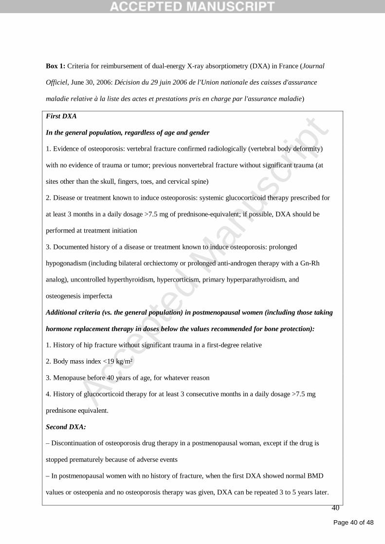
Page 40 of 48
Accep
ted
Man
uscr
ipt
40
Box 1: Criteria for reimbursement of dual-energy X-ray absorptiometry (DXA) in France (Journal
Officiel, June 30, 2006: Décision du 29 juin 2006 de l'Union nationale des caisses d'assurance
maladie relative à la liste des actes et prestations pris en charge par l'assurance maladie)
First DXA
In the general population, regardless of age and gender
1. Evidence of osteoporosis: vertebral fracture confirmed radiologically (vertebral body deformity)
with no evidence of trauma or tumor; previous nonvertebral fracture without significant trauma (at
sites other than the skull, fingers, toes, and cervical spine)
2. Disease or treatment known to induce osteoporosis: systemic glucocorticoid therapy prescribed for
at least 3 months in a daily dosage >7.5 mg of prednisone-equivalent; if possible, DXA should be
performed at treatment initiation
3. Documented history of a disease or treatment known to induce osteoporosis: prolonged
hypogonadism (including bilateral orchiectomy or prolonged anti-androgen therapy with a Gn-Rh
analog), uncontrolled hyperthyroidism, hypercorticism, primary hyperparathyroidism, and
osteogenesis imperfecta
Additional criteria (vs. the general population) in postmenopausal women (including those taking
hormone replacement therapy in doses below the values recommended for bone protection):
1. History of hip fracture without significant trauma in a first-degree relative
2. Body mass index <19 kg/m²
3. Menopause before 40 years of age, for whatever reason
4. History of glucocorticoid therapy for at least 3 consecutive months in a daily dosage >7.5 mg
prednisone equivalent.
Second DXA:
– Discontinuation of osteoporosis drug therapy in a postmenopausal woman, except if the drug is
stopped prematurely because of adverse events
– In postmenopausal women with no history of fracture, when the first DXA showed normal BMD
values or osteopenia and no osteoporosis therapy was given, DXA can be repeated 3 to 5 years later.
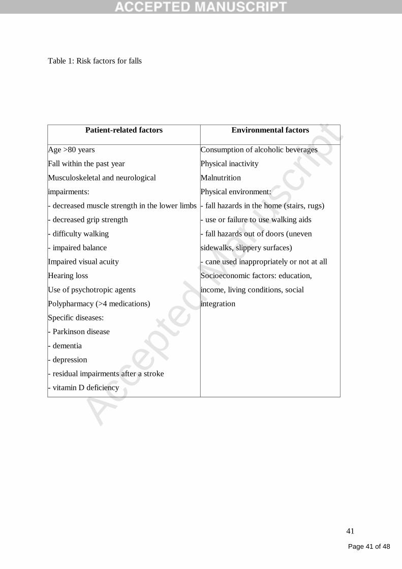
Page 41 of 48
Accep
ted
Man
uscr
ipt
41
Table 1: Risk factors for falls
Patient-related factors Environmental factors
Age >80 years
Fall within the past year
Musculoskeletal and neurological
impairments:
- decreased muscle strength in the lower limbs
- decreased grip strength
- difficulty walking
- impaired balance
Impaired visual acuity
Hearing loss
Use of psychotropic agents
Polypharmacy (>4 medications)
Specific diseases:
- Parkinson disease
- dementia
- depression
- residual impairments after a stroke
- vitamin D deficiency
Consumption of alcoholic beverages
Physical inactivity
Malnutrition
Physical environment:
- fall hazards in the home (stairs, rugs)
- use or failure to use walking aids
- fall hazards out of doors (uneven
sidewalks, slippery surfaces)
- cane used inappropriately or not at all
Socioeconomic factors: education,
income, living conditions, social
integration
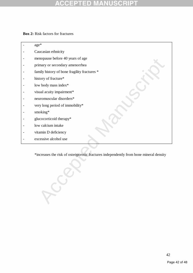
Page 42 of 48
Accep
ted
Man
uscr
ipt
42
Box 2: Risk factors for fractures
- age*
- Caucasian ethnicity
- menopause before 40 years of age
- primary or secondary amenorrhea
- family history of bone fragility fractures *
- history of fracture*
- low body mass index*
- visual acuity impairment*
- neuromuscular disorders*
- very long period of immobility*
- smoking*
- glucocorticoid therapy*
- low calcium intake
- vitamin D deficiency
- excessive alcohol use
*increases the risk of osteoporotic fractures independently from bone mineral density
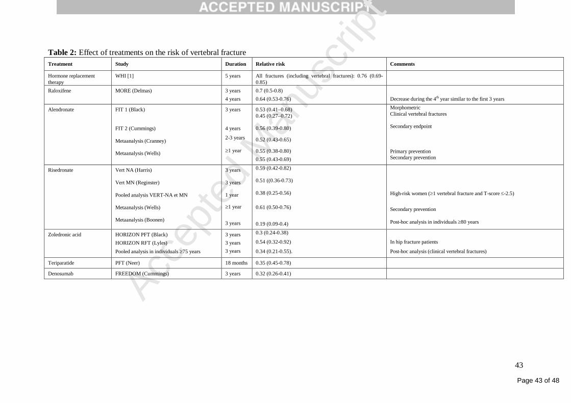
Page 43 of 48
Accep
ted
Man
uscr
ipt
43
Table 2: Effect of treatments on the risk of vertebral fracture Treatment Study Duration Relative risk Comments
Hormone replacement therapy
WHI [1] 5 years All fractures (including vertebral fractures): 0.76 (0.69-0.85)
Raloxifene MORE (Delmas) 3 years 4 years
0.7 (0.5-0.8) 0.64 (0.53-0.76)
Decrease during the 4th year similar to the first 3 years
Alendronate FIT 1 (Black) FIT 2 (Cummings) Metaanalysis (Cranney) Metaanalysis (Wells)
3 years 4 years
2-3 years 1 year
0.53 (0.41–0.68) 0.45 (0.27–0.72) 0.56 (0.39-0.80)
0.52 (0.43-0.65)
0.55 (0.38-0.80) 0.55 (0.43-0.69)
Morphometric Clinical vertebral fractures Secondary endpoint Primary prevention Secondary prevention
Risedronate Vert NA (Harris) Vert MN (Reginster) Pooled analysis VERT-NA et MN Metaanalysis (Wells) Metaanalysis (Boonen)
3 years 3 years 1 year
1 year 3 years
0.59 (0.42-0.82) 0.51 ((0.36-0.73) 0.38 (0.25-0.56) 0.61 (0.50-0.76) 0.19 (0.09-0.4)
High-risk women (1 vertebral fracture and T-score -2.5)
Secondary prevention Post-hoc analysis in individuals 80 years
Zoledronic acid HORIZON PFT (Black) HORIZON RFT (Lyles) Pooled analysis in individuals 75 years
3 years 3 years 3 years
0.3 (0.24-0.38)
0.54 (0.32-0.92)
0.34 (0.21-0.55).
In hip fracture patients
Post-hoc analysis (clinical vertebral fractures)
Teriparatide PFT (Neer) 18 months 0.35 (0.45-0.78)
Denosumab FREEDOM (Cummings) 3 years 0.32 (0.26-0.41)
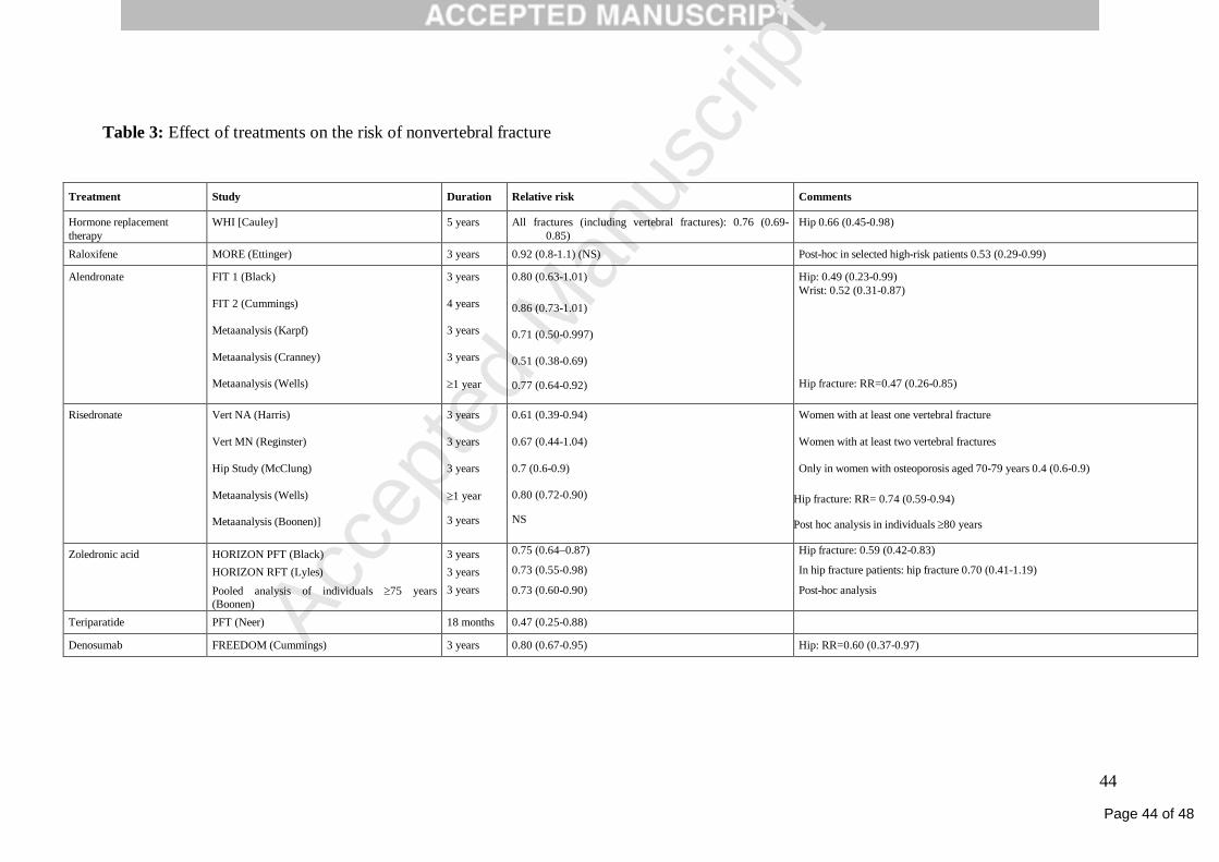
Page 44 of 48
Accep
ted
Man
uscr
ipt
44
Table 3: Effect of treatments on the risk of nonvertebral fracture
Treatment Study Duration Relative risk Comments
Hormone replacement therapy
WHI [Cauley] 5 years All fractures (including vertebral fractures): 0.76 (0.69-0.85)
Hip 0.66 (0.45-0.98)
Raloxifene MORE (Ettinger) 3 years 0.92 (0.8-1.1) (NS) Post-hoc in selected high-risk patients 0.53 (0.29-0.99)
Alendronate FIT 1 (Black) FIT 2 (Cummings) Metaanalysis (Karpf) Metaanalysis (Cranney) Metaanalysis (Wells)
3 years 4 years 3 years 3 years 1 year
0.80 (0.63-1.01) 0.86 (0.73-1.01) 0.71 (0.50-0.997) 0.51 (0.38-0.69)
0.77 (0.64-0.92)
Hip: 0.49 (0.23-0.99) Wrist: 0.52 (0.31-0.87) Hip fracture: RR=0.47 (0.26-0.85)
Risedronate Vert NA (Harris) Vert MN (Reginster) Hip Study (McClung) Metaanalysis (Wells) Metaanalysis (Boonen)]
3 years 3 years 3 years 1 year
3 years
0.61 (0.39-0.94) 0.67 (0.44-1.04) 0.7 (0.6-0.9) 0.80 (0.72-0.90)
NS
Women with at least one vertebral fracture Women with at least two vertebral fractures Only in women with osteoporosis aged 70-79 years 0.4 (0.6-0.9)
Hip fracture: RR= 0.74 (0.59-0.94)
Post hoc analysis in individuals 80 years
Zoledronic acid HORIZON PFT (Black) HORIZON RFT (Lyles) Pooled analysis of individuals 75 years (Boonen)
3 years 3 years 3 years
0.75 (0.64–0.87)
0.73 (0.55-0.98)
0.73 (0.60-0.90)
Hip fracture: 0.59 (0.42-0.83)
In hip fracture patients: hip fracture 0.70 (0.41-1.19)
Post-hoc analysis
Teriparatide PFT (Neer) 18 months 0.47 (0.25-0.88)
Denosumab FREEDOM (Cummings) 3 years 0.80 (0.67-0.95) Hip: RR=0.60 (0.37-0.97)
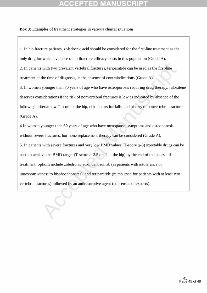
Page 45 of 48
Accep
ted
Man
uscr
ipt
45
Box 3: Examples of treatment strategies in various clinical situations
1. In hip fracture patients, zoledronic acid should be considered for the first-line treatment as the
only drug for which evidence of antifracture efficacy exists in this population (Grade A).
2. In patients with two prevalent vertebral fractures, teriparatide can be used as the first-line
treatment at the time of diagnosis, in the absence of contraindications (Grade A).
3. In women younger than 70 years of age who have osteoporosis requiring drug therapy, raloxifene
deserves considerations if the risk of nonvertebral fractures is low as indicated by absence of the
following criteria: low T-score at the hip, risk factors for falls, and history of nonvertebral fracture
(Grade A).
4 In women younger than 60 years of age who have menopausal symptoms and osteoporosis
without severe fractures, hormone replacement therapy can be considered (Grade A).
5. In patients with severe fractures and very low BMD values (T-score ≤-3) injectable drugs can be
used to achieve the BMD target (T-score >-2.5 or -2 at the hip) by the end of the course of
treatment; options include zoledronic acid, denosumab (in patients with intolerance or
unresponsiveness to bisphosphonates), and teriparatide (reimbursed for patients with at least two
vertebral fractures) followed by an antiresorptive agent (consensus of experts).
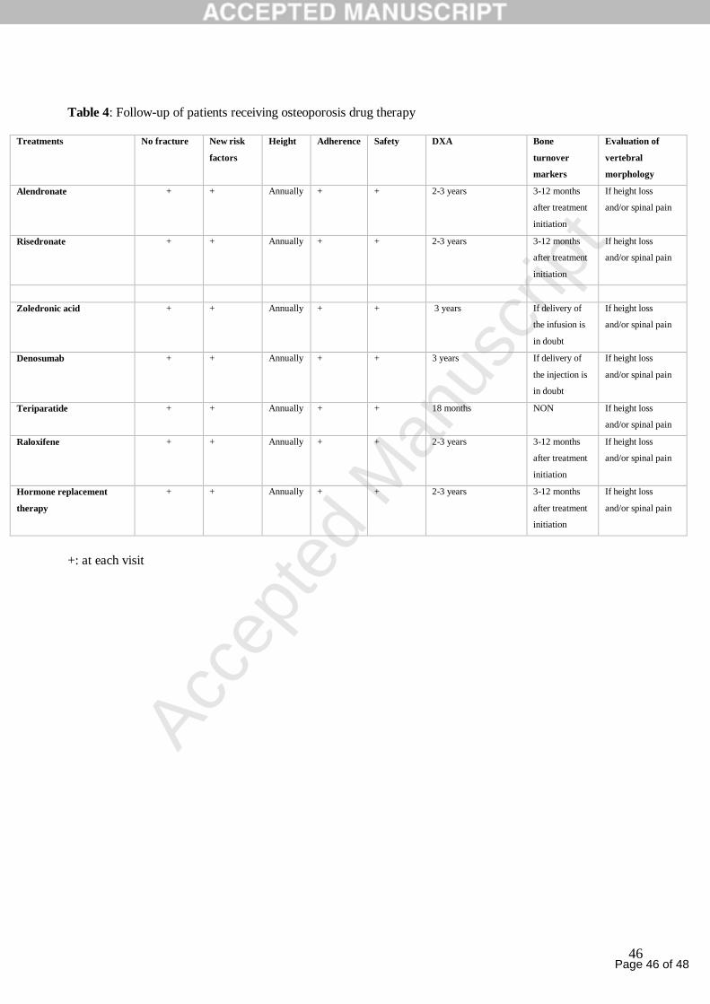
Page 46 of 48
Accep
ted
Man
uscr
ipt
46
Table 4: Follow-up of patients receiving osteoporosis drug therapy
Treatments No fracture New risk
factors
Height Adherence Safety DXA Bone
turnover
markers
Evaluation of
vertebral
morphology
Alendronate + + Annually + + 2-3 years 3-12 months
after treatment
initiation
If height loss
and/or spinal pain
Risedronate + + Annually + + 2-3 years 3-12 months
after treatment
initiation
If height loss
and/or spinal pain
Zoledronic acid + + Annually + + 3 years If delivery of
the infusion is
in doubt
If height loss
and/or spinal pain
Denosumab + + Annually + + 3 years If delivery of
the injection is
in doubt
If height loss
and/or spinal pain
Teriparatide + + Annually + + 18 months NON If height loss
and/or spinal pain
Raloxifene + + Annually + + 2-3 years 3-12 months
after treatment
initiation
If height loss
and/or spinal pain
Hormone replacement
therapy
+ + Annually + + 2-3 years 3-12 months
after treatment
initiation
If height loss
and/or spinal pain
+: at each visit

Page 47 of 48
Accep
ted
Man
uscr
ipt
47
Figure 1: FRAX® probability of major osteoporotic fracture cutoffs for osteoporosis drug therapy according
to
patient
age in
France
Figure
2:
Indicatio
ns of
drug
therapy
for
postmen
opausal
osteopor
osis
Table S1: Calcium content of foods (www.grio.org)
*mean calcium content for several foods belonging to the same group
Calcium content of foods The calcium contents are reported in mg for a standard portion, whose weight (uncooked) or volume is given in parentheses. Large portion = medium portion x 1.5 Small portion = medium portion x 0.5
1 Milk, 100 mL 120 mg 2 1 yogurt or custard (125 g) 150 mg 3 Cottage or cream cheese, 100 g 95 mg*
Small 28 mg 4 Petit suisse cream cheese Large 56 mg 5 Hard cheese, 30 g 263 mg* 6 Soft cheese, 30 g 120 mg* 7 Meat, fish, 120 g 15 mg* 8 1 egg 28 mg 9 Potatoes, 200 g 20 mg
10 Fries, 160 g 96 mg 11 Pasta or semolina, 50g 10 mg 12 Legumes, 75g 53 mg* 13 Green vegetables, 200g 94 mg*
100g 25 mg 200g 50 mg
14 Bread
1 rusk, 10g 4 mg 15 1 fruit, 150g 25 mg*
Milk chocolate, white chocolate, 20 g 43 mg Dark chocolate, 20 g 13 mg Milk chocolate, white chocolate, 100 g 214 mg
16&17
Chocolate
Dark chocolate, 100 g 63 mg 18 Tap water, 100 mL 9 mg
Badoit 22 mg Evian 8 mg Contrexéville 46 mg Perrier 14 mg Vichy Saint Yorre 8 mg Hépar 55 mg Vittel Grande Source 20 mg*
19
Bottled water, 100 mL
Other or not specified 10 mg* 20 Other beverages: 1 glass of wine, beer, cider, or fruit juice 7 mg
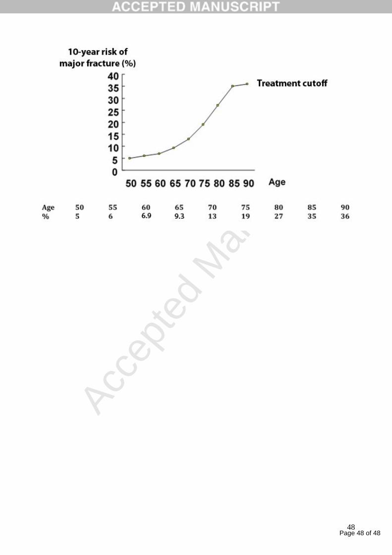
Page 48 of 48
Accep
ted
Man
uscr
ipt
48


![Centum Electronics acquires French Adetel Group [Company Update]](https://img.pdfslide.us/doc/110x75/577c79371a28abe05491d821/centum-electronics-acquires-french-adetel-group-company-update.jpg)