Embed Size (px)
Citation preview
TODAY’S VETERINARY PRACTICE | July/August 2015 | tvpjournal.com
A PRACTITIONER’S GUIDE TO FRACTURE MANAGEMENTPeer Reviewed
18
Fractures occur commonly in both dogs and cats. While typically fractures occur after a traumatic incident, such as being hit by a car or falling from a height, some fractures occur following a pathologic weakening of the bone, which is seen with certain neoplastic conditions, such as osteosarcoma.
Since fractures are frequently seen in general practice, it is important for veterinarians to understand fracture biomechanics as well as how to: • Classify and diagnose fractures• Choose the correct fixation to ensure proper bone
healing• Identify bone healing • Address complications, if they occur.
In Part 1 of this 2-part series, fracture biomechanics, fracture classifi cation and diagnosis, and factors to consider when selecting a fi xation technique are discussed. In Part 2, selection of fi xation technique and specifi c techniques, identifi cation of bone healing, and potential complications will be addressed.
FRACTURE BIOMECHANICS Bone is an amazing tissue with complex properties that allow it to adapt to its environment but typically conserve its general structure and shape. By following Wolff ’s law, bone adapts and changes in areas of high stress, while minimizing changes in areas of low stress. Basically, bone is shaped for the greatest strength while, at the same time, minimizing bone mass that would contribute to increased weight of the animal.
Qualities of BoneBone is considered both viscoelastic and anisotropic:
• The viscoelastic property of bone states that the strength of bone depends on the rate upon which it is loaded. For example, bone is stronger when it is loaded rapidly versus slowly (ie, the more rapidly bone is loaded, the more inflexible it becomes). This property is advantageous because most injuries are inflicted by impact with high loading rate forces. Therefore, fewer fractures actually occur because the more rapidly bone is stressed, the stiffer it becomes. However, once any bone reaches a failure point of loading, it will fracture.1
• The anisotropic nature of bone suggests that bone strength is dependent on the direction in which it is loaded. For example, bone is stronger when loaded longitudinally versus transversely, which explains why bone is more likely to fracture with sudden high impact placed transversely upon the bone.
Fracture ForcesBone is subject to many forces; a fracture occurs when the sum of these forces exceeds the ultimate strength of the bone. Therefore, understanding the forces placed upon bone is crucial to counteracting these forces when stabilizing a fracture.
PART 1: DIAGNOSING FRACTURES & CHOOSING A FIXATION TECHNIQUE
A Practitioner’s Guide to Fracture ManagementMeredith Kapler, DVMNorth Carolina State University
David Dycus, DVM, MS, Diplomate ACVS (Small Animal)Veterinary Orthopedic & Sports Medicine Group, Annapolis Junction, Maryland
TABlE 1. Five Main Forces That Act on BoneThe most common forces acting on bone, and those that MUST be counteracted with bone fi xation are:
1. Bending2. Compression 3. Shearing 4. Tension5. Torsion
Wolff’s law—a theory developed by German anatomist and surgeon Julius Wolff—states that bone in a healthy person or animal will adapt to the loads under which it is placed or, more simply, bone adapts to pressure, or a lack of it.
tvpjournal.com | July/August 2015 | TODAY’S VETERINARY PRACTICE
A PRACTITIONER’S GUIDE TO FRACTURE MANAGEMENT Peer Reviewed
19
The 5 main forces that act on bone are listed in Table 1. • Tensile (tension) forces
act to lengthen the bone, while compressive forces shorten the bone.
• Shearing forces are typically parallel or tangential to the bone, while torsional forces act to twist bone about its long axis.
• Bending forces create a convex side of the bone (bone loaded in tension on the convex side) and a concave side (bone loaded in compression on the concave side). Bending forces are typically referred to as moments.
FRACTURE DIAGNOSIS & CLASSIFICATIONPatient StabilizationBecause most fractures result from trauma, it is important to ensure patient stability prior to focusing on the fracture. Ideally, for any patient that presents after a traumatic event: 1. Check and stabilize vitals (temperature, pulse
quality and heart rate, respiration rate, blood pressure, pulse oximetry), if needed.
2. Perform thorough physical, orthopedic, and neurologic examinations.
3. Pursue initial diagnostics, including blood analysis, thoracic and abdominal radiographs, and an AFAST ultrasound.
4. Resolve any life-threatening issues, which means that surgery may need to be delayed for several days due to conditions, such as pulmonary contusions or hypovolemia.
5. Administer proper analgesia as soon as possible: Ideal analgesics are pure mu opioids, such as:
• Morphine: Dogs, 0.5 to 2 mg/kg; cats, 0.05 to 0.4 mg/kg; IV or IM Q 6 to 8 H
• Hydromorphone: Dogs/cats, 0.05 to 0.2 mg/kg IV or IM Q 6 to 8 H
• Oxymorphone: Dogs/cats, 0.05 to 0.2 mg/kg IV or IM Q 6 to 8 H
• Methadone: Dogs, 0.05 to 0.3 mg/kg; cats, 0.1 up to 0.3 mg/kg; IV or IM Q 6 to 8 H
• Fentanyl: Dogs/cats, 2 mcg/kg loading dose followed by 2 to 10 mcg/kg/H CRI (caution with cats).
Other opioids, such as butorphanol, do not typically provide adequate analgesia.
Post Stabilization DiagnosticsOnce the patient is deemed stable: 1. Obtain a thorough history: It is important to
separate traumatic from pathologic fractures by determining the cause of the fracture, such as hit by car or fall from a height.
2. Evaluate physical examination fi ndings: Signs of fractures include pain, swelling, reluctance to bear weight, crepitus, and/or angulation deformities.
3. If the patient is nonambulatory on the affected limb, perform a complete neurologic examination to rule out any neurologic defects, such as: » Radial nerve damage—seen with distal humeral
fractures » Sciatic nerve damage—seen with ilial fractures.
Radiographs are the mainstay for determining fracture type and location. Key radiographic projections are orthogonal views, including lateral, craniocaudal, and oblique (if needed) views.
If only a lateral view is taken, some fractures, such as T-Y humeral fractures that involve both the articular surface of the distal humeral condyle and the supracondylar region, will be missed (Figure 1).
Fracture Classifi cationTo improve communication between veterinarians and clients, and between veterinarians themselves,
Bone is strongerwhen loaded rapidly versus slowly, and when loaded longitudinally versus transversely. Therefore, bone is stiffer and stronger when loaded rapidly and longitudinally.
FIGURE 1. Lateral radiograph of the antebrachium in a young dog presenting for right forelimb lameness; this radiograph does not reveal any evidence of fracture or pathology (A). Craniocaudal radiograph of the same right forelimb, revealing a Salter Harris IV fracture of the distal humeral condyle (B).
A
B
Today’s VeTerinary PracTice | July/august 2015 | tvpjournal.com
a PracTiTioner’s Guide To FracTure ManaGeMenTPeer reviewed
20
fractures must be correctly identified and classified. Fractures are initially classified by anatomic
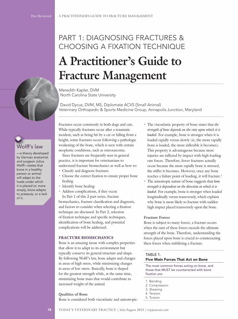
location, such as articular, physeal, epiphyseal, metaphyseal, or diaphyseal (Figure 2). Certain fractures are further subclassified based on anatomic locations, such as condylar, supracondylar, trochanteric, or subtrochanteric.
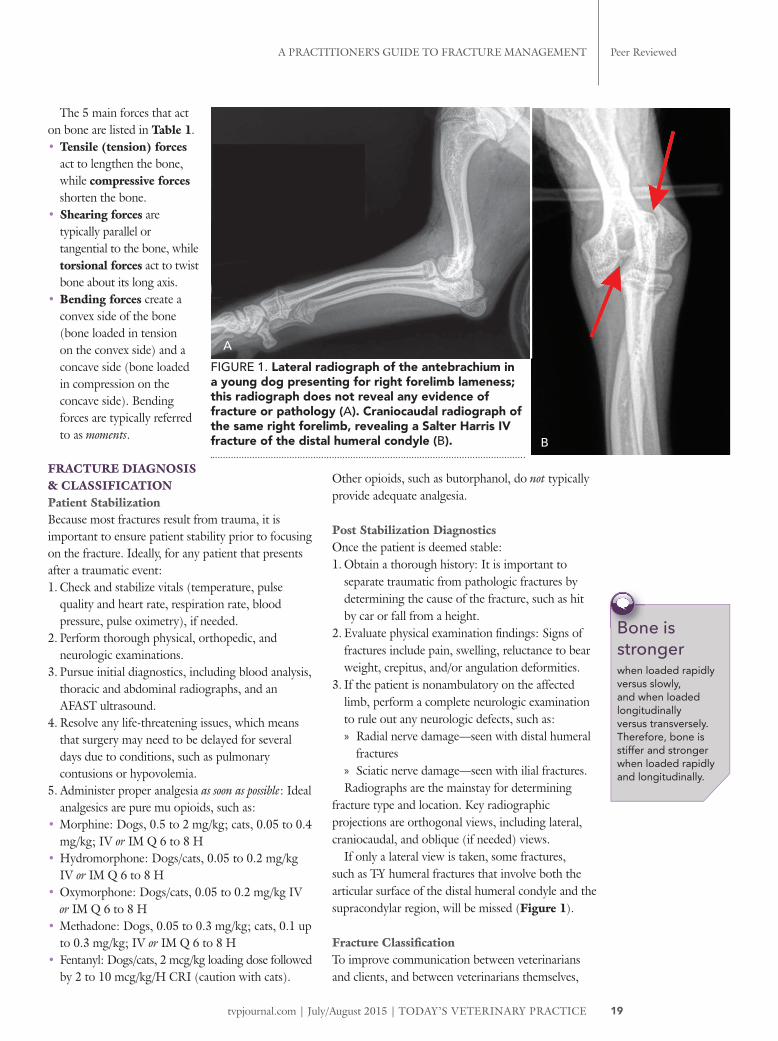
Based on radiographs, fractures are classified by severity, such as: • Incomplete: Fracture through only one cortex• Complete: Fracture through both cortices• Comminuted: Multiple fragments (Figure 3)• Segmental: Two or more separate fractures. The term compound fracture is no longer used for fracture classification.
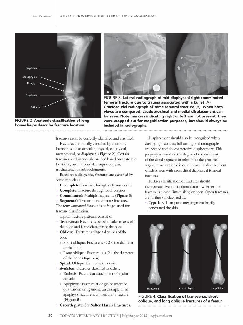
Typical fracture patterns consist of: • Transverse: Fracture is perpendicular to axis of
the bone and is the diameter of the bone • Oblique: Fracture is diagonal to axis of the
bone » Short oblique: Fracture is < 2× the diameter
of the bone » Long oblique: Fracture is > 2× the diameter
of the bone (Figure 4). • Spiral: Oblique fracture with a twist • Avulsion: Fractures classified as either:
» Enthesis: Fracture at attachment of a joint capsule
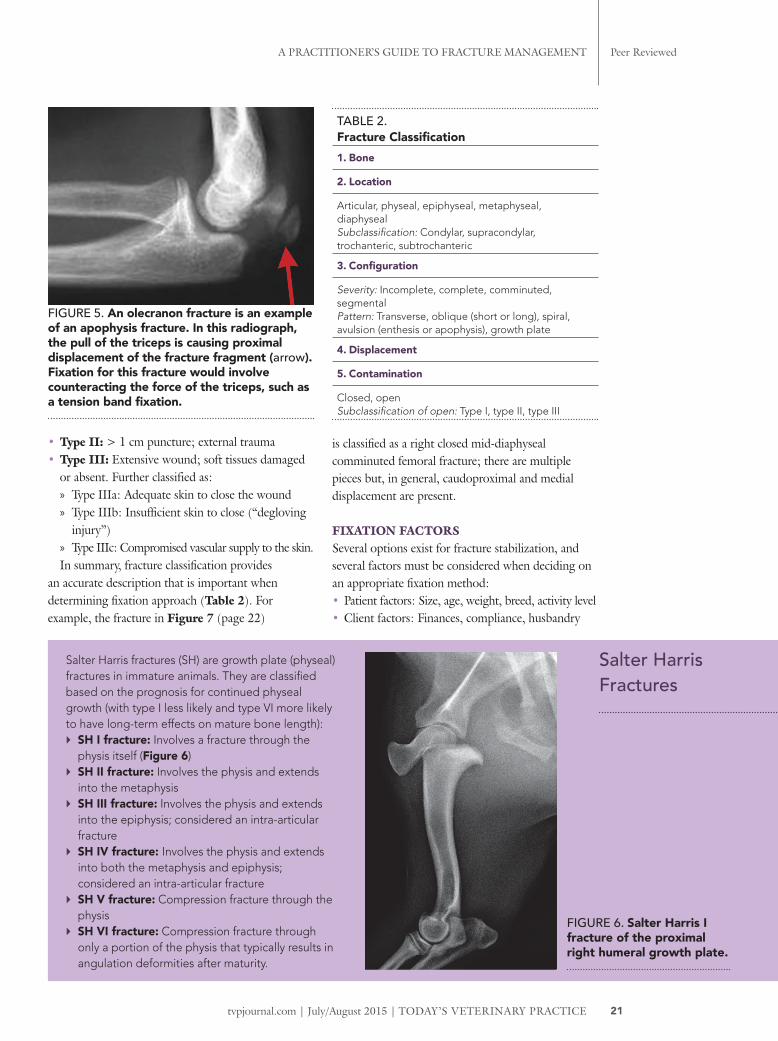
» Apophysis: Fracture at origin or insertion of a tendon or ligament; an example of an apophysis fracture is an olecranon fracture (Figure 5)
• Growth plate: See Salter Harris Fractures.
Displacement should also be recognized when classifying fractures; full orthogonal radiographs are needed to fully characterize displacement. This property is based on the degree of displacement of the distal segment in relation to the proximal segment. An example is caudoproximal displacement, which is seen with most distal diaphyseal femoral fractures.
Further classification of fractures should incorporate level of contamination—whether the fracture is closed (intact skin) or open. Open fractures are further subclassified as: • Type I: < 1 cm puncture; fragment briefly
penetrated the skin
FIGURE 2. Anatomic classification of long bones helps describe fracture location.
FIGURE 3. Lateral radiograph of mid-diaphyseal right comminuted femoral fracture due to trauma associated with a bullet (A). Craniocaudal radiograph of same femoral fracture (B). When both views are compared, caudoproximal and medial displacement can be seen. Note markers indicating right or left are not present; they were cropped out for magnification purposes, but should always be included in radiographs.
FIGURE 4. Classification of transverse, short oblique, and long oblique fractures of a femur.
A B
tvpjournal.com | July/August 2015 | TodAy’s VeTerinAry PrAcTice
A PrAcTiTioner’s Guide To FrAcTure MAnAGeMenT Peer reviewed
21
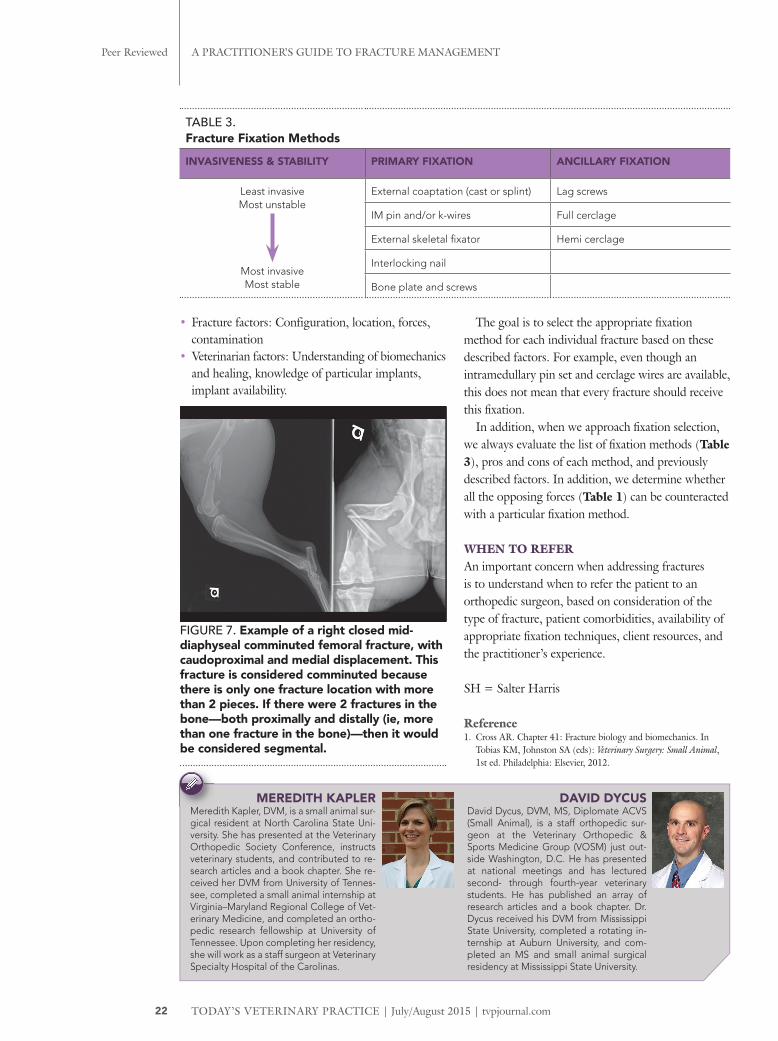
Salter Harris Fractures
Salter Harris fractures (SH) are growth plate (physeal) fractures in immature animals. They are classified based on the prognosis for continued physeal growth (with type I less likely and type VI more likely to have long-term effects on mature bone length): ` SH I fracture: Involves a fracture through the
physis itself (Figure 6) ` SH II fracture: Involves the physis and extends
into the metaphysis ` SH III fracture: Involves the physis and extends
into the epiphysis; considered an intra-articular fracture
` SH IV fracture: Involves the physis and extends into both the metaphysis and epiphysis; considered an intra-articular fracture
` SH V fracture: Compression fracture through the physis
` SH VI fracture: Compression fracture through only a portion of the physis that typically results in angulation deformities after maturity.
FIGURE 6. Salter Harris I fracture of the proximal right humeral growth plate.
• Type II: > 1 cm puncture; external trauma• Type III: Extensive wound; soft tissues damaged
or absent. Further classified as: » Type IIIa: Adequate skin to close the wound » Type IIIb: Insufficient skin to close (“degloving
injury”) » Type IIIc: Compromised vascular supply to the skin.
In summary, fracture classification provides an accurate description that is important when determining fixation approach (Table 2). For example, the fracture in Figure 7 (page 22)
is classified as a right closed mid-diaphyseal comminuted femoral fracture; there are multiple pieces but, in general, caudoproximal and medial displacement are present.
FIXATION FACTORSSeveral options exist for fracture stabilization, and several factors must be considered when deciding on an appropriate fixation method: • Patient factors: Size, age, weight, breed, activity level• Client factors: Finances, compliance, husbandry
TABlE 2. Fracture Classification1. Bone
2. Location
Articular, physeal, epiphyseal, metaphyseal, diaphysealSubclassification: Condylar, supracondylar, trochanteric, subtrochanteric
3. Configuration
Severity: Incomplete, complete, comminuted, segmentalPattern: Transverse, oblique (short or long), spiral, avulsion (enthesis or apophysis), growth plate
4. Displacement
5. Contamination
Closed, openSubclassification of open: Type I, type II, type III
FIGURE 5. An olecranon fracture is an example of an apophysis fracture. In this radiograph, the pull of the triceps is causing proximal displacement of the fracture fragment (arrow). Fixation for this fracture would involve counteracting the force of the triceps, such as a tension band fixation.
TODAY’S VETERINARY PRACTICE | July/August 2015 | tvpjournal.com
A PRACTITIONER’S GUIDE TO FRACTURE MANAGEMENTPeer Reviewed
22
• Fracture factors: Configuration, location, forces, contamination
• Veterinarian factors: Understanding of biomechanics and healing, knowledge of particular implants, implant availability.
The goal is to select the appropriate fi xation method for each individual fracture based on these described factors. For example, even though an intramedullary pin set and cerclage wires are available, this does not mean that every fracture should receive this fi xation.
In addition, when we approach fi xation selection, we always evaluate the list of fi xation methods (Table 3), pros and cons of each method, and previously described factors. In addition, we determine whether all the opposing forces (Table 1) can be counteracted with a particular fi xation method.
WHEN TO REFERAn important concern when addressing fractures is to understand when to refer the patient to an orthopedic surgeon, based on consideration of the type of fracture, patient comorbidities, availability of appropriate fi xation techniques, client resources, and the practitioner’s experience.
SH = Salter Harris
Reference1. Cross AR. Chapter 41: Fracture biology and biomechanics. In
Tobias KM, Johnston SA (eds): Veterinary Surgery: Small Animal, 1st ed. Philadelphia: Elsevier, 2012.
TABlE 3. Fracture Fixation Methods
INVASIVENESS & STABILITY PRIMARY FIXATION ANCILLARY FIXATION
Least invasive Most unstable
Most invasiveMost stable
External coaptation (cast or splint) Lag screws
IM pin and/or k-wires Full cerclage
External skeletal fi xator Hemi cerclage
Interlocking nail
Bone plate and screws
MEREDITH KAPLERMeredith Kapler, DVM, is a small animal sur-gical resident at North Carolina State Uni-versity. She has presented at the Veterinary Orthopedic Society Conference, instructs veterinary students, and contributed to re-search articles and a book chapter. She re-ceived her DVM from University of Tennes-see, completed a small animal internship at Virginia–Maryland Regional College of Vet-erinary Medicine, and completed an ortho-pedic research fellowship at University of Tennessee. Upon completing her residency, she will work as a staff surgeon at Veterinary Specialty Hospital of the Carolinas.
DAVID DYCUS David Dycus, DVM, MS, Diplomate ACVS (Small Animal), is a staff orthopedic sur-geon at the Veterinary Orthopedic & Sports Medicine Group (VOSM) just out-side Washington, D.C. He has presented at national meetings and has lectured second- through fourth-year veterinary students. He has published an array of research articles and a book chapter. Dr. Dycus received his DVM from Mississippi State University, completed a rotating in-ternship at Auburn University, and com-pleted an MS and small animal surgical residency at Mississippi State University.
Meredith Kapler, DVM, is a small animal sur-
FIGURE 7. Example of a right closed mid-diaphyseal comminuted femoral fracture, with caudoproximal and medial displacement. This fracture is considered comminuted because there is only one fracture location with more than 2 pieces. If there were 2 fractures in the bone—both proximally and distally (ie, more than one fracture in the bone)—then it would be considered segmental.