Embed Size (px)
Citation preview
2014 Recommendations for the follow up of Patients with
DTC Using Initial and Ongoing Risk Assessment to guide
Clinical Management
Vilma M Rabell-Vilches
SPED Ponce
April,2016
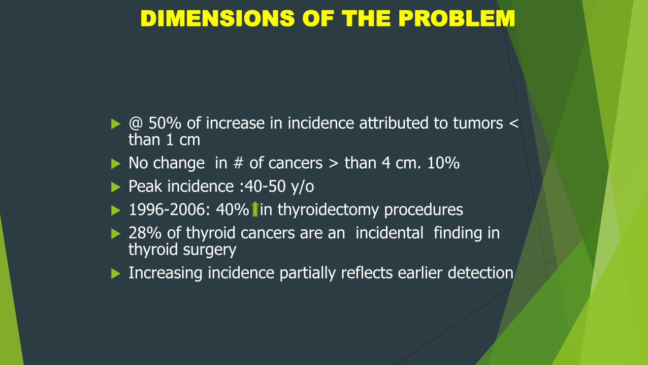
DIMENSIONS OF THE PROBLEM
@ 50% of increase in incidence attributed to tumors < than 1 cm
No change in # of cancers > than 4 cm. 10%
Peak incidence :40-50 y/o
1996-2006: 40% in thyroidectomy procedures
28% of thyroid cancers are an incidental finding in thyroid surgery
Increasing incidence partially reflects earlier detection
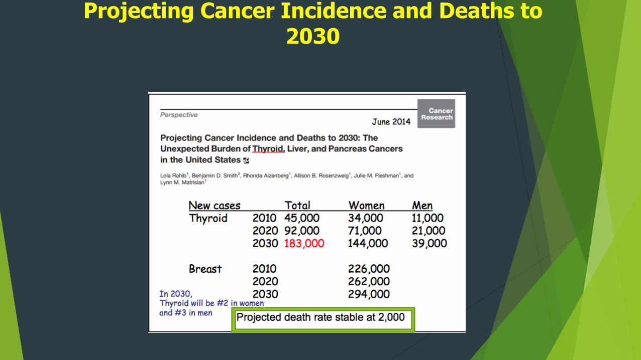
Projecting Cancer Incidence and Deaths to 2030
Differentiated Thyroid Cancer Guidelines
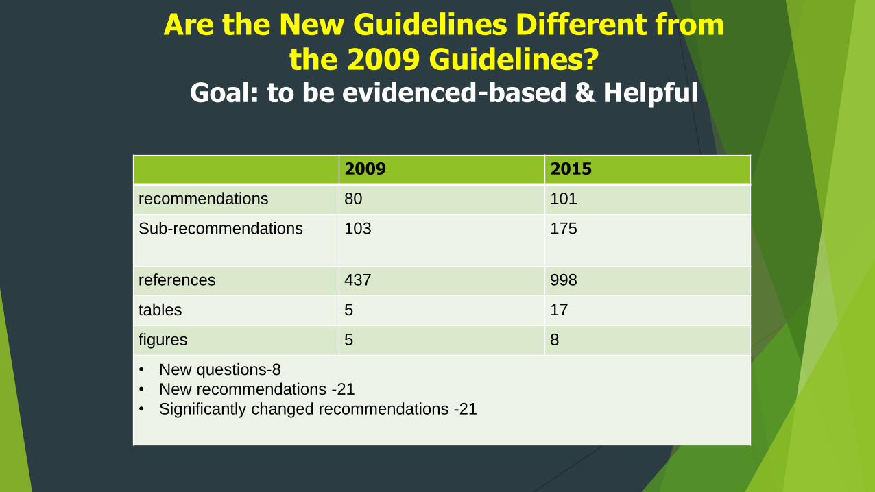
Are the New Guidelines Different from the 2009 Guidelines?
Goal: to be evidenced-based & Helpful
2009 2015
recommendations 80 101
Sub-recommendations 103 175
references 437 998
tables 5 17
figures 5 8
• New questions-8
• New recommendations -21
• Significantly changed recommendations -21
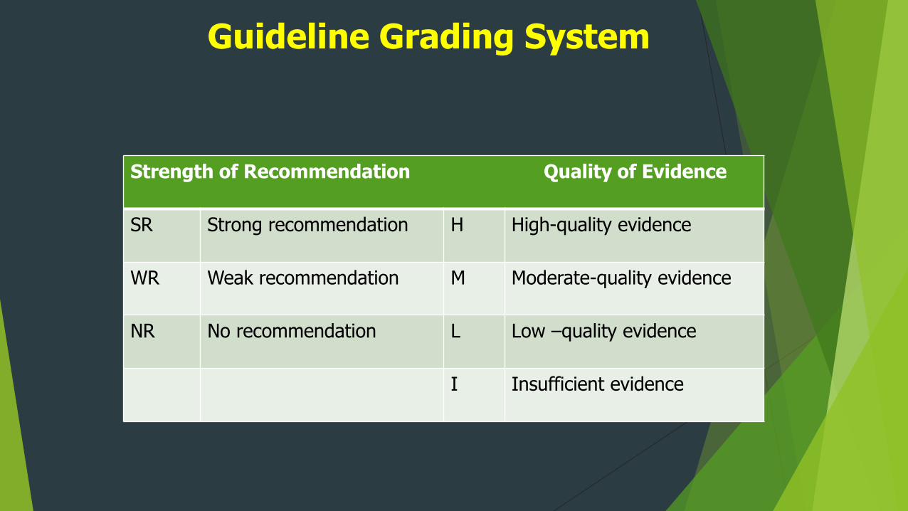
Guideline Grading System
Strength of Recommendation Quality of Evidence
SR Strong recommendation H High-quality evidence
WR Weak recommendation M Moderate-quality evidence
NR No recommendation L Low –quality evidence
I Insufficient evidence
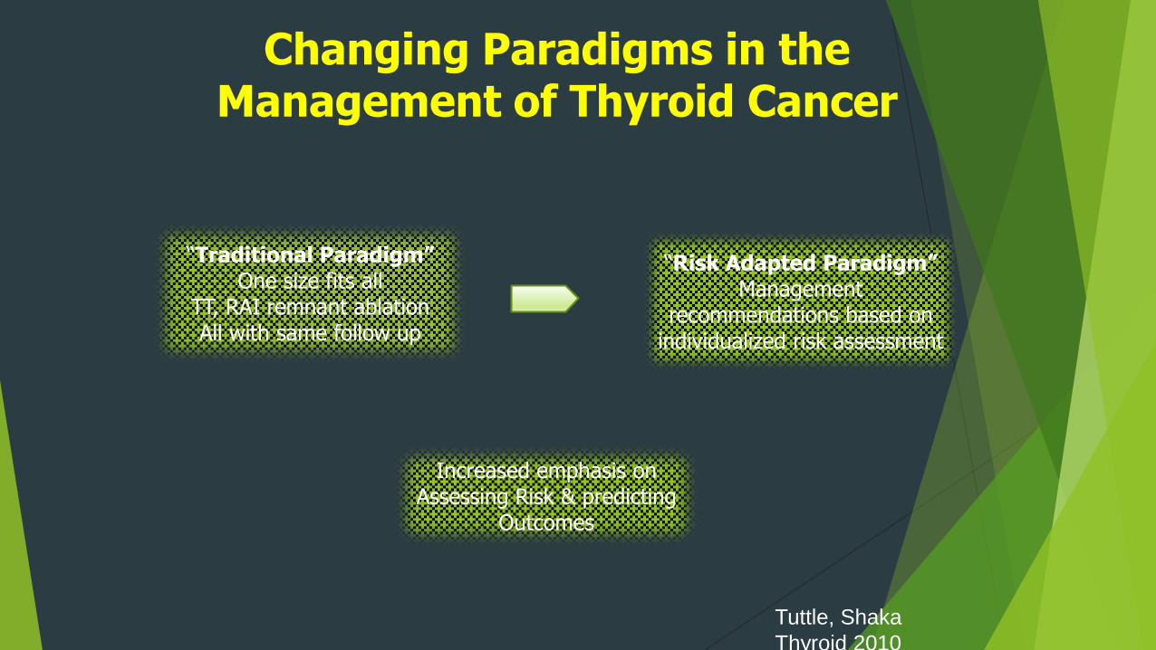
Changing Paradigms in the Management of Thyroid Cancer
“Traditional Paradigm”One size fits all
TT, RAI remnant ablationAll with same follow up
“Risk Adapted Paradigm”Management
recommendations based on individualized risk assessment
Increased emphasis on Assessing Risk & predicting
Outcomes
Tuttle, Shaka
Thyroid 2010
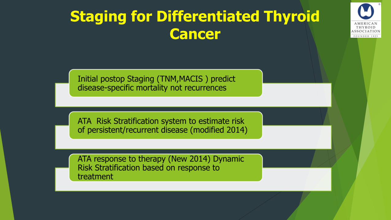
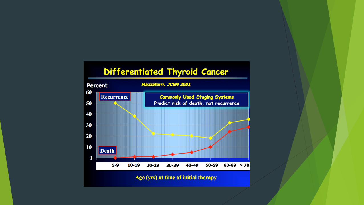
Staging for Differentiated Thyroid Cancer
Initial postop Staging (TNM,MACIS ) predictdisease-specific mortality not recurrences
ATA Risk Stratification system to estimate risk of persistent/recurrent disease (modified 2014)
ATA response to therapy (New 2014) Dynamic Risk Stratification based on response to treatment
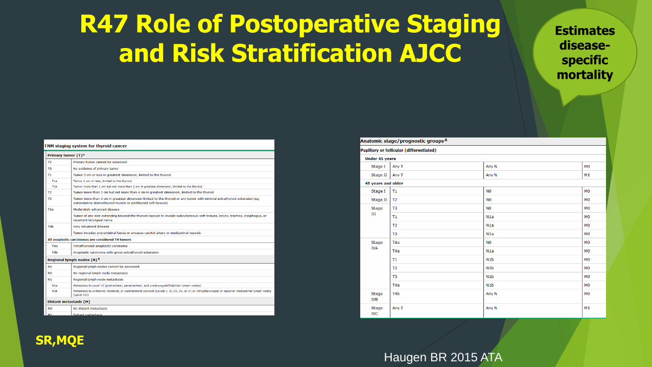
R47 Role of Postoperative Staging and Risk Stratification AJCC
Estimates disease-specific
mortality
Haugen BR 2015 ATA
SR,MQE
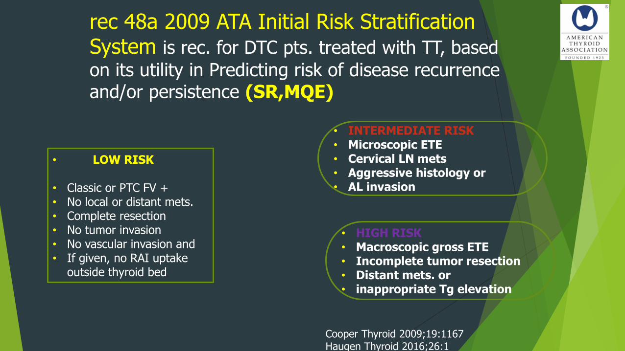
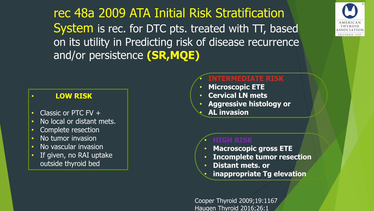
rec 48a 2009 ATA Initial Risk Stratification System is rec. for DTC pts. treated with TT, based
on its utility in Predicting risk of disease recurrence and/or persistence (SR,MQE)
• LOW RISK
• Classic or PTC FV +• No local or distant mets.• Complete resection• No tumor invasion• No vascular invasion and • If given, no RAI uptake
outside thyroid bed
• INTERMEDIATE RISK• Microscopic ETE • Cervical LN mets• Aggressive histology or• AL invasion
• HIGH RISK• Macroscopic gross ETE • Incomplete tumor resection• Distant mets. or• inappropriate Tg elevation
Cooper Thyroid 2009;19:1167Haugen Thyroid 2016;26:1
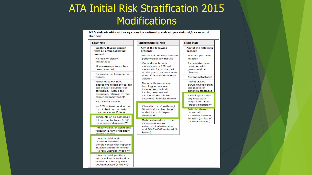
ATA Initial Risk Stratification 2015 Modifications
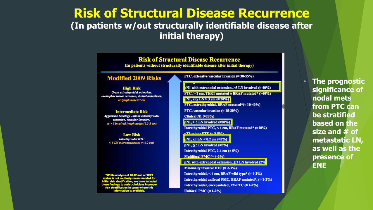
Risk of Structural Disease Recurrence (In patients w/out structurally identifiable disease after
initial therapy)
• The prognostic significance of nodal metsfrom PTC can be stratified based on the size and # of metastatic LN, as well as the presence of ENE
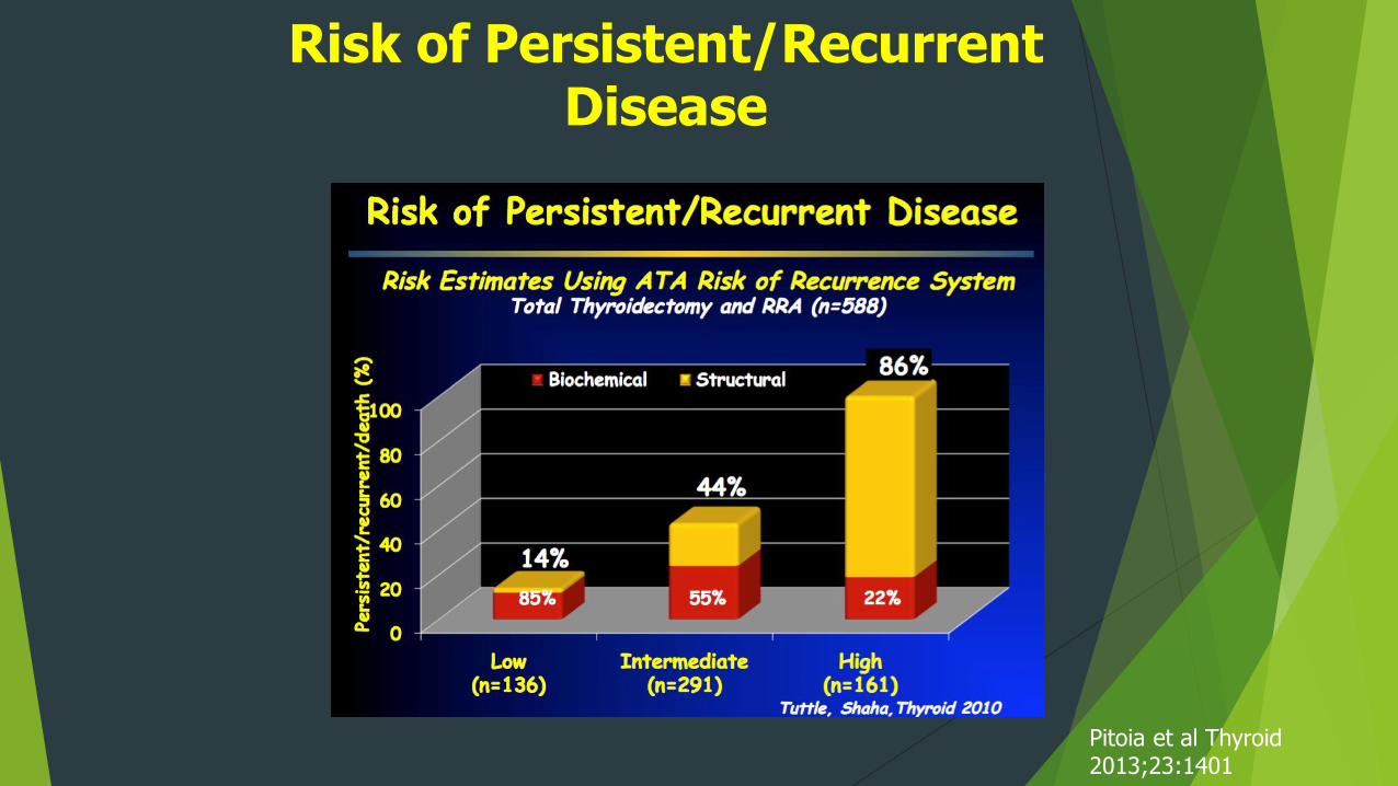
Risk of Persistent/Recurrent Disease
Pitoia et al Thyroid 2013;23:1401
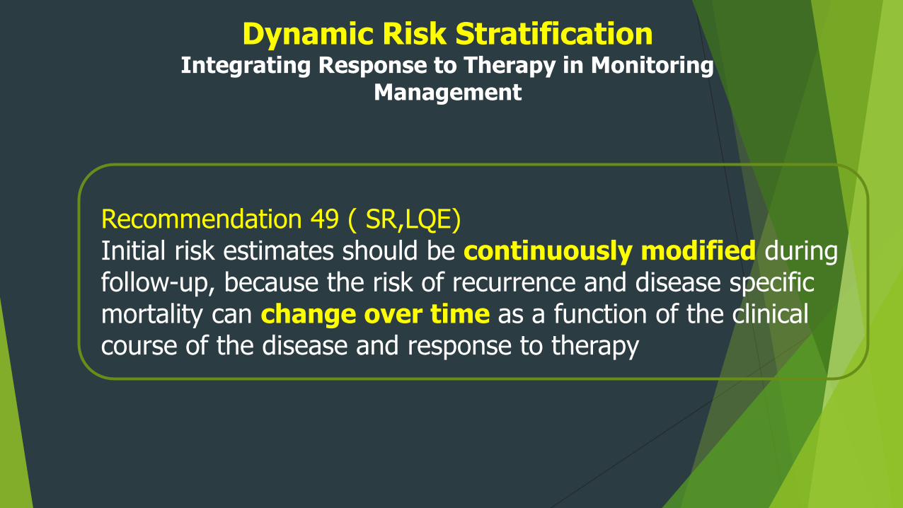
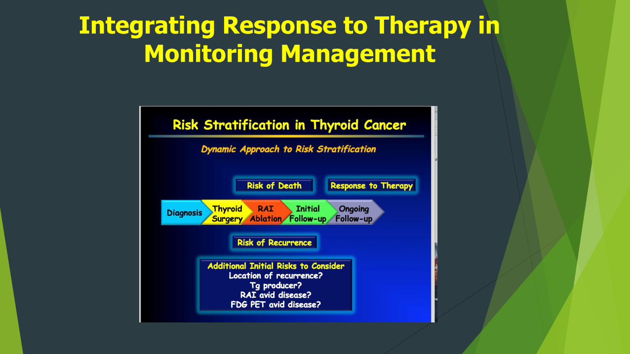
Dynamic Risk StratificationIntegrating Response to Therapy in Monitoring
Management
Recommendation 49 ( SR,LQE)Initial risk estimates should be continuously modified during follow-up, because the risk of recurrence and disease specific mortality can change over time as a function of the clinical course of the disease and response to therapy
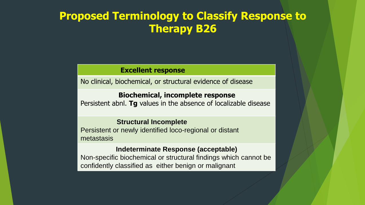
Proposed Terminology to Classify Response to Therapy B26
Excellent response
No clinical, biochemical, or structural evidence of disease
Biochemical, incomplete responsePersistent abnl. Tg values in the absence of localizable disease
Structural Incomplete
Persistent or newly identified loco-regional or distant
metastasis
Indeterminate Response (acceptable)
Non-specific biochemical or structural findings which cannot be
confidently classified as either benign or malignant
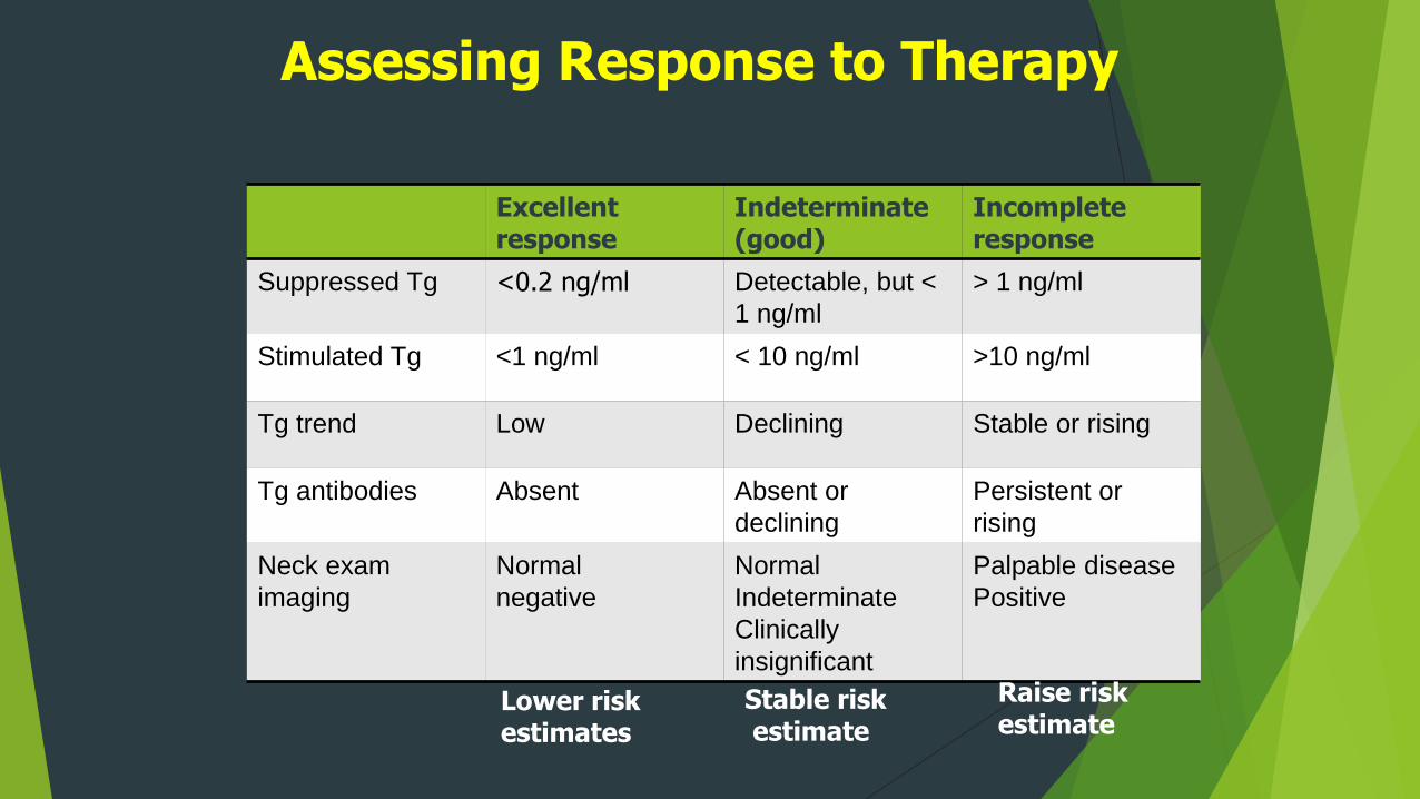
Excellent response
Indeterminate (good)
Incompleteresponse
Suppressed Tg <0.2 ng/ml Detectable, but <
1 ng/ml
> 1 ng/ml
Stimulated Tg <1 ng/ml < 10 ng/ml >10 ng/ml
Tg trend Low Declining Stable or rising
Tg antibodies Absent Absent or
declining
Persistent or
rising
Neck exam
imaging
Normal
negative
Normal
Indeterminate
Clinically
insignificant
Palpable disease
Positive
Assessing Response to Therapy
Lower risk estimates
Stable riskestimate
Raise risk estimate
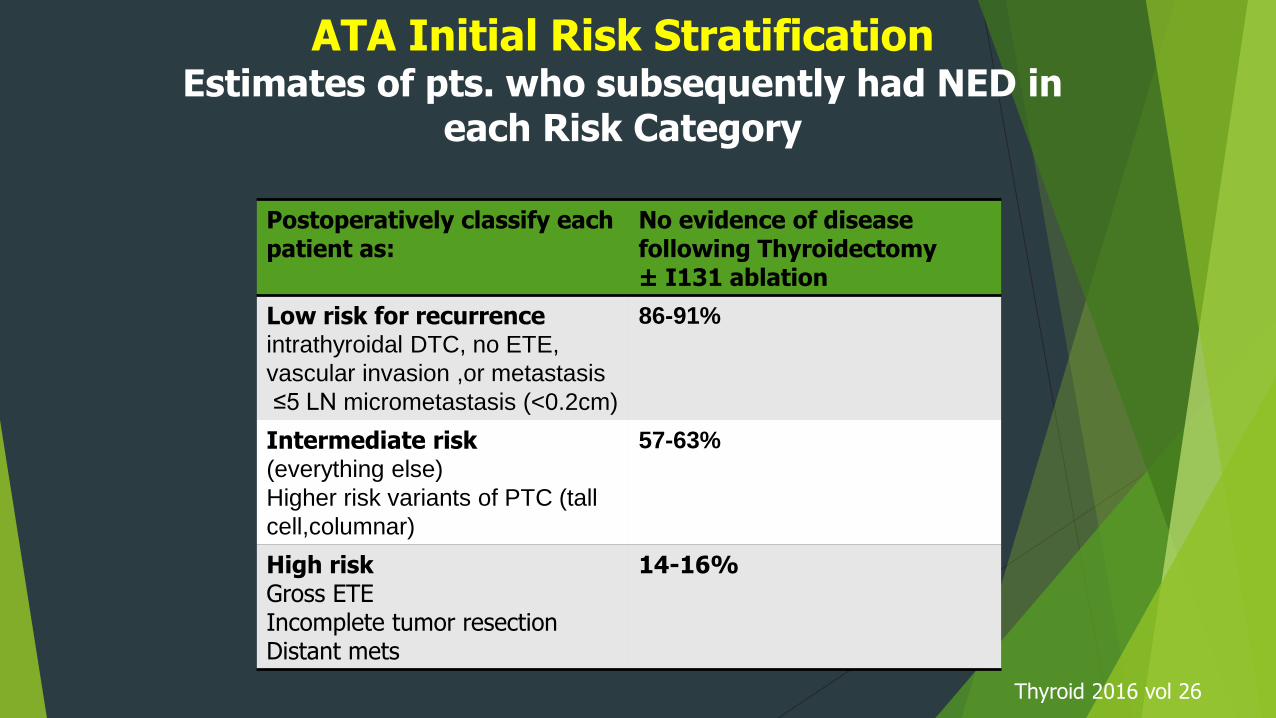
ATA Initial Risk Stratification Estimates of pts. who subsequently had NED in
each Risk Category
Postoperatively classify each patient as:
No evidence of disease following Thyroidectomy± I131 ablation
Low risk for recurrenceintrathyroidal DTC, no ETE,
vascular invasion ,or metastasis
≤5 LN micrometastasis (<0.2cm)
86-91%
Intermediate risk(everything else)
Higher risk variants of PTC (tall
cell,columnar)
57-63%
High riskGross ETEIncomplete tumor resectionDistant mets
14-16%
Thyroid 2016 vol 26
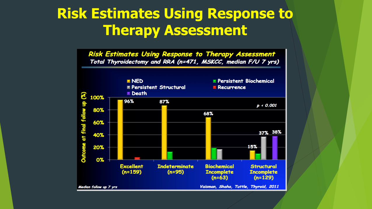
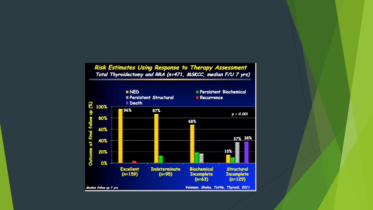
Risk Estimates Using Response to Therapy Assessment
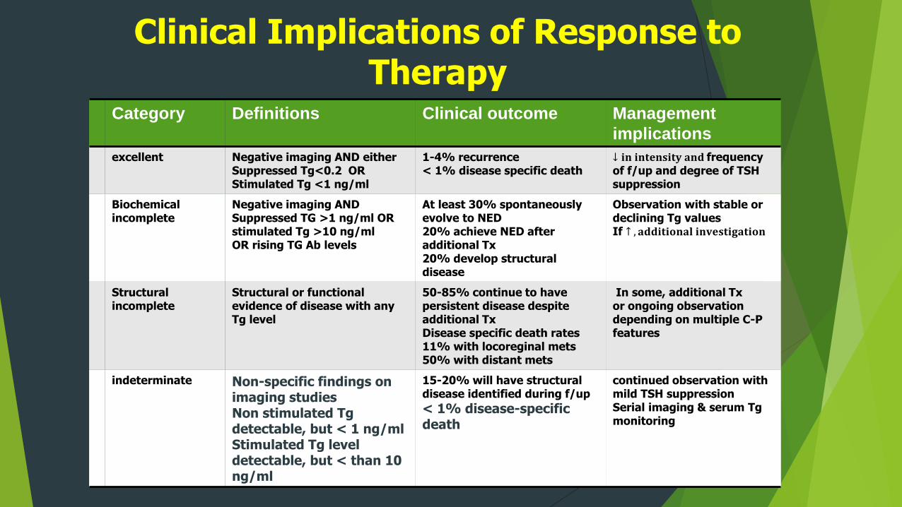
Clinical Implications of Response to Therapy
Category Definitions Clinical outcome Management
implications
excellent Negative imaging AND eitherSuppressed Tg<0.2 ORStimulated Tg <1 ng/ml
1-4% recurrence < 1% disease specific death
↓ 𝐢𝐧 𝐢𝐧𝐭𝐞𝐧𝐬𝐢𝐭𝐲 𝐚𝐧𝐝 frequency of f/up and degree of TSH suppression
Biochemical incomplete
Negative imaging ANDSuppressed TG >1 ng/ml OR stimulated Tg >10 ng/mlOR rising TG Ab levels
At least 30% spontaneously evolve to NED20% achieve NED after additional Tx20% develop structural disease
Observation with stable or declining Tg valuesIf ↑ , 𝐚𝐝𝐝𝐢𝐭𝐢𝐨𝐧𝐚𝐥 𝐢𝐧𝐯𝐞𝐬𝐭𝐢𝐠𝐚𝐭𝐢𝐨𝐧
Structural incomplete
Structural or functional evidence of disease with anyTg level
50-85% continue to have persistent disease despite additional TxDisease specific death rates 11% with locoreginal mets50% with distant mets
In some, additional Txor ongoing observation depending on multiple C-P features
indeterminate Non-specific findings on imaging studiesNon stimulated Tgdetectable, but < 1 ng/mlStimulated Tg level detectable, but < than 10 ng/ml
15-20% will have structural disease identified during f/up
< 1% disease-specific death
continued observation with mild TSH suppression Serial imaging & serum Tgmonitoring

Initial treatment
Goals of Initial Therapy
Remove the primary tumor, disease that has extended beyond the capsule, and c LN mets
Completeness of surgical resection is an imp. determinant of outcome.
Minimize risk of disease recurrence and metastatic spread
Adequate surgery is the most imp. variable influencing prognosis.
Facilitate postoperative Tx with RAI, where appropriate.
Permit accurate staging and risk stratification of the disease.
Accurate post-op risk assessment is a crucial element in the Txof DTC
Permit accurate long-term surveillance for disease recurrence
Minimize treatment-related morbidity
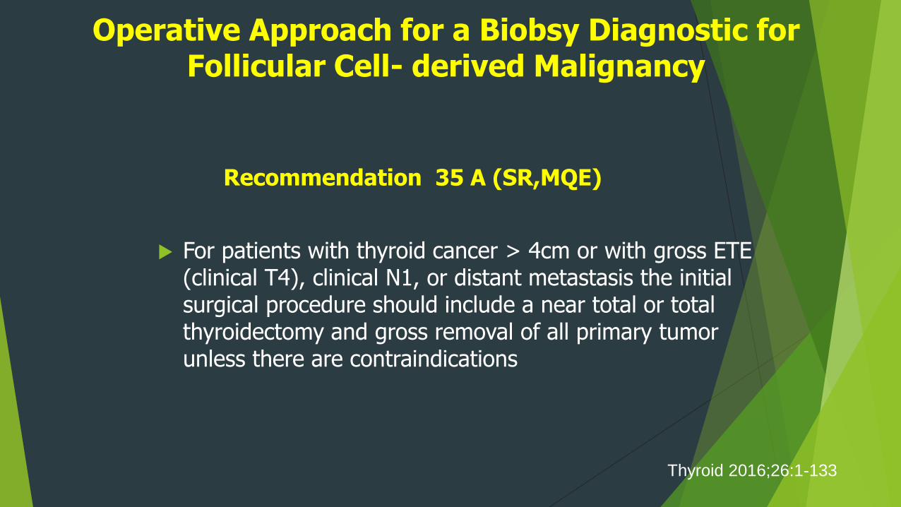
Operative Approach for a Biobsy Diagnostic for Follicular Cell- derived Malignancy
Recommendation 35 A (SR,MQE)
For patients with thyroid cancer > 4cm or with gross ETE (clinical T4), clinical N1, or distant metastasis the initial surgical procedure should include a near total or total thyroidectomy and gross removal of all primary tumor unless there are contraindications
Thyroid 2016;26:1-133
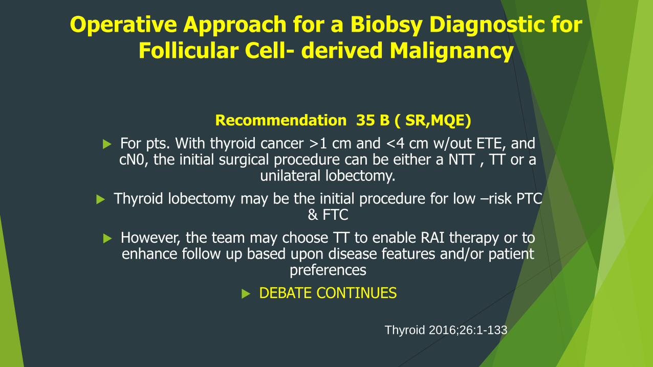
Operative Approach for a Biobsy Diagnostic for Follicular Cell- derived Malignancy
Recommendation 35 B ( SR,MQE)
For pts. With thyroid cancer >1 cm and <4 cm w/out ETE, and cN0, the initial surgical procedure can be either a NTT , TT or a
unilateral lobectomy.
Thyroid lobectomy may be the initial procedure for low –risk PTC & FTC
However, the team may choose TT to enable RAI therapy or to enhance follow up based upon disease features and/or patient
preferences
DEBATE CONTINUES
Thyroid 2016;26:1-133
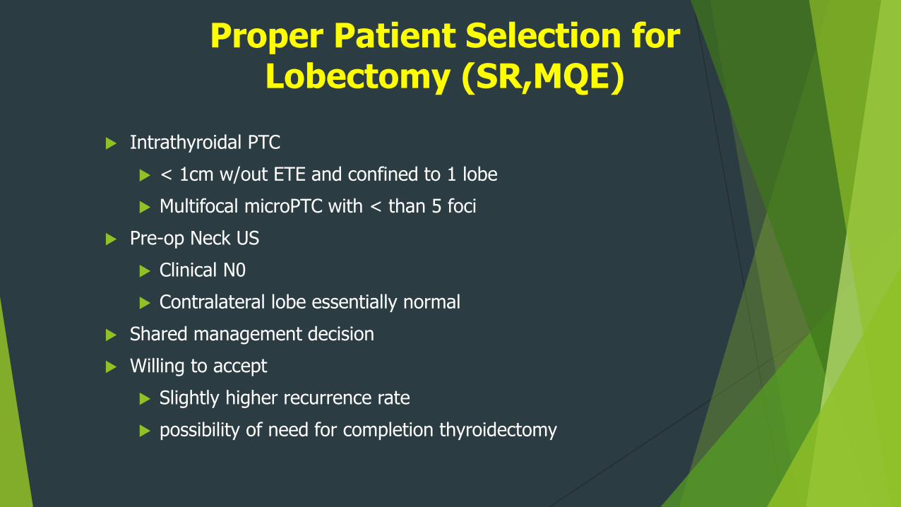
Proper Patient Selection for Lobectomy (SR,MQE)
Intrathyroidal PTC
< 1cm w/out ETE and confined to 1 lobe
Multifocal microPTC with < than 5 foci
Pre-op Neck US
Clinical N0
Contralateral lobe essentially normal
Shared management decision
Willing to accept
Slightly higher recurrence rate
possibility of need for completion thyroidectomy
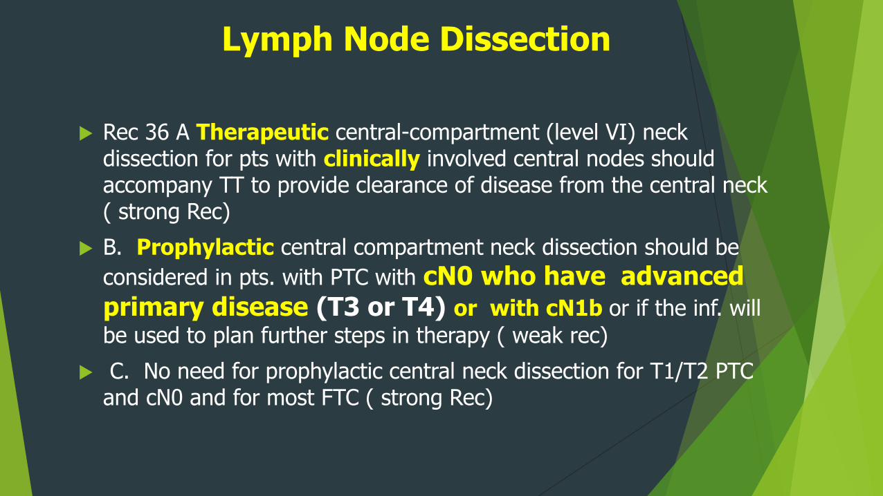
Lymph Node Dissection
Rec 36 A Therapeutic central-compartment (level VI) neck dissection for pts with clinically involved central nodes should accompany TT to provide clearance of disease from the central neck ( strong Rec)
B. Prophylactic central compartment neck dissection should be
considered in pts. with PTC with cN0 who have advanced primary disease (T3 or T4) or with cN1b or if the inf. will
be used to plan further steps in therapy ( weak rec)
C. No need for prophylactic central neck dissection for T1/T2 PTC and cN0 and for most FTC ( strong Rec)
Lymph Node Dissection
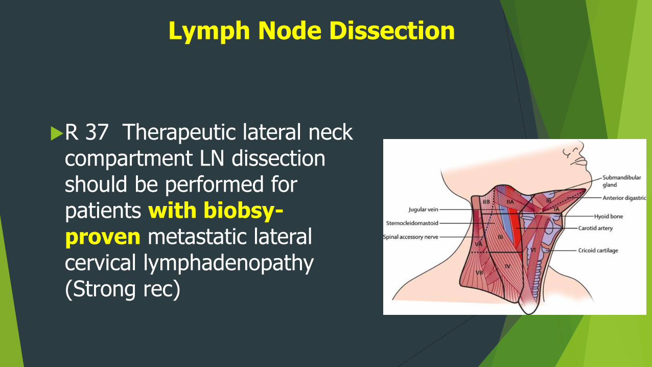
R 37 Therapeutic lateral neck compartment LN dissection should be performed for patients with biobsy-proven metastatic lateral cervical lymphadenopathy (Strong rec)

Radioiodine Treatment
Goals of Radioiodine
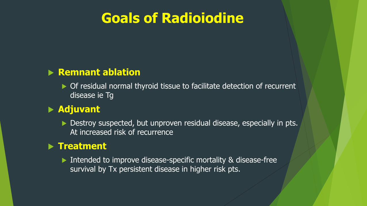
Remnant ablation
Of residual normal thyroid tissue to facilitate detection of recurrent disease ie Tg
Adjuvant
Destroy suspected, but unproven residual disease, especially in pts. At increased risk of recurrence
Treatment
Intended to improve disease-specific mortality & disease-free survival by Tx persistent disease in higher risk pts.
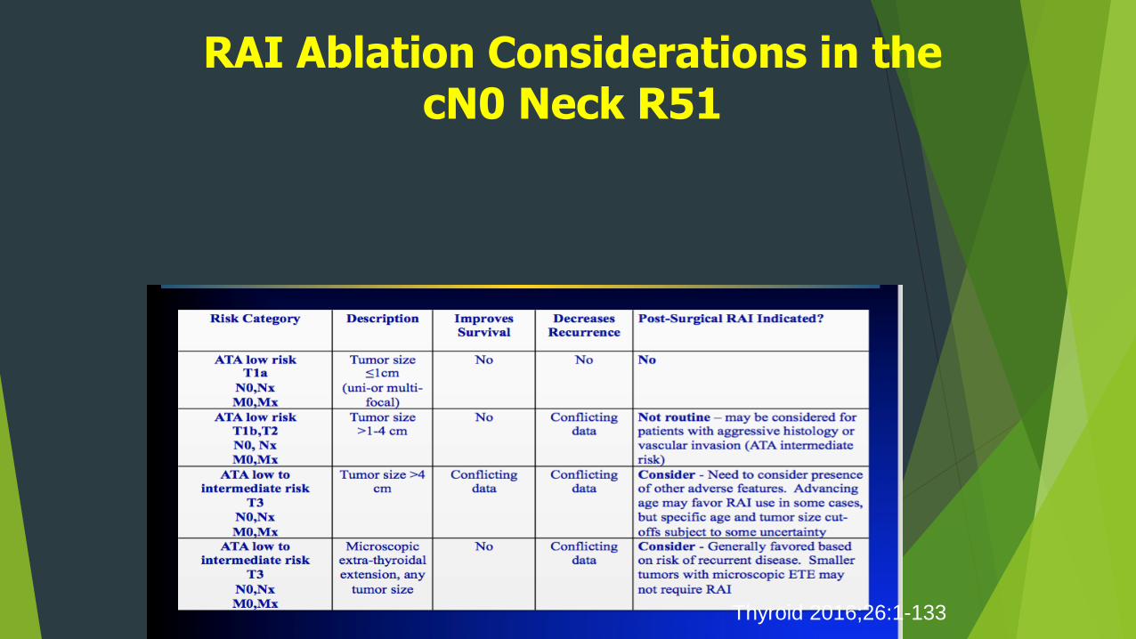
RAI Ablation Considerations in the cN0 Neck R51
Thyroid 2016;26:1-133
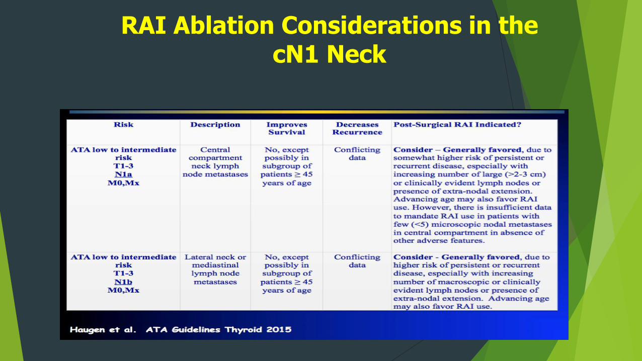
RAI Ablation Considerations in the cN1 Neck
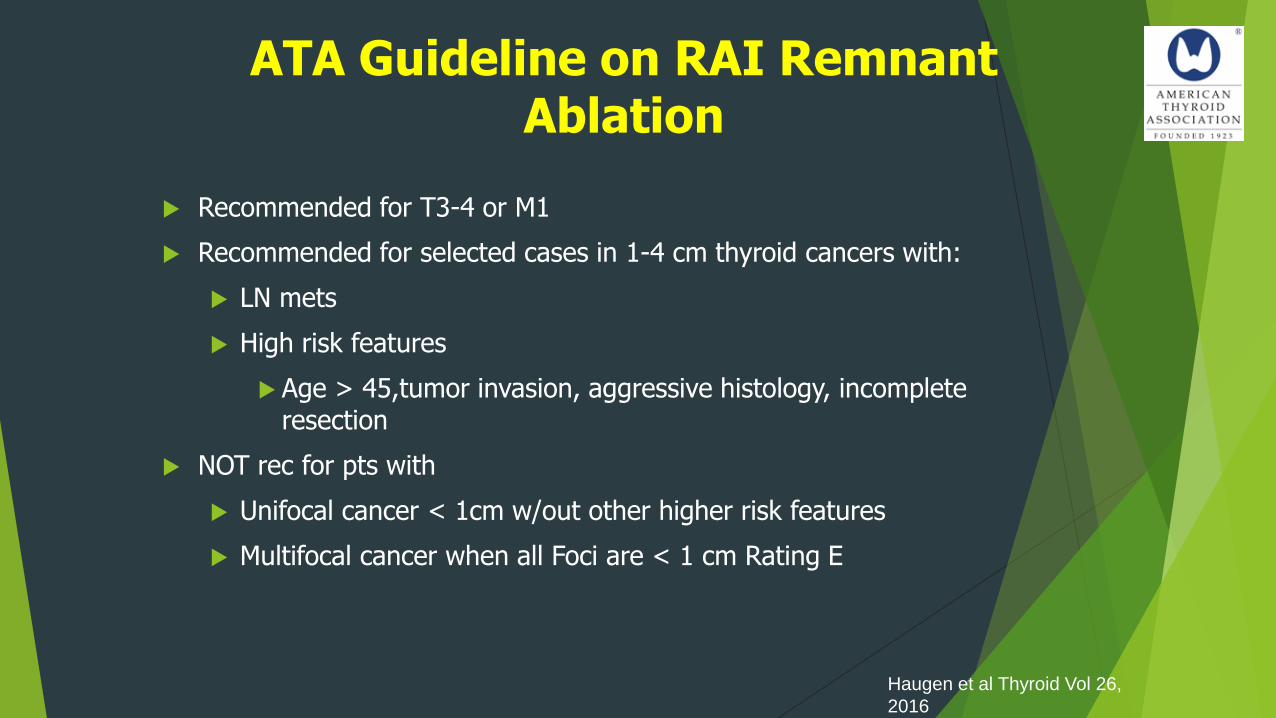
ATA Guideline on RAI Remnant Ablation
Recommended for T3-4 or M1
Recommended for selected cases in 1-4 cm thyroid cancers with:
LN mets
High risk features
Age > 45,tumor invasion, aggressive histology, incomplete resection
NOT rec for pts with
Unifocal cancer < 1cm w/out other higher risk features
Multifocal cancer when all Foci are < 1 cm Rating E
Haugen et al Thyroid Vol 26,
2016

Long-Term Management
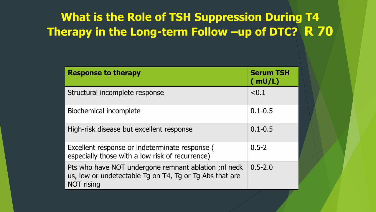
What is the Role of TSH Suppression During T4
Therapy in the Long-term Follow –up of DTC? R 70
Response to therapy Serum TSH ( mU/L)
Structural incomplete response <0.1
Biochemical incomplete 0.1-0.5
High-risk disease but excellent response 0.1-0.5
Excellent response or indeterminate response ( especially those with a low risk of recurrence)
0.5-2
Pts who have NOT undergone remnant ablation ;nl neck us, low or undetectable Tg on T4, Tg or Tg Abs that are NOT rising
0.5-2.0
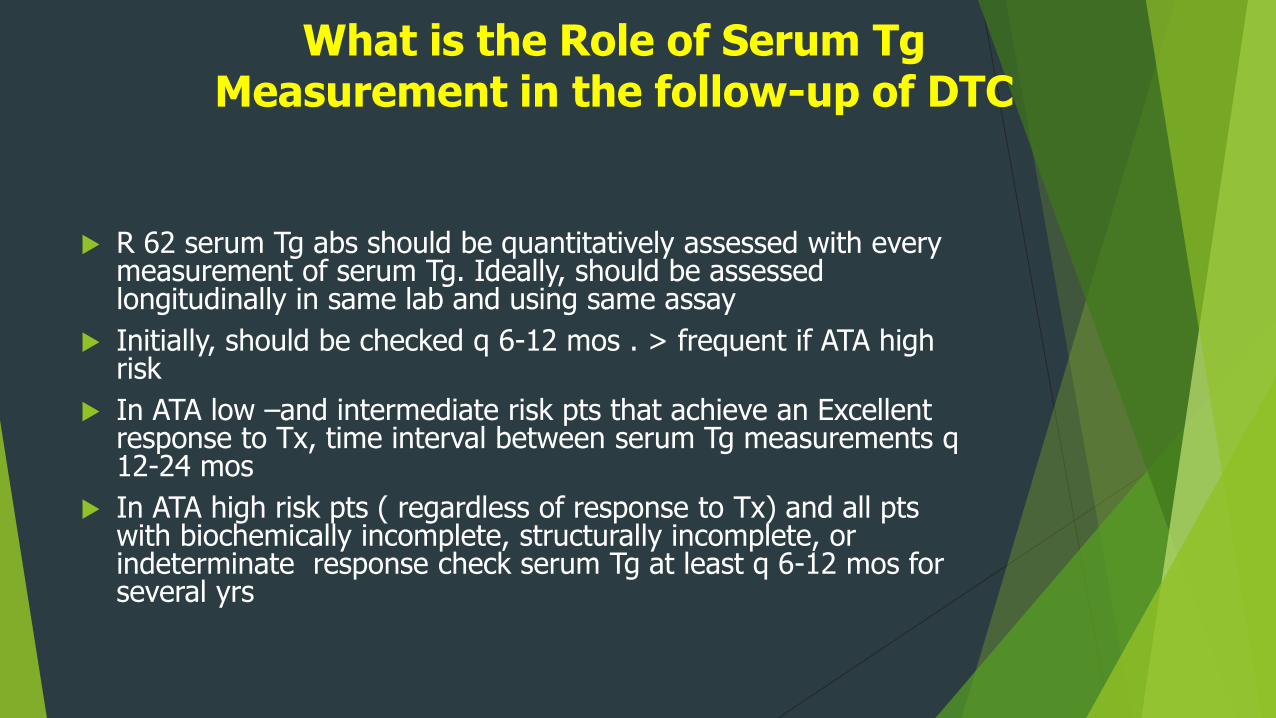
What is the Role of Serum TgMeasurement in the follow-up of DTC
R 62 serum Tg abs should be quantitatively assessed with every measurement of serum Tg. Ideally, should be assessed longitudinally in same lab and using same assay
Initially, should be checked q 6-12 mos . > frequent if ATA high risk
In ATA low –and intermediate risk pts that achieve an Excellent response to Tx, time interval between serum Tg measurements q 12-24 mos
In ATA high risk pts ( regardless of response to Tx) and all pts with biochemically incomplete, structurally incomplete, or indeterminate response check serum Tg at least q 6-12 mos for several yrs
What is the Role of Serum TgMeasurement in follow-up of DTC ?
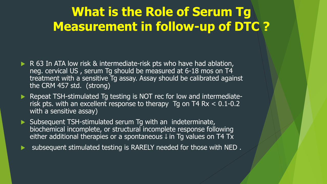
R 63 In ATA low risk & intermediate-risk pts who have had ablation, neg. cervical US , serum Tg should be measured at 6-18 mos on T4 treatment with a sensitive Tg assay. Assay should be calibrated against the CRM 457 std. (strong)
Repeat TSH-stimulated Tg testing is NOT rec for low and intermediate-risk pts. with an excellent response to therapy Tg on T4 Rx < 0.1-0.2 with a sensitive assay)
Subsequent TSH-stimulated serum Tg with an indeterminate, biochemical incomplete, or structural incomplete response following either additional therapies or a spontaneous ↓ in Tg values on T4 Tx
subsequent stimulated testing is RARELY needed for those with NED .
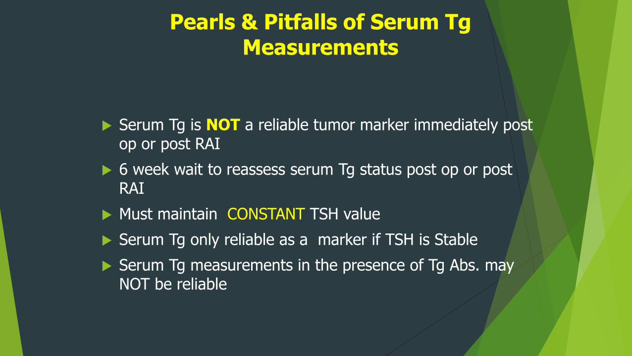
Pearls & Pitfalls of Serum TgMeasurements
Serum Tg is NOT a reliable tumor marker immediately post op or post RAI
6 week wait to reassess serum Tg status post op or post RAI
Must maintain CONSTANT TSH value
Serum Tg only reliable as a marker if TSH is Stable
Serum Tg measurements in the presence of Tg Abs. may NOT be reliable
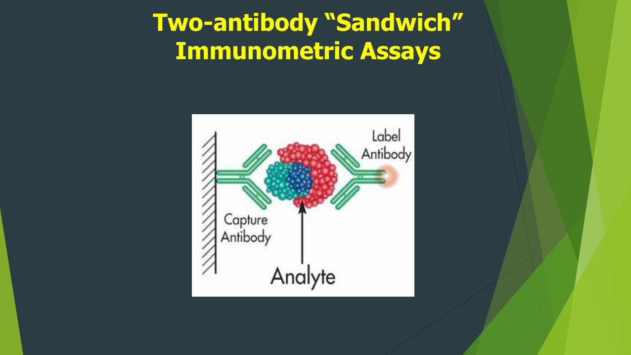
Two-antibody “Sandwich” Immunometric Assays
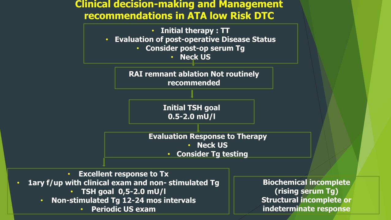
• Excellent response to Tx• 1ary f/up with clinical exam and non- stimulated Tg
• TSH goal 0,5-2.0 mU/l• Non-stimulated Tg 12-24 mos intervals
• Periodic US exam
• Initial therapy : TT• Evaluation of post-operative Disease Status
• Consider post-op serum Tg• Neck US
RAI remnant ablation Not routinely recommended
Initial TSH goal 0.5-2.0 mU/l
Evaluation Response to Therapy• Neck US
• Consider Tg testing
Biochemical incomplete(rising serum Tg)
Structural incomplete or indeterminate response
Clinical decision-making and Management recommendations in ATA low Risk DTC
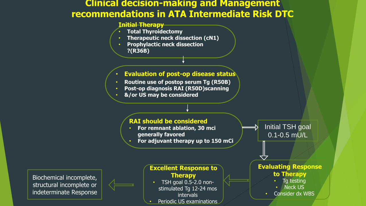
Initial Therapy• Total Thyroidectomy• Therapeutic neck dissection (cN1)• Prophylactic neck dissection
?(R36B)
RAI should be considered• For remnant ablation, 30 mci
generally favored• For adjuvant therapy up to 150 mCi
• Routine use of postop serum Tg (R50B)• Post-op diagnosis RAI (R50D)scanning • &/or US may be considered
• Evaluation of post-op disease status
Evaluating Response to Therapy • Tg testing• Neck US
• Consider dx WBS
Excellent Response to Therapy
• TSH goal 0.5-2.0 non-stimulated Tg 12-24 mos
intervals• Periodic US examinations
Initial TSH goal
0.1-0.5 mU/L
Biochemical incomplete, structural incomplete or indeterminate Response
Clinical decision-making and Management recommendations in ATA Intermediate Risk DTC
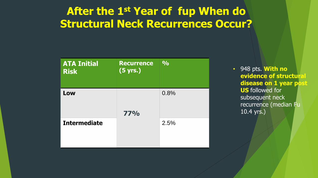
After the 1st Year of fup When do Structural Neck Recurrences Occur?
ATA Initial Risk
Recurrence (5 yrs.)
%
Low 0.8%
Intermediate 2.5%
77%
• 948 pts. With no evidence of structural disease on 1 year post US followed for subsequent neck recurrence (median Fu 10.4 yrs.)
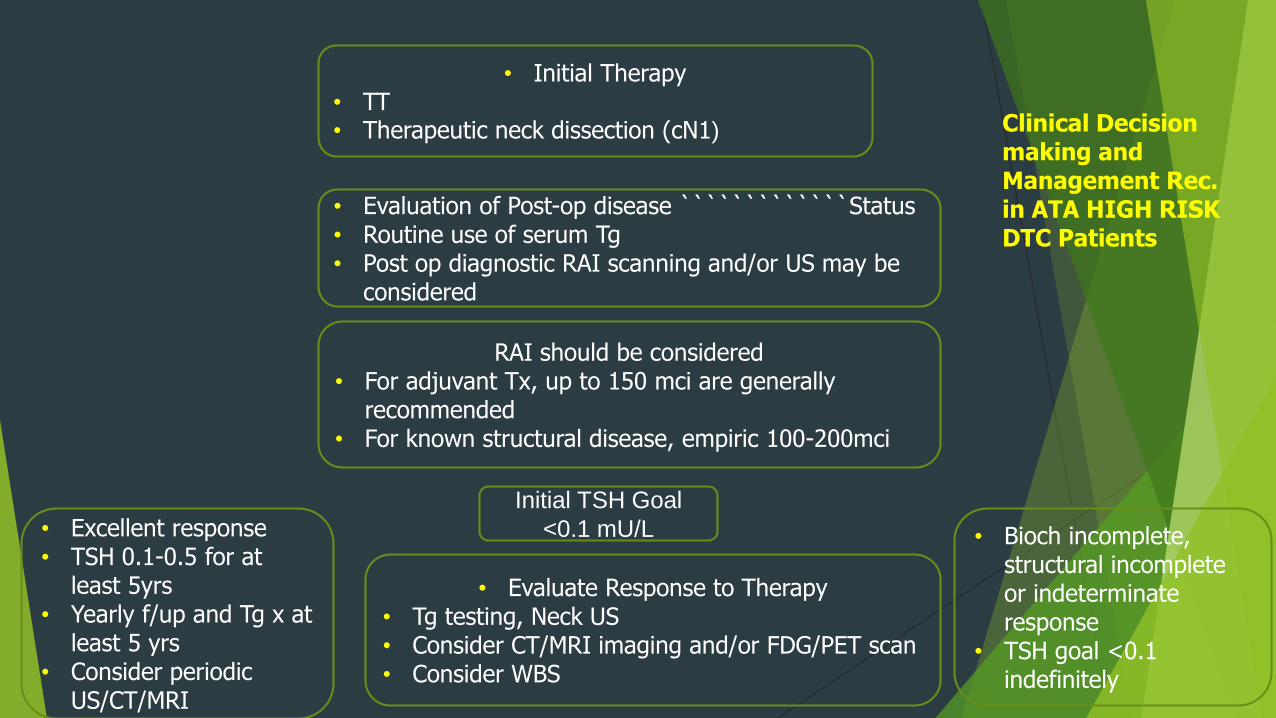
• Evaluate Response to Therapy• Tg testing, Neck US• Consider CT/MRI imaging and/or FDG/PET scan• Consider WBS
• Evaluation of Post-op disease `````````````Status• Routine use of serum Tg• Post op diagnostic RAI scanning and/or US may be
considered
RAI should be considered• For adjuvant Tx, up to 150 mci are generally
recommended• For known structural disease, empiric 100-200mci
• Excellent response• TSH 0.1-0.5 for at
least 5yrs• Yearly f/up and Tg x at
least 5 yrs• Consider periodic
US/CT/MRI
• Bioch incomplete, structural incomplete or indeterminate response
• TSH goal <0.1 indefinitely
Initial TSH Goal
<0.1 mU/L
• Initial Therapy • TT• Therapeutic neck dissection (cN1) Clinical Decision
making and Management Rec. in ATA HIGH RISK DTC Patients
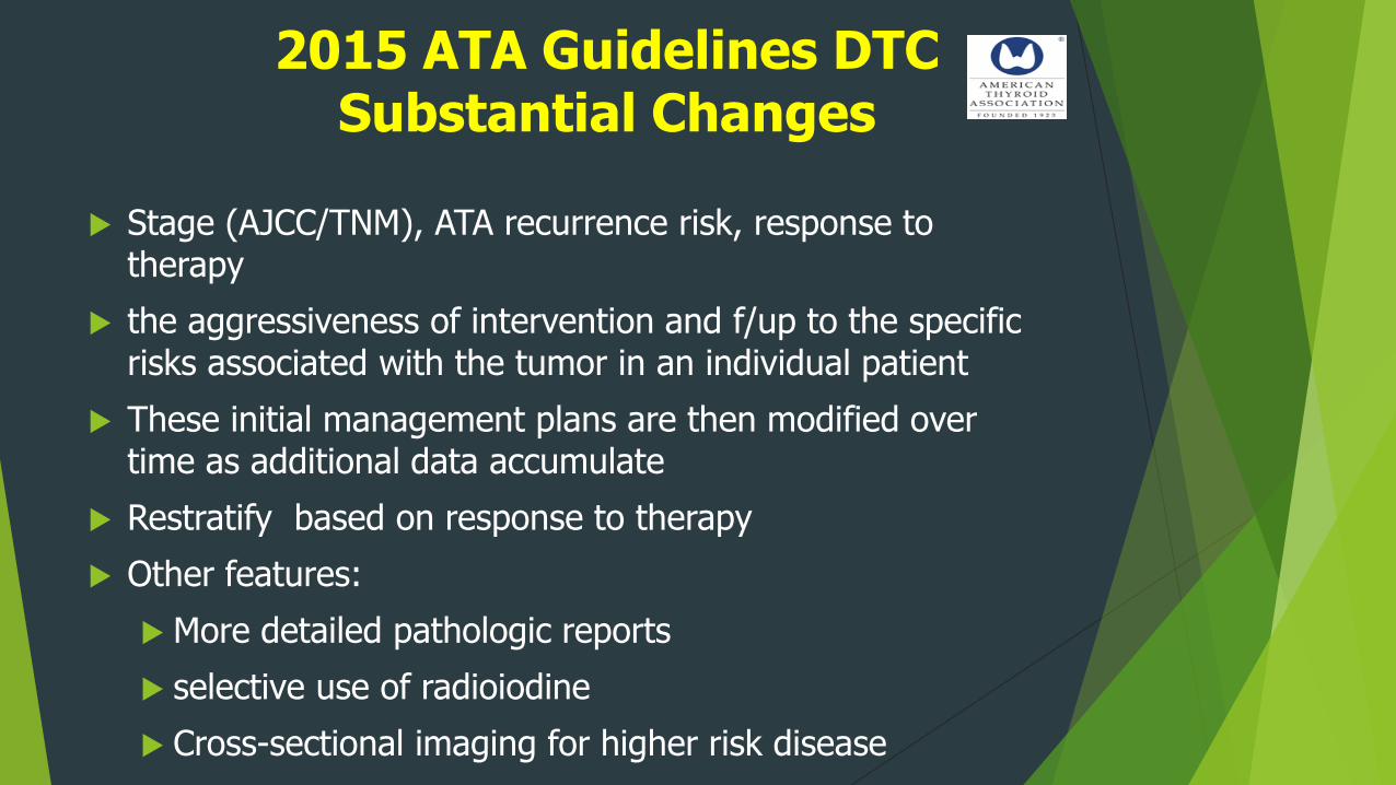
2015 ATA Guidelines DTCSubstantial Changes
Stage (AJCC/TNM), ATA recurrence risk, response to therapy
the aggressiveness of intervention and f/up to the specific risks associated with the tumor in an individual patient
These initial management plans are then modified over time as additional data accumulate
Restratify based on response to therapy
Other features:
More detailed pathologic reports
selective use of radioiodine
Cross-sectional imaging for higher risk disease

Thank You!
rec 48a 2009 ATA Initial Risk Stratification System is rec. for DTC pts. treated with TT, based
on its utility in Predicting risk of disease recurrence and/or persistence (SR,MQE)
• LOW RISK
• Classic or PTC FV +• No local or distant mets.• Complete resection• No tumor invasion• No vascular invasion• If given, no RAI uptake
outside thyroid bed
• INTERMEDIATE RISK• Microscopic ETE • Cervical LN mets• Aggressive histology or• AL invasion
• HIGH RISK• Macroscopic gross ETE • Incomplete tumor resection• Distant mets. or• inappropriate Tg elevation
Cooper Thyroid 2009;19:1167Haugen Thyroid 2016;26:1
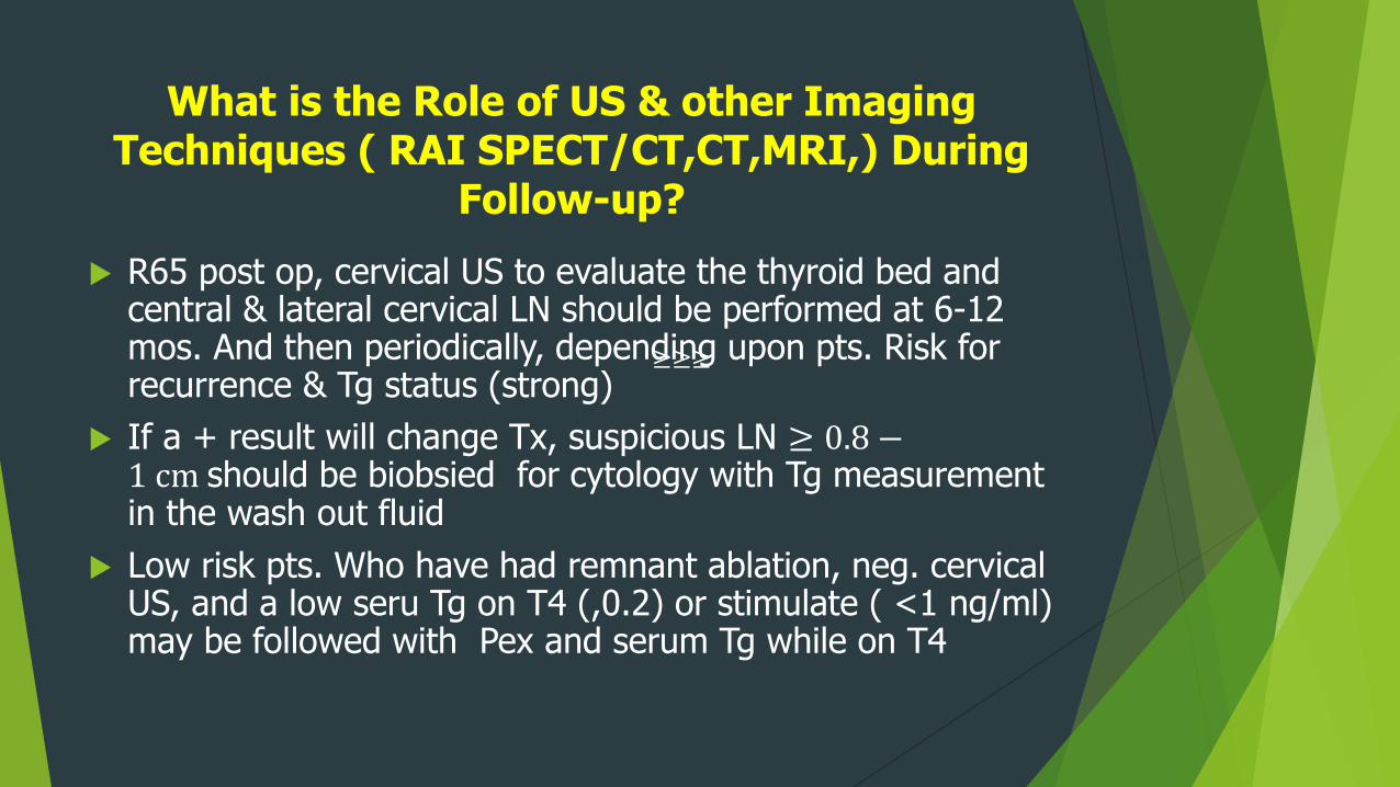
What is the Role of US & other Imaging Techniques ( RAI SPECT/CT,CT,MRI,) During
Follow-up?
R65 post op, cervical US to evaluate the thyroid bed and central & lateral cervical LN should be performed at 6-12 mos. And then periodically, depending upon pts. Risk for recurrence & Tg status (strong)
If a + result will change Tx, suspicious LN ≥ 0.8 −1 cm should be biobsied for cytology with Tg measurement in the wash out fluid
Low risk pts. Who have had remnant ablation, neg. cervical US, and a low seru Tg on T4 (,0.2) or stimulate ( <1 ng/ml) may be followed with Pex and serum Tg while on T4
≥≥≥
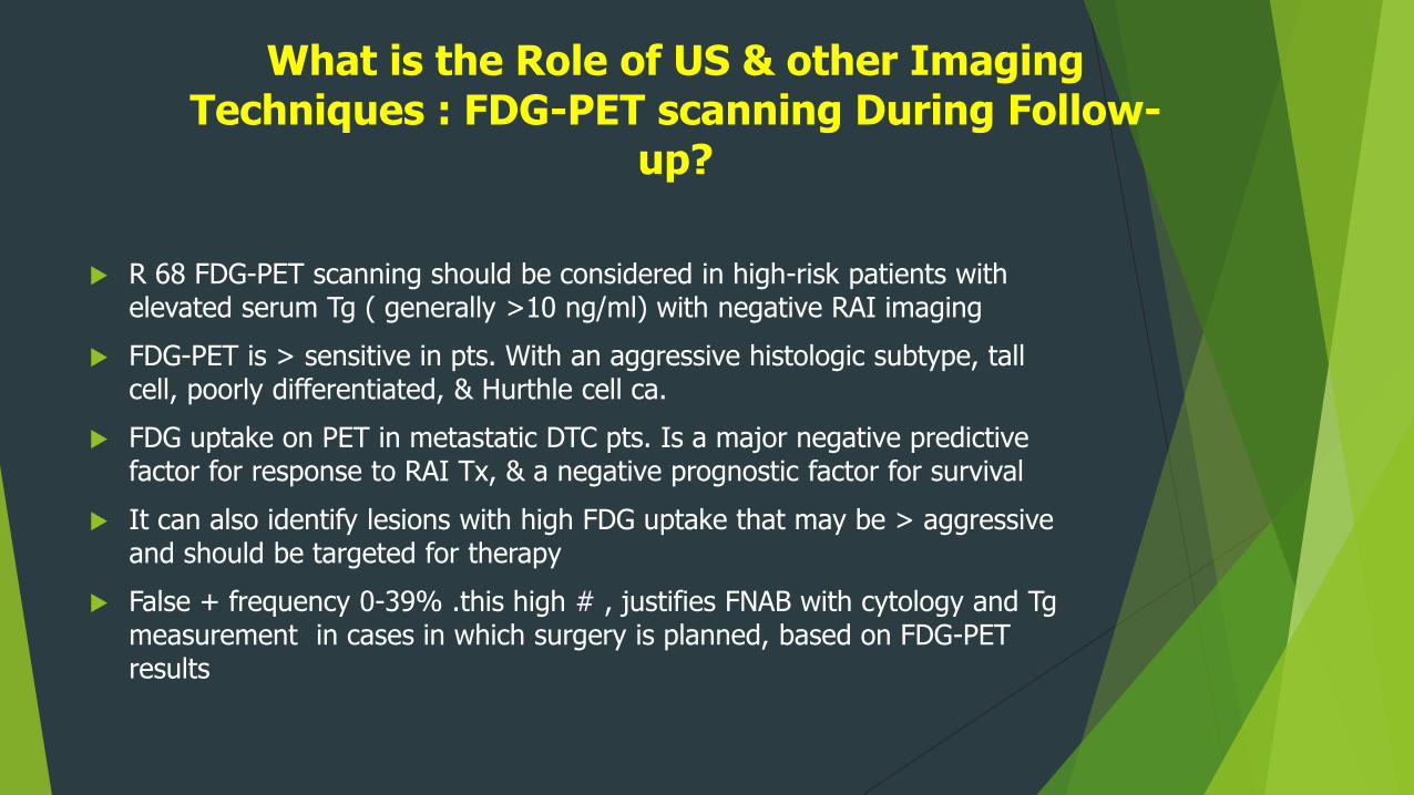
R 68 FDG-PET scanning should be considered in high-risk patients with elevated serum Tg ( generally >10 ng/ml) with negative RAI imaging
FDG-PET is > sensitive in pts. With an aggressive histologic subtype, tall cell, poorly differentiated, & Hurthle cell ca.
FDG uptake on PET in metastatic DTC pts. Is a major negative predictive factor for response to RAI Tx, & a negative prognostic factor for survival
It can also identify lesions with high FDG uptake that may be > aggressive and should be targeted for therapy
False + frequency 0-39% .this high # , justifies FNAB with cytology and Tgmeasurement in cases in which surgery is planned, based on FDG-PET results
What is the Role of US & other Imaging Techniques : FDG-PET scanning During Follow-
up?
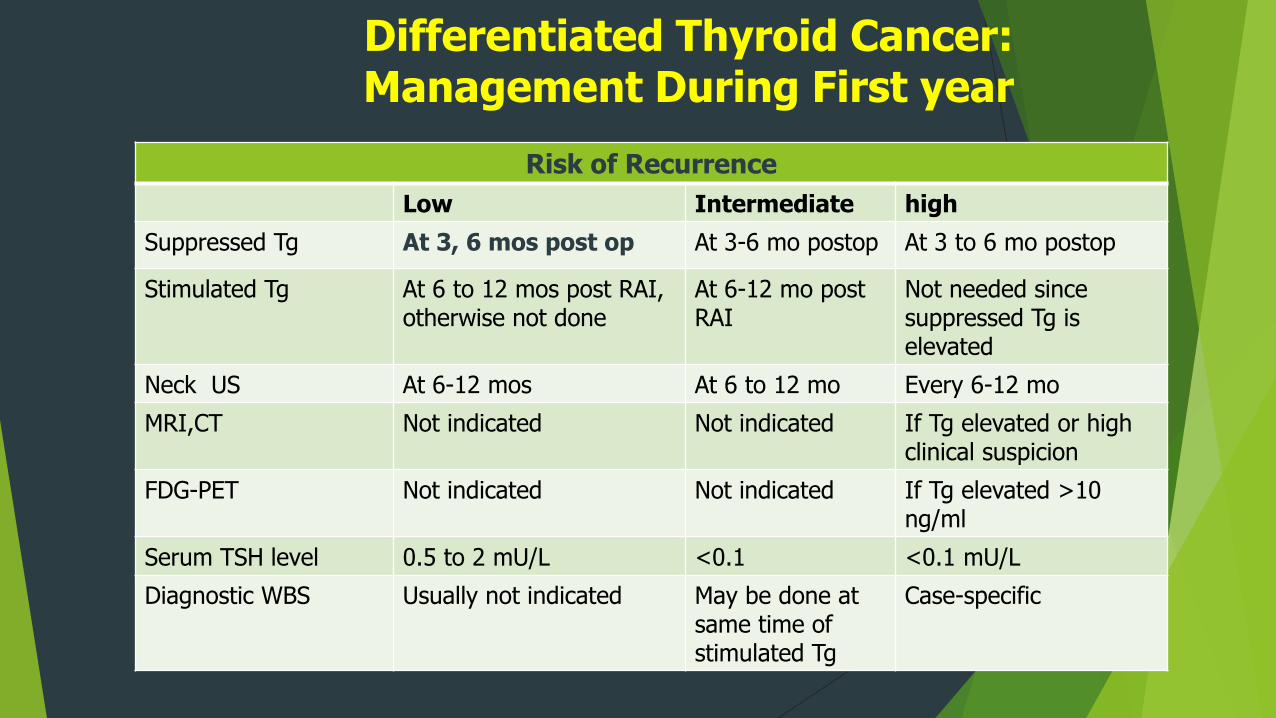
Differentiated Thyroid Cancer: Management During First year
Risk of Recurrence
Low Intermediate high
Suppressed Tg At 3, 6 mos post op At 3-6 mo postop At 3 to 6 mo postop
Stimulated Tg At 6 to 12 mos post RAI, otherwise not done
At 6-12 mo post RAI
Not needed since suppressed Tg is elevated
Neck US At 6-12 mos At 6 to 12 mo Every 6-12 mo
MRI,CT Not indicated Not indicated If Tg elevated or high clinical suspicion
FDG-PET Not indicated Not indicated If Tg elevated >10 ng/ml
Serum TSH level 0.5 to 2 mU/L <0.1 <0.1 mU/L
Diagnostic WBS Usually not indicated May be done at same time ofstimulated Tg
Case-specific
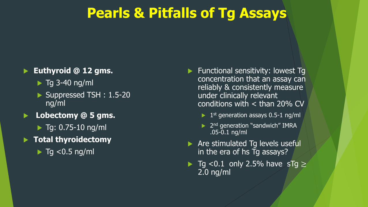
Pearls & Pitfalls of Tg Assays
Euthyroid @ 12 gms.
Tg 3-40 ng/ml
Suppressed TSH : 1.5-20 ng/ml
Lobectomy @ 5 gms.
Tg: 0.75-10 ng/ml
Total thyroidectomy
Tg <0.5 ng/ml
Functional sensitivity: lowest Tgconcentration that an assay can reliably & consistently measure under clinically relevant conditions with < than 20% CV
1st generation assays 0.5-1 ng/ml
2nd generation “sandwich” IMRA .05-0.1 ng/ml
Are stimulated Tg levels useful in the era of hs Tg assays?
Tg <0.1 only 2.5% have sTg ≥2.0 ng/ml
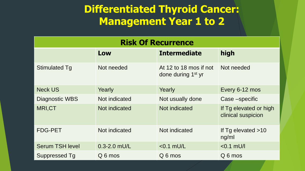
Differentiated Thyroid Cancer: Management Year 1 to 2
Risk Of Recurrence
Low Intermediate high
Stimulated Tg Not needed At 12 to 18 mos if not
done during 1st yr
Not needed
Neck US Yearly Yearly Every 6-12 mos
Diagnostic WBS Not indicated Not usually done Case –specific
MRI,CT Not indicated Not indicated If Tg elevated or high
clinical suspicion
FDG-PET Not indicated Not indicated If Tg elevated >10
ng/ml
Serum TSH level 0.3-2.0 mU/L <0.1 mU/L <0.1 mU/l
Suppressed Tg Q 6 mos Q 6 mos Q 6 mos
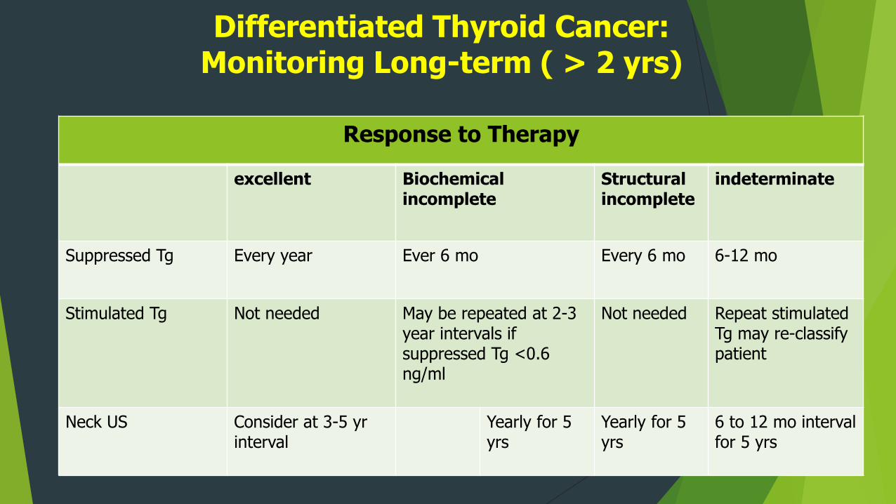
Differentiated Thyroid Cancer: Monitoring Long-term ( > 2 yrs)
Response to Therapy
excellent Biochemical incomplete
Structural incomplete
indeterminate
Suppressed Tg Every year Ever 6 mo Every 6 mo 6-12 mo
Stimulated Tg Not needed May be repeated at 2-3 year intervals if suppressed Tg <0.6 ng/ml
Not needed Repeat stimulated Tg may re-classify patient
Neck US Consider at 3-5 yrinterval
Yearly for 5 yrs
Yearly for 5 yrs
6 to 12 mo interval for 5 yrs
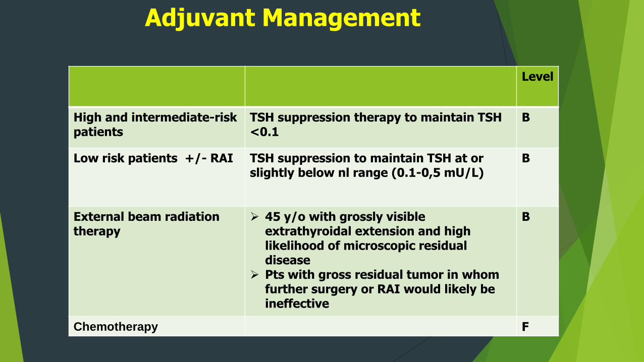
Adjuvant Management
Level
High and intermediate-risk patients
TSH suppression therapy to maintain TSH <0.1
B
Low risk patients +/- RAI TSH suppression to maintain TSH at or slightly below nl range (0.1-0,5 mU/L)
B
External beam radiation therapy
45 y/o with grossly visible extrathyroidal extension and high likelihood of microscopic residual disease
Pts with gross residual tumor in whom further surgery or RAI would likely be ineffective
B
Chemotherapy F
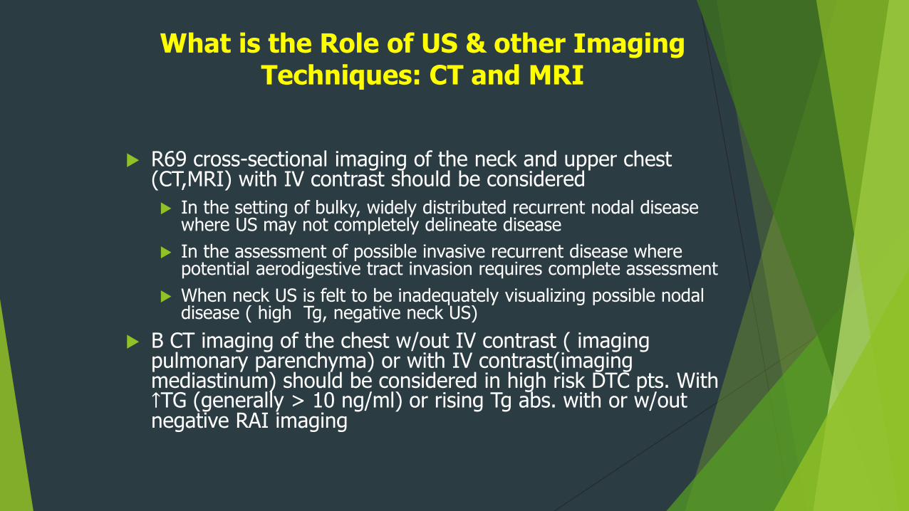
What is the Role of US & other Imaging Techniques: CT and MRI
R69 cross-sectional imaging of the neck and upper chest (CT,MRI) with IV contrast should be considered
In the setting of bulky, widely distributed recurrent nodal disease where US may not completely delineate disease
In the assessment of possible invasive recurrent disease where potential aerodigestive tract invasion requires complete assessment
When neck US is felt to be inadequately visualizing possible nodal disease ( high Tg, negative neck US)
B CT imaging of the chest w/out IV contrast ( imaging pulmonary parenchyma) or with IV contrast(imaging mediastinum) should be considered in high risk DTC pts. With ↑TG (generally > 10 ng/ml) or rising Tg abs. with or w/out negative RAI imaging
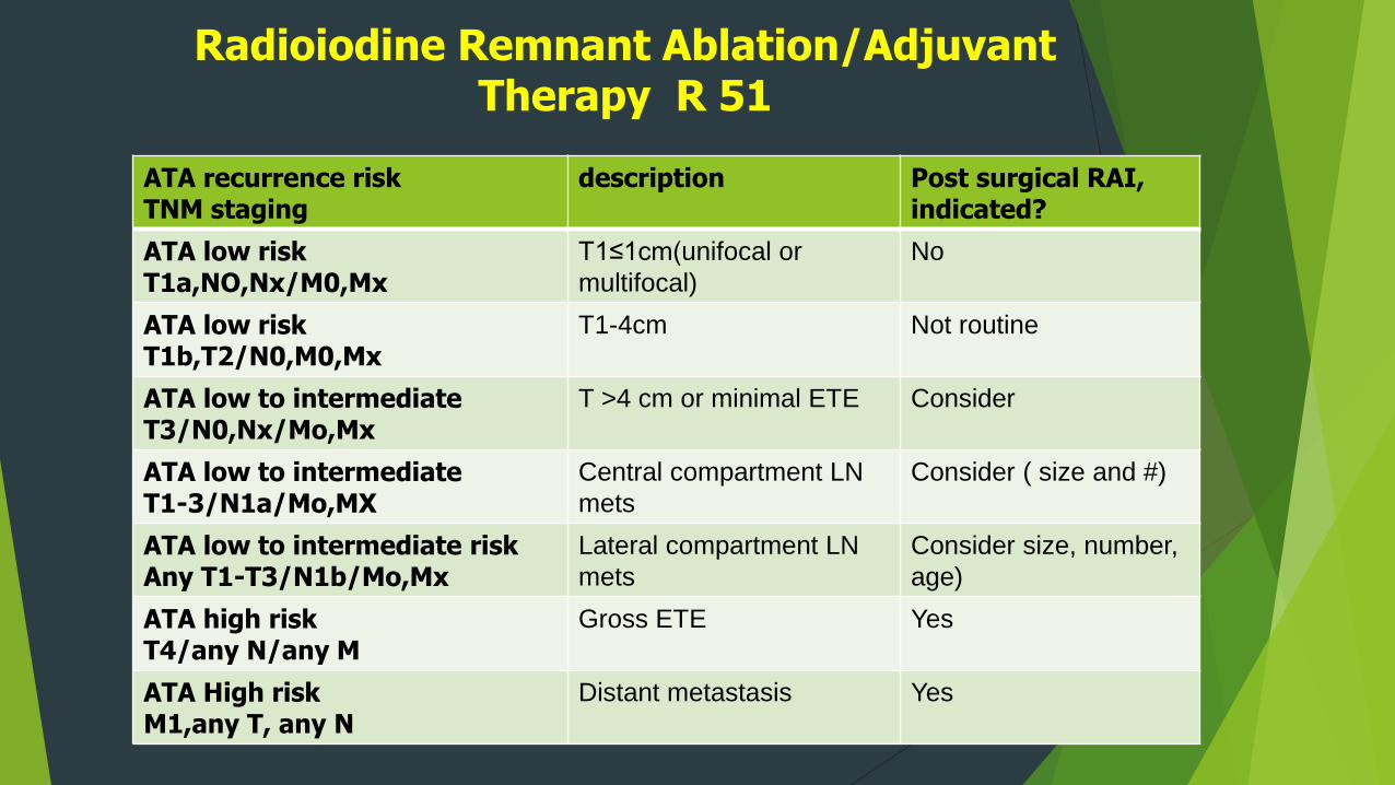
Radioiodine Remnant Ablation/Adjuvant Therapy R 51
ATA recurrence riskTNM staging
description Post surgical RAI, indicated?
ATA low riskT1a,NO,Nx/M0,Mx
T1≤1cm(unifocal or
multifocal)
No
ATA low riskT1b,T2/N0,M0,Mx
T1-4cm Not routine
ATA low to intermediateT3/N0,Nx/Mo,Mx
T >4 cm or minimal ETE Consider
ATA low to intermediateT1-3/N1a/Mo,MX
Central compartment LN
mets
Consider ( size and #)
ATA low to intermediate riskAny T1-T3/N1b/Mo,Mx
Lateral compartment LN
mets
Consider size, number,
age)
ATA high risk T4/any N/any M
Gross ETE Yes
ATA High riskM1,any T, any N
Distant metastasis Yes
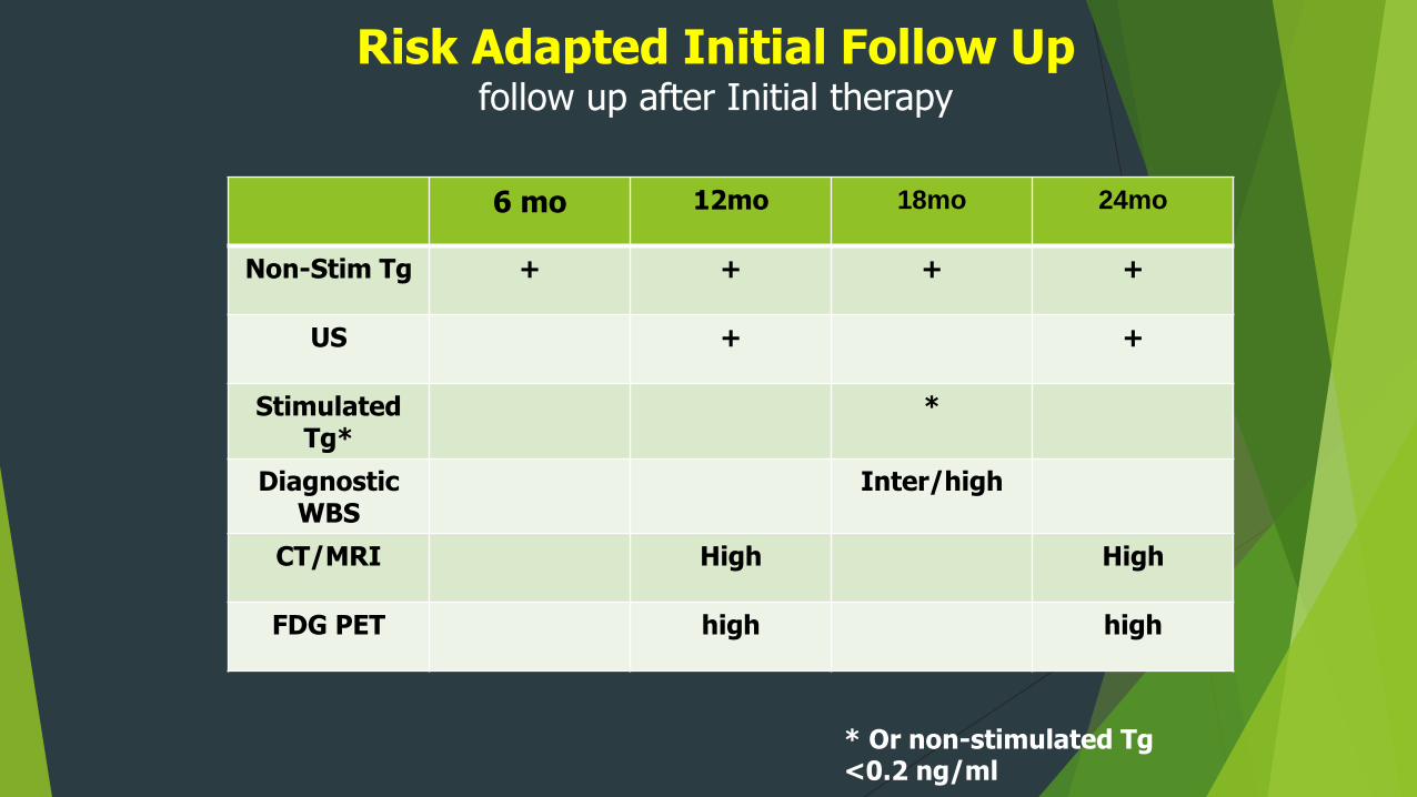
Risk Adapted Initial Follow Upfollow up after Initial therapy
6 mo 12mo 18mo 24mo
Non-Stim Tg + + + +
US + +
Stimulated Tg*
*
Diagnostic WBS
Inter/high
CT/MRI High High
FDG PET high high
* Or non-stimulated Tg<0.2 ng/ml
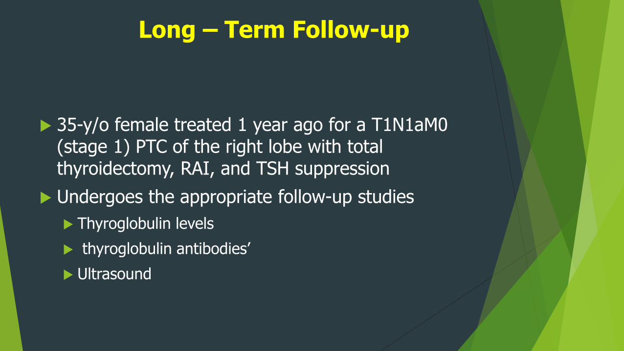
Long – Term Follow-up
35-y/o female treated 1 year ago for a T1N1aM0 (stage 1) PTC of the right lobe with total thyroidectomy, RAI, and TSH suppression
Undergoes the appropriate follow-up studies
Thyroglobulin levels
thyroglobulin antibodies’
Ultrasound
Integrating Response to Therapy in Monitoring Management
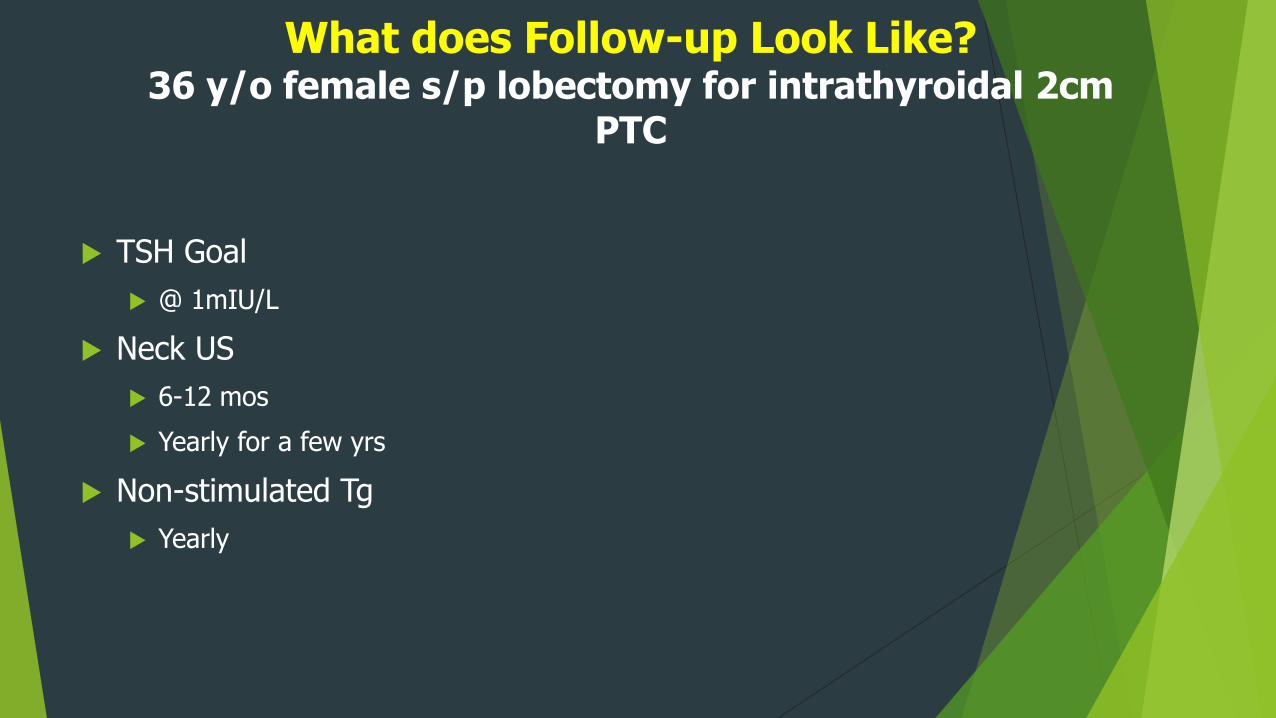
What does Follow-up Look Like?36 y/o female s/p lobectomy for intrathyroidal 2cm
PTC
TSH Goal
@ 1mIU/L
Neck US
6-12 mos
Yearly for a few yrs
Non-stimulated Tg
Yearly
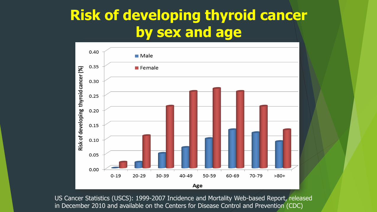
Risk of developing thyroid cancer by sex and age
US Cancer Statistics (USCS): 1999-2007 Incidence and Mortality Web-based Report, released in December 2010 and available on the Centers for Disease Control and Prevention (CDC)

What does Follow-up Look Like?36 y/o female s/p lobectomy for intrathyroidal 2cm
PTC
TSH Goal
@ 1mIU/L
Neck US
6-12 mos
Yearly for a few yrs
Non-stimulated Tg
Yearly
R 46 ( new) Pathology reports should include:
TNM criteria
Vascular invasion & # of vessels
Number of LN examined and involved
Size of the largest metastatic LN focus
Extranodal extension
( strong recommendation, moderate quality evidence )
Central to Risk Stratification Improved & standardized Histopathologic
Assessment
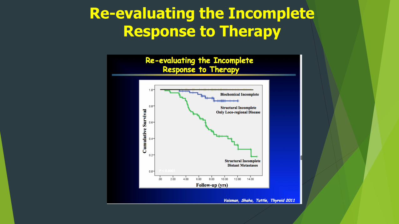
Re-evaluating the Incomplete Response to Therapy
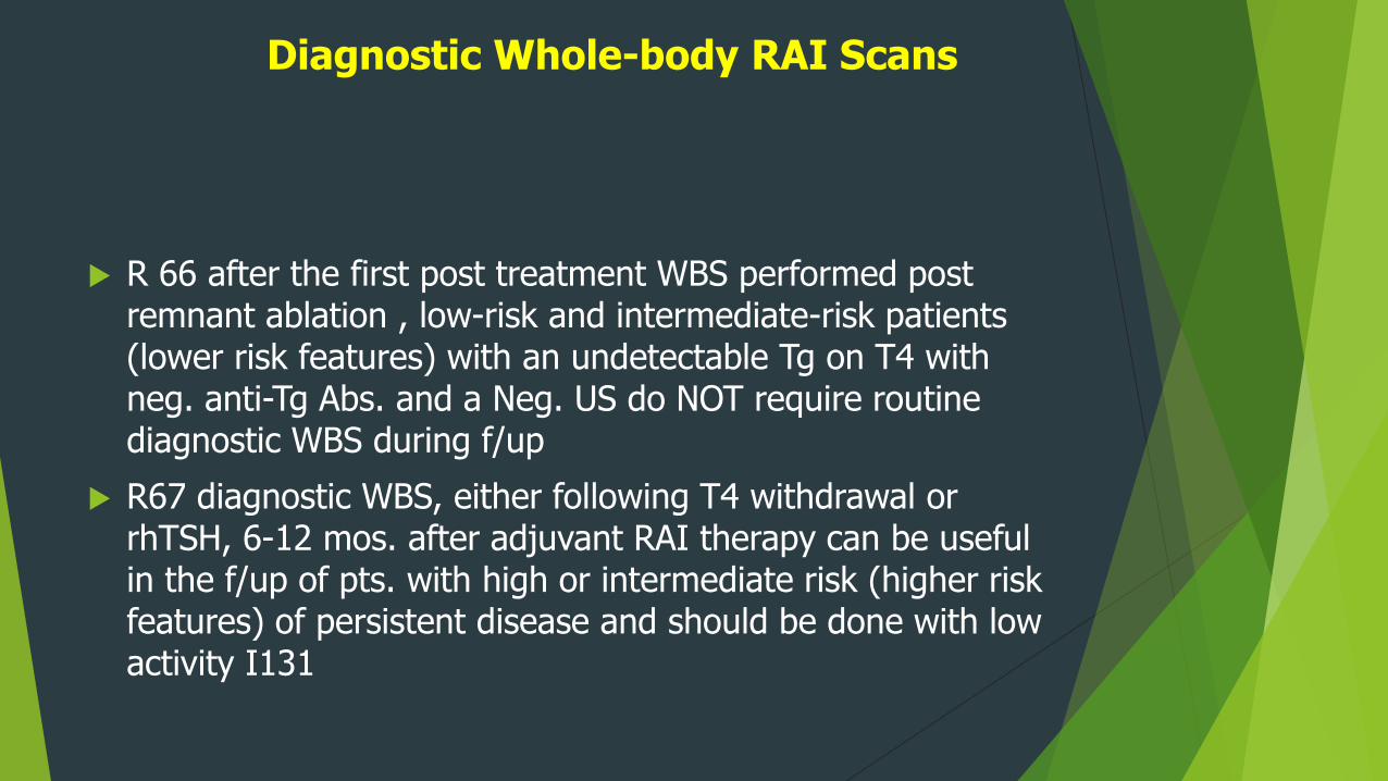
Diagnostic Whole-body RAI Scans
R 66 after the first post treatment WBS performed post remnant ablation , low-risk and intermediate-risk patients (lower risk features) with an undetectable Tg on T4 with neg. anti-Tg Abs. and a Neg. US do NOT require routine diagnostic WBS during f/up
R67 diagnostic WBS, either following T4 withdrawal or rhTSH, 6-12 mos. after adjuvant RAI therapy can be useful in the f/up of pts. with high or intermediate risk (higher risk features) of persistent disease and should be done with low activity I131