Embed Size (px)
Citation preview
7/27/2019 2012 03 20 Chapter 11c Uterine Inversion
http://slidepdf.com/reader/full/2012-03-20-chapter-11c-uterine-inversion 1/9
7/27/2019 2012 03 20 Chapter 11c Uterine Inversion
http://slidepdf.com/reader/full/2012-03-20-chapter-11c-uterine-inversion 2/9
SA Perinatal Practice Guideline: Chapter 11c Uterine inversion
Document title: Uterine inversionFirst developed: 21 July 2004Subsequent updates: 25 January 2005; 25 March 2008Last reviewed: 20 March 2012ISBN number: Replaces document: New documentAuthor: South Australian Perinatal Practice Guideline Workgroup
Audience: Medical, midwifery and allied health staff in South Australia public and private maternity services
Endorsed by: South Australian Perinatal Practice GuidelinesWorkgroup
Contact: South Australian Perinatal Practice Guidelinesworkgroup at: [email protected]
Disclaimer
This guideline provides advice of a general nature. This statewide guideline has beenprepared to promote and facilitate standardisation and consistency of practice, using amultidisciplinary approach. The guideline is based on a review of published evidence andexpert opinion.
Information in this statewide guideline is current at the time of publication.
SA Health does not accept responsibility for the quality or accuracy of material onwebsites linked from this site and does not sponsor, approve or endorse materials onsuch links.
Health practitioners in the South Australian public health sector are expected to reviewspecific details of each patient and professionally assess the applicability of the relevantguideline to that clinical situation.
If for good clinical reasons, a decision is made to depart from the guideline, theresponsible clinician must document in the patient’s medical record, the decision made,by whom, and detailed reasons for the departure from the guideline.
This statewide guideline does not address all the elements of clinical practice andassumes that the individual clinicians are responsible for:
Discussing care with consumers in an environment that is culturally appropriate andwhich enables respectful confidential discussion. This includes the use of interpreter services where necessary
Advising consumers of their choice and ensuring informed consent is obtained Providing care within scope of practice, meeting all legislative requirements andmaintaining standards of professional conduct and,
Documenting all care in accordance with mandatory and local requirements
Refer to online version, destroy printed copies after useLast reviewed: 20/03/12
Page 2 of 9
7/27/2019 2012 03 20 Chapter 11c Uterine Inversion
http://slidepdf.com/reader/full/2012-03-20-chapter-11c-uterine-inversion 3/9
SA Perinatal Practice Guideline: Chapter 11c Uterine inversion
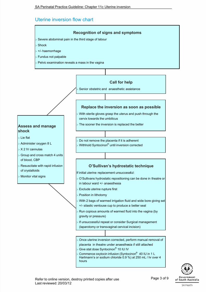
Uterine inversion flow chart
Uterine Inversion
Assess and manageshock
Lie flat
Administer oxygen 8 L
X 2 IV cannulas
Group and cross match 4 units
of blood, CBP
Resuscitate with rapid infusion
of crystalloids
Monitor vital signs
Once uterine inversion corrected, perform manual removal of
placenta in theatre under anaesthesia if still attached Give stat dose Syntocinon ® 10 IU IV Commence oxytocin infusion (Syntocinon ® 40 IU in 1 LHartmann’s or sodium chloride 0.9 %) at 250 mL / hr over 4hours
Do not remove the placenta if it is adherent Withhold Syntocinon ® until inversion corrected
O’Sullivan’s hydrostatic technique
If initial uterine replacement unsuccessful:
O’Sullivans hydrostatic repositioning can be done in theatre or
in labour ward +/- anaesthesia
Exclude uterine rupture first
Position in lithotomy
With 2 bags of warmed irrigation fluid and wide bore giving set
+/- silastic ventouse cup to produce a better seal
Run copious amounts of warmed fluid into the vagina (by
gravity or pressure)
If unsuccessful repeat or consider Surgical management
(laparotomy or transvaginal cervical incision)
Replace the inversion as soon as possible
With sterile gloves grasp the uterus and push through the
cervix towards the umbilicus
The sooner the inversion is replaced the better
Call for help
Senior obstetric and anaesthetic assistance
Recognition of signs and symptoms
Severe abdominal pain in the third stage of labour
Shock
+/- haemorrhage Fundus not palpable
Pelvic examination reveals a mass in the vagina
Refer to online version, destroy printed copies after useLast reviewed: 20/03/12
Page 3 of 9
7/27/2019 2012 03 20 Chapter 11c Uterine Inversion
http://slidepdf.com/reader/full/2012-03-20-chapter-11c-uterine-inversion 4/9
SA Perinatal Practice Guideline: Chapter 11c Uterine inversion
Abbreviations
CBP Complete blood picturee.g. For examplehr Hour % PercentageIU International Units® Registered trademarkIV IntravenousL Litre(s)mL Millilitre(s)URL Uniform resource Locator +/- Plus or minus
Refer to online version, destroy printed copies after useLast reviewed: 20/03/12
Page 4 of 9
7/27/2019 2012 03 20 Chapter 11c Uterine Inversion
http://slidepdf.com/reader/full/2012-03-20-chapter-11c-uterine-inversion 5/9
SA Perinatal Practice Guideline: Chapter 11c Uterine inversion
Table of contents
Disclaimer
Uterine inversion flow chart
Abbreviations
Introduction
Definition
Recognition
Management
Guideline for O’Sullivan’s hydrostatic techniqueSurgical management
References
Refer to online version, destroy printed copies after useLast reviewed: 20/03/12
Page 5 of 9
7/27/2019 2012 03 20 Chapter 11c Uterine Inversion
http://slidepdf.com/reader/full/2012-03-20-chapter-11c-uterine-inversion 6/9
SA Perinatal Practice Guideline: Chapter 11c Uterine inversion
Introduction Uterine inversion is almost always caused by applying cord traction before the uterushas contracted firmly and placental separation has occurred. Teaching shouldemphasise the maxim that the uterus must be palpated to confirm that it is contracted
before applying any traction on the cord Uterine inversion is often associated with acute lower abdominal pain and severe shockof neurogenic and haemorrhagic origin. The shock is often out of proportion to thedegree of blood loss (Blood loss may not occur if the placenta remains attached)
Bimanual examination will confirm the diagnosis and also reveal the degree of inversion
Definition 1 Uterine inversion is the folding of the fundus into the uterine cavity in varying degrees
First degree: The uterus is partially turned out
Second degree: The fundus has passed through the cervix but notoutside the vagina
Third degree: The fundus is prolapsed outside the vagina
Fourth degree: The uterus, cervix and vagina are completely turnedinside out and are visible
Acute inversion occurs within 24 hours of birth
Subacute inversion occurs between 24 hours and 30 days postpartum
Chronic inversion occurs after 30 days postpartum and is rare
Recognition 2 Early recognition is key to initiate prompt treatment and reduce associated morbidity
Symptoms and signs include:
Severe lower abdominal pain in the third stage of labour
Haemorrhage (present in 94 % of cases)
Severe shock
Placenta may or may not be attached
Uterine fundus is not palpable abdominally or in mild degrees there may be a dimple inthe fundal area
Pelvic examination reveals a mass in the vagina (first or second degree) or outside theintroitus (third degree) 2
Management 1, 2, 5
Call for assistance – both senior obstetric and anaesthetic assistance
Immediately try to correct the inversion
With sterile gloves on, grasp the uterus and push it through the cervixtowards the umbilicus to its normal position, using the other hand tosupport the uterus
Keep the hand in the uterus until firm contraction of the uterus is feltSimultaneous maternal resuscitation:
Refer to online version, destroy printed copies after useLast reviewed: 20/03/12
Page 6 of 9
7/27/2019 2012 03 20 Chapter 11c Uterine Inversion
http://slidepdf.com/reader/full/2012-03-20-chapter-11c-uterine-inversion 7/9
SA Perinatal Practice Guideline: Chapter 11c Uterine inversion
Withhold Syntocinon ® until after successful correction of inversion
Do not attempt to remove the placenta from an inverted uterus (danger of massivehaemorrhage)
Administer oxygen via face mask
Ensure the head of the bed is flat. (The woman may remain with her legs bent or inlithotomy)
Commence monitoring immediately, including automated blood pressure recording,pulse, respirations, SpO 2
Assess for clinical signs of shock e.g. cool, clammy, pale, rapid pulse, decreased bloodpressure
Insert intravenous access x 2 using 16 gauge cannulas
Group and cross match at least 4 units of blood, complete blood picture
Resuscitate with appropriate intravenous fluid, e.g. sodium chloride 0.9 %, Hartmann’ssolution (crystalloids) or Gelafusine ® (gelatin – based colloid). When using crystalloid,the ratio of resuscitative intravenous fluid required to blood lost is 3:1
To resuscitate more quickly, administer intravenous fluids using a pressure infusiondevice
Insert indwelling catheter without hindering resuscitation
If the uterus is successfully returned to its normal position then the placenta can bemanually removed in theatre under anaesthesia
Following removal of the placenta, administer 10 IU of Syntocinon ® intravenouslyfollowed by an oxytocic infusion ( Syntocinon ® 40 IU in 1000 mL Hartmann’s solution or sodium chloride 0.9 % at 250 mL / hour over 4 hours)
If the above measures are unsuccessful then employ O’Sullivan’s hydrostatictechnique. Failure to reduce a uterine inversion may be the result of contraction of the
cervix once the uterus has prolapsed through it. This leaves insufficient room for theprolapsed uterus to be inverted back through the cervix
Guideline for O'Sullivan's hydrostatic technique 1, 2, 5
Hydrostatic reduction is a method of correcting the inversion by infusing warm salineinto the vagina
Exclude uterine rupture before performing the procedure
If immediate uterine replacement is unsuccessful, consider using a uterine relaxingagent such as:
Glycereryl trinitrate spray 400 micrograms - sublingual (works within 2
minutes and has a short half life) OR
Intravenous salbutamol up to 250 micrograms OR
Subcutaneous terbutaline 250 micrograms (for further information see ch101 Tocolysis for uterine hypercontractility )
Arrange theatre to reduce / correct the inversion. Once the uterine inversion iscorrected perform a manual removal of placenta if necessary
The hydrostatic method does not always require anaesthesia and may be done in thelabour and delivery room while waiting for theatre or on the way to theatre
Position the woman in lithotomy
Use 2 x 1 litre bags of warmed irrigation fluid (e.g. sodium chloride 0.9 %) attached to awide bore giving set (or cystoscopy irrigation set).
Refer to online version, destroy printed copies after useLast reviewed: 20/03/12
Page 7 of 9
7/27/2019 2012 03 20 Chapter 11c Uterine Inversion
http://slidepdf.com/reader/full/2012-03-20-chapter-11c-uterine-inversion 8/9
SA Perinatal Practice Guideline: Chapter 11c Uterine inversion
The open end of the tubing may be inserted into the vagina and the introitus sealed byholding the labia tightly around the forearm, using the other hand, to prevent thewarmed fluid from leaking out (may require an assistant)
OR
The open end of the tubing may be attached to a 6 cm silastic ventouse cup. Thesilastic ventouse suction cup is positioned in the lower vagina at the inner aspect of the
introitus to create a seal Run in copious amounts of the warmed fluid by gravity or by pressure on the bag. Up tofour litres may be required
In most cases this will reduce the inversion, with rapid resolution of the shock. Theplacenta can then be removed under anaesthesia
Thereafter contraction of the uterus must be maintained by appropriate oxytocictreatment
Surgical management If manual / hydrostatic methods are unsuccessful, resuscitate and anaesthetise the
woman (halogenated gases may be needed to provide full uterine relaxation). Onceanaesthetised and with aid of uterine relaxants, repeat the procedure
If this fails again proceed to trans vaginal cervical incision and repair or laparotomy tocorrect the defect
At laparotomy, the uterine position may be corrected by traction on the round ligaments.If this fails the retraction ring at the level of the cervix should be incised. The incisionsshould be made at 12 o’clock and 6 o’clock to avoid the uterine vessels. In the transcervical approach the bladder and rectum are also vulnerable
Uterotonic drugs are then given to maintain uterine contraction and to preventreinversion
Refer to online version, destroy printed copies after useLast reviewed: 20/03/12
Page 8 of 9
7/27/2019 2012 03 20 Chapter 11c Uterine Inversion
http://slidepdf.com/reader/full/2012-03-20-chapter-11c-uterine-inversion 9/9
SA Perinatal Practice Guideline: Chapter 11c Uterine inversion
Refer to online version, destroy printed copies after useL t i d 20/03/12
Page 9 of 9
References1. Belfort MA, Dildy III GA. Postpartum hemorrhage and other problems of the third
stage. In: James DK, Weiner CP, Steer PJ, Gonik B, Crowther C, Robson SC,editors. High risk pregnancy management options. 4 th ed. St Louis: Elsevier Saunders; 2011. p. 1307-1308.
2. Wykes CB. Uterine inversion. In: Grady K, Howell C, Cox C, editors. ManagingObstetric Emergencies and Trauma. Second ed. London: RCOG Press; 2009. p.238-241.
3. World Health Organization (WHO). Managing complications in pregnancy andchildbirth: a guide for midwives and doctors. Section 3: Correcting uterineinversion. P. 91. Department of Reproductive Health and Research, WHO, Geneva.
Available at URL: http://whqlibdoc.who.int/hq/2000/WHO_RHR_00.7.pdf 4. Catanzarite VA, Moffitt KD, Longmire Baker M, Awadalla SG, Argubright KF, Perkins
RP. New approaches to the management of acute puerperal uterine inversion.Obstet Gynecol 1986; 68: Suppl 7S – 10S.
5. Sibai BM. Evaluation and management of postpartum hemorrhage. In: Sibai BM.Management of acute obstetric emergencies. Philadelphia: Elsevier; 2011. p. 63-70.


![Clinical Practice Guidelines: Obstetrics/Uterine inversion · uterine inversion. • Evidence of shock is common. [2] • Severe abdominal/pelvic pain occurs due to excessive traction](https://img.pdfslide.us/doc/110x75/5fa20d6dcb8c686e684ea4d5/clinical-practice-guidelines-obstetricsuterine-inversion-uterine-inversion-a.jpg)