Embed Size (px)
Citation preview
Mar. Drugs 2011, 9, 2605-2621; doi:10.3390/md9122605
Marine Drugs ISSN 1660-3397
www.mdpi.com/journal/marinedrugs
Article
Fucose-Containing Sulfated Polysaccharides from Brown Seaweeds Inhibit Proliferation of Melanoma Cells and Induce Apoptosis by Activation of Caspase-3 in Vitro
Marcel Tutor Ale 1, Hiroko Maruyama 2, Hidekazu Tamauchi 3, Jørn D. Mikkelsen 1 and
Anne S. Meyer 1,*
1 Center for Bioprocess Engineering, Department of Chemical and Biochemical Engineering,
Technical University of Denmark (DTU), Soeltoft Plads Bldg. 229, 2800 Kgs. Lyngby, Denmark;
E-Mails: [email protected] (M.T.A.); [email protected] (J.D.M.) 2 Department of Pathology, School of Allied Science, Kitasato University, Kitasato 1-15-1,
Sagamihara, Kanagawa 252-0373, Japan; E-Mail: [email protected] 3 Department of Microbiology, School of Medicine, Kitasato University, Kitasato 1-15-1,
Sagamihara, Kanagawa 252-0373, Japan; E-Mail: [email protected]
* Author to whom correspondence should be addressed; E-Mail: [email protected];
Tel.: +45-4525-2800; Fax: +45-4593-2906.
Received: 11 October 2011; in revised form: 21 November 2011 / Accepted: 6 December 2011 /
Published: 13 December 2011
Abstract: Fucose-containing sulfated polysaccharides (FCSPs) extracted from seaweeds,
especially brown macro-algae, are known to possess essential bioactive properties, notably
growth inhibitory effects on tumor cells. In this work, we conducted a series of in vitro
studies to examine the influence of FCSPs products from Sargassum henslowianum
C. Agardh (FSAR) and Fucus vesiculosus (FVES), respectively, on proliferation of
melanoma B16 cells and to investigate the underlying apoptosis promoting mechanisms.
Cell viability analysis showed that both FCSPs products, i.e., FSAR and FVES, decreased
the proliferation of the melanoma cells in a dose-response fashion, with FSAR being more
potent at lower dosages, and FVES being relatively more anti-proliferative than FSAR at
higher dosages. Flow cytometric analysis by Annexin V staining of the melanoma cells
exposed to the FCSPs products confirmed that both FSAR and FVES induced apoptosis.
The FCSPs-induced apoptosis was evidenced by loss of plasma membrane asymmetry and
translocation of the cell membrane phospholipids and was accompanied by the activation
of caspase-3. The FCSPs bioactivity is proposed to be attributable to distinct structural
features of the FCSPs, particularly the presence of sulfated galactofucans (notably in
OPEN ACCESS
Mar. Drugs 2011, 9
2606
S. henslowianum) and sulfated fucans (notably in F. vesiculosus). This study thus indicates
that unfractionated FCSPs may exert bioactive effects on skin cancer cells via induction of
apoptosis through cascades of reactions that involve activation of caspase-3.
Keywords: fucoidan; anti-tumor; sulfated polysaccharides; bio-activity; apoptosis; fucose
1. Introduction
Fucose-containing sulfated polysaccharides (FCSPs) designate a group of diverse polysaccharides
that can be extracted from brown seaweeds of the class Phaeophyceae. This seaweed class includes the
order Fucales, in which seaweed species such as Fucus sp. and Sargassum sp. belong. The most studied
FCSPs, originally called fucoidin, fucoidan or just fucans, have a backbone built of (1→3)-linked
α-L-fucopyranosyl residues or of alternating (1→3)- and (1→4)-linked α-L-fucopyranosyl residues [1,2].
These fucopyranosyl residues may be substituted with short fucoside side chains or sulfate groups at
C-2 or C-4, and may also carry other minor substitutions, e.g., acetate, xylose, mannose, glucuronic
acid, galactose, or glucose [3–5]. Brown seaweed FCSPs also include sulfated galactofucans with
backbones built of (1→6)-β-D-galacto- and/or (1→2)-β-D-mannopyranosyl units. In addition to sulfate
these backbone residues may be substituted with fucosides, single fucose substitutions, and/or
glucuronic acid, xylose or glucose substitutions [4]. Recently it has been understood that the
compositional and structural features of FCSPs differ significantly among seaweed species and that
these features are markedly influenced by the conditions used to extract them [3,6].
FCSPs of different degrees of purity and composition, extracted from brown seaweeds such as
Sargassum sp. and Fucus sp., have been documented to have a wide range of biological activities
including anticoagulant [7,8], antithrombotic [8], anti-inflammatory [9], anti-viral [10,11]; and notably
anti-tumoral effects [8,12,13]. Unfractionated FCSPs have thus specifically been found to reduce cell
proliferation of lung carcinoma and melanoma cells in vitro; to exert immunopotentiating effects in
tumor bearing animals; and to activate natural killer cells in mice leading to increased anti-tumor
effectiveness [13–16]. Kim et al. [17] applied a crude polysaccharide composed predominantly of
sulfated fucose from Fucus vesiculosus to human colon cancer cells in vitro, and concluded that this
crude brown seaweed polysaccharide extract can induce apoptosis, and provided data that suggested
that the apoptosis was induced via activation of caspases. Moreover, commercially available crude
FCSPs (“fucoidan”) extracted from F. vesiculosus have been reported to inhibit proliferation and
induce apoptosis on human lymphoma HS-Sultan cells lines by activation of caspase-3 [18]. Recently,
we have reported that crude FCSPs extracted from a Sargassum sp. and from F. vesiculosus,
respectively, induce growth inhibition and apoptosis of melanoma B16 cells in vitro [13]. When
injected intraperitoneally into mice over four days, these same unfractionated FCSPs were found to
induce enhanced natural killer cells (NK cells) activity to result in specific lysis of YAC-1 cells
(a murine T-lymphoma cell line sensitive to NK cells) [13]. Previous reports with human HS-Sultan
cells and MCF-7 cells, respectively, have suggested that the FCSPs induced apoptosis initiation may
take place via activation of caspase-3 and caspase-8 dependent pathways, respectively [18,19], but no
firm evidence has been established regarding the exact mechanism responsible for the apoptotic action
Mar. Drugs 2011, 9
2607
of the FCSPs. The objective of the present study was, therefore, to examine whether the
anti-proliferative action and apoptosis of melanoma B16 cells induced by FCSPs derived from
Sargassum henslowianum C. Agardh and Fucus vesiculosus, are accompanied by increased caspase-3
activity. We also wanted to evaluate whether any structural features of the FCSPs might be crucial for
bioactivity. In this study, we present the different structural features of the FCSPs derived from
S. henslowianum and F. vesiculosus as assessed by IR and 1H NMR spectroscopy and show that these
FCSPs exert bioactive effects that inhibit the proliferation of melanoma B16 cells by apoptosis. We also
show that the antiproliferative effects and the apoptosis are accompanied by activation of caspase-3.
2. Results
2.1. FCSPs Chemical Composition
The compositional analysis of the fucose-containing sulfated polysaccharide products from
S. henslowianum C. Agardh (FSAR) and F. vesiculosus (FVES), respectively, showed that the FSAR
product was mainly made up of uronic acid and fucose, with a significant level of sulfate, and minor
amounts of other monosaccharides, mainly galactose and mannose (Table 1). The FVES product had a
similar monosaccharide profile and a similar sulfation level, but the amounts of fucose, galactose and
xylose were significantly higher than in FSAR; whereas the uronic acid and mannose levels were
lower (Table 1).
Table 1. Monosaccharide composition and sulfate content of the fucose-containing sulfated
polysaccharides: Sargassum henslowianum C. Agardh (FSAR) derived from S. henslowianum
C. Agardh and Fucus vesiculosus (FVES) derived from F. vesiculosus, respectively.
Samples Monosaccharide Composition * in mg/g DW
Fuc ** Rha Ara Gal ** Glc Xyl ** Man ** UA ** Sulfate
FSAR 31 ± 2 1.6 ± 0.1 0.2 ± 0.1 14 ± 1 4.2 ± 0.1 4.2 ± 0.3 5.7 ± 0.5 123 ± 7 384 ± 26
FVES 139 ± 5 2.0 ± 0.6 2.8 ± 0.2 28 ± 1 2.5 ± 1.8 13 ± 2 0.2 ± 0.4 19 ± 2 342 ± 45
* Monosaccharide composition: Fuc = fucose, Rha = rhamnose, Ara = arabinose, Gal = galactose,
Glc = glucose, Xyl = xylose, Man = mannose, UA = uronic acid; ** Significantly different levels among
FSAR and FVES at P ≤ 0.05, number of replicates = 4.
2.2. IR and 1H NMR Spectra of FCSPs
The FCSPs were analyzed to determine if their infrared absorption properties were similar to the
previously reported fucoidan IR absorption data [2,20]. The spectra of the FSAR and FVES samples
scanned between wavenumbers 4000 and 400 cm−1 both exhibited major absorption bands at around
3340 and 3420 cm−1 that were interpreted as being due to O–H stretching (data not shown). The IR
spectra between 1800 and 500 cm−1 (Figure 1a,b) revealed small but distinct bands for both the
samples at 1720 cm−1 which indicated the presence of O-acetyl groups [21], whereas the absorption
bands at ~1610 to 1620 cm−1 (Figure 1a,b), most pronounced for the FSAR sample, indicated uronic
acid [20]. The FSAR sample showed an intense IR band at around 1400–1470 cm−1 which could be
attributable to scissoring vibration of CH2 (galactose, mannose) and asymmetric bending vibration of CH3
(fucose, O-acetyls) as suggested previously for absorption at around 1455 cm−1 by Synytsya et al. [22]. The
Mar. Drugs 2011, 9
2608
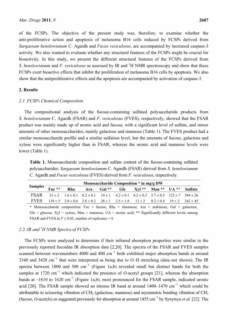
absorption band at 1240 cm−1 observed for both samples, but being particularly prevalent for the FVES
sample, was assigned as S=O stretching vibration, indicating the presence of esterified sulfate [20]. A
similar absorption pattern around 820–840 cm−1 was observed for both FCSPs: The FSAR infrared
spectrum showed an absorption band at 817 cm−1 (Figure 1a) whereas the FVES infrared spectrum
displayed a broader absorption band at 838 cm−1 and a small shoulder of absorption at 822 cm−1
(Figure 1b). Since IR adsorption at 840 cm−1 has been reported to be due to sulfate groups at the axial
C-4 position whereas sulfate groups at the equatorial C-2 and/or C-3 positions have been reported to
give a small absorption at 820 cm−1 [2], the observed absorption bands at 820–840 cm−1 were
interpreted as being indicative of sulfate groups.
Figure 1. Infrared analysis of fucose-containing sulfated polysaccharides (FCSPs) from
(a) Sargassum henslowianum C. Agardh (FSAR) and (b) Fucus vesiculosus (FVES)
scanned between 1800 and 550 cm−1.
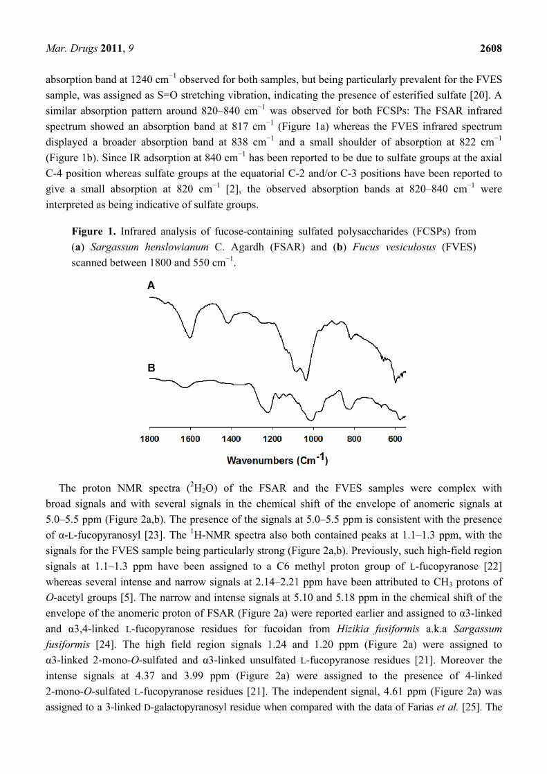
The proton NMR spectra (2H2O) of the FSAR and the FVES samples were complex with
broad signals and with several signals in the chemical shift of the envelope of anomeric signals at
5.0–5.5 ppm (Figure 2a,b). The presence of the signals at 5.0–5.5 ppm is consistent with the presence
of α-L-fucopyranosyl [23]. The 1H-NMR spectra also both contained peaks at 1.1–1.3 ppm, with the
signals for the FVES sample being particularly strong (Figure 2a,b). Previously, such high-field region
signals at 1.1–1.3 ppm have been assigned to a C6 methyl proton group of L-fucopyranose [22]
whereas several intense and narrow signals at 2.14–2.21 ppm have been attributed to CH3 protons of
O-acetyl groups [5]. The narrow and intense signals at 5.10 and 5.18 ppm in the chemical shift of the
envelope of the anomeric proton of FSAR (Figure 2a) were reported earlier and assigned to α3-linked
and α3,4-linked L-fucopyranose residues for fucoidan from Hizikia fusiformis a.k.a Sargassum
fusiformis [24]. The high field region signals 1.24 and 1.20 ppm (Figure 2a) were assigned to
α3-linked 2-mono-O-sulfated and α3-linked unsulfated L-fucopyranose residues [21]. Moreover the
intense signals at 4.37 and 3.99 ppm (Figure 2a) were assigned to the presence of 4-linked
2-mono-O-sulfated L-fucopyranose residues [21]. The independent signal, 4.61 ppm (Figure 2a) was
assigned to a 3-linked D-galactopyranosyl residue when compared with the data of Farias et al. [25]. The
Mar. Drugs 2011, 9
2609
FVES had an intense signal at 5.45 ppm (Figure 2b), which was assigned to α3-linked 2-mono-O-sulfated
L-fucopyranose residues, whereas the signals at 5.40, 4.58 and 4.39 ppm (Figure 2b) were assigned to
be due to di-sulfated residues, i.e., α3-linked 2,4-di-O-sulfated L-fucopyranose residues [26]. Hence in
general, the 1H NMR confirmed the anticipated FCSPs structures of the two samples.
Figure 2. One-dimensional 1H NMR spectra of crude FCSPs from (a) Sargassum
henslowianum C. Agardh (FSAR) and (b) Fucus vesiculosus (FVES) in D2O obtained
using an INOVA 600 NMR spectrometer (Agilent Technologies, Tokyo, Japan).
2.3. Anti-Proliferative Effects of the FCSPs
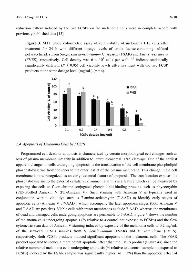
The viability of melanoma B16 cells treated with the FSAR and FVES products, respectively, was
determined via measurement of cell proliferation using an MTT based colorimetric assay. Both FCSPs
products (FSAR and FVES) decreased the viability of melanoma B16 cells in a dose-dependent
fashion, after 24 h of incubation of 6 × 10⁴ cells density per well (Figure 3). In particular, a pronounced
cell viability reduction was noticed after the addition of low levels, 0.1 mg/mL, of FSAR, producing a
cell viability of ~80% of the control, and cell proliferation was halted gradually as the FCSPs dosage
level increased (Figure 3) indicating moderate cytotoxicity. The FVES treated cells showed the same
trend, but the FVES product generally induced a lower anti-proliferative effect than the FSAR product
at the lower FCSP addition levels (P ≤ 0.05), but a significantly higher effect than FSAR (P ≤ 0.05) at
the higher addition level, producing a drastic reduction of the proliferation of melanoma B16 cells
leaving only ~6% of the cells viable at an FCSPs addition level of 1 mg/mL (Figure 3). The viability
Mar. Drugs 2011, 9
2610
reduction pattern induced by the two FCSPs on the melanoma cells were in complete accord with
previously published data [13].
Figure 3. MTT based colorimetric assay of cell viability of melonama B16 cells after
treatment for 24 h with different dosage levels of crude fucose-containing sulfated
polysaccharides from Sargassum henslowianum C. Agardh (FSAR) and Fucus vesiculosus
(FVES), respectively. Cell density was 6 × 10⁴ cells per well. a,b indicate statistically
significantly different (P ≤ 0.05) cell viability levels after treatment with the two FCSP
products at the same dosage level (mg/mL) (n = 4).
2.4. Apoptosis of Melanoma Cells by FCSPs
Programmed cell death or apoptosis is characterized by certain morphological cell changes such as
loss of plasma membrane integrity in addition to internucleosomal DNA cleavage. One of the earliest
apparent changes in cells undergoing apoptosis is the translocation of the cell membrane phospholipid
phosphatidylserine from the inner to the outer leaflet of the plasma membrane. This change in the cell
membrane is now recognized as an early, essential feature of apoptosis. The translocation exposes the
phosphatidylserine to the external cellular environment and this is a feature which can be measured by
exposing the cells to fluorochrome-conjugated phospholipid-binding proteins such as phycoerythin
(PE)-labelled Annexin V (PE-Annexin V). Such staining with Annexin V is typically used in
conjunction with a vital dye such as 7-amino-acticomycin (7-AAD) to identify early stages of
apoptotic cells (Annexin V+, 7-AAD−) which accompany the later apoptosis stages (both Annexin V
and 7-AAD are positive). Viable cells with intact membranes exclude 7-AAD, whereas the membranes
of dead and damaged cells undergoing apoptosis are permeable to 7-AAD. Figure 4 shows the number
of melanoma cells undergoing apoptosis (% relative to a control not exposed to FCSPs) and the flow
cytometric scan data of Annexin V staining induced by exposure of the melanoma cells to 0.2 mg/mL
of the seaweed FCSPs samples from S. henslowianum (FSAR) and F. vesiculosus (FVES),
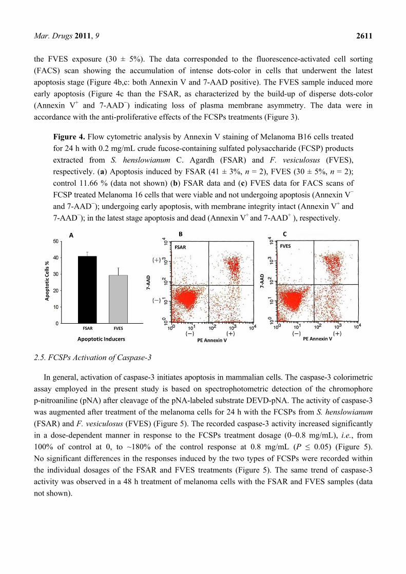
respectively. Both FCSPs products induced significant apoptosis of the melanoma cells: The FSAR
product appeared to induce a more potent apoptotic effect than the FVES product (Figure 4a) since the
relative number of melanoma cells undergoing apoptosis (% relative to a control sample not exposed to
FCSPs) induced by the FSAR sample was significantly higher (41 ± 3%) than the apoptotic effect of
Mar. Drugs 2011, 9
2611
the FVES exposure (30 ± 5%). The data corresponded to the fluorescence-activated cell sorting
(FACS) scan showing the accumulation of intense dots-color in cells that underwent the latest
apoptosis stage (Figure 4b,c: both Annexin V and 7-AAD positive). The FVES sample induced more
early apoptosis (Figure 4c than the FSAR, as characterized by the build-up of disperse dots-color
(Annexin V+ and 7-AAD−) indicating loss of plasma membrane asymmetry. The data were in
accordance with the anti-proliferative effects of the FCSPs treatments (Figure 3).
Figure 4. Flow cytometric analysis by Annexin V staining of Melanoma B16 cells treated
for 24 h with 0.2 mg/mL crude fucose-containing sulfated polysaccharide (FCSP) products
extracted from S. henslowianum C. Agardh (FSAR) and F. vesiculosus (FVES),
respectively. (a) Apoptosis induced by FSAR (41 ± 3%, n = 2), FVES (30 ± 5%, n = 2);
control 11.66 % (data not shown) (b) FSAR data and (c) FVES data for FACS scans of
FCSP treated Melanoma 16 cells that were viable and not undergoing apoptosis (Annexin V−
and 7-AAD−); undergoing early apoptosis, with membrane integrity intact (Annexin V+ and
7-AAD−); in the latest stage apoptosis and dead (Annexin V+ and 7-AAD+ ), respectively.
2.5. FCSPs Activation of Caspase-3
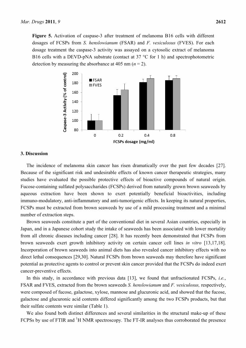
In general, activation of caspase-3 initiates apoptosis in mammalian cells. The caspase-3 colorimetric
assay employed in the present study is based on spectrophotometric detection of the chromophore
p-nitroaniline (pNA) after cleavage of the pNA-labeled substrate DEVD-pNA. The activity of caspase-3
was augmented after treatment of the melanoma cells for 24 h with the FCSPs from S. henslowianum
(FSAR) and F. vesiculosus (FVES) (Figure 5). The recorded caspase-3 activity increased significantly
in a dose-dependent manner in response to the FCSPs treatment dosage (0–0.8 mg/mL), i.e., from
100% of control at 0, to ~180% of the control response at 0.8 mg/mL (P ≤ 0.05) (Figure 5).
No significant differences in the responses induced by the two types of FCSPs were recorded within
the individual dosages of the FSAR and FVES treatments (Figure 5). The same trend of caspase-3
activity was observed in a 48 h treatment of melanoma cells with the FSAR and FVES samples (data
not shown).
Mar. Drugs 2011, 9
2612
Figure 5. Activation of caspase-3 after treatment of melanoma B16 cells with different
dosages of FCSPs from S. henslowianum (FSAR) and F. vesiculosus (FVES). For each
dosage treatment the caspase-3 activity was assayed on a cytosolic extract of melanoma
B16 cells with a DEVD-pNA substrate (contact at 37 °C for 1 h) and spectrophotometric
detection by measuring the absorbance at 405 nm (n = 2).
3. Discussion
The incidence of melanoma skin cancer has risen dramatically over the past few decades [27].
Because of the significant risk and undesirable effects of known cancer therapeutic strategies, many
studies have evaluated the possible protective effects of bioactive compounds of natural origin.
Fucose-containing sulfated polysaccharides (FCSPs) derived from naturally grown brown seaweeds by
aqueous extraction have been shown to exert potentially beneficial bioactivities, including
immuno-modulatory, anti-inflammatory and anti-tumorigenic effects. In keeping its natural properties,
FCSPs must be extracted from brown seaweeds by use of a mild processing treatment and a minimal
number of extraction steps.
Brown seaweeds constitute a part of the conventional diet in several Asian countries, especially in
Japan, and in a Japanese cohort study the intake of seaweeds has been associated with lower mortality
from all chronic diseases including cancer [28]. It has recently been demonstrated that FCSPs from
brown seaweeds exert growth inhibitory activity on certain cancer cell lines in vitro [13,17,18].
Incorporation of brown seaweeds into animal diets has also revealed cancer inhibitory effects with no
direct lethal consequences [29,30]. Natural FCSPs from brown seaweeds may therefore have significant
potential as protective agents to control or prevent skin cancer provided that the FCSPs do indeed exert
cancer-preventive effects.
In this study, in accordance with previous data [13], we found that unfractionated FCSPs, i.e.,
FSAR and FVES, extracted from the brown seaweeds S. henslowianum and F. vesiculosus, respectively,
were composed of fucose, galactose, xylose, mannose and glucuronic acid, and showed that the fucose,
galactose and glucuronic acid contents differed significantly among the two FCSPs products, but that
their sulfate contents were similar (Table 1).
We also found both distinct differences and several similarities in the structural make-up of these
FCPSs by use of FTIR and 1H NMR spectroscopy. The FT-IR analyses thus corroborated the presence
Mar. Drugs 2011, 9
2613
of sulfate groups in both the FSAR and the FVES sample (Figure 1). The IR spectra indicated that the
sulfate substitutions of the FCSPs extracted from the Sargassum sp. (FSAR) were located in the
equatorial C-2 and/or C-3 positions as depicted by absorption bands at 817 cm−1. This finding was in
agreement with data reported for fucoidan fractions isolated from Sargassum stenophyllum [4].
However, Duarte et al. [4] also reported that two other saccharide fractions from S. stenophyllum had
an absorption band at 837 cm−1 indicating sulfate groups at the C-4 positions of the structural
monosaccharides [4]. The spectra of the FCSPs from F. vesiculosus (FVES) displayed an absorption
band at 838 cm−1 with a small shoulder at ~822 cm−1 indicating sulfate groups at both the C-4 and the
C-2 position (Figure 1). This finding corresponds to previously reported 1H NMR data of FCSPs from
F. vesiculosus that have indicated a typical structure of algal fucoidan consisting of
α3-linked 2-mono-O-sulfated L-fucopyranose residues, and/or α3-linked 2,4-di-O-sulfated L-fucopyranose
residues [2,26]. Small disparities in the IR spectra from different published reports can be due to
factors such as sample handling and the FCSPs extraction procedure employed.
The present study also aimed at establishing whether crude FCSPs extracted from Sargassum
henslowianum C. Agardh (FSAR) contained fucoidan-like structures composed of α-3-linked or/and
α-3,4-linked L-fucopyranose residues. Even though signals consistent with the presence of
α-L-fucopyranose entities were recorded (with 1H NMR signals at 5.10 and 5.18 ppm, Figure 2a), the
probability that the FSAR may contain a cocktail of polysaccharides is likely. Hence, the 1H NMR
spectra also showed that the FSAR sample contained 3-linked D-galactopyranose residues as indicated
by an independent signal at 4.61 ppm (Figure 2a). β-(1→)3-linked galactopyranose residues are known
to be a typical structural feature of seaweed polysaccharides, from e.g., Laminaria angustata var.
longissima, Botryocladia occidentalis [25,31]. However, another possibility might be that the FSAR
sample was not composed of a mixture of different types of polysaccharides but rather, that the sample
consisted of one type of a highly complex hetero-polysaccharide as suggested by Duarte et al. [4] for
the fucoidans from Sargassum stenophyllum. It can safely be said that the 1H NMR spectra of the
FCSPs samples were complex and overlapping. It is therefore difficult to draw any definite
conclusions about the detailed structural features and differences among the two FCSPs; the detailed
elucidations of the definite structural details were also beyond the scope of this present study, but
clearly deserve further investigation. Nonetheless, the data confirmed that the diversity, i.e., the
compositional and structural complexity of (potentially bioactive) algal fucose-containing sulfated
polysaccharides, is much wider than originally believed.
The biological activities of the FCSPS against skin cancer cells were investigated in vitro, and the
results revealed that both FSAR and FVES can exert anti-proliferative effects on melanoma B16 cells
in vitro. The FSAR sample induced more significant reductions of the cell viability of melanoma cells
than the FVES sample at low dosage levels (Figure 3). At higher dosages, the FSAR treatment still
induced gradually more loss of cell viability, but the FVES had more potent anti-proliferative effects at
higher dosages than FSAR which could indicate direct cell toxicity. The bioactivities of these FCSPs
may be attributable to their distinct structural features, notably the level of sulfation (charge density),
the distribution (e.g., random versus clustered) and bonding of the sulfate substitutions, as well as other
specific structural features of the sulfated fucans and the sulfated galactofucan complexes. The sulfate
groups of FVES were substituted at the C-2 and C-4 position of the fucose substituents, typical for
fucoidan from F. vesiculosus, and consistent with previously published data indicating that the sulfate
Mar. Drugs 2011, 9
2614
groups were substituted at C-2 of α3-linked L-fucopyranose residues in fucoidan from e.g.,
Fucus evanescens [21]. In contrast, the sulfate substitutions in the FSAR were interpreted to be mainly
at the C-2 and/or C-3 positions of the monosaccharides according to the IR spectra (Figure 1); the
observation of C-2 linked sulfate groups agreed with the data mentioned above for fucoidan from
Fucus spp. [3,21], but is also in agreement with the proposition that the sulfate groups were substituted
at C-2 on the 3-linked galactopyranose residues [25]. The possible presence of sulfated, 3-linked
galactan in the structure of FSAR may contribute to the efficacy of FSAR to induce anti-proliferative
effects as it has been reported that 2-O-sulfated 3-linked galactan is more bioactive than 2-O-sulfated
3-linked fucans and 3-O-sulfated 4-linked galactan [32–34].
The findings that the FCSPs induced apoptosis of the melanoma B16 cells in vitro were in
agreement with recent reports [13,16,35], but the differential apoptotic efficacies, and the dose-response
effects of differently structured FCSPs (Figure 4) have not been reported earlier. In particular, it is a
novel finding that significantly different sulfated, polysaccharide structures from brown seaweeds—as
evaluated in the present work—exert relatively similar apoptotic effects on melanoma cells. The
results of this work thus indicate that not only the well-studied, classical type of FCSPs having a
backbone made up of (1→3)-linked α-L-fucopyranosyl or of alternating (1→3)- and (1→4)-linked
α-L-fucopyranosyl residues have potential tumor-preventing effecs, but also that the more complex
sulfated fucose-rich galacto-mannans from Sargassum spp. exert promising cancer-preventive effects.
The principal objective of this study was to assess whether any structural features of the FCSPs might
be crucial for bioactivity, and the data suggest that the sulfate substitutions, and not necessarily only
the fucose-backbone structure itself, confer this decisive bioactivity. It is however important to
investigate whether other differently structured FCSPs may exert similar growth inhibitory and
apoptosis inducing effects on cancer cells.
In this work we noted that both FCSPs activate caspase-3 in a dosage-response fashion (Figure 5).
These findings affirmed the results reported previously which have shown that FCSPs (“fucoidan”)
from F. vesiculosus induce apoptosis in human lymphoma HS-Sultan cell lines and in HT-29 and
HCT116 human colon cancer cells in vitro, and moreover that the exposure of these cells to the
F. vesiculosus FCSPs appear to activate caspase-3 [17,18]. The F. vesiculosus FCSPs treatment was
also shown to enhance mitochondrial membrane permeability of human colon cancer cells in vitro, and
to induce cytochrome c and Smac/Diablo release from the mitochondria [17]. It has also been reported
that pretreatment of HT-29 and HCT116 colon cancer cells with individual caspase-8 or caspase-9
inhibitors (Z-IETD-FMK and Z-LEHD-FMK, respectively) prior to fucoidan exposure reduced the
levels of caspases, including caspase-3 [17]. It has likewise been shown that pretreatment of human
lymphoma HS Sultan cells with a pan-caspase inhibitor, z-VAD-FMK, reduced fucoidan-induced
apoptosis [18]. Hence, the available data support the proposition that fucoidan-induced apoptosis
occurs through caspase activation pathways. The cascade mechanism by which the caspase-activation
is presumed to take place via the mitochondria-mediated apoptotic pathway is illustrated in Figure 6.
Loss of plasma membrane is one of the earliest features of apoptosis and Annexin V staining can
identify apoptosis at an early stage. However this assay does not distinguish between cells that have
undergone apoptotic death versus those that have died as a result of a necrotic pathway, because in
either case the dead cells will stain with both Annexin V and 7-AAD. Both early and later apoptosis
stages were observed by the FACS scanning indicating that the FCSPs had a direct apoptotic effect on
Mar. Drugs 2011, 9
2615
the melanoma B16 cells (in vitro) (Figure 4). The direct apoptotic action of the FCSPs was probably
due to the interaction of the highly negative charge density of the FCSPs with the melanoma B16 cells
(as a result of the sulfation). Recently, we reported that crude fucoidan from Sargassum sp. could
trigger apoptosis indirectly by enhancing the activity of natural killer (NK) cells activity in vivo [13].
NK cells produce immunologically important cytokines, notably IFN-γ, which can promote the
activation of T-cells to produce interleukin-2 and -12 that in turn further enhance the NK cell
activation [14,36].
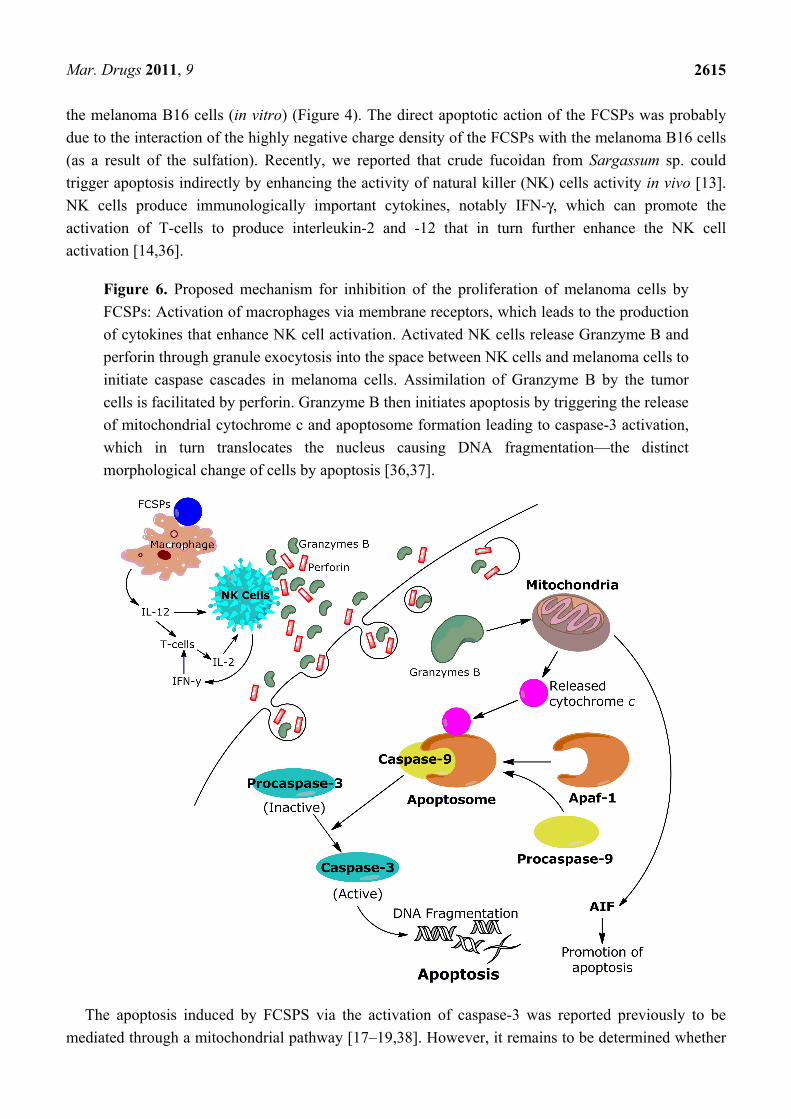
Figure 6. Proposed mechanism for inhibition of the proliferation of melanoma cells by
FCSPs: Activation of macrophages via membrane receptors, which leads to the production
of cytokines that enhance NK cell activation. Activated NK cells release Granzyme B and
perforin through granule exocytosis into the space between NK cells and melanoma cells to
initiate caspase cascades in melanoma cells. Assimilation of Granzyme B by the tumor
cells is facilitated by perforin. Granzyme B then initiates apoptosis by triggering the release
of mitochondrial cytochrome c and apoptosome formation leading to caspase-3 activation,
which in turn translocates the nucleus causing DNA fragmentation—the distinct
morphological change of cells by apoptosis [36,37].
The apoptosis induced by FCSPS via the activation of caspase-3 was reported previously to be
mediated through a mitochondrial pathway [17–19,38]. However, it remains to be determined whether
Mar. Drugs 2011, 9
2616
differences in FCSPs structures will influence the apoptotic mechanism, including the mitochondrial
pathway apoptosis cascade. The route of the mitochondrially dependent apoptotic pathway is the
release of apoptosis-inducing factor (AIF) and cytochrome c from the inner mitochondrial membrane
into the cytosol. Cytochrome c interacts with Apaf-1 (apoptotic protease activating factor 1) and
procaspase-9 to form the active apoptosome. The apoptosome then initiates the cleavage of
procaspase-3, producing active caspase-3, which initiates the execution phase of apoptosis by
proteolysis of substances whose cleavage commits the cell to apoptosis [39] (Figure 6). The influence
of the different FCSPs structures on the mitochondrial membrane permeability and electric potential
requires further study. We hope in the future to investigate the bioactivity and mechanism of FCSPs on
certain degenerative diseases in vivo and to further elucidate specific molecular targets of FSCPs for
inhibition of cancer cells.
4. Experimental Section
4.1. Chemicals
Dried S. henslowianum C. Agardh was obtained from Viet Delta Ltd. (Ho Chi Menh, Vietnam) and
the Fucose-containing sulfated polysaccharides (FCSPs) from the S. henslowianum (FSAR) were
extracted in our laboratory (see below). Crude fucoidan from F. vesiculosus (FVES) was obtained
from Sigma-Aldrich (Steinheim, Germany); according to the product description the FVES had been
prepared from F. vesiculosus via the extraction method described by Black and Dewar [40].
Hydrochloric acid (37%), D-glucose and D-xylose were purchased from Merck (Darmstadt, Germany).
Trifluoracetic acid (99%, TFA), trichloroacetic acid (99%, TCA), CaCl2, Na2SO4, BaCl2, arabinose,
rhamnose, D-galactose and L-fucose were from Sigma–Aldrich Co. (Steinheim, Germany). Agarose D-2
was obtained from Hispanagar (Burgos, Spain). Caspase-3 colorimetric assay kit was obtained from
Biovision, Inc. (Mountain View, CA. USA). Minimal essential medium eagle (MEM-eagle) cell
culture media was purchased from Sigma–Aldrich Co. (Steinheim, Germany); foetal bovine serum
(FBS) was from Flow Laboratories (North Ryde, N.S.W., Australia); streptomycin–penicillin and
Trypan Blue were from Gibco (Canada). Cell Proliferation Kit 1 was obtained from Roche Applied
Science, Germany. The PE Annexin V Apoptosis Detection Kit 1 was obtained from BD Biosciences
(Franklin Lakes, NJ, USA). All chemicals used were analytical grade.
4.2. Extraction of FCSPs from S. henslowianum C. Agardh
The Sargassum FCSP product (FSAR) used was extracted from S. henslowianum C. Agardh by use
of an optimized single-step extraction procedure described previously [6]. Briefly, the dried
S. henslowianum seaweed was ground and sieved to pass through a 500 µm sieve and 100 g of dried
ground seaweed was extracted in 2 L of 0.03 M HCl with continuous stirring at 200 rpm for 4 h at
90 °C water bath (Julabo, Germany). The suspended seaweed was filtered, and the extract was
precipitated using 60% ethanol, the precipitate collected after centrifugation at 10,600 rpm for 10 min
(Sigma Laboratory Centrifuge 4K15, VWR, Denmark), and the resulting pellet was freeze dried. This
freeze dried pellet constituted the fucose-containing sulfated polysaccharides (FSAR).
Mar. Drugs 2011, 9
2617
4.3. Acid Hydrolysis and FCSPs Composition Analysis
The freeze dried FSAR and FVES samples (20 mg) were hydrolyzed separately in 2 M TFA (final
concentration) at 121 °C for 2 h, then the hydrolyzed mixture were freeze dried at −57 °C (Heto Lyolab
3000, England). Each dried powder sample was resolubilized in doubly distilled water and centrifuged
at 10,000 rpm for 10 min to collect the supernatant (Sigma Laboratory Centrifuge 4K15, VWR,
Denmark). Each supernatant was filtered through a 0.2 µm syringe tip filter (SUN-Sri, Rockwood, TN)
prior to injection into the HPAEC-PAD for monosaccharide analysis [41]. Analysis of sulfate content
was done according to the method described by Jackson and McCandless [42].
4.4. 1H NMR and FTIR Spectroscopy
The 1H NMR spectra were obtained using an INOVA 600 NMR spectrometer (Agilent Technologies
Japan, Ltd., Tokyo Japan) equipped with a 1H[15N-31P] pulse field gradient indirect-detecting probe.
Standard pulse sequences were used in all operations. The 1H chemical shift (δH) was referenced to
HOD (δH 4.76 ppm, 2H2O). The 1H NMR spectrum was assigned through the 1H–1H decoupling
technique. An NMR spectrum of L-fucose was utilized as a reference for chemical shift assignment.
The lyophilized FCSPs powders were dissolved in deuterium oxide (2H2O) and evaporated to exchange
the unstable 1H with 2H. The evaporation and dissolution step was repeated five times, and the samples
(10 mg) were finally dissolved in 0.75 mL 2H2O and then subjected to NMR spectroscopy. The IR
spectra were obtained using a Spectrum One FT-IR spectrometer (Perkin Elmer, Waltham, MA, USA)
equipped with universal attenuated total reflectance (UATR) accessories. Analysis of each of the
FSAR and FVES powders, ~1 mg of each, was done using diffuse reflectance infrared transform
spectroscopy (DRIFTS) and the spectrum was evaluated by Perkin Elmer Spectrum software version 5
(Perkin Elmer, Waltham, MA, USA).
4.5. Cell Culture and Anti-Proliferative Assay
Melanoma B16 cells (MC) were grown in MEM eagle medium supplemented with 10% (v/v) heat
inactivated FBS, 1% (w/v) streptomycin–penicillin and 1% (v/v) of 200 mM L-glutamine at 37 °C
under 5% CO2. Monolayer cultivation was carried out by adding 100 µL of the cell-MEM-FBS mixture
into separate wells in 96-flat well plates at a density of 6 × 10⁴ cells per well followed by incubation
for 24 h in 5% CO2 at 37 °C. For the anti-proliferation assay the medium was removed after the 24 h of
monolayer cell cultivation and replaced with 100 µL of MEM medium containing 2% FBS and
varying concentrations (0.1–1.0 mg/mL) of the crude FCSPs, i.e., FSAR and FVES, respectively, and
the mixtures were then incubated for 24 h. Quantification of cell proliferation was carried out using a
tetrazolium salt (MTT (3-(4,5-dimethyl-thiazolyl-2)-2,5-diphenyltetrazolium bromide)) based
colorimetric assay following the protocol supplied with the Cell Proliferation Kit 1 (Roche Applied
Science, Germany). Briefly, 20 µL MTT solution (5 mg/mL) was added to the cell cultures after the
24 h of incubation with the FCSPs, and the cell cultures were then re-incubated for 4 h. Finally,
100 µL of stabilization solution was added to each well and the plates were incubated overnight at
37 °C under 5% CO2. Absorbance was measured using an Elisa reader at 550–690 nm.
Mar. Drugs 2011, 9
2618
4.6. Cell Culture and Caspase-3 Assay
Melanoma B16 cells (MC) were grown in MEM eagle culture medium supplemented with
10% (v/v) heat inactivated FBS, 1% (w/v) streptomycin–penicillin and 1% (v/v) of 200 mM
L-glutamine maintained at 37 °C under 5% CO2. For the caspase-3 assay, monolayer cultivation was
carried out in a petri dish (60 × 15 mm) by adding 5 mL culture medium containing melanoma cells at
a density of 1 × 10⁵ per mL and varying concentrations (0.2, 0.4 and 0.8 mg/mL) of the FSAR and
FVES, respectively. The mixtures were then incubated for 24 and 48 h in 5% CO2 at 37 °C. The
caspase-3 assay was performed according to the protocol supplied with the assay kit (Biovision Inc.,
Mountain View, CA, USA) used to assay the activity of caspases that recognize the amino acid
sequence DEVD. The assay was based on spectrophotometric detection of the chromophore
p-nitroaniline (pNA) after cleavage from the labeled substrate DEVD-pNA. Concisely, the melanoma
B16 cells exposed to FSAR and FVES, respectively, were harvested and resuspended in 50 µL of cell
lysis buffer and incubated on ice for 10 min. and the mixture centrifuged for 1 min (14,000 × g, 4 °C).
Each supernatant was then transferred to a fresh tube, and reaction buffer (50 µL) and 4 mM DEVD-pNA
substrate (5 µL) were added, and this reaction mixture was then incubated at 37 °C for 1 h. Absorbance
of pNA light emission was quantified using a microtiter plate reader at 405 nm.
4.7. Apoptosis Assay by Fluorescence-Activated Cell Sorting (FACS)
After 24 h of monolayer cultivation of melanoma B16 cells with 0.2 mg/mL of FSAR or FVES, and
no FCSPs addition as control, the culture medium was removed, and the cells harvested by addition of
1 mL Trypsin-EDTA. The harvested cells were washed twice with 0.1 M PBS and then resuspended in
binding buffer according to the protocol for the Annexin V Apoptosis Detection Kit I (BD Biosciences,
Franklin Lakes, NJ, USA). 100 µL of this solution at 1 × 10⁵ cells was transferred to a culture tube and
5 µL of Annexin V and 5 µL of 7-amino-actinomycin (7-ADD) were added, and the mixture incubated
at room temperature for 25 min. Then, 400 μL of binding buffer was added and the extent of apoptosis
and staining pattern of the cells were tracked by flow cytometric analysis on a FACScan instrument
(Becton Dickinson).
5. Conclusions
The tumor inhibitory bioactivity of fucose-containing sulfated polysaccharides (FCSPs) from
Sargassum henslowianum C. Agardh (FSAR) and F. vesiculosus (FVES) was demonstrated through
evaluation of inhibition of melanoma cell proliferation, activation of caspase-3, and apoptosis of
melanoma B-16 cells in vitro. The structural traits of the FCSPs products were shown to be complex
and to differ among the two FCSPs making it delicate to draw definite conclusions about structural
effects and mechanisms. However, since the sulfate levels were relatively high as well as relatively
similar among the two FCSPs, we propose that the bioactivity effects of the FSAR and FVES might be
attributable to the sulfation (charge density), positioning and bonding of the sulfate substitutions in the
FCSPs. The work clearly indicates that unfractionated fucose-containing sulfated polysaccharides from
both Sargassum henslowianum C. Agardh and Fucus vesiculosus may have therapeutic potential as
skin-cancer preventive agents.
Mar. Drugs 2011, 9
2619
Acknowledgments
The authors would like to express their gratitude to D. Tsubokawa, H. Kitasato and
K. Ishihara—School of Allied Health Science, Kitasato University—Sagamihara, Kanagawa, Japan for
sharing their laboratory facilities; and for their untiring assistance, support and encouragement during
the entire experimental activities of this present study.
References
1. Percival, E.; McDowell, R. Chemistry and Enzymology of Marine Algal Polysaccharides;
Academic Press: London, UK, 1967; p. 157.
2. Patankar, M.S.; Oehninger, S.; Barnett, T.; Williams, R.L.; Clark, G.F. A revised structure for
fucoidan may explain some of its biological activities. J. Biol. Chem. 1993, 268, 21770–21776.
3. Bilan, M.I.; Usov, A.I. Structural analysis of fucoidans. Nat. Prod. Comm. 2008, 3, 1639–1648.
4. Duarte, M.E.; Cardoso, M.A.; Noseda, M.D.; Cerezo, A.S. Structural studies on fucoidans from
the brown seaweed Sargassum stenophyllum. Carbohydr. Res. 2001, 333, 281–293.
5. Tako, M.; Yoza, E.; Tohma, S. Chemical characterization of acetyl fucoidan and alginate from
commercially cultured Cladosiphon okamuranus. Bot. Mar. 2000, 43, 393–398.
6. Ale, M.T.; Mikkelsen, J.D.; Meyer, A.S. Designed optimization of a single-step extraction of
fucose-containing sulfated polysaccharides from Sargassum sp. J. Appl. Phycol. 2011,
doi:10.1007/s10811-011-9690-3.
7. Nardella, A.; Chaubet, F.; Boisson-Vidal, C.; Blondin, C.; Durand, P.; Jozefonvicz, J. Anticoagulant
low molecular weight fucans produced by radical process and ion exchange chromatography of
high molecular weight fucans extracted from the brown seaweed Ascophyllum nodosum.
Carbohydr. Res. 1996, 289, 201–208.
8. Jiao, G.; Yu, G.; Zhang, J.; Ewart, S.E. Chemical structure and bioactivities of sulfated
polysaccharides from marine algae. Mar. Drugs 2011, 9, 196–223.
9. Blondin, C.; Fischer, E.; Boisson-Vidal, C.; Kazatchkine, M.D.; Jozefonvicz, J. Inhibition of
complement activation by natural sulfated polysaccharides (fucans) from brown seaweed.
Mol. Immunol. 1994, 31, 247–253.
10. Adhikari, U.; Mateii, C.G.; Chattopadhyay, K.; Pujol, C.A.; Damonte, E.B.; Ray, B. Structure and
antiviral activity of sulfated fucans from Stoechospermum marginatum. Phytochemistry 2006, 67,
2474–2482.
11. Trinchero, J.; Ponce, N.M.A.; Cordoba, O.L.; Flores, M.L.; Pampuro, S.; Stortz, C.A.;
Salomon, H.; Turk, G. Antiretroviral activity of fucoidans extracted from the brown seaweed
Adenocystis utricularis. Phytother. Res. 2009, 23, 707–712.
12. Zhuang, C.; Itoh, H.; Mizuno, T.; Ito, H. Antitumor active fucoidan from brown seaweed,
Umitoranoo (Sargassum thunbergii). Biol. Biotechnol. Biochem. 1995, 59, 563–567.
13. Ale, M.T.; Maruyama, H.; Tamauchi, H.; Mikkelsen, J.D.; Meyer, A.S. Fucoidan from Sargassum sp.
and Fucus vesiculosus reduces cell viability of lung carcinoma and melanoma cells in vitro and
activates natural killer cells in mice in vivo. Int. J. Biol. Macromol. 2011, 49, 331–336.
Mar. Drugs 2011, 9
2620
14. Maruyama, H.; Tamauchi, H.; Iizuka, M.; Nakano, T. The role of NK cells in antitumor activity of
dietary fucoidan from Undaria pinnatifida sporophylls (Mekabu). Planta Med. 2006, 72,
1415–1417.
15. Takahashi, M. Studies on the mechanism of host mediated antitumor action of fucoidan from a
brown alga Eisenia bicyclis. J. Jpn. Soc. Reticuloendothel. Syst. 1983, 22, 269–283.
16. Foley, S.A.; Mulloy, B.; Tuohy, M.G. An unfractionated fucoidan from Ascophyllum nodosum:
Extraction, characterization, and apoptotic effects in vitro. J. Nat. Prod. 2011, doi:10.1021/
np200124m.
17. Kim, E.J.; Park, S.Y.; Lee, J.Y.; Park, J.H. Fucoidan present in brown algae induces apoptosis of
human colon cancer cells. BMC Gastroenterol. 2010, 10, doi:10.1186/1471-230X-10-96.
18. Aisa, Y.; Miyakawa, Y.; Nakazato, T.; Shibata, H.; Saito, K.; Ikeda, Y.; Kizaki, M. Fucoidan
induces apoptosis of human HS-sultan cells accompanied by activation of caspase-3 and
down-regulation of ERK pathways. Am. J. Hematol. 2005, 78, 7–14.
19. Yamasaki-Miyamoto, Y.; Yamasaki, M.; Tachibana, H.; Yamada, K. Fucoidan induces apoptosis
through activation of caspase-8 on human breast cancer MCF-7 cells. J. Agric. Food Chem.
2009, 57, 8677–8682.
20. Mori, H.; Nisizawa, K. Sugar constituents of sulfated polysaccharides from the fronds of Sargassum
ringgoldianum. Bull. Jpn. Soc. Sci. Fish. 1982, 48, 981–986.
21. Bilan, M.I.; Grachev, A.A.; Ustuzhanina, N.E.; Shashkov, A.S.; Nifantiev, N.E.; Usov, A.I.
Structure of a fucoidan from brown seaweed Fucus evanescens. Carbohydr. Res. 2002, 337,
719–730.
22. Synytsya, A.; Kim, W.J.; Kim, S.M.; Pohl, R.; Synytsya, A.; Kvasnicka, F.; Copikova, J.;
Park, YI. Structure and antitumor activity of fucoidan isolated from sporophyll of Korean brown
seaweed Undaria pinnatifida. Carbohydr. Pol. 2010, 81, 41–48.
23. Mulloy, B.; Ribeiro, A.C.; Alves, A.P.; Vieira, R.P.; Mourao, P.A.S. Sulfated fucans from
Echinoderms have a regular tetrasaccharide repeating unit defined by specific patterns of sulfation
at the 0-2 and 0-4 positions. J. Biol. Chem. 1994, 269, 22113–22123.
24. Shiroma, R.; Konishi, T.; Uechi, S.; Tako, M. Structural study of fucoidan from the brown
seaweed Hizikia fusiformis. Food Sci. Technol. Res. 2008, 14, 176–182.
25. Farias, W.R.L.; Valente, A.P.; Pereira, M.S.; Maurão, P.A.S. Structure and anticoagulant activity
of sulfated galactan. J. Biol. Chem. 2000, 275, 29299–29307.
26. Periera, M.S.; Mulloy, B.; Mourão, P.A.S. Structure and anticoagulant activity of sulfated fucans.
J. Biol. Chem. 1999, 274, 7656–7667.
27. Garibyan, L.; Fisher, D.E. How sunlight causes melanoma. Curr. Oncol. Rep. 2010, 12, 319–326.
28. Iso, H.; Kubota, Y. Nutrition and disease in the Japan Collaborative Cohort Study for Evaluation
of Cancer (JACC). Asian Pac. J. Cancer Prev. 2007, 8, 35–80.
29. Teas, J.; Harbison, M.L.; Gelman, R.S. Dietary seaweed (Laminaria) and mammary carcinogenesis
in rates. Cancer Res. 1984, 7, 2758–2761.
30. Yamamoto, I.; Maruyama, H. Effect of dietary seaweed preparations on 1,2-dimethylhydrazine-
induced intestinal carcinogenesis in rats. Cancer Lett. 1985, 26, 241–251.
Mar. Drugs 2011, 9
2621
31. Nishino, T.; Nishioka, C.; Ura, H.; Nagumo, T. Isolation and partial characterization of a novel
amino sugar-containing fucan sulfate from commercial Fucus vesiculosus fucoidan. Carbohydr.
Res. 1994, 255, 213–224.
32. Berteau, O.; Mulloy, B. Sulfated fucans, fresh perspectives: Structures, functions, and biological
properties of sulfated fucans and an overview of enzymes active toward this class of
polysaccharide. Glycobiology 2003, 13, 29–40.
33. Periera, M.S.; Vilela-Silva, A.E.S.; Valente, A.; Mourão, P.A.S. A 2-sulfated, 3-linked
α-L-galactan is an anticoagulant polysaccharide. Carbohydr. Res. 2002, 337, 2231–2238.
34. Periera, M.S.; Melo, F.R.; Mourão, P.A.S. Is there a correlation between stricture and
anticoagulant action of sulfated galactans and sulfated fucans? Glycobiology 2002, 12, 573–580.
35. Silva Costa, L.; Silva Telles, C.B.; Oliveira, R.M.; Nobre, L.T.D.B.; Dantas-Santos, N.; Camara,
R.B.G.; Costa, M.S.S.P.; Almeida-Lima, J.; Melo-Silveira, R.F.; Albuquerque, I.R.L.; et al.
Heterofucan from Sargassum filipendula induces apoptosis in Hela cells. Mar. Drugs 2011, 9,
603–614.
36. Ale, M.T.; Mikkelsen, J.D.; Meyer, A.S. Important determinants for fucoidan bioactivity: A
critical review of structure-function relations and extraction methods for fucose-containing
sulfated polysaccharides from brown seaweeds. Mar. Drugs 2011, 9, 2106–2130.
37. Kindt, T.J.; Goldsby, R.A.; Osborne, B.A. Cell-Mediated Cytotoxic Responses. In Immunology
Sixth Edition, 6th ed.; Kuby, J., Ed.; W.H. Freeman and Company: New York, NY, USA, 2007;
Chapter 14, p. 360.
38. Teruya, T.; Konishi, T.; Uechi, S.; Tamaki, H.; Tako, M. Anti-proliferative activity of oversulfated
fucoidan from commercially cultured Cladosiphon okamuranus TOKIDA in U937 cells. Int. J.
Biol. Macromol. 2007, 41, 221–226.
39. Kaufmann, S.H.; Hengartner, M.O. Programmed cell death: Alive and well in the new
millennium. Trends Cell. Biol. 2001, 11, 526–534.
40. Black, W.A.P.; Dewar, E.T.; Woodward, F.N. Manufacturing of algal chemicals 4: Laboratory
scale isolation of fucoidan from brown marine algae. J. Sci. Food Agric. 1952, 3,122–129.
41. Arnous, A.; Meyer, A.S. Comparison of methods for compositional characterization of grape
(Vitis vinifera) and apple (Malus domestica) skins. Food Bioprod. Proc. 2008, 86, 79–86.
42. Jackson, S.G.; McCandless, E.L. Simple, rapid, turbidometric determination of inorganic sulfate
and/or protein. Anal. Biochem. 1978, 90, 802–808.
Samples Availability: Available from the authors.
© 2011 by the authors; licensee MDPI, Basel, Switzerland. This article is an open access article
distributed under the terms and conditions of the Creative Commons Attribution license
(http://creativecommons.org/licenses/by/3.0/).


![Sulfated zirconia[1]](https://img.pdfslide.us/doc/110x75/5568f2ecd8b42aff2e8b4932/sulfated-zirconia1.jpg)