Embed Size (px)
Citation preview

Universidade de São Paulo
2010-12
Biosensors for efficient diagnosis of
Leishmaniasis: innovations in bioanalytics for
a neglected disease Analytical Chemistry,Washington, DC : American Chemical Society - ACS,v. 82, n. 23, p. 9763-9768,
Dec. 2010http://www.producao.usp.br/handle/BDPI/49712
Downloaded from: Biblioteca Digital da Produção Intelectual - BDPI, Universidade de São Paulo
Biblioteca Digital da Produção Intelectual - BDPI
Departamento de Física e Ciências Materiais - IFSC/FCM Artigos e Materiais de Revistas Científicas - IFSC/FCI

Biosensors for Efficient Diagnosis ofLeishmaniasis: Innovations in Bioanalytics for aNeglected Disease
Angelo C. Perinoto,† Rafael M. Maki,‡ Marcelle C. Colhone,§ Fabiana R. Santos,§
Vanessa Migliaccio,§ Katia R. Daghastanli,| Rodrigo G. Stabeli,⊥ Pietro Ciancaglini,§
Fernando V. Paulovich,‡ Maria C. F. de Oliveira,‡ Osvaldo N. Oliveira, Jr.,† andValtencir Zucolotto*,†
Instituto de Fısica de Sao Carlos, USP, CP 369, 13560-970 Sao Carlos, SP, Brazil, Instituto de Ciencias Matematicase de Computacao, USP, CP 668, 13560-970 Sao Carlos, SP, Brazil, Faculdade de Filosofia, Ciencias e Letras deRibeirao Preto, USP, Ribeirao Preto, SP, Brazil, Departamento de Biofísica da Escola Paulista de Medicina,UNIFESP, Sao Paulo, SP, Brazil, and Universidade Federal de Rondonia (UNIR) and Fundacao Oswaldo Cruz -Fiocruz Noroeste, Rondonia, Brazil
The need for reliable, fast diagnostics is closely linked tothe need for safe, effective treatment of the so-called“neglected” diseases. The list of diseases with no field-adapted diagnostic tools includes leishmaniasis, shigella,typhoid, and bacterial meningitis. Leishmaniasis, in par-ticular, is a parasitic disease caused by Leishmania spp.transmitted by infected phlebotomine sandfly, whichremains a public health concern in developing countrieswith ca. 12 million people infected and 350 million at riskof infection. Despite several attempts, methods for diag-nosis are still noneffective, especially with regard tospecificity due to false positives with Chagas’ diseasecaused by Trypanosoma cruzi. Accepted golden standardsfor detecting leishmaniasis involve isolation of parasiteseither microscopically, or by culture, and in both methodsspecimens are obtained by invasive means. Here, we showthat efficient distinction between cutaneous leishmaniasisand Chagas’ disease can be obtained with a low-costbiosensor system made with nanostructured films con-taining specific Leishmania amazonensis and T. cruziantigens and employing impedance spectroscopy as thedetection method. This unprecedented selectivity wasafforded by antigen-antibody molecular recognition pro-cesses inherent in the detection with the immobilizedantigens, and by statistically correlating the electricalimpedance data, which allowed distinction between realsamples that tested positive for Chagas’ disease andleishmaniasis. Distinction could be made of blood serumsamples containing 10-5 mg/mL of the antibody solu-tion in a few minutes. The methods used here aregeneric and can be extended to any type of biosensor,which is important for an effective diagnosis of manyother diseases.
Leishmaniasis is a parasitic neglected disease that remains asa major public health concern, especially for its high incidence indeveloping countries. It affects around 12 million people, and 350million are at risk of infection.1 The disease is caused byLeishmania spp., transmitted by infected phlebotomine sandfly.Depending on the type of parasite causing the infection, leish-maniasis may be classified as visceral leishmaniasis (VL), diffusecutaneous leishmaniasis, mucocutaneous leishmaniasis, and cu-taneous leishmaniasis (CL).2,3 Many research groups have focusedon novel strategies for treatment and for low-cost, effectivediagnosis systems.4,5 Despite such efforts, diagnosis methods arestill noneffective, with limitations regarding cost, sensitivity,difficulty in using under field conditions, and specificity.6 The latterlimitation, in particular, applies to cases of cross-reaction withChagas’ disease,7,8 and to a lesser extent to tuberculosis andleprosy.5 Cutaneous leishmaniasis (CL) is frequently diagnosedby classic clinical investigation, or via laboratorial diagnosis undermicroscopic examination of smears or biopsies of skin lesions.9,10
Culturing of replicative Leishmania specimens from the skin lesionborder may also be performed.4,10,11 These methods, however,often lead to misdiagnosis; the culture assays are positive in onlyca. 70% when the patient has a severe disease.11 Moreover,contaminations may affect culture procedures, whereas the his-
* To whom correspondence should be addressed. E-mail: [email protected].† Instituto de Fısica de Sao Carlos.‡ Instituto de Ciencias Matematicas e de Computacao.§ Faculdade de Filosofia, Ciencias e Letras de Ribeirao Preto.| Departamento de Biofísica da Escola Paulista de Medicina.⊥ Universidade Federal de Rondonia and Fundacao Oswaldo Cruz.
(1) Usdin, M.; Guillerm, M.; Chirac, P. Nature 2006, 441, 283–284.(2) Desjeux, P. Nat. Rev. Microbiol. 2004, 2, 692–693.(3) Calderon, L. A.; Silva-Jardim, I.; Zuliani, J. P.; Silva, A. A.; Ciancaglini, P.;
da Silva, L. H. P.; Stabeli, R. G. J. Braz. Chem. Soc. 2009, 20, 1011–1023.(4) Vega-Lopez, F. Curr. Opin. Infect. Dis. 2003, 16, 97–101.(5) Singh, S.; Sivakumar, R. J. Postgrad. Med. (Bombay) 2003, 49, 55–60.(6) Romero, H. D.; Silva, L. A.; Silva-Vergara, M. L.; Rodriguez, V.; Costa, R. T.;
Guimaraes, S. F.; Alecrim, W.; Moraes-Souza, H.; Prata, A. Am. J. Trop.Med. Hyg. 2009, 81, 27–33.
(7) Nouir, N. B.; Gianinazzi, C.; Gorcii, M.; Muller, N.; Nouri, A.; Babba, H.;Gottstein, B. Trans. R. Soc. Trop. Med. Hyg. 2009, 103, 355–364.
(8) Castro, E. A.; Thomaz-Soccol, V.; Augur, C.; Luz, E. Exp. Parasitol. 2007,117, 13–21.
(9) Boggild, A. K.; Valencia, B. M.; Espinosa, D.; Veland, N.; Ramos, A. P.;Arevaldo, J.; Llanos-Cuentas, A.; Low, D. E. Clin. Infect. Dis. 2010, 50,1–6.
(10) Herwaldt, B. L. Lancet 1999, 354, 1191–1199.(11) Weigle, K. A.; Labrada, L. A.; Lozano, C.; Santrich, C.; Barker, D. C. J. Clin.
Microbiol. 2002, 40, 601–606.
Anal. Chem. 2010, 82, 9763–9768
10.1021/ac101920t 2010 American Chemical Society 9763Analytical Chemistry, Vol. 82, No. 23, December 1, 2010Published on Web 11/02/2010

topathological examination is often difficult due to dehydrationand deformation of amastigotes.2,4 All of these procedures areexpensive and time-consuming, causing delay in the diagnosis andhindering precocious treatment of the patient.
The low sensitivity and time-consuming limitations exhibitedby cell culture and microscopic examination of skin lesions maybe overcome by using immunodetection assays, in which antigensare applied for direct detection of specific antileishmania antibod-ies. Diagnosis of visceral leishmaniasis has been performed inimmunoblotting assays with Leishmania antigens.12 However,such methods are limited in the case of cutaneous leishmaniasis,for anti-Leishmania antibodies are present in the serum at verylow levels.5 Molecular analyses have also been applied to detectDNA and RNA from leishmania parasites, with PCR methodsbeing by far the most used.13,14 Despite the high sensitivity andspecificity of the PCR methodology, through which specificsequences of the parasites can be determined, the method is notcost-effective, being unsuitable for field diagnosis.
The limitations found in immunoassays and molecular diag-nosis have motivated the development of integrated electronicbiosensors systems, which in most cases employ nanostructuredbiomaterials. These systems are capable of detecting analytes viaspecific recognition based upon interaction between protein andligands or antigens and antibodies.15,16 Integrated biosensors mayexhibit high sensitivity and specificity, as exemplified with thedetection of pasteurelosis.16 Detection is performed via electricalmeasurements, following the concept of a taste sensor, in whichthe bioreceptor material is immobilized on the gaps of aninterdigitated electrode and immersed in aqueous solutionscontaining different concentrations of the analyte.16 Differencesin the electrical capacitance of the electrodes are correlated tothe type and concentration of the analytes using equivalent electriccircuits. These circuits represent the experimental system, thatis, metallic tracks covered with a layer of biological bioreceptors,immersed in an electrolyte.17 The latter biodetection systems areuser-friendly and are capable of providing in-field diagnosis withina short period of time. These technical characteristics areadvantageous for the high throughput inherent in new drug anddiagnosis tests.
In this Article, we present a nanostructured biosensor systemto detect specific anti-leishmania antibodies using capacitancemeasurements as the detection method. The system is comprisedof phospholipid liposomes18 incorporating membrane antigenicproteins as the immobilized phase, which had been anchored tothe surface of interdigitated electrodes. The electrodes containingantigenic proteins were used to detect antibodies, in which thebiological reaction was converted into variations in the electrical
response (capacitance). Here, we selected a pool of membraneantigenic proteins from L. amazonensis because of the highincidence of this species in the north region of Brazil. However,the system described here may be applied to any other Leishmaniaspecies.
The proteoliposomes incorporating the antigenic proteins wereprepared according to a well-established methodology,18 whichis described in the Experimental Section. The freshly preparedproteoliposomes were immobilized on interdigitated electrodesusing the layer-by-layer technique,19,20 in conjunction with polya-midoamine dendrimers generation 4 (PAMAM). Because of theirbranched, porous structure, dendrimers have been used forimmobilizing proteins in biosensors, with enhanced sensitivity andshort response times due to diffusion of analytes through themultilayer structure.21 Furthermore, the porous architecture ofthe film is crucial for confining electrical charges within itsstructure (e.g., charges generated in the biological reaction),which are responsible for the detectable changes in the electricalresponse.21
EXPERIMENTAL SECTIONMaterials. All solutions were prepared using Millipore (Bed-
ford, MA) DirectQ ultrapure water. Tris(hydroxymethyl)amino-methane (TRIS), bovine serum albumine (BSA), Schneider’s insectmedium, ethylenediaminetetraacetic (EDTA), phenylmethylsul-fonyl fluoride (PMSF), trans-epoxysuccinyl-L-leucinamido-(4-guani-dino)btutane (E-64), 1,10-phenanthroline dipalmitoylphosphati-dylcholine(DPPC),dipalmitoylphosphatidylserine(DPPS),cholesterol,and sodium dodecyl sulfate (SDS) were purchased from Sigma(St. Louis, MO); calbiosorb resin was from Calbiochem (SanDiego, CA). Polyamidoamine generation 4 dendrimer (PAMAM)and poly(allylamine hydrochloride) (PAH) (used as polycations)and poly(vinyl) sulfonic acid (PVS) (polyanion) were purchasedfrom Aldrich and used without further purification. Gold (Au)interdigitated electrodes were lithographically fabricated at theNational Synchroton Laboratory facilities, Campinas, Brazil. Autracks (10 µm wide × 70 nm thick) were deposited onto BK7 glasssubstrates with a separation distance of 10 µm among the tracks.
Parasites. The parasite strain IFLA/BR/67/PH8 of Leishma-nia amazonensis was maintained in BALB/c mice (Nunes et al.,1997; Noronha et al., 1998; Santos et al., 2006).
Serum Preparation of L. amazonensis and T. cruzi. Bloodsamples were collected from the BALB/c mice naıve, miceinfected subcutaneously in the hind footpad with 106 stationarygrowth phase L. amazonensis promastigotes, and mice infectedintraperitoneally with a nonlethal dose of 300 blood-formtrypomastigotes of the Y strain of T. cruzi. The serum againsttotal L. amazonensis and T. cruzi antigenic determinants wasobtained from coagulated blood by centrifugation and frozenat -20 °C until use. The total IgG of positive and negativeserum samples was purified with the Protein G Sepharose 4Fast Flow (GE Healthcare).
(12) Santos-Gomes, G.; Gomes-Pereira, S.; Campino, Araujo, L. M. A.; Abranches,P. J. Clin. Microbiol. 2000, 38, 175–178.
(13) Romero, G. A. S.; Noronha, E. F.; Pirmez, C.; Pires, F. E. S. S.; Fernandes,O.; Nehme, N. S.; Cupolillo, E.; Firoozmand, L.; da Graca, G. C.; Volpini,A.; Santos, S. L.; Romanha, A. J. Acta Trop. 2009, 109, 74–77.
(14) Reithinger, R.; Dujardin, J.-C. J. Clin. Microbiol. 2007, 45, 21–25.(15) Zucolotto, V.; Pinto, A. P. A.; Tumolo, T.; Moraes, M. L.; Baptista, M. S.;
Riul, A., Jr.; Araujo, A. P. U.; Oliveira, O. N., Jr. Biosens. Bioelectron. 2006,21, 1320–1326.
(16) Zucolotto, V.; Daghastanli, K. R. P.; Hayasaka, C. O.; Riul, A., Jr.; Ciancaglini,P.; Oliveira, O. N., Jr. Anal. Chem. 2007, 79, 2163–2167.
(17) Taylor, D. M.; MacDonald, A. G. J. Phys. D: Appl. Phys. 1987, 20, 1277–1283.
(18) Santos, F. R.; Ferraz, D. B.; Daghastanli, K. R. P.; Ramalho-Pinto, F. J.;Ciancaglini, P. J. Membr. Biol. 2006, 210, 173–181.
(19) Mattoso, L. H. C.; Zucolotto, V.; Patterno, L. G.; Van Griethuijsen, R.;Ferreira, M.; Campana, S. P.; Oliveira, O. N., Jr. Synth. Met. 1995, 71,2037–2038.
(20) Decher, G. Science 1997, 277, 1232–1237.(21) Fernandes, E. G. R.; Vieira, N. C. S.; de Queiroz, A. A. A.; Guimaraes,
F. E. G.; Zucolotto, V. J. Phys. Chem. C 2010, 114, 6478–6483.
9764 Analytical Chemistry, Vol. 82, No. 23, December 1, 2010

Solubilization of L. amazonensis Membrane Proteins andProteoliposomes Preparation. The procedures to obtain thecrude extract and dissolve the membrane protein of L. amazon-ensis were performed as described in Santos et al.18 Briefly, thefrozen pellets of amastigotes were resuspended in 5 mM TRIS-HCl buffer, pH 7.5, containing a protease inhibitor cocktail (1 mMEDTA, 1.6 mM PMSF, 0.1 mM E-64, and 1 mM 1,10-phenanthro-line). The suspension was sonicated at 4 °C with three 30 s blastsat 60 W. SDS was added to a sample with 0.5 mg/mL in protein
of this crude extract of the parasite to reach 0.1% (w/v) at 4 °C.The solubilization of the crude extract was carried out instanta-neously, followed by separation with ultracentrifugation at 100 000gfor 1 h. The solubilized protein concentration was estimated inthe supernatant, in the presence of SDS 2.0% (w/v), usingcrystallized BSA as standard. Proteoliposomes were prepared bythe cosolubilization method, using a DPPC:DPPS:cholesterol ratioof 5:1:4 (w/w) as described elsewere.22
Electrode Preparation. PAMAM and proteoliposomes wereused at 1 and 0.7 mg/mL, respectively, in a 5 mM Tris-HCl pH7.5 buffer solution. Nanostructured LbL films containing up to 12PAMAM/proteoliposome bilayers were assembled on quartzslides for UV-vis measurements, and 5-bilayer PAMAM/proteo-liposome films were deposited onto gold interdigitated electrodesfor capacitance measurements. All substrates were previouslycleaned in a NH4OH/H2O2/H2O (5:1:1 v/v) bath for 20 min.Deposition of the multilayers was carried out by immersingthe quartz slides or interdigitated electrodes alternately intothe PAMAM (polycationic) and proteoliposomes (polyanionic)solutions for 5 and 15 min, respectively. After deposition of eachlayer, the substrate/film system was immersed for 1 min inthe buffer solution. The deposition process was monitored ateach deposited layer using a quartz crystal microbalance(QCM).
Electrical Detection of Antiserum. Capacitance measure-ments were performed with a Solartron impedance/gain phaseanalyzer (model 1260A). Unlike the electrochemical experiments,the in-plane capacitance of the film deposited between the tracks
Figure 1. Amount of PAMAM and proteoliposome adsorbed at eachdeposition step. Inset: Frequency change and adsorbed mass forPAMAM first layer as a function of deposition time, as measured witha QCM.
Figure 2. Capacitance versus frequency curves for three electrodes immersed into 10-5 mg/mL antibody solutions, as indicated in the legend.The electrodes comprised a bare electrode (upper left), an electrode containing 5 bilayers of PAMAM/PVS (upper right), and the electrodecontaining 5 bilayers of PAMAM/proteoliposome (lower right).
9765Analytical Chemistry, Vol. 82, No. 23, December 1, 2010
of the interdigitated electrode was collected in a frequency rangefrom 10 Hz to 1 MHz, with no need of a reference electrode. Allmeasurements were taken with the films deposited onto goldinterdigitated electrodes immersed in buffer, and in positive IgGantibody solutions at different concentrations from 10-2 to 10-10
mg/mL. The negative IgG solutions were comprised of purifiedIgGs anti-T. cruzi and were employed at the same concentrationrange. Capacitance curves were taken three times for eachsensor, after soaking for 20 min in the analytical solutions. Aftereach measurement, the sensors were rinsed in the buffersolution. For comparison, the same sets of experiments werecarried out using bare interdigitated electrodes and interdigi-tated electrodes coated with a 5-bilayer PAMAM/PVS film (inthe absence of proteoliposomes).
RESULTS AND DISCUSSIONThe immobilization process occurred via the alternate immer-
sion of interdigitated electrodes into the PAMAM or proteolipo-some buffer solutions, with adsorption taking place spontaneouslywithin 5 min for PAMAM and ca. 15 min for the proteoliposomes.The adsorption process is depicted in Figure 1, in which thealternate adsorption of PAMAM (cationic) and the proteoliposome(anionic) layers was followed by QCM. Adsorption is quiteefficient, with deposition of ca. 500 ng of proteoliposome in eachdeposition step, while for the PAMAM layer the deposited masswas ca. 200 ng. The electrodes containing the PAMAM/proteo-liposome LbL film were used to detect and distinguish betweenL. amazonensis and T. cruzi purified antibodies (purified IgGs)via impedance measurements. The electrodes were immersed indifferent antibodies solutions at several concentrations, and thechanges in the capacitance of the electrodes, under an appliedAC signal, were recorded as a function of frequency, as reportedfor capacitance biosensors.15,16 Purified anti-L. amazonensis, T.cruzi, and negative (mouse serum in the absence of antibodiesagainst parasites) antibodies solutions were employed at concen-trations ranging from 10-1 to 10-10 mg/mL. For comparison,measurements were also taken in a buffer solution, containingno antibodies, as the control, in a mixture of anti-L. amazonensisand anti-T. cruzi, and negative antibody solutions.
As a proof-of-concept, the ability of the electrode bearing theproteoliposomes to distinguish among different antibodies isshown in Figure 2, which also brings the response of a bareelectrode and of an electrode containing PAMAM adsorbed inconjunction with poly(vinyl sulfonic acid) (PVS). The latterelectrode was used to verify the ability of an electrode containingan organic film (where no specific interactions are expected) todetect the specific antibodies. There is little change in capacitancefor the bare electrode and the PAMAM/PVS electrode uponimmersing into the solutions with 10-5 mg/mL antibodies. Incontrast, the capacitance curve changed significantly for thePAMAM/proteoliposome electrode, especially in the frequencyregion from 102 to 105 Hz. Of special interest is the frequencyregion between 1 and 10 kHz, for which the selectivity of thePAMAM/proteoliposome electrode was highest. The capaci-tance for the mixture of antibodies was practically the sameas that for the positive anti-L. amazonensis IgGs (at 10 kHz),which reveals that specific interactions occur upon immersionof the electrode in the mixture of IgGs, with only the positiveanti-L. amazonensis antibodies binding to the electrode. This
selectivity also applies to the negative antibodies, for thecapacitance of the electrode immersed in the buffer solutionwas the same as that of the electrodes immersed in the solutionwith negative antibodies (at 10 kHz). Furthermore, with thePAMAM/proteoliposome electrode, it is also possible todistinguish the capacitance signal of the anti-T. cruzi IgGssolution, which is separated from the other systems. The latterproves that the electrode is quite selective for anti-L. amazon-ensis IgGs, and no-cross reaction with T. cruzi occurs at thisfrequency region.
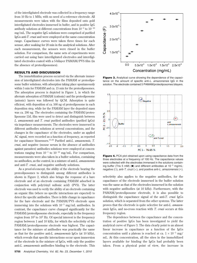
The dependence between the capacitance and the concen-tration of positive IgGs has been investigated to yield theanalytical curve of Figure 3. The data clearly point to an almostlinear increase in capacitance as a function of the IgGsconcentration until a plateau is reached at ca. 1 × 10⁻⁵ mg/mL. At this concentration, all the sites in the immobilizedlayers available for binding the IgGs had probably beentaken. From a physical point of view, the increase in
Figure 3. Analytical curve showing the dependence of the capaci-tance on the amount of specific anti-L. amazonensis IgG in thesolution. The electrode contained 5 PAMAM/proteoliposomes bilayers.
Figure 4. PCA plot obtained upon using capacitance data from thethree electrodes at a frequency of 100 Hz. The capacitance valueswere collected with the electrodes immersed in the solutions contain-ing buffer (Tris 5 mM) (b) and different antibodies at 10-5 mg/mL:negative (O), anti-T. cruzi (×), and positive anti-L. amazonensis (+).
9766 Analytical Chemistry, Vol. 82, No. 23, December 1, 2010

capacitance may be related to changes in the dielectricconstant of the material, in this case, the PAMAM/proteo-liposome film, deposited on the gaps of gold tracks in theinterdigitated electrode. The increase in capacitance isprobably related to the generation of additional interfacesin the dielectric region due to adsorption of antibodies.
To improve the distinguishing ability of the biosensing system,we combined the capacitance values at 1 kHz obtained with thethree electrodes, (i) bare electrode, (ii) electrode containing 5PAMAM/PVS bilayers, and (iii) electrode containing 5 PAMAM/proteoliposome bilayers, and analyzed using principal componentanalysis (PCA). The PCA plot in Figure 4 demonstrates that allanalytes can be distinguished for a concentration of 10-5 mg/mL of IgGs.
To reduce costs for sample preparation and IgGs purification,as required for in field diagnosis, we investigated the ability ofthe electrodes to detect specific antibodies in total serum samplesfrom mice infected with L. amazonensis, and also from miceinfected with T. cruzi (see details in the Experimental Section).The total serum had been diluted in buffer at concentrations from10-2 to 10-10 mg/mL. Note that the positive serum concentra-tion at 10-10 mg/mL implies a much lower aliquot for positiveanti-L. amzonensis IgGs. Surprisingly, the electrodes could stilldistinguish between the two total serum samples, as depictedin the PCA plot of Figure 5. The data from negative sampleswere located in the left part of the plot, whereas data from positivesamples were located in the right side of the chart. The latterindicated the capacity of the methods to detect positive anti-L.amazonensis in the total serum, as well as the ability to distinguishthe data from anti-T. cruzi IgGs. Therefore, no cross-reactionscould be noted, and the performance of the diagnosis system isoptimized.
Figure 5. PCA plot built with capacitance data from the three electrodes immersed in total serum samples from mice infected with L. amazonensis(positive total serum) and from mice infected with T. cruzi (negative total serum). The total serum in both cases was diluted from 10-2 to 10-10
mg/mL.
Figure 6. Sammon’s mapping projection for all samples, with thestandardization procedure applied to the m-dimensional data samples.In this case, analyses were performed using data from four sensors,including a sensor based on immobilized proteoliposomes containingT. cruzi antigens.
9767Analytical Chemistry, Vol. 82, No. 23, December 1, 2010

We also stress that use can be made of multidimensionalprojection techniques as described elsewhere23 to visualize theimpedance data for all the samples tested in the same plot. Figure6 shows that a complete separation is reached for the varioussamples when the Sammon’s mapping method is employed.Further details of the application of projection techniques to thisbiosensing data can be found in ref 24.
CONCLUSIONSThe need for cost-effective, reliable diagnosis methods, through
which leishmaniasis can be identified and treated in the very earlystages, is evidenced by the fact that first and second-line drugsused for treating it present high toxicity, several side effects, andrequire long-term management. In this Article, we showed a
promising way for rapid, accurate diagnosis of Leishmania infec-tions using interdigitated electrodes containing immobilized pro-teoliposomes. Electrical capacitance measurements allowed thedetection of specific anti-L. amazonensis antibodies at concentra-tions down to 10-5 mg/mL. The use of PCA to statisticallycorrelate the capacitance data permitted the distinction betweenpositive and negative total serum samples. The methods usedhere are generic and may be extended to the diagnosis ofbacterial, protozoan, and helminth infectious diseases affectingdeveloping countries, including tuberculosis, malaria, and otherneglected diseases, such as the African sleeping sickness,onchocerciasis, lymphatic filariasis, and schistosomiasis.
ACKNOWLEDGMENTThis work was supported by FAPESP, CNPq, and Capes
(Brazil).
Received for review July 20, 2010. Accepted October 4,2010.
AC101920T
(22) Daghastanli, K. R. P.; Ferreira, R. B.; Thedei, G., Jr.; Maggio, B.; Ciancaglini,P. Colloids Surf., B 2004, 36, 127–137.
(23) Siqueira, J. R., Jr.; Maki, R. M.; Paulovich, F. V.; Werner, C. F.; Poghossian,A.; Oliveira, M. C. F.; Zucolotto, V.; Oliveira, O. N., Jr.; Schoning, M. J.Anal. Chem. 2010, 82, 61–65.
(24) Paulovich, F. V.; Maki, R. M.; de Oliveira, M. C. F.; Colhone, M. C.; Santos,F. R.; Migliaccio, V.; Ciancaglini, P.; Daghastanli, K. R.; Stabeli, R. G.;Perinotto, A. C.; Oliveira, O. N., Jr.; Zucolotto, V. Anal. Chem., submitted.
9768 Analytical Chemistry, Vol. 82, No. 23, December 1, 2010