Embed Size (px)
Citation preview
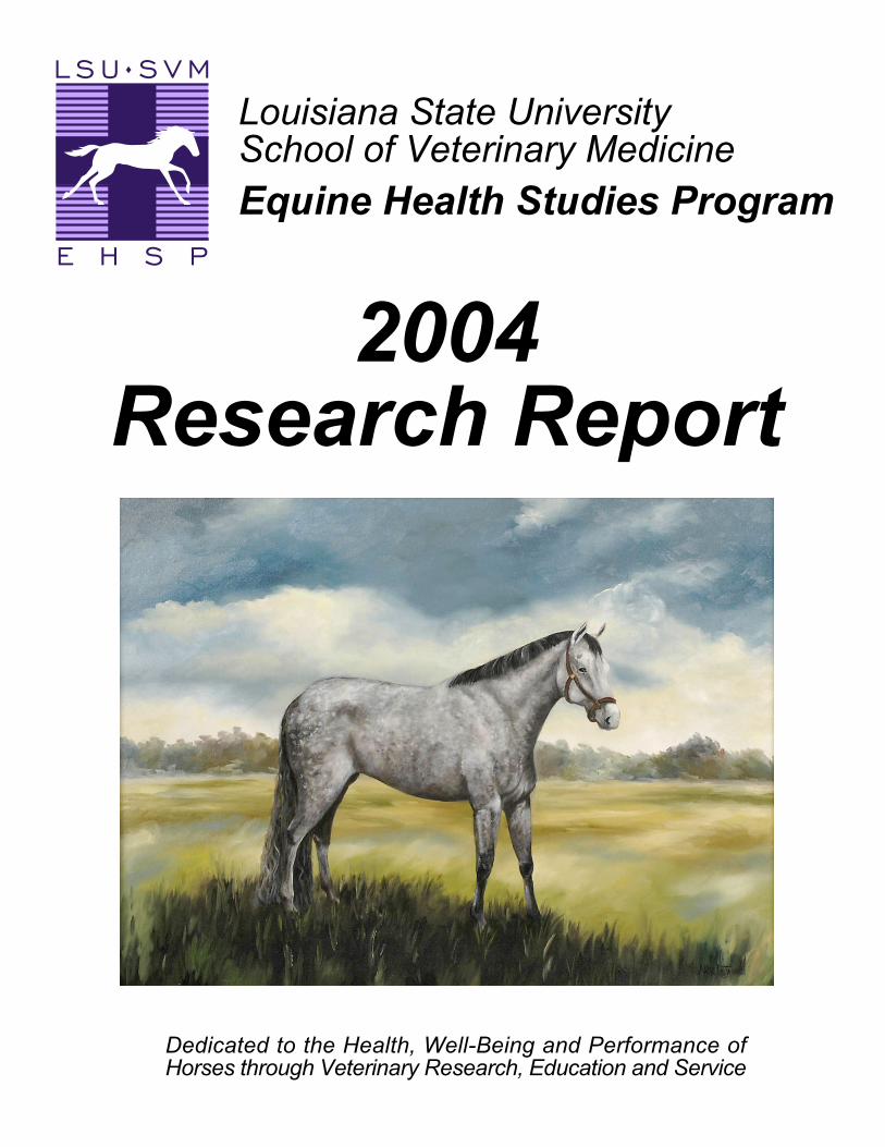
Dedicated to the Health, Well-Being and Performance ofHorses through Veterinary Research, Education and Service
2004Research Report
Equine Health Studies Program
Louisiana State UniversitySchool of Veterinary Medicine
L S U
E H S P
♦ S V M

Director’s Message
Dr. Rustin M. Moore, DirectorEquine Health Studies ProgramSchool of Veterinary MedicineLouisiana State University
The purpose of this Equine Health Studies Program (EHSP)Research Report is to document the activities pertaining toscientific investigations involving equine health and diseaseconducted at the LSU School of Veterinary Medicine. Thecontents of this inaugural issue of the EHSP Research Reportrepresent equine biomedical scientific investigations completedbetween 1998 and 2000. The second issue will follow soonhereafter and will contain information related to studies completedbetween 2001 and 2003. The report demonstrates the diverseand extensive equine biomedical research conducted by themultidisciplinary faculty, advanced studies students and technicalstaff that comprise the EHSP. The quantity and quality of work isespecially noteworthy considering the limited intramural fundsavailable during this period. These works demonstrate theprogram’s commitment to the health, well-being and performanceof horses through scientific investigations. Additionally, the reporthighlights information pertaining to research facilities andequipment, scientific manuscripts and abstracts published, andgrants and contracts awarded.
The state-of-the-art laboratory facilities and equipment highlighted in this report represent majoradvances made in the EHSP as a result of acquisition of funds from the Louisiana Governor’sBiotechnology Initiative, the Louisiana Board of Regents Enhancement Grants Program, andrecurrent Legislative funding obtained in 2003. Our research facilities are becoming second-to-none and provide faculty, students and staff an enriching and stimulating atmosphere to conductleading-edge equine biomedical research. This helps to facilitate a better understanding of equinediseases and to assist with finding methods for improved treatment of critically ill and injuredhorses. We are also indebted to numerous organizations and individuals for their support of thebasic and applied equine research activities.
The important findings of the research studies outlined in this report would not have been possiblewithout the horses utilized for these studies. It should be noted that all research activities areconducted following federal guidelines for the care and use of animals. We do not take them forgranted; we value their life and dignity, and we treat them with the utmost humane care. Theiravailability and utilization is necessary for progress toward the future care and treatment of ill andinjured horses.
We are proud of the accomplishments we have made thus far and look to the future with optimismand excitement! We are well on our way to achieving and maintaining our mission of becoming anelite equine biomedical program.
Sincerely,
Rustin M. Moore, DVM, Ph.D., DACVSDirector

Published by the Equine Health Studies Program, School of Veterinary Medicine, Louisiana StateUniversity, Baton Rouge, Louisiana.
Dean, School of Veterinary Medicine: Michael G. Groves, DVM, MPH, DACVM, DACVPM,DAVPM
Executive Associate Dean: Peter F. Haynes, DVM, MS, DACVS
Associate Dean for Research and Advanced Studies: Thomas R. Klei, Ph.D.
Director, Equine Health Studies Program: Rustin M. Moore, DVM, Ph.D., DACVS
On the Cover: “Blue”, a commissioned oil painting by Anita Lejeune, a local artist from Lakeland,Louisiana, of “So Blue Riva”, a Quarter Horse mare owned by Ms. Sydney L. Hines of PassChristian, Mississippi. “Blue” was successfully treated for a severe injury and infection of her rightfront foot at the LSU Equine Clinic in 2000, and subsequently gave birth to her first foal on April 1st
of this year. Mrs. Jeanne Hines McDaniel, Sydney’s mother, gave a major charitable gift in honor of“Blue” and on behalf of her daughter and granddaughter, Ms. Jeanne L. Pitre. The gift assisted withconstruction of a new, 10-stall state-of-the-art Equine Intensive Care Unit, which opened inSeptember 2004 in the LSU Veterinary Hospital’s Equine Clinic. To view more of the artist’s equineand other works or to inquire about commissioned works, visit her website, www.anitalejeune.com,and help support the LSU Equine Health Studies Program (EHSP). The artist donates 20% of allsales to the EHSP.
The Equine Health Studies Program is supported with funds provided by the Louisiana StateUniversity School of Veterinary Medicine, the State of Louisiana, and contributions from privatedonors.
Louisiana State University does not discriminate in any of its policies, procedures or practices.The University is an affirmative action/equal opportunity employer.
Our Mission: The LSU Equine Health Studies Program will become a premier equine biomedicalcenter in the 21st century through leading-edge research of equine diseases, contemporaryinstruction of professional veterinary students and veterinarians in advanced studies programs, andenhanced continuing education of the horse-owning public and private equine practitioners with theultimate goal of providing state-of-the-art diagnostic and therapeutic capabilities for critically ill andinjured horses and optimal clinical service to horsemen in Louisiana and the surrounding region.
2004 Research ReportEquine Health Studies Program
School of Veterinary MedicineLouisiana State University
Baton Rouge, Louisiana 70803Phone: (225) 578-9500
Fax: (225) 578-9605E-mail: [email protected]
Website: www.equine.vetmed.lsu.edu

2 Equine Health Studies Program 2004 Research Report
Table of ContentsDean’s Message - Dr. Michael G. Groves .................................................................................... 4EHSP Faculty ............................................................................................................................... 5Interdepartmental Equine Health Studies Program ....................................................................11EHSP Grants and Contracts ........................................................................................................12EHSP Selected Scientific Publications ......................................................................................15EHSP Selected Published Scientific Abstracts ...........................................................................18EHSP Development ................................................................................................................... 21EHSP Facilities and Equipment ..................................................................................................22
RESEARCHFoals
Plasma magnesium concentrations in neonatal foal admitted to the intensive care unit ................... 28
Gastrointestinal TractIn vitro effects of oxytocin, acepromazine, detomidine, xylazine, butorphanol, terbutaline,
isoproterenol, and dantrolene on smooth and skeletal muscles of the equineesophagus ..................................................................................................................................30
Effects of treatment with oxytocin, xylazine butorphanol, guaifenesin, acepromazine,and detomidine on esophageal manometric pressure in conscious horses ................................. 31
Plasma magnesium concentrations in critically ill horses affected with gastrointestinaltract disease ............................................................................................................................... 32
In vitro effects of an electron transport inhibitor on resistance, permeability, and adeninenucleotide content of equine colonic mucosal tissue ...................................................................33
The effect of Black Walnut extract on equine colonic histopathology and in vitro transport ................ 35Parasitized equine colonic epithelium exhibits an increased chloride secretory response
compared to non-parasitized epithelieal tissue in vitro ................................................................ 36Detection and comparison of nitric oxide in clinically healthy horses and those with
naturally acquired large colon and small intestinal strangulating obstruction ............................... 37Quantitation of adenine nucleotides in equine colonic mucosal tissue using high
performance liquid chromatography ............................................................................................38Pharmacological evaluation of endothelin-antagonists in blocking constriction of equine
colonic vessels induced by endothelin-1 ..................................................................................... 40
HemodynamicTransient weakness, ataxia, and recumbency associated with catheterization of the
right side of the heart in 3 horses ............................................................................................... 41Cardiopulmonary and sedative effects of intravenous administration of low doses of
medetomidine and xylazine to adult horses .................................................................................42Hemodynamic and metabolic alterations associated with intravenous infusion of a
combination of adenosine triphosphate and magnesium chloride in conscious horses ............... 43Pharmacokinetics, clinical signs and hemodynamic effects of an intravenous bolus dose
of Nù -nitro-L-arginine methyl ester (L-NAME) in healthy conscious horses ................................. 44In vitro responses of equine colonic arterial and venous rings to adenosine triphosphate ................. 45
InflammatoryPlasma and urine nitric oxide concentration in horses given a low dose of endotoxin ....................... 47Nitric oxide concentrations in cerebrospinal fluid of horses with or without neurologic
disease .......................................................................................................................................48Cephalic venous plasma endothelin-1 concentrations and digital blood flow responses
to ATP-MgCl2 administration after low-dose endotoxin infusion in horses .................................... 49Temporal effects of freezing on plasma nitric oxide concentrations in ponies .................................... 50
Table of Contents
Equine Health Studies Program 2004 Research Report 3
Table of Contents
Effects of adenosine triphosphate-magnesium chloride on cardiopulmonary andclinicopathologic variables, cytokine activity, and endothelin concentration in horsesadministered a low dose of endotoxin .........................................................................................52
MusculoskeletalAlterations of nitric oxide and endothelin-1 subsequent to experimental lipopolysaccharide
induced acute synovitis in horses ................................................................................................54Synovial fluid and plasma nitric oxide and endothelin-1 concentrations in horses with
and without joint disease .............................................................................................................55Pharmacological control of digital vasculature in the prevention and treatment of
laminitis in horses ....................................................................................................................... 56The effects of endothelin-1 and nitric oxide on digital hemodynamics of normal horses .....................58
ParasitologyAlterations in histologic and immunohistochemical staining for nitric oxide alterations
in parasite free ponies infected with Strongylus vulgaris ............................................................. 60Clinical signs, hematologic, cytokine, and plasma nitric oxide alterations in response to
Strongylus vulgaris infection in ponies ........................................................................................ 61Epidemiology of small strongyle infections in southern Louisiana .................................................... 62Immunity to strongyle nematodes in ponies ....................................................................................... 64
ReproductionProperly timed teasing can aid uterine clearance .............................................................................. 67Effect of dose and day of treatment on uterine response to oxytocin in mares .................................. 68Prostaglandin F2á metabolatie (PGFM) response to exogenous oxytocin and determination
of the half-life of oxytocin in nonpregnant mares ................................................................................68Role of nitric oxide in ovulation in the mare .............................................................................................. 69“In-vitro” produced foals ................................................................................................................... 70Reproductive parameters of Miniature horse stallions ....................................................................... 71The effects of pH, osmolarity and urine contamination on equine spermatozoal motility .................... 72Inducing earlier cyclicity in mares ...................................................................................................... 72Using acupuncture to advance the onset of cyclicity ......................................................................... 73Using acupuncture to aid uterine clearance ...................................................................................... 74
RespiratoryCytokine gene expression: Is it altered in horses affected with summer pasture-associated
obstructive pulmonary disease?...................................................................................................75Correlation of clinical score, pleural pressure, bronchoalveolar lavage fluid cytology, and
pulmonary histology in horses with summer pasture-associated obstructive pulmonarydisease .......................................................................................................................................76
Plasma and bronchoalveolar fluid concentrations of nitric oxide and histochemical andimmunohistochemical localization of nitric oxide synthesis in lungs of horses withsummer pasture-associated obstructive pulmonary disease ....................................................... 77
Comparative responses of bronchial rings to mediators of airway hyper-reactivity inhealthy horses and those affected with summer pasture-associated obstructivepulmonary disease ..................................................................................................................... 79
SurgeryNd:YAG laser assisted modified Forssell’s procedure for treatment of cribbing (crib-biting)
in horses..................................................................................................................................... 80Cecal amputation within the right ventral colon for surgical treatment of nonreducible cecocolic
intussusception in eight horses ................................................................................................. 81Factors associated with racing performance of Thoroughbreds undergoing lag screw repair of
condylar fractures of the third metacarpal or metatarsal bone .................................................. 82Career racing performance in Thoroughbreds treated with prosthetic laryngeoplasty for laryngeal
neuropathy: 52 cases (1981-1989) ...........................................................................................83
4 Equine Health Studies Program 2004 Research Report
Dean’s Message
Dean’s Message
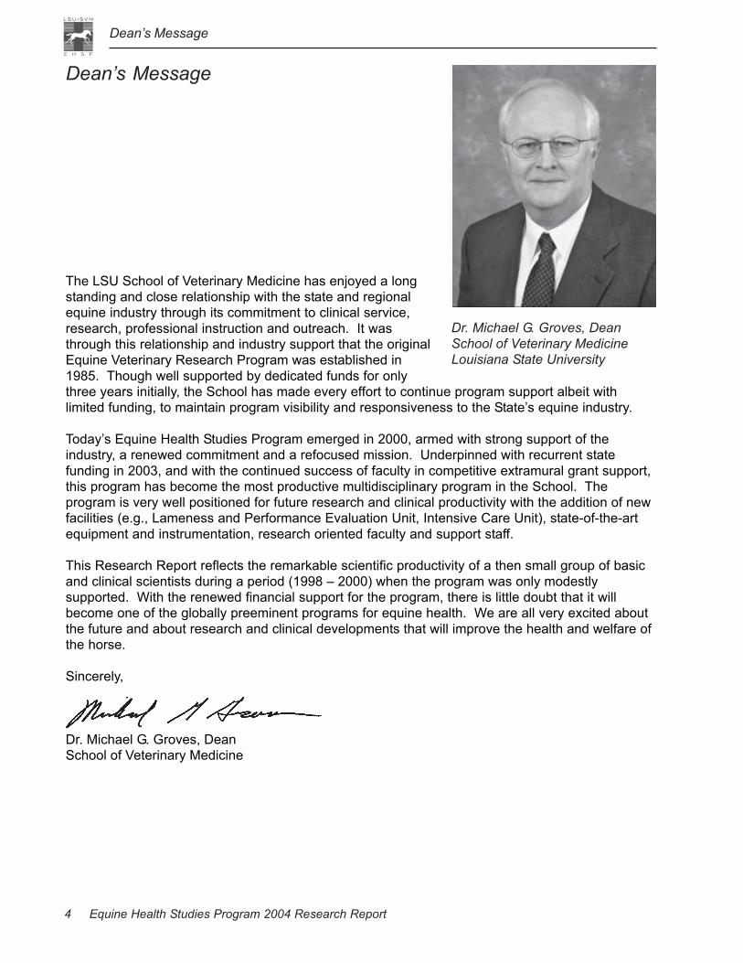
Dr. Michael G. Groves, DeanSchool of Veterinary MedicineLouisiana State University
The LSU School of Veterinary Medicine has enjoyed a longstanding and close relationship with the state and regionalequine industry through its commitment to clinical service,research, professional instruction and outreach. It wasthrough this relationship and industry support that the originalEquine Veterinary Research Program was established in1985. Though well supported by dedicated funds for onlythree years initially, the School has made every effort to continue program support albeit withlimited funding, to maintain program visibility and responsiveness to the State’s equine industry.
Today’s Equine Health Studies Program emerged in 2000, armed with strong support of theindustry, a renewed commitment and a refocused mission. Underpinned with recurrent statefunding in 2003, and with the continued success of faculty in competitive extramural grant support,this program has become the most productive multidisciplinary program in the School. Theprogram is very well positioned for future research and clinical productivity with the addition of newfacilities (e.g., Lameness and Performance Evaluation Unit, Intensive Care Unit), state-of-the-artequipment and instrumentation, research oriented faculty and support staff.
This Research Report reflects the remarkable scientific productivity of a then small group of basicand clinical scientists during a period (1998 – 2000) when the program was only modestlysupported. With the renewed financial support for the program, there is little doubt that it willbecome one of the globally preeminent programs for equine health. We are all very excited aboutthe future and about research and clinical developments that will improve the health and welfare ofthe horse.
Sincerely,
Dr. Michael G. Groves, DeanSchool of Veterinary Medicine
Equine Health Studies Program 2004 Research Report 5
EHSP Faculty
FacultyAbolghasem Baghian, Assistant Professor, Veterinary Microbiology & ParasitologyDr. Baghian received his M.S. in Microbiology from Southeastern Louisiana University in 1981, and hereceived his Ph.D. in 1985 from Arizona State University. Dr. Baghian was a postdoctoral researcher at theSchool of Veterinary Medicine at Louisiana State University, where he later became an instructor and iscurrently an assistant professor. Dr. Baghian’s research focuses on investigating the sturcture and functionof herpes simplex virus glycoprotein K and the structure and function of Kaposi’s sarcoma associatedherpesvirus (KSHV) glycoproteins gH, gL, and gB to those other herpesviruses.
Steven A. Barker, Professor, Veterinary Physiology, Pharmacology & ToxicologySteven A. Barker is a professor of veterinary physiology, pharmacology and toxicology at the School ofVeterinary Medicine in the department of Comparative Biomedical Sciences. He received his B.S. in 1971,his M.S. in 1973, and his Ph.D. in 1978, all from the University of Alabama. Dr. Barker is also the directorof the Analytical Systems Laboratory.
Rallph E. Beadle, Professor Emeritus, Equine MedicineDr. Beadle was born and raised in Montana. He completed his pre-veterinary and veterinary education atColorado State University, where he was awarded a DVM degree in 1967. He spent the next five years atthe University of Georgia, where he worked in the Equine Clinic and obtained a Ph.D. degree in VeterinaryPhysiology. After a period of two years spent at Michigan State University, he has been at Louisiana StateUniversity for the rest of his professional career. During the first seven years at LSU, he was in theDepartment of Veterinary Physiology, Pharmacology and Toxicology where he taught both physiology andpharmacology. From that time until September of 1999, he was in the Department of Veterinary ClinicalSciences where he worked in the Medicine Section of the Equine Clinic. He retired in September of 1999,but since that time has continued to be involved in the activities of the Department of Veterinary ClinicalSciences as a professor emeritus. His research interests involve non-sweating horses, horses withrecurrent airway disease and horses affected with acute and chronic laminitis.
Aloisio C. D. Bueno, Clinical Instructor, Equine SurgeryDr. Bueno, originally from Brazil, obtained his veterinary medical degree from Unoeste University and thencompleted two years of work in a private equine practice in Brazil. He then completed a one-year internshipin large animal medicine and surgery followed by a two-year M.S. program at the LSU School of VeterinaryMedicine. Dr. Bueno then completed a one-year fellowship in large animal surgery at Oregon StateUniversity before going to the University of California-Davis for a three-year residency in equine surgery.Upon completion of his residency, Dr. Bueno returned to LSU as a clinical instructor of equine surgery andprovides the majority of the equine emergency surgery service. Dr. Bueno is investigating thepathophysiology, prevention and treatment of laminitis.
Daniel J. Burba, Professor, Equine SurgeryDr. Daniel J. Burba was born near Punxsutawney, Pa., on a dairy farm. He and his parents moved toGrayson, a small town in eastern Kentucky, when he was 13. His family raised Quarter Horses, and he stillowns Quarter Horses and competes in team penning in the southern regional organization with family wholive in Florida. He completed his pre-veterinary studies at Morehead State University in Kentucky. Hereceived his DVM from Auburn University in 1986, and then completed a large animal internship (1987) andequine surgical residency (1990) at Oklahoma State University. He is board certified by the AmericanCollege of Veterinary Surgeons. His clinical interests include lameness and orthopedic surgery and lasersurgery. His research interests include musculoskeletal injuries, such as joint disease.
Dr. Sharon Chirgwin, Assistant Professor, ResearchDr. Sharon Chirgwin was born and raised in Australia. She obtained a Bachelor of Science with Honors,majoring in Biochemistry and Zoology, from James Cook University, in Townsville, Queensland. Dr.Chirgwin then completed a Ph.D. in Molecular Parasitology at the Queensland Institute of Medical
6 Equine Health Studies Program 2004 Research Report
Research, before joining the laboratory of Dr. Thomas Klei at LSU, where she works on both human andhorse parasites. Her research interests include the molecular characterization of the early infection eventsof parasitism. Dr. Chirgwins’ current position in the EHSP involves teaching students, staff and facultymolecular biological techniques, and advising on the contribution this technology can make to equineresearch.
Doo Youn Cho, Professor, Veterinary PathologyDr. Doo Youn Cho is a professor of veterinary pathology in the department of Pathobiological Sciences atthe School of Veterinary Medicine. Dr. Cho is also the section chief for necroscopy/surgical biopsy in theSchool’s Veterinary Teaching Hospital and Clinics. He received his DVM in 1966 and his M.V.Sc. in 1970,both from Seoul National University in Korea. In 1973, he received his M.S., and in 1978, he received hisPh.D., both from Kansas State University.
Susan C. Eades, Professor, Equine MedicineDr. Susan Eades graduated from the LSU School of Veterinary Medicine, then completed an internship inlarge animal medicine and surgery, and a residency in large animal internal medicine at the University ofPennsylvania’s New Bolton Center. She then moved to Athens, Ga., and completed a Ph.D. program inVeterinary Physiology at the University of Georgia. Her doctoral studies concentrated on intestinal vascularand nonvascular smooth muscle physiology and pharmacology. Upon completion of her Ph.D., Dr. Eadesbegan as an assistant professor of large animal medicine at the University of Georgia College of VeterinaryMedicine, where she remained through 1997. She returned to LSU in 1997 as an associate professor ofequine medicine. Dr. Eades’ clinical interests include equine internal medicine; however, she has a specialinterest in cardiology and ultrasound. Her research interests include intestinal disease and laminitis.
Bruce E. Eilts, Professor, TheriogenologyDr. Bruce E. Eilts is originally from the Minneapolis/St. Paul area in Minnesota. He graduated from highschool in West St. Paul, Minn., and then attended the University of Minnesota as pre-veterinary medicinestudent. He obtained a B.S. in veterinary science in 1975 and his DVM in 1977, both from the University ofMinnesota. He was in private practice for one year before returning to the University of Minnesota to obtainan M.S. in theriogenology in 1982. After two and one half years in private practice in southern California, hecame to Louisiana State University as an assistant professor in 1984. He became board certified in theAmerican College of Theriogenologists in 1986. His main clinical interest is basic reproductionmanagement in the horse. His main research interest is intrafollicular insemination in the mare.
Timothy P. Foster, Research Assistant Professor, Molecular Virology and Cell BiologyDr. Timothy Foster was born in San Francisco, CA and obtained a BS degree in Biochemistry and a BSdegree in Microbiology/Zoology from Louisiana State University in 1995. In 1999, he received a PhD inVeterinary Medical Sciences with an emphasis in Biochemistry and Molecular Virology from the LouisianaState University Departments of Biochemistry and Veterinary Microbiology and Parasitology. Dr. Foster iscurrently a Research Assistant Professor in the Division of Biotechnology and Molecular Medicine at theLSU School of Veterinary Medicine. His primary interests are deciphering the molecular interplay betweenhost cells and various pathogens, as well as translational investigations that transition primary bench workscience rapidly into the clinical environment.
Dennis D. French, Professor, Veterinary ScienceDr. Dennis D. French, originally from Chatfield, Minn., obtained his B.S. and DVM degrees from theUniversity of Minnesota in 1976 and 1979, respectively. He is a Diplomate of the American Board ofVeterinary Practitioners, certified in equine practice. His clinical interests include equine herd health andsport horse medicine. His research interests include equine parasitology, immunology and exercisephysiology in horses. Dr. French is currently a professor of veterinary science at the LSU School ofVeterinary Medicine, and provides equine ambulatory services for the Veterinary Teaching Hospital &Clinics. Dr. French is a past president of the Louisiana Veterinary Medical Association. Dr. French and hisfamily are active in many equine and equestrian activities throughout the state.
EHSP Faculty
Equine Health Studies Program 2004 Research Report 7
William G. Henk, Professor, Veterinary Anatomy and Cell BiologyDr. William G. Henk is a professor of veterinary anatomy and cell biology in the department of ComparativeBiomedical Sciences at the School of Veterinary Medicine. He is also the chief of the Electron MicrosocpyLaboratory. Dr. Henk received his B.S. in 1967, His M.Ed. in 1971, and his Ph.D. in 1977, all from theUniversity of Georgia.
Jeremy D. Hubert, Assistant Professor, Equine SurgeryDr. Jeremy D. Hubert, was born in Wales, but grew up on a ranch in Zimbabwe, where he received hisveterinary degree (BVSc.). After two years of mixed animal practice in Zimbabwe and the United Kingdom,he completed an internship in equine medicine and surgery at LSU. This was followed by a year in equinepractice in the U.K. before embarking on a combined equine surgery residency and M.S. program, whichhe completed in July 1999. He became board certified by the American College of Veterinary Surgeons in2000. He worked as a clinical instructor in Equine Surgery for one year and accepted a position as anassistant professor of equine surgery at LSU in October 2001. His clinical interests include upperrespiratory tract disease, as well as lameness and orthopedics. He is currently involved in scientificinvestigations involving extracorporeal shockwave therapy, bone density, and the role of eosinophils ingastrointestinal tract disease.
Jill R. Johnson, Professor, Equine MedicineDr. Jill R. Johnson is a native of South Dakota. She graduated from veterinary school at the University ofMinnesota, then stayed on and completed a M.S. degree in Veterinary Surgery and Radiology. She joinedthe faculty of the School of Veterinary Medicine at LSU in 1977. She is a specialist in internal medicine(Diplomate, American College of Veterinary Internal Medicine) and Equine Practice (Diplomate, AmericanBoard of Veterinary Practitioners). Past research activities have centered on immuogenetics andimmunology. Current research activities include evaluation of methods of quantifying exercise trainingusing the global positioning system (GPS) and development of tissue culture models to study laminitis andchronic obstructive pulmonary disease using microgravity methods.
Dae Y. Kim, Instructor, Pathobiological SciencesDr. Dae Y. Kim is an instructor in the department of Pathobiological Sciences at the School of VeterinaryMedicine. He received his DVM from Seoul National University in Korea, and he received his Ph.D. fromLouisiana State University.
Thomas R. Klei, Boyd Professor, Parasitology and Veterinary ScienceDr. Thomas Klei obtained his B.S. and Ph.D. degrees in biology and zoology from Northern MichiganUniversity and Wayne State University in 1965 and 1971, respectively. He then completed postdoctoraltraining at the National Institute of Health. He joined the faculty at the LSU School of Veterinary Medicine in1975. He became a Boyd Professor in Parasitology and Veterinary Science at LSU in 1992. Dr. Klei hasconducted leading-edge investigations into and has contributed greatly to our current understanding ofequine parasitology. Dr. Klei is currently serving as the associate dean for Research and Advanced Studiesat the School of Veterinary Medicine.
Konstantin G. Kousoulas, Professor, Veterinary VirologyDr. Konstantin G. Kousoulas is a professor of veterinary virology in the department of PathobiologicalSciences at the School of Veterinary Medicine. He is also a professor of poultry science and an adjunctprofessor of biological sciences. Dr. Kousoulas is the directorof of the School of Veterinary Medicine’sDivision of Biotechnology & Molecular Medicine. He received his B.S. in 1975 from Fairleigh Dickinson. In1977, he received his M.S. and in 1981, he received his Ph.D., both from Pennsylvania State University.
Mandi J. Lopez, Assistant Professor, Equine and Comparative OrthopedicsDr. Mandi Lopez was born and raised in the Pacific Northwest. She attended veterinary school at theUniversity of California, Davis and then completed an internship at Kansas State University prior to going tothe University of Wisconsin, where she completed a residency in large animal surgery and obtained bothan M.S. and Ph.D. degrees. Her area of interest and expertise is comparative orthopedic research andsurgery. She is board-certified by the American College of Veterinary Surgeons. She came to LSU inJanuary 2004 and heads the Laboratory for Equine and Comparative Orthopedic Research.
EHSP Faculty
8 Equine Health Studies Program 2004 Research Report
EHSP Faculty
Sara K. Lyle, Clinical Instructor, TheriogenologyDr. Sara K. Lyle was born and raised in Gainesville, Fla. She obtained her B.S. in Chemistry at DukeUniversity and her DVM from the University of Florida. She completed a residency in theriogenology in1989 and a M.S. in reproduction in 1991 at the University of Florida. She is board certified by the AmericanCollege of Theriogenologists. Her clinical interests include mare infertility and assisted reproductivetechnologies. Her research interests include reproductive immunology (equine) and assisted reproductivetechnologies in horses.
Rebecca S. McConnico, Assistant Professor, Equine MedicineDr. Rebecca S. McConnico is originally from north central Ohio, where she lived for 18 years. She obtainedher B.S. in Animal Science from the University of Arkansas, her DVM degree from Louisiana StateUniversity, and her Ph.D. and clinical residency in large animal internal medicine from North Carolina StateUniversity. She is board certified in Equine Internal Medicine and her clinical interests are in equine criticalcare and internal Medicine. Her research interests include inflammatory disease of the equine largeintestine and infectious diseases and the effects on mucosal physiology and permeability.
Rustin M. Moore, Professor, Equine SurgeryDr. Rustin M. Moore, professor of equine surgery, currently serves as director of the Equine Health StudiesProgram and service chief of Equine Medicine and Surgery. He is originally from West Virginia and earnedhis B.S. from West Virginia University. He obtained his DVM and Ph.D. from The Ohio State University andcompleted his equine surgical residency at the same institution. He is board certified by the AmericanCollege of Veterinary Surgeons. Dr. Moore began at the LSU School of Veterinary Medicine in October1994. Some of his clinical interests include lameness, surgery and colic and its associated complications.Dr. Moore’s research focuses on vascular and nonvascular smooth muscle physiology and pharmacologyand the pathophysiology and treatment of colic, laminitis, endotoxemia and heaves.
Claudio C. Natalini, Assistant Professor, Veterinary AnesthesiologyDr. Claudio C. Natalini is originally from Rio de Janeiro, Brazil, where he attended the Universidade FederalFluminense (UFF) and graduated in veterinary medicine in 1984. From 1985 to 1986, Dr. Natalini enrolledin a residency program in veterinary surgery and medicine at the Universidade Federal do Rio Grande doSul (UFRGS), Brazil. He worked from 1986 to 1992 as a staff surgeon/anesthesiologist at UFRGS. In 1991,Dr. Natalini completed an M.S. program in veterinary anesthesiology at Universidade Federal de SantaMaria (UFSM), Brazil. In 1992 he became an assistant professor of veterinary anesthesiology at UFSM. In1994 he obtained his board certification from the Brazilian College of Veterinary Surgeons andAnesthesiologists (CBCAV) and served as CBCAV secretary for one year. In 1996, Dr. Natalini enrolled in aPh.D./residency program at the University of Minnesota, earning his degree in 2000 working with opioidspinal mediated analgesia in the equine. In 2002 Dr. Natalini joined the Department of Veterinary ClinicalSciences at the LSU SVM. Dr. Natalini clinical interests are small and large animal pain management withemphasis in spinal analgesia and local anesthesia. His research interest includes the pharmacology andphysiology of spinal administration of analgesic in horses.
Marlene Orandle, Assistant Professor, VirologyDr. Marlene Orandle was born and raised in Baltimore, MD. She obtained her B.A. in Biology from SaintMary’s College of Maryland in 1987. Following four years of research experience at Johns HopkinsUniversity School of Medicine, Dr. Orandle completed her DVM from Iowa State University in 1995 and herPh.D. in Veterinary Pathology from the University of Florida in 1999. Since receiving her Ph.D., she hascompleted Postdoctoral Fellowships at both the New England and Tulane National Primate ResearchCenters where she studied the pathogenesis of simian immunodeficiency virus (SIV) infection in the brainas a model for AIDS dementia. Dr. Orandle joined the faculty within the Department of PathobiologicalSciences at the LSU School of Veterinary Medicine in 2004. Her research interest is in the study ofcomparative lentiviral pathogenesis with a specific focus on factors contributing to the development ofneurological disease. Ongoing research in her laboratory is focused on understanding the mechanismsinvolved in trafficking of virus-infected cells across the blood-brain barrier in SIV-infected rhesus macaquesand in EIAV-infected horses.
Equine Health Studies Program 2004 Research Report 9
EHSP Faculty
Dale L. Paccamonti, Professor, TheriogenologyDr. Dale L. Paccamonti, originally from Kankakee, Ill., completed his undergraduate and veterinaryeducation at Michigan State University, receiving his DVM in 1981. After four years in a mixed practice inChestertown, Md., he pursued advanced training at the University of Florida, where he completed aresidency in Theriogenology and received his M.S. degree in 1988. Dr. Paccamonti is a Diplomate in theAmerican College of Theriogenologists. He joined the faculty at the LSU School of Veterinary Medicine in1988, where he is currently a full professor of theriogenology in the Department of Veterinary ClinicalSciences. Dr. Paccamonti’s primary research interests include the study of infertility in mares, assistedreproduction techniques in horses, factors affecting sperm motility in stallions, semen cryopreservation installions and the process of fetal maturation and parturition in mares. He also collaborates in reproductiveresearch in other domestic species. In addition to research endeavors, his duties include teachingtheriogenology to third and fourth year veterinary students. He shares responsibility for clinicaltheriogenology cases in all species presented to the Veterinary Teaching Hospital & Clinics.
Daniel B. Paulsen, Professor, Veterinary PathologyDr. Paulsen received his B.S. in 1975, his D.V.M. in 1977, and his M.S. in 1978, all from Kansas StateUniversity. In 1989, he received his Ph.D. from Oklahoma State University. Dr. Paulsen major areas ofresearch interest are bovine respiratory disease with emphasis on Mannheimia haemolytica, Pasteurellamultocida, Haemophilus somnus, bovine virus diarrhea, and bovine respiratory coronavirus; pathogenesis,bacterial genetics, respiratory immunity and vaccinology; toxicologic pathology associated with inhaledtoxins and effects of inhaled substances on the pathogenesis of asthma; and applicatio nofimmunohistochemical techniques in equine respiratory disease and laminitis and in cancer biology.
Glenn R. Pettifer, Assistant Professor, Veterinary AnesthesiologyDr. Pettifer was born and raised in Ontario, Canada. He received his DVM from the Ontario VeterinaryCollege at the University of Guelph. Following this he worked with the Equine Ambulatory Service at theOntario Veterinary College and then with Humber Equine Clinic in Toronto, Ontario. He then returned to theOntario Veterinary College, where he completed residency training and DVSc in Veterinary Anesthesiology.Since that time he has held instructor positions at the University of Saskatchewan and the University ofGeorgia. Most recently, Dr. Pettifer was assistant professor of veterinary anesthesiology at the OntarioVeterinary College before coming to LSU in the fall of 1999. His current research interests include painmanagement in all species, particularly horses. Dr. Pettifer is the recipient of Morris Animal Foundationsupport to investigate the analgesic effects of transdermally administered fentanyl in horses.
Changaram S. Venugopal, Professor, Veterinary Physiology & Pharmacology Dr. Changaram S. Venugopal is a veterinarian who graduated from Kerala Veterinary College andResearch Institute of Kerala University. After practicing as a veterinarian on the Kamadhenu Dairy Farm for5 years he pursued and received his M.Sc. degree in neuropharmacology from Calicut University, India. Hereceived his M.S. degree in cardiovascular pharmacology and Ph.D. in pulmonary pharmacology fromMassachusetts College of Pharmacy and Allied Health Sciences in a cooperative program with HarvardUniversity in Boston. Then he worked as a postdoctoral fellow at Harvard Medical School before he joinedthe faculty at Louisiana State University School of Veterinary Medicine in 1981. Dr. Venugopal is currently aprofessor of Pharmacology in the Department of Comparative Biomedical Sciences. He received his NewInvestigator Award grant from NIH in 1983 and the Beecham Award for Research Excellence in 1985. Hisresearch interests include the physiology and pharmacology of vascular and nonvascular smooth musclephysiology and pharmacology, and the pathophysiology of summer pasture associated obstructivepulmonary disease.
Dr. Ashley M. Stokes, Research Assistant ProfessorDr. Ashley M. Stokes was born in Baton Rouge and moved to Tuscaloosa, Alabama, to complete herbachelor’s degree from the University of Alabama. She returned to Baton Rouge to work in Oceanographyfor LSU for three years before her veterinary training. She completed the DVM/PhD program at the LSUSchool of Veterinary Medicine in the Department of Comparative Biomedical Sciences in 2001 and 2003,respectively. She completed a one-year post-doctorate research fellowship in the summer of 2004 whereshe continued her doctoral work on the vascular pathophysiology of equine laminitis. As a research

10 Equine Health Studies Program 2004 Research Report
EHSP Faculty
assistant professor within the EHSP, Dr. Stokes will continue focusing her efforts in cardiovascularphysiology with special emphasis on equine diseases.
H. Wayne Taylor, Professor, Veterinary PathologyDr. H. Wayne Taylor is a professor of veterinary pathology in the department of Pathobiological Sciences atthe School of Veterinary Medicine. He is also a veterinary pathologist and the director of the LouisianaVeterinary Medical Diagnostic Laboratory. Dr. Taylor received his DVM from Auburn University in 1967. In1969, he received his M.S. from the University of Missouri, where he also received his Ph.D. in 1971. Dr.Taylor is a Diplomate of the American College of Veterinary Pathologists.
Honor Ame Walesby, Assistant Professor, Equine SurgeryDr. Honor Ame Walesby, originally from Baltimore, Maryland, attended Virginia Tech for her BS in AnimalScience, Virginia Maryland Regional College of Veterinary Medicine for her DVM, and Iowa State Universityfor her MS in Veterinary Surgery. She became board certified in Large Animal Surgery with an equineemphasis in 2000. Her clinical interests lie in the filed of soft tissue and reconstructive surgery, abdominalsurgery, lameness, and ultrasonography. Her research interests include pharmacokinetics and endotoxin-related abortion in late gestation mares.
R. Wayne Waguespack, Assistant Professor, Equine SurgeryDr. Wayne Waguespack, graduated from Tuskegee University, completed an internship in large animalmedicine and surgery at the University of Georgia, and then completed an equine surgery residency andMS degree at Auburn University. Dr. Waguespack has clinical interests in soft tissue and orthopedicsurgery. He also has several research interests, including studying laminitis and potential ways to preventand treat this devastating disease, as well as the effects of extracorporeal shockwave therapy on tendon/ligament healing.
Equine Health Studies Program 2004 Research Report 11
Interdepartmental Equine Health Studies Program
Interdepartmental Equine HealthStudies ProgramThe Equine Health Studies Program (EHSP) is one of four recognized priority research programs in the LSU School ofVeterinary Medicine. Horses and equestrian activities are an important economic and recreational commodity inLouisiana and the surrounding region. Approximately 200,000 horses were owned by an estimated 60,000 people in thestate, with a total direct economic impact of the equine industry in Louisiana of 1.4 billion dollars annually. Scientificinvestigation into the prevention and treatment of equine disease is critical to maintaining the health, well-being andperformance of horses, and thus, is important for sustaining the equine industry. Substantial resources, includingmultidisiplinary, interdepartmental faculty, technical staff, facilities, and equipment provide an excellent environment foreither graduate or clinical advanced studies.
Graduate ProgramsStudents in the School of Veterinary Medicine’s interdepartmental Equine Health Studies Program can obtain Master ofScience (M.S.) and Doctor of Philosophy (Ph.D.) degrees in Veterinary Medical Sciences through the school’s academicdepartments: Comparative Biomedical Sciences, Pathobiological Sciences and Veterinary Clinical Sciences.
Current Research Interests• Gastrointestinal tract disease (colic)
- Intestinal ischemia-reperfusion- Ulcerative disease- Iintestinal motility disorders- Inflammatory bowel disease
• Effect of gastrointestinal tract inflammation on mucosal permeability• Effect of NSAIDs on colonic mucosal permeability• Summer pasture-associated obstructive pulmonary disease/COPD and other respiratory tract diseases• Nonvascular smooth muscle physiology, pharmacology, and pathobiology
- Gastrointestinal-Bronchial- Uterine
• Vascular smooth muscle physiology, pharmacology, and pathobiology• Analgesia and pain management• Medication surveillance• Synovitis and arthritis• Acupuncture• Laminitis• Parasitology• Endotoxemia• Virology• Use of global positioning system technology for equine epidemiologic studies• Inflammatory mediators, including nitric oxide, endothelin and cytokines• Mare reproductive physiology, infertility and placentitis• Improving freezing methods for stallion semen• Advancing the onset of the breeding season in mares• Intrafollicular insemination of mares• Equine embryo biotechnology• Assisted reproduction techniques in horses• Endotoxin-induced late gestation abortion in mares• Musculoskeletal injuries and other diseases causing poor performance• Comparative orthopedics• Effects of extracorporeal shockwave therapy on bone, tendon, ligament and nerve
12 Equine Health Studies Program 2004 Research Report
EHSP Grants and Contracts
EHSP Grants and Contracts2000Costa LRR, Gaunt S, O’Reilly KL, Horohov DW, Moore RM: In vitro identification of mold allergens and cytokinesinvolved in neutrophil chemotaxis in horses affected with summer-pasture associated obstructive pulmonary disease.$4,000.00. Comparative Respiratory Society, August 2000.
Costa LRR, Blackmer JJM, Truax R, Horohov DW, Moore RM: In vitro effects of IL-4 on mucus and endothelin-1production by equine bronchial epithelial cells. $2,000.00. LSU Equine Health Studies Program, June 2000.
Curtis LA, Moore RM, Eades SC, Truax R: $5,000.00. LSU School of Veterinary Medicine Merck-Merial StudentResearch Grant Program, March 2000.
Holm AS, Eades SC, LeBlanc C, Moore RM: Characterization of the role of endothelin-1 and nitric oxide in platelet-neutrophil aggregation and its association with acute laminitis in horses. $5,000.00. LSU School of Veterinary MedicineMerck-Merial Student Research Grant Program, March 2000.
Klei TR: Dose conformation trial of a combination of moxidectin and praziquantel against Gasterophilus andAnoplocephala in horses. Fort Dodge- Animal Health. $69,900, 2000-2001.
Klei TR: Prevalence of anthelmintic resistance on horse farms in the southern United State. Merial, Inc. $12,000,2000-2002.
Klei TR: USDA-National Needs Fellowship, Biotechnology-Animal. USDA - $138,000,2000 2003.
Moore RM, Eades SC, Venugopal CS, Holm AS, Oliver JL, LeBLanc C: Pathophysiologic and therapeutic implicationsfor endothelin in equine laminitis. $216,000.00. USDA-NRICGP, November 2000.
Tetens J, Moore RM, Eades SC: Cephalic venous endothelin-1 concentrations and digital blood flow response to ATP-MgCl2 administration after low-dose endotoxin infusion in horses. $8,675.00. American Horse Show Association, March2000.
Venugopal CS, KrishKumar, Moore RM: Comparative in vitro responses of airways of clinically healthy and SPAOPD-affected horses to endothelin-1 in the presence and absence of endothelin receptor antagonists. $4,000.00. LSUEquine Health Studies Program, June 2000.
1999Burba DJ, Dela Calle J, Moore RM, Goad ME, Goad D, LeBlanc C, Hosgood G, Horohov DW: Endothelin-1 inexperimentally induced acute synovitis in horses - A pilot study. $3,724.00. LSU Equine Veterinary Research Program,June 1999.
Horohov DW, Hannant D: Investigations into age-associated immune dysfunction in horses, Home of Rest for Horses,UK, 10,870. July 1999.
Horohov DW: The effect of aging on the immune response to influenza vaccination. Burroughs Wellcome Fund.Research Travel Grant. $13,800. July 1999 - December 1999.
Horohov DW, Beadle RE: Does cytokine production correlate with reversible airway obstruction in SPAOPD horses?Grayson Jockey Club Research Foundation, Inc. April 1999 - March 2001.
Horohov DW, Beadle RE: The role of type 2 cytokines in equine recurrent airway obstruction (RAO). USDA-NRICGP.$200,000. October 1999 - September 2001.
Klei TR, Horohov DW, Moore RM, Taylor HW, Elzer PH: Equine T-cell responses to nematode parasites. $238,000.00.United Sates Department of Agriculture NRICGP, June 1999.
Klei TR: Molecular Pathogenesis of Disease Induction. Louisiana Board of Regents Graduate Fellows Program,$285,000. 1999-2004.
Equine Health Studies Program 2004 Research Report 13
EHSP Grants and Contracts
Klei TR, Horohov DW: Equine T-cell responses to nematode parasites. USDA-National Research Initiative, $230,000,1999-2003.
Klei TR: Efficacy trial of EQUIMAX against equine tapeworms. Virbac Inc, $73,000, 1999-2000.
Moore RM, Eades SC, Holm AS, Venugopal CS, Oliver JL: Role of endothelin and nitric oxide in equine laminitis.$89,892.00 over two years. Grayson-Jockey Club Research Foundation, Inc., March 1999.
Pinto CRF, Eades SC, Barker SA, Moore RM: Pharmacokinetics and hemodynamics of NÙ-nitro arginine methyl ester(L-NAME) administered intravenously in horses. $3,000.00. LSU Veterinary Clinical Sciences Organized ResearchFund, September 1999.
Tetens J, Moore RM, Eades SC, Cornick-Seahorn JL, Hosgood G, Horohov DW: The effects of ATP-MgCl2 during low-dose endotoxin infusion in conscious horses. $28,280.00. Morris Animal Foundation, October 1999.
Tetens J, Moore RM, Henk WG, Hosgood G: Quantification of colonic epithelial cell tight junction damage secondary toATP depletion. $3,000.00. LSU Veterinary Clinical Sciences Organized Research Fund, September 1999.
Tetens J, Moore RM, Barker SA, Hosgood G, Waguespacjk M: In vitro depletion and repletion of adenosinetriphosphate in equine colonic mucosal tissue. $2,000.00. LSU Equine Veterinary Research Program, June 1999.
Tetens J, Moore RM, Henk WG, Hosgood G, Eades SC, Venugopal CS: Role of ATP in maintenance of equine colonicepithelial cell tight junction integrity. $5,000.00. Comparative Gastroenterology Society, June 1999.
Tetens J, Moore RM, Eades SC: Characterization of contractile purinergic receptors in equine colonic vessels and theirin vitro response to exogenously administered adenosine triphosphate. $3,000.00. LSU School of Veterinary MedicineResearch Funds, February 1999.
Truax RE, Curtis L, Oliver JL, Eades SC, Moore RM: A study of the role of endothelin-1 and nitric oxide in equinelaminitis using digital endothelial cell culture. $4,620.00. LSU Equine Veterinary Research Program, June 1999.
Wooldridge AA, Eades SC, Moore RM: The effects of oxytocin, acepromazine, detomidine, and xylazine/butorphanolcombination on in vivo esophageal pressure in adult horses. $2,000.00. LSU Equine Veterinary Research Program,June 1999.
1998Burba DJ, Del la Calle J, Moore RM, Williams J, VanSteenhouse J, Hosgood G: Synovial fluid and plasma nitric oxideand endothelin-1 concentrations in horses with and without joint disease. $5,876.00. Houston Equine ResearchOrganization, October 1998.
Del la Calle J, Burba DJ, Moore RM, Williams J, VanSteenhouse J, Hosgood G: Synovial fluid and plasma nitric oxideconcentrations in horses with joint disease. $1,529.00. Veterinary Clinical Sciences Organized Research Fund,September 1998.
Eades SC, Moore RM, Holm AS: Role of endothelin and nitric oxide in equine laminitis. $5,822.00. Louisiana StateUniversity School of Veterinary Medicine USDA 1433 Formula Fund, November 1998.
Eades SC, Moore RM, Venugopalan CS, Ramaswamy CM: Quantification of plasma endothelin concentrations inhorses with colic. $4,814.00. LSU Equine Veterinary Research Program, July 1998.
Goad ME, Moore RM, Ramaswamy CM, Hosgood G: Endothelin-1 immunohistochemical staining in the gastrointestinaltract of clinically normal horses and those with naturally acquired intestinal ischemia. $1,950.00. Louisiana StateUniversity School of Veterinary Medicine USDA 1433 Formula Fund, November 1998.
Holm AS, Moore RM, Venugopalan CS: Pharmacologic control of digital vasculature in the prevention and treatment oflaminitis in horses. $5,000.00. Frontiers for Veterinary Medicine Summer Grants for Veterinary Students. April 1998.
Horohov DW: Molecular basis of stress-induced immune modulation. USDA-NRICGP. $180,000. October 1998 -September 2000.
14 Equine Health Studies Program 2004 Research Report
EHSP Grants and Contracts
Klei TR: Ivermectin efficacy against endoparasites of horses. Merial Inc.$53,000, 1998-1999.
Klei TR: Dose confirmation study of moxidectin against encysted cyathostomes. Fort-Dodge Animal Health, $105,000.1998-1999.
Klei TR: Dose confirmation study of moxidectin against naturally acquired infections of Onchocerca cervicalismicrofilariae. Fort- Dodge Animal Health, $51,000, 1998-1999.
Ramaswamy CM, Moore RM, Seahorn TL, Venugopal CS, Eades SC: Quantification of plasma endothelinconcentrations in horses before and after low-dose endotoxin infusion. $2,000.00. LSU Equine Veterinary ResearchProgram, July 1998.
Tetens J, Moore RM, Barker SA, Eades SC, Hosgood G: Colonic mucosal ATP and colonic venous plasma nitric oxideconcentrations in horses during intravenous infusion of ATP-MgCl2. $5,850.00. Louisiana State University School ofVeterinary Medicine USDA 1433 Formula Fund, November 1998.
Tetens J, Eades SC, Moore RM, Venugopalan CS: Characterization of purinergic receptors in the vasculature of theascending colon in horses. $4,283.00. American Association of Equine Practitioners, October 1998.
Tetens JL, Moore RM, Barker SA: Validation of a method to quantify ATP in equine colonic mucosal tissue. $1,500.00.Veterinary Clinical Sciences Organized Research Fund, September 1998.
Tetens J, Moore RM, Eades SC, Cornick-Seahorn JL, Hosgood G: Colonic and digital hemodynamic and metabolicalterations during ATP-MgCl2 infusion in anesthetized horses. $5,000.00. LSU Equine Veterinary Research Program,July 1998.
Tetens J, Moore RM, Cornick-Seahorn JL, Hosgood G: Hemodynamic and metabolic effects of intravenous infusion ofATP-MgCl2 in conscious horses. $3,968.00. Louisiana State University School of Veterinary Medicine USDA 1433Formula Funds, January 1998.
Venugopalan CS, Moore RM: Pharmacologic evaluation of endothelin antagonists in blocking constriction of equinecolonic vessels induced by endothelin-1. $4,000.00. Louisiana State University School of Veterinary Medicine USDA1433 Formula Funds, January 1998.

Equine Health Studies Program 2004 Research Report 15
EHSP Selected Scientific Publications
EHSP Selected Scientific Publications2000Baudena MA, Chapman MR, Larsen M, Klei TR. Efficacy of the nematophagous fungus Duddingtonia flagrans inreducing equine cyathostome larvae on pasture in south Louisiana. Vet Parasitol 2000;28;89(3):219-230.
Baudena MA, Chapman MR, French DD, Klei TR. Seasonal development and survival of equine cyathostome larvaeon pasture in south Louisiana. Vet Parasitol 2000;29:88(1-2):51-60.
Benarafa, C., Cunningham FM, Hamblin AS, Horohov DW, Collins ME. Cloning of equine chemokines eotaxin,monocyte chemoattractant protein (MCP)-1, MCP-2 and MCP-4, mRNA expression in tissues and induction by IL-4 indermal fibroblasts. Vet Immunol Immunopathol 2000;76:283-298.
Bueno AC, Seahorn TL, Moore RM. Diagnostic and therapeutic approach to horses with right dorsal colitis. CompendContin Educ Pract Vet 2000;22 (2) 173-181.
Costa LRR, Seahorn TL, Moore RM, Taylor HW, Gaunt SD, Beadle RE. Correlation of clinical score, intrapleuralpressure, cytologic findings of bronchoalveolar fluid, and histopathologic lesions of pulmonary tissue in horses withsummer pasture-associated obstructive pulmonary disease. Am J Vet Res 2000;61 (2)167-173.
Dohmann K, Wagner B, Horohov DW, Leibold W. Expression and characterization of equine interleukin 2 andinterleukin 4. Vet Immunol Immunopathol 2000;77:243.
Gilger BC, Malok E, Stewart T, Horohov D, Ashton P, Smith T, Jaffe GJ, Allen JB. Effect of an intravitreal cyclosporineimplant on experimental uveitis in horses. Vet Immunol Immunopathol 2000;76:239-255.
Horohov DW, Lunn DP, Townsend HG, Wilson D. Equine vaccination. J Vet Intern Med;14:221-2.
Horohov DW. Equine T-cell cytokines. Protection and pathology. Vet Clin North Am Equine Pract 2000;16:1-14.
Hubert JD, Hardy J, Holcombe SJ, Moore RM. Cecal amputation within the right ventral colon for surgical treatment ofnonreducible cecocolic intussusception in 8 horses. Vet Surg 2000;29(4)317-325.
Moore RM. Joint medications in horses with osteochondrosis. Compend Contin Educ Pract Vet 2000;22(12)1127-1130.
Swiderski CE, Soboll G, Lunn DP, Horohov DW. Molecular cloning, sequencing, and expression of equine interleukin-6. Vet Immunol Immunopathol 2000;77:213.
Tetens J, Hubert JD, Eddy AL, Moore RM. Dynamic tracheal collapse as a cause of exercise intolerance in aThoroughbred. J Am Vet Med Assoc 2000;216(5)722-724.
1999Bueno AC, Moore RM, Seahorn TL, Cornick-Seahorn J, Koch CE. Transient weakness, ataxia, and recumbencyassociated with catheterization of the right side of the heart in three horses. J Equine Vet Sci 1999;19(11):711-714.
Bueno AC, Cornick-Seahorn JL, Seahorn TL, Hosgood G, Moore RM. Cardiopulmonary and sedative effects ofintravenous administration of low doses of medetomidine and xylazine to adult horses. Am J Vet Res 1999;60(11):1371-1376.
Bueno AC, Seahorn TL, Cornick-Seahorn JL, Horohov DW, Moore RM. Plasma and urine nitric oxide concentrations inhorses administered a low dose of endotoxin. Am J Vet Res 1999;60(8):969 976.
Chapman, MR, Kearney, MT, Klei, TR. An experimental evaluation of methods used to enumerate mucosalcyathostome larvae in ponies. Vet Parasitol 1999;86:191-202.
16 Equine Health Studies Program 2004 Research Report
EHSP SelectedScientific Publications
CochranR, ReggioB, Carter J, Hylan D, Paccamonti D, Pinto C, Eilts B, Guitreau A, Godke RA . Twin pregnanciesresulting from the transfer of sperm-injected equine oocytes harvested from altrenogest-treated mares. Theriogenology1999;51(1,1 Jan), 281.
Eysker M, Klei TR. Mucosal larval recovery techniques of cyathostomes Can they be standardized? Vet Parasitol1999;86:203-211.
Gilger BC, Malok E, Cutter KV, Stewart T, Horohov DW, Allen JB. Characterization of T-lymphocytes in the anterior uveaof eyes with chronic equine recurrent uveitis. Vet Immunol Immunopathol 1999;71:17-28.
Hammond SA, Horohov DW, Montelaro RC. Functional characterization of equine dendritic cells propagated ex vivousing recombinant human GM-CSF and recombinant equine IL-4. Vet Immunol Immunopathol 1999;71197-214.
Klei TR,Chapman MR. Immunity in equine cyathostome infections. VetParasitol 1999;86:191 202.
Le Jambre LF, Gray GD, Klei TR. Workshop on irradiated larval vaccines. Int J Parasitol 1999;29(1):193-8.
Mirza MH, Oliver JL, Seahorn TL, Hosgood G, Moore RM. Detection and comparison of nitric oxide in clinically normalhorses and those with small intestinal strangulation obstruction. Can J Vet Res 1999;63:230-240.
Rao UR, Chapman MR, Singh RN, Mehta K, Klei TR. Transglutamase activity in equine strongyles and its potential rolein growth and development. Parasite 1999;6:131-139.
Sedrish SA, Venugopalan CS, Holmes EP, Koch CE, Moore RM. In vitro response of large colon arterial and venousrings to vasodilating drugs in horses. Am J Vet Res 1999;60(2)204-210.
Swiderski CE, Klei TR, Horohov DW. Quantitative measurement of equine cytokine mRNA expression by polymerasechain reaction using target-specific standard curves. J Immunol Method 1999;222:155-169.
Swiderski CE, Klei TR, Folsom RW, Pourciau SS, Chapman A, Chapman MR, Moore RM, McClure JR, Taylor HW,Horohov DW. Vaccination against Strongylus vulgaris in ponies: Comparison of the humoral and cytokine responses ofvaccinates and nonvaccinates. Adv Vet Med 1999;41:389-404.
Swiderski CE, Klei TR, Pourciau SS, Chapman A., Chapman MR, Moore RM, McClure JR, Horohov DW. T cell cytokineresponses to Strongylus vulgaris in infected and vaccinated ponies. Equine Infectious Diseases. Newmarket: R&WPublications, Ltd., 1999;206-210.
Swiderski CE, Klei TR, Horohov DW. Quantitative measurement of cytokine mRNA expression by polymerase chainreaction using target-specific standard curves. J Imm Meth 1999;222:155-169.
Tetens J, Bueno AC, Cornick-Seahorn JL, Hosgood G, Eades SC, Moore RM. Hemodynamic and metabolic alterationsassociated with intravenous infusion of ATP-MgCl2 in conscious horses. Am J Vet Res 1999;60(9):1140-1147.
Wooldridge AA, Gill MS, Lemarchand T, Eilts B, Taylor HW, Otterson T. Gynecomastia and mammary glandadenocarcinoma in a Nubian buck. Can Vet J 1999;40:663-5.
1998Hubert J, Williams J, Moore RM. What is your diagnosis? Fetlock subluxation associated with avulsion of the medialcollateral ligament in a horse. J Am Vet Med Assoc 1998;213(2)203-204.
Ramirez S, McClure JJ, Moore RM, Wolfsheimer KJ, Gaunt SD, Mirza MH, HW Taylor. Hyperthyroidism associated witha thyroid adenocarcinoma in a 21-year-old gelding. J Vet Intern Med 1998;12(6)475-477.
Monahan CM, Chapman MR, French DD, Taylor HW,Klei TR. Experimental cyathostome challenge of poniesmaintained with or without benefit of daily pyrantel tartrate feed additive: Comparisons of parasite burdens,immunityand colonic pathology. Vet Parasit 1998;74: 229 241.
Moore RM, Muir WW, Rush BR. Systemic and colonic venous biochemical alterations in horses during low-flowischemia and reperfusion of the large colon. Can J Vet Res 1998;62:14-20.
Equine Health Studies Program 2004 Research Report 17
EHSP Selected Scientific Publications
Moore RM, Muir WW, Bertone AL, Oliver JA. Effect of platelet activating factor antagonist L 691,880 on large colonischemia-reperfusion injury in horses. Vet Surg 1998;27 (1) 37-48.
Pinto RF, Eilts BE, Paccamonti DL, Burba DJ. Theriogenology Question of the Month: Torsion of the spermatic cord. JAm Vet Med Assoc 1998;213(2):205-6.
Ramirez S, Wolfsheimer KJ, Moore RM, Mora F, Bueno AC, Mirza T. Duration of effect of phenylbutazone on serumtotal thyroxine and free thyroxine concentrations in horses. J Vet Intern Med 1998;11(6)371-374.
Scholl PJ,Chapman MR, French DD, Klei TR. Efficacy of moxidectin 2% oral gel against 2ND and 3RD instars ofGasterophilus intestinalus De Geer. J Parasitol 1998;84:656-657.
Sedrish SA, Moore RM, Kelly K, Martin GS, Burba DJ. In vitro pullout strength of screws inserted in foal thirdmetacarpal bone after overdrilling a 4.5mm threaded insertion hole. Vet Comp Orthop Traumatol 1998;1:200-204.
Sedrish SA, Moore RM, Kelly K, Martin GS, Burba DJ. In-vitro pullout strength of screws inserted in adult equine thirdmetacarpal bone after overdrilling a 4.5-mm threaded insertion hole. Vet Surg 1998;27(2)143-149.
Swiderski CE, Klei TR, Folsom RW, Pourciau SS, Chapman MR, Moore RM, McClure JR, Taylor HW, Horohov DW.Vaccination against Strongylus vulgaris in ponies: Comparison of the humoral and cytokine responses of vaccinatesand nonvaccinates. Adv Vet Med 1998;41:389-404.
Venugopalan CS, Moore RM, Sedrish SA, Holmes EP, Koch KE. Biphasic responses of equine colonic vessel rings tovasoactive inflammatory mediators. J Autonom Pharmacol 1998;18(4) 231-237.
18 Equine Health Studies Program 2004 Research Report
EHSP Selected Published Scientific Abstracts
EHSP Selected PublishedScientific Abstracts2000Baudena MA, Kara M, Chapman MR, Klei TR. Equine Antibody Responses to Somatic L3 and Surface antigens ofcyathostomes, in Proceedings. Am Assoc Vet Parasitol 2000;64.
Costa LRR, Seahorn TL, Moore RM, Oliver J, Beadle RE. Nitric oxide as an inflammatory mediatory in horses withsummer pasture-associated obstructive pulmonary disease. J Vet Intern Med 2000;14 (3) 371.
Eades SC, Holm AS, Venugopal CS, Moore RM. Endothelin-1 induces vasoconstriction in the equine digit. FASEB J2000;14(4)A688.
Holm AS, Eades SC, Venugopal CS, Moore RM. Endothelin-1 induces vasoconstriction in the normal equine digit. J VetIntern Med 2000;14(3)369.
Johnson JD, Horohov DW, Moore RM, Chapman MR, Pourciau SS, Mistric LA, Klei TR. Modulation of equine t-cellresponses to Strongylus vulgaris, in Proceedings. Am Assoc Vet Parasitol 2000;65.
Kaplan RM, Chapman MR, Tolliver SC, Lyons ET, Klei TR. Characterization of tubulin genes from cyathostomepopulations with differing sensitivities to benzimidazole anthelmintics, in Proceedings. Am Assoc Vet Parasitol 2000;75.
Polkes AC, Mackay RJ, Moore RM, Lester GD, Carroll LL, Hernandez J. Plasma nitric oxide concentrations in sickneonatal foals as compared to healthy neonatal foals. J Vet Intern Med 2000;14 (3) 333.
Wooldridge AA, Eades SC, Moore RM. The effects of oxytocin, acepromazine, detomidine, and xylazine/butorphanolcombination on in vivo esophageal pressure in adult horses. J Vet Intern Med 2000;14 (3) 329.
1999Benbow CA, Henk WA, Ward-Rainey N, Rainey FA, Chapman MR, Klei TR. Description and prevelance of a segmentedfilimantous bacteria on adult Strongylus edentatus from ponies in Louisiana, in Proceedings. Am Assoc Vet Parasitol1999;74.
Chapman MR, French DD, Klei TR. Intestinal helminths of ponies; A comparison of species prevelent in Louisiana pre-and post-ivermectin, in Proceedings. Am Assoc Vet Parasitol 1999;74.
Chapman MR,SchollPJ, French DD, Klei TR. Efficacy of moxidectin against encysted stages of equine cyathostomes, inProceedings. World Assoc Adv Vet Parasitol 1999;107.
Costa LRR, Goad MEP, Seahorn TL, Moore RM. Endothelin-1 expression in horses affected with summer pasture-associated obstructive pulmonary disease. Vet Pathol 1999;36(5)490.
Costa LRR, Seahorn TL, Moore RM, Taylor HW, Gaunt SD, Beadle RE. Correlation of clinical score, bronchoalveolarlavage fluid analysis, and pulmonary histology in horses with summer pasture-associated pulmonary obstructive diseaseresiding in Louisiana, in Proceedings. 17th Symposium of the Comparative Respiratory Society 1999;100.
Costa LRR, Seahorn TL, Moore RM, Oliver J, Beadle RE. Nitric oxide as an inflammatory mediatory in horses withsummer pasture-associated obstructive pulmonary disease, in Proceedings. 17th Symposium of the ComparativeRespiratory Society 1999;101.
Costa LRR, Seahorn TL, Moore RM, Taylor HW, Gaunt SD, Beadle RE. Correlation of clinical score, bronchoalveolarlavage fluid analysis, and pulmonary histology in horses with summer pasture-associated pulmonary obstructive diseaseresiding in Louisiana. J Vet Intern Med 1999;13(3)271.
Costa LRR, Eades SC, Tulley RT, Richard SD, Seahorn TL, Moore RM. Plasma magnesium concentrations in horses withgastrointestinal tract disease. J Vet Intern Med 1999;13 (3) 274.
Equine Health Studies Program 2004 Research Report 19
EHSP Selected Published Scientific Abstracts
French DD, Chapman MR, Scholl PJ, Klei TR. Confirmation of efficacy of moxidectin against encysted stages of equinecyathostomes, in Proceedings. Am Assoc Vet Parasitol 1999;75.
Hubert JD, Seahorn TL, Horohov DW, Klei TR, Hosgood G, Moore RM. The relationship of serum cytokines and nitricoxide production in the inflammatory response to Strongylus vulgaris infection in the gastrointestinal tract of parasite freeponies. BEVA Congress 1999.
Hubert JD, Hardy J, Holcombe SJ, Moore RM. Cecal amputation within the right ventral colon for surgical treatment ofnonreducible cecocolic intussusception in 8 horses. BEVA Congress 1999.
Hubert JD, Hardy J, Holcombe SJ, Moore RM. Cecal amputation within the right ventral colon for surgical treatment ofnonreducible cecocolic intussusception in 8 horses. Vet Surg 1999;28(5)395.
Hubert JD, Seahorn TL, Horohov DW, Klei TR, Hosgood G, Moore RM. Relationship of serum cytokines and nitric oxideproduction in the inflammatory response to Strongylus vulgaris infection in the gastrointestinal tract of ponies rearedparasite free. Vet Surg 1999;28(5)395.
Johnson JD, Horohov DW, Chapman MR, Pourciau S, Antoku K, Snedden K, Klei TR. Decreased humoral and cellularimmune responses to a heteralogus protein in heavily parasitized ponies, in Proceedings. Am Assoc Vet Parsitol 1999;75.
Klei TR, Vanderheyden C, Pourciau S, Chapman MR, Moore RM, Taylor HW, Horohov DW. Protective immunity toStrongylus vulgaris involves a Type 2 cytokine response, in Proceedings. Am Assoc Vet Parasitol 1999;76.
Klei TR, Vanderheyden C, Pourciau S, Chapman MR, Moore RM, Taylor HW, Horohov DW. Immunity to Strongylusvulgaris involves a Type 2 cytokine response, in Proceedings. World Assoc Adv Vet Parasitol 1999;a205.
Moore RM, Bueno AC, Seahorn TL, Cornick-Seahorn JL, Horohov DW. Plasma and urine nitric oxide in horsesadministered a low dose of endotoxin. Advances in Veterinary Shock Research, Fourth International Shock Congress1999; 23.
Tetens J, Bueno AC, Cornick-Seahorn JL, Hosgood G, Eades SC, Moore RM. Adenosine triphosphate-magnesiumchloride infusion in conscious horses - Hemodynamic and metabolic effects. Vet Surg 1999;28(5)406.
Tetens J, Eades SC, Hosgood G, Koch CE, Moore RM. Local colonic and systemic hemodynamic alterations during IVinfusion of adenosine triphosphate-magnesium chloride in anesthetized horses. Vet Surg 1999;28(5) 406.
Tetens J, Eades SC, Hosgood G, Koch CE, Moore RM. Colonic and systemic hemodynamic alterations during IV infusionof ATP-MgCl2. Shock 1999;11 (Supplement) 18.
Tetens J, Bueno AC, Cornick-Seahorn JL, Hosgood G, Eades SC, Moore RM. Intravenous infusion of ATP-MgCl2 inconscious horses. Advances in Veterinary Shock Research, Fourth International Shock Congress 1999;9.
Wooldridge AA, Eades SC, Moore RM. Effect of oxytocin, acepromazine, isoproterenol, xylazine, and dantrolene onelectrical field stimulated smooth and striated esophageal muscle of horses. J Vet Intern Med 1999;13 (3)229.
1998Baudena MA, Larsen M, Klei TR. Efficacy of Duddingtonia flagrans in reducing infective equine cyathostome L3 onpasture in southern Louisiana, in Proceedings. Am Assoc Vet Parasitol 1998;58.
Bueno AC, Cornick-Seahorn JL, Moore RM, Seahorn TL. Cardiopulmonary effects of low-dose medetomidine andxylazine administered intravenously in endotoxin-challenged horses, in Proceedings. Sixth Colic Research Symposium1998;33.
Bueno AC, Moore RM, Seahorn TL, Cornick-Seahorn JL, Horohov DW. Quantification of plasma and urine nitric oxide(NO) concentrations in experimentally induced endotoxemia in horses, in Proceedings. Sixth Colic Research Symposium1998;16.
Bueno AC, Moore RM, Seahorn TL, Cornick-Seahorn JL, Horohov DW. Quantification of plasma and urine nitric oxideconcentrations in experimentally induced endotoxemia in horses. Vet Surg 1998;27 (5)502.
Costa LRR, Seahorn TL, Moore RM. Nitric oxide concentrations in cerebrospinal fluid in horses with and withoutneurologic disease. J Vet Intern Med 1998;12(3)255.
20 Equine Health Studies Program 2004 Research Report
EHSP Selected Published Scientific Abstracts
Holm AS, Venugopal CS, Holmes EP, Koch CE, Moore RM. Inhibition of endothelin-1 (ET-1) induced contraction ofequine colonic vessels by potential ET-1 receptor antagonists, in Proceedings. Sixth Colic Research Symposium 1998;30.
Horohov DW, Vanderheyden C, Pourciau S, Chapman M, Klei TR. Immunity to equine nematodes involves a Type 2cytokine response. 5th International Veterinary Immunology Symposium 1998.
Hubert JD, Hardy J, Holcombe SJ, Moore RM. Cecal amputation via a right ventral colon enterotomy for correction ofnonreducible cecocolic intussusception in 8 horses, in Proceedings. Sixth Colic Research Symposium 1998;62.
Hubert JD, Seahorn TL, Klei TR, Hosgood G, Moore RM. Clinical signs, hematologic, cytokine, and plasma nitric oxidealterations in response to Strongylus vulgaris infection in ponies reared parasite free, in Proceedings. Sixth ColicResearch Symposium 1998;17.
Klei TR, Swiderski CP, Chapman MR, Pourciau, Moore R, McClure JR, Horohov DW. Cytokine gene expression during aprotective immune response to a nematode infection, in Proceedings. Sixth Colic Research Symposium 1998;14.
Klei TR, Swiderski CP, Chapman MR, Moore R, McClure RJ, Horohov DW. Cytokine gene expression to Strongylusvulgaris in immune and nonimmune ponies, in Proceedings. Am Assoc Vet Parasitol 1998;55.
Mirza MH, Seahorn TL, Oliver JL, Moore RM. Detection and comparison of nitric oxide in clinically normal horses andthose with large colon volvulus, in Proceedings. Sixth Colic Research Symposium 1998;30.
Moore RM, Venugopalan CS, Sedrish SA, Holmes EP. Role of endothelium and nitric oxide in the in vitro response ofequine large colon arterial and venous rings to vasoconstrictors, in Proceedings. Sixth Colic Research Symposium1998;49.
Moore RM, Venugopalan CS, Sedrish SA, Holmes EP, Koch CE. Role of endothelium and nitric oxide in the in vitroresponse of equine colonic vessel rings to neuropeptides, in Proceedings. Sixth Colic Research Symposium 1998;39.
Sedrish SA, Moore RM, Kelly K, Martin GS, Burba DJ. In vitro pullout strength of screws inserted in foal third metacarpalbone after overdrilling a 4.5mm threaded insertion hole. Vet Surg 1998;27(5)530.
Sedrish SA, Venugopalan CS, Holmes EP, Koch CE, Moore RM. In vitro vasomotor response of equine large colonarterial and venous rings to vasodilating drugs, in Proceedings. Sixth Colic Research Symposium 1998;50.
Sobol G, Swiderski CE, Horohov DW, McGregor M, Larsen D, Olsen CW, Lunn DP. Development of an equine IL-6plasmid for use as an adjuvant in DNA vaccination, in Proceedings. 16th ACVIM Forum 1998;132.
Swiderski CE, Klei TR, Horohov DW. Type 1 and Type 2 cytokine responses in horses, in Proceedings. 16th ACVIMForum 1998;189.
Swiderski CE, Klei TR, Pourciau MR, Chapman MR, Moore RM, McClure JR, Horohov DW. Cytokine responses toStrongylus vulgaris in infected and vaccinated ponies. J Vet Intern Med 1998;12 (3)230.
Swiderski CE, Klei TR, Pourciau MR, Chapman MR, Moore RM, McClure JR, Horohov DW. Cytokine responses toStrongylus vulgaris in infected and vaccinated ponies, in Proceedings. 16th Am Coll Vet Intern Med Forum 1998.
Swiderski CE, Klei TR, Folsom RW, Pourciau SS, Chapman MR, Moore RM, McClure JR, Taylor HW, Horohov DW.Vaccination against Strongylus vulgaris in ponies: Comparison of the humoral and cytokine responses of vaccinates andnonvaccinates. International Veterinary Vaccine and Diagnostics Conference 1998.
Tetens J, Bueno AC, Cornick-Seahorn JL, Hosgood G, Moore RM. Hemodynamic and metabolic alterations associatedwith intravenous infusion of ATP-MgCl2 in conscious horses, in Proceedings. Sixth Colic Research Symposium 1998;31.
Venugopalan CS, Moore RM, Holmes EP, Koch CE. Effect of endothelin antagonists on the in vitro contractile responsesof equine colonic vessels to endothelin-1 (ET-1). FASEB J 1998;12(4)A383.
Williams JC, Gasbarre LC, Klei TR, Derosa A, Chapman MR. Effect of previous exposure on establishment of cattlenematodes after natural challenge exposure on contaminated pasture, in Proceedings. Am Assoc Vet Parasitol 1998;49.

Equine Health Studies Program 2004 Research Report 21
EHSP Development
How You Can Support the EHSP andEnhance the Health, Well-Being andPerformance of HorsesThere are many ways individuals or companies can help support the Louisiana State University (LSU) School ofVeterinary Medicine (SVM) Equine Health Studies Program (EHSP). The EHSP is “Dedicated to the health, well-beingand performance of horses through veterinary research, education and service.” In order to fulfill our mission ofbecoming one of the premier equine biomedical centers in the country, we have initiated a capital campaign togenerate funds to enhance all aspects of our program.
The LSU School of Veterinary Medicine is a relatively young institution, with only 28 years of graduates. Ourendowment is comparatively small, so each gift is extremely special to us and will make an important and immediateimpact on our programs. Our fund-raising efforts have been principally through private, charitable, tax-deductible giftsas well as some other special events and activities. All gifts are completely tax-deductible and can be pledged with aportion being given annually over a period of a few years. We hope that you will give consideration to assisting us withour fund raising efforts for facility enhancements, endowed/distinguished professorships and chairs, and/or scientificinvestigation.
An endowed gift is a permanent gift. The principal is invested and returns annual interest. Part of the annualinterest is reinvested to increase the principal, and part is used for the purpose intended (such as a professorship/chairor research activities). Endowed funds are usually named for the benefactor or for a designated honoree. Someexamples of how your endowed gifts can advance the EHSP and its research, education and service missions includeprofessorships, chairs, research, and facility construction.
Professorships and Chairs: The state of Louisiana has a matching program for Endowed Professorships andEndowed Chairs. The school currently does not have any Endowed Chairs and only three Endowed Professorships,neither of which is in the area of equine clinical or biomedical science. An Endowed Chair in equine biomedicalsciences would be distinguished by being the first and only endowed chair in the School of Veterinary Medicine. Theseendowed positions are vital to move our instructional and investigational programs forward. The individuals in thesepositions will serve as leaders of teams of equine clinicians and investigators that conduct leading-edge scientificinvestigations to improve prevention and treatment of equine diseases.
Equine Biomedical Research: Private gifts can provide funds for conducting leading-edge scientific investigation intothe cause, prevention and treatment of illnesses and injuries afflicting horses. With the limited amount of state andfederal funding available for equine scientific investigations, it is vital to the health, well-being and performance ofhorses that we provide funds through private, charitable gifts to investigate and improve our ability to successfullyprevent and treat illnesses and injuries of horses that can be performance-limiting, career-ending and even life-threatening.
General EHSP Support Fund: Gifts can be made into the General EHSP Support Fund, a non-endowed account, thatis used to purchase new or replacement equipment in the Equine Clinic for scientific investigations. Additionally, thesefunds are often used for continuing educational activities for the horse-owning public and private equine veterinarianswho rely upon us for consultation and referral services. These funds also are used to assist with other educational,promotional and fundraising activities.
Memorial Gifts and Naming Opportunities: Your gift may be used to honor or memorialize a beloved horse, familymember, or friend. Naming opportunities exist for endowed gifts such as scholarships, professorships, and chairs.Construction projects such as the Equine Intensive Care Unit, Equine Isolation Unit, Equine Reproduction Unit, EquineLameness and Performance Evaluation Unit and research laboratories offer a wide variety of naming opportunities.
Again, any gift will be very special to the LSU School of Veterinary Medicine’s Equine Health Studies Program and willmake a dramatic and immediate impact on our teaching, service and scientific investigation programs. We thank you inadvance for your generosity and support. To learn more about how your gift will assist the EHSP with its mission,please visit our website (www.equine.vetmed.lsu.edu) or contact Dr. Rustin Moore via telephone (225-578-9500) or e-mail ([email protected]).
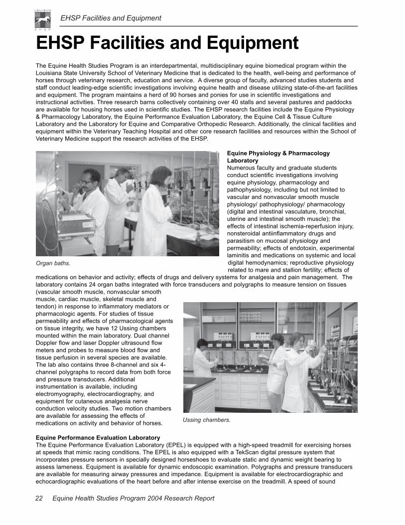
22 Equine Health Studies Program 2004 Research Report
EHSP Facilities and Equipment
EHSP Facilities and EquipmentThe Equine Health Studies Program is an interdepartmental, multidisciplinary equine biomedical program within theLouisiana State University School of Veterinary Medicine that is dedicated to the health, well-being and performance ofhorses through veterinary research, education and service. A diverse group of faculty, advanced studies students andstaff conduct leading-edge scientific investigations involving equine health and disease utilizing state-of-the-art facilitiesand equipment. The program maintains a herd of 90 horses and ponies for use in scientific investigations andinstructional activities. Three research barns collectively containing over 40 stalls and several pastures and paddocksare available for housing horses used in scientific studies. The EHSP research facilities include the Equine Physiology& Pharmacology Laboratory, the Equine Performance Evaluation Laboratory, the Equine Cell & Tissue CultureLaboratory and the Laboratory for Equine and Comparative Orthopedic Research. Additionally, the clinical facilities andequipment within the Veterinary Teaching Hospital and other core research facilities and resources within the School ofVeterinary Medicine support the research activities of the EHSP.
Equine Physiology & PharmacologyLaboratoryNumerous faculty and graduate studentsconduct scientific investigations involvingequine physiology, pharmacology andpathophysiology, including but not limited tovascular and nonvascular smooth musclephysiology/ pathophysiology/ pharmacology(digital and intestinal vasculature, bronchial,uterine and intestinal smooth muscle); theeffects of intestinal ischemia-reperfusion injury,nonsteroidal antiinflammatory drugs andparasitism on mucosal physiology andpermeability; effects of endotoxin, experimentallaminitis and medications on systemic and localdigital hemodynamics; reproductive physiologyrelated to mare and stallion fertility; effects of
medications on behavior and activity; effects of drugs and delivery systems for analgesia and pain management. Thelaboratory contains 24 organ baths integrated with force transducers and polygraphs to measure tension on tissues(vascular smooth muscle, nonvascular smoothmuscle, cardiac muscle, skeletal muscle andtendon) in response to inflammatory mediators orpharmacologic agents. For studies of tissuepermeability and effects of pharmacological agentson tissue integrity, we have 12 Ussing chambersmounted within the main laboratory. Dual channelDoppler flow and laser Doppler ultrasound flowmeters and probes to measure blood flow andtissue perfusion in several species are available.The lab also contains three 8-channel and six 4-channel polygraphs to record data from both forceand pressure transducers. Additionalinstrumentation is available, includingelectromyography, electrocardiography, andequipment for cutaneous analgesia nerveconduction velocity studies. Two motion chambersare available for assessing the effects ofmedications on activity and behavior of horses.
Equine Performance Evaluation LaboratoryThe Equine Performance Evaluation Laboratory (EPEL) is equipped with a high-speed treadmill for exercising horsesat speeds that mimic racing conditions. The EPEL is also equipped with a TekScan digital pressure system thatincorporates pressure sensors in specially designed horseshoes to evaluate static and dynamic weight bearing toassess lameness. Equipment is available for dynamic endoscopic examination. Polygraphs and pressure transducersare available for measuring airway pressures and impedance. Equipment is available for electrocardiographic andechocardiographic evaluations of the heart before and after intense exercise on the treadmill. A speed of sound
Organ baths.
Ussing chambers.
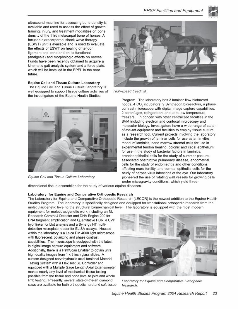
Equine Health Studies Program 2004 Research Report 23
EHSP Facilities and Equipment
ultrasound machine for assessing bone density isavailable and used to assess the effect of growth,training, injury, and treatment modalities on bonedensity of the third metacarpal bone of horses. Afocused extracorporeal shock wave therapy(ESWT) unit is available and is used to evaluatethe effects of ESWT on healing of tendon,ligament and bone and on its functional(analgesia) and morphologic effects on nerves.Funds have been recently obtained to acquire akinematic gait analysis system and a force plate,which will be installed in the EPEL in the nearfuture.
Equine Cell and Tissue Culture LaboratoryThe Equine Cell and Tissue Culture Laboratory iswell equipped to support tissue culture activities ofthe investigators of the Equine Health Studies
Program. The laboratory has 3 laminar flow biohazardhoods, 4 CO2 incubators, 9 Synthecon bioreactors, a phasecontrast microscope with digital image capture capabilities,2 centrifuges, refrigerators and ultra-low temperaturefreezers. In concert with other centralized faculties in theSVM including electron and confocal microscopy andmolecular biology, investigators have a wide range of state-of-the-art equipment and facilities to employ tissue cultureas a research tool. Current projects involving the laboratoryinclude the growth of laminar cells for use as an in vitromodel of laminitis, bone marrow stromal cells for use inexperimental tendon healing, colonic and cecal epitheliumfor use in the study of bacterial factors in laminitis,bronchoepithelial cells for the study of summer pasture-associated obstructive pulmonary disease, endometrialcells for the study of endometritis and other conditionsaffecting mare fertility, and corneal epithelial cells for thestudy of herpes virus infections of the eye. Our laboratorypioneered the use of rotating wall vessels for growing cellsunder microgravity conditions, which yield three-
dimensional tissue assemblies for the study of various equine diseases.
Laboratory for Equine and Comparative Orthopedic ResearchThe Laboratory for Equine and Comparative Orthopedic Research (LECOR) is the newest addition to the Equine HealthStudies Program. The laboratory is specifically designed and equipped for translational orthopedic research from themolecular/genetic level to the structural biomechanical level. The laboratory is equipped with the most modernequipment for molecular/genetic work including an MJResearch Chromo4 Detector and DNA Engine 200 forDNA fragment amplification and Quantitative PCR, a UVPhybrilinker for blot analysis and a Synergy HT multi-detection microplate reader for ELISA assays. Housedwithin the laboratory is a Leica DM 4000 light microscopewith fluorescent, polarizing and phase contrastcapabilities. The microscope is equipped with the latestin digital image capture equipment and software.Additionally, there is a PathScan Enabler to obtain ultrahigh quality images from 1 x 3 inch glass slides. Acustom-designed servohydraulic axial torsional MaterialTesting System with a Flex Test SE Controller andequipped with a Multiple Gage Length Axial Extensometermakes nearly any level of mechanical tissue testingpossible from the tissue and bone level to joint and wholelimb testing. Presently, several state-of-the-art diamondsaws are available for both orthopedic hard and soft tissue
High-speed treadmill.
Equine Cell and Tissue Culture Laboratory.
Laboratory for Equine and Comparative OrthopedicResearch.
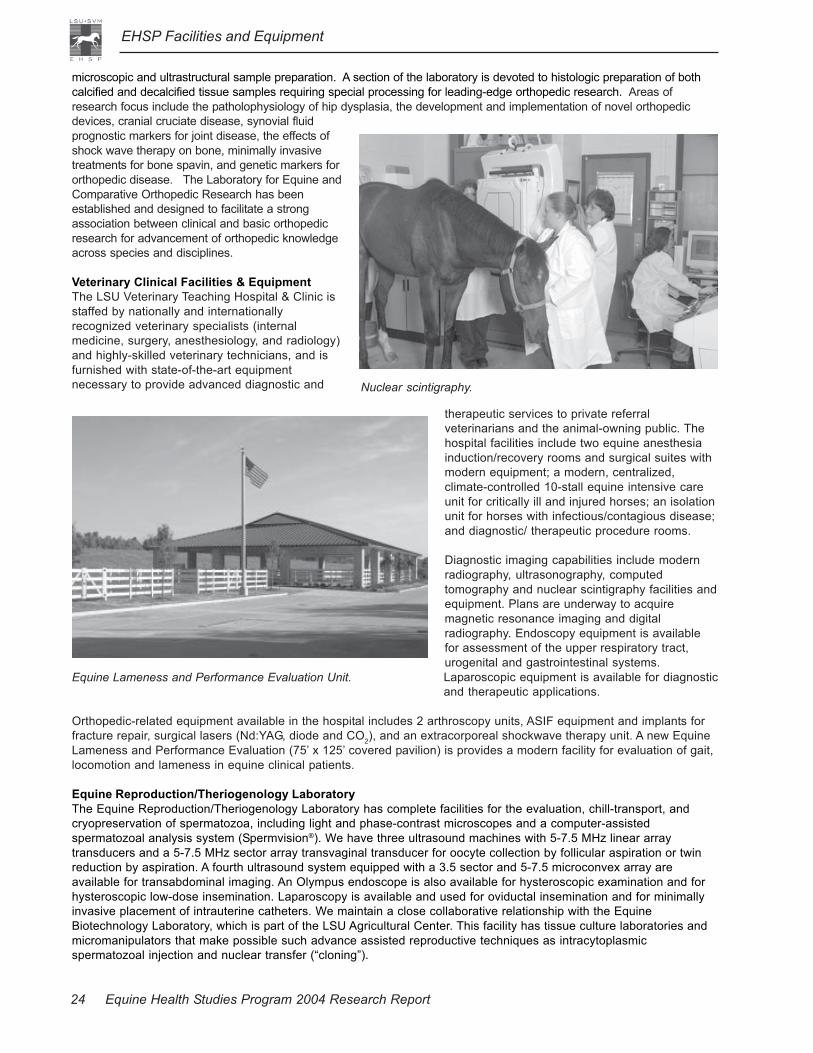
24 Equine Health Studies Program 2004 Research Report
EHSP Facilities and Equipment
microscopic and ultrastructural sample preparation. A section of the laboratory is devoted to histologic preparation of bothcalcified and decalcified tissue samples requiring special processing for leading-edge orthopedic research. Areas ofresearch focus include the patholophysiology of hip dysplasia, the development and implementation of novel orthopedicdevices, cranial cruciate disease, synovial fluidprognostic markers for joint disease, the effects ofshock wave therapy on bone, minimally invasivetreatments for bone spavin, and genetic markers fororthopedic disease. The Laboratory for Equine andComparative Orthopedic Research has beenestablished and designed to facilitate a strongassociation between clinical and basic orthopedicresearch for advancement of orthopedic knowledgeacross species and disciplines.
Veterinary Clinical Facilities & EquipmentThe LSU Veterinary Teaching Hospital & Clinic isstaffed by nationally and internationallyrecognized veterinary specialists (internalmedicine, surgery, anesthesiology, and radiology)and highly-skilled veterinary technicians, and isfurnished with state-of-the-art equipmentnecessary to provide advanced diagnostic and
therapeutic services to private referralveterinarians and the animal-owning public. Thehospital facilities include two equine anesthesiainduction/recovery rooms and surgical suites withmodern equipment; a modern, centralized,climate-controlled 10-stall equine intensive careunit for critically ill and injured horses; an isolationunit for horses with infectious/contagious disease;and diagnostic/ therapeutic procedure rooms.
Diagnostic imaging capabilities include modernradiography, ultrasonography, computedtomography and nuclear scintigraphy facilities andequipment. Plans are underway to acquiremagnetic resonance imaging and digitalradiography. Endoscopy equipment is availablefor assessment of the upper respiratory tract,urogenital and gastrointestinal systems.Laparoscopic equipment is available for diagnosticand therapeutic applications.
Orthopedic-related equipment available in the hospital includes 2 arthroscopy units, ASIF equipment and implants forfracture repair, surgical lasers (Nd:YAG, diode and CO2), and an extracorporeal shockwave therapy unit. A new EquineLameness and Performance Evaluation (75’ x 125’ covered pavilion) is provides a modern facility for evaluation of gait,locomotion and lameness in equine clinical patients.
Equine Reproduction/Theriogenology LaboratoryThe Equine Reproduction/Theriogenology Laboratory has complete facilities for the evaluation, chill-transport, andcryopreservation of spermatozoa, including light and phase-contrast microscopes and a computer-assistedspermatozoal analysis system (Spermvision®). We have three ultrasound machines with 5-7.5 MHz linear arraytransducers and a 5-7.5 MHz sector array transvaginal transducer for oocyte collection by follicular aspiration or twinreduction by aspiration. A fourth ultrasound system equipped with a 3.5 sector and 5-7.5 microconvex array areavailable for transabdominal imaging. An Olympus endoscope is also available for hysteroscopic examination and forhysteroscopic low-dose insemination. Laparoscopy is available and used for oviductal insemination and for minimallyinvasive placement of intrauterine catheters. We maintain a close collaborative relationship with the EquineBiotechnology Laboratory, which is part of the LSU Agricultural Center. This facility has tissue culture laboratories andmicromanipulators that make possible such advance assisted reproductive techniques as intracytoplasmicspermatozoal injection and nuclear transfer (“cloning”).
Nuclear scintigraphy.
Equine Lameness and Performance Evaluation Unit.

Equine Health Studies Program 2004 Research Report 25
BIOMMED – Biotechnology and Molecular MedicineThe Division of Biotechnology and Molecular Medicine (BIOMMED), a division within the LSU SVM, is organized intothree different Laboratories: 1) GeneLab; 2) Viral Vectors Laboratory (VVL); 3) Protein and Antibody Production andPurification Laboratory (PAPPL). GENELAB: GeneLab produces synthetic oligonucleotides including biotinylated,fluoresceinated, phosphorylated and phosphorothioate (antisense) oligonucleotides up to 200-bases long. Additionalmolecular biology services include cloning and automated sequencing of genes, real-time quantitative PCR, automatedpreparation of chromosomal and plasmid DNAs, cDNA library construction, and microarray production and analysis.VVL: This laboratory provides custom baculovirus, adenovirus, vaccinia virus, herpes, and other recombinant virusconstruction for heterologous gene expression, and vaccine and gene therapy studies. PAPPL: This laboratoryconcentrates on the production and purification of proteins and antibodies. The laboratory produces monospecificantibodies in rabbits and mice using conventional immunization methodologies using purified protein immunogens aswell as genetic immunization methods. Antibodies are concentrated and purified using standard methodologies. Thelaboratory also provides on a limited basis the production and characterization of monoclonal antibodies.
GeneLab operates a new bioinformatics module, which is equipped with three Macintosh G4 computers and two DELLPC computers. A new WEB-based system direct communication between researchers and GeneLab staff has beenrecently purchased. Available software include: Primer Express (PE Biosystems) for the design of TaqMan probes forreal-time PCR, MacVector (Genetics Computer Group, Inc.) for analysis of DNA and protein sequences, Oligo(Molecular Biology Insights, Inc.) for the design of PCR primers, Quantity One (BioRad, Inc.) for the visualization andanalysis of images such as those produced by ethidium bromide agarose electrophoresis of DNA fragments, SDS-PAGE analysis of proteins, immunoblots, etc. GeneLab has also additional software for assembly of large DNAsequences (Sequencher), analysis of blots (alpha Innotech) and microarrays (Alpha Innotech 6000). Automatedordering for synthetic DNA and other reagents is assisted via a WEB-based Information System (DNA LIMS).BIOMMED has 3 automated sequences (ABI377, 310, 310), three real-time PCR equipment (PE 7900, 7400),LightCycler (Roche), 2 MagnaPure automated nucleic acid extractors (Roche), microarray OmniGrid spotter (GeneMachines), microarray reader (AlphaReader 6000, Alpha Innotech), Imager station (Alpha Innotech Fluorochem 8000),5 PCR (thermocyclers, a New Brunswick Fermentor, AktaExplorer Chromatography System, Cyclone phosphorimager,2 four column DNA synthesizers (ABI), one Synergy peptide synthesizer (ABI), and other equipment.
Immunology LaboratoryThree laboratories (~1200 sq ft) located on the third floor of the School of Veterinary Medicine (SVM) are designatedfor the Immunology Laboratory. These labs contain necessary equipment for immunological assays and the in vitrocultivation of lymphocytes, including; laminar flow biosafety cabinets, CO2 incubators, microscopes, water baths, a pHmeter, low speed centrifuges, mixers, stir plates, refrigerators and freezers. One of the laboratories is dedicatedspecifically for molecular biology procedures and contains all of the equipment and materials for the isolation of andanalysis of RNA, DNA and proteins. Separate refrigerators and freezers for molecular biology samples are found in thislaboratory. Spectrophotometers, pH meters, electronic balances, refrigerators and freezer and other small equipmentitems are also available in this laboratory.
The Flow Cytometry Facility is a core laboratory located on the third floor of the School of Veterinary Medicine buildingon the campus of Louisiana State University. The facility features a Becton Dickinson FACScan flow cytometer capableof measuring two light scatter parameters and three fluorescence emissions. Immunophenotyping, cell cycle analysis,apoptosis studies, and measurements of cellular function are examples of applications, which are performed routinelyin this laboratory. A newly acquired FACS Aria Dual Laser Flow Cytometer, which is capable of high performance cellsorting of up to 30,000 cells per second and separation of 1 to 4 distinct cell populations. Additionally, multicolorimmunophenotyping and cell functional assayscan expand to seven-color analysis. This uniquecentralized facility provides analytical capabilitiesfor investigators throughout the Louisiana StateUniversity System, including the LSU AgriculturalCenter and LSU Pennington Biomedical ResearchCenter. Both PC and Macintosh computers areutilized in data acquisition and analysis and are allconnected to the Veterinary Medicine and LSUnetworks.
Microscopy CenterThe SVM Microscopy Center has state-of-the-artequipment for light and electron microscopy,including: Light Microscopes: Zeiss Axioplan,Zeiss Axiovert with heated stage, ZeissPhotomicroscope III, Olympus Vanox, PALM
EHSP Facilities and Equipment
Laser Scanning Confocal Microscope.

26 Equine Health Studies Program 2004 Research Report
Laser Microdissection System, Laser Scanning Confocal Microscope. Electron Microscopes: FEI Quanta 200 ESEM,Zeiss EM-10C TEM. Cryo: Balzers 400K Freeze Fracture Device, Balzers JFD30 Propane Jet Freezer, Balzers FTD010Plunge Freezer, Balzers FSU010 Freeze Substitution. Camera systems: Dage 330C, Spot RT, Nikon CoolPix 5000,Nikon CoolPix 995, Hamamatsu 2400 Video. Image capture systems: Scion Image, IP Lab, Panasonic GX4 VCRs andEditors. Ancillary equipment: Ultramicrotomes, Vacuum Evaporators, Critical Point Apparatus, DC Sputtering Devices.Image Manipulation Hardware: Imaging Workstations (2), Wacom 8x12 Graphics Tablet, Wacom Graphire 3x4 Tablet,Image Manipulation Software: IP Lab, Scion Image, Image Pro +, SigmaPlot, VoxBlast, Corel PhotoPaint, QuatroPro,Photoshop 7, IP Toolkit, Fovea Pro, PowerPoint, HazeBuster, and CorelDraw.
The Center features three new powerful microscopes: The laser capture and microdissection microscope (PALMZeissMicroBeam-Axiovert 200 System) allows researchers to dissect out parts of tissue on a slide and transfer it to acontainer for genetic, gene expression and proteomic analysis, which enables researchers to determine what genesare present and what genes and proteins are being expressed. The scanning laser confocal microscope (LEICA TCSSP2 AOBS) provides excellent quality three-dimensional reconstructions from cells and relatively thick sections oftissues, which enable researchers to examine cells and cell components in three-dimension and allows researchers toconduct co-localization studies to mark multiple proteins within the cell simultaneously. The environmental scanningelectron microscope (FEI Quanta 200) provides a detailed evaluation of the surfaces with or without dehydrating thesamples. This microscope also has an energy dispersive x-ray spectrometer that allows researchers to determineelemental composition.
Analytical Systems LaboratoriesThe Analytical Systems Laboratories arecentral service, comprehensive analyticallaboratories, consisting of The Laboratory forDrug Residue Studies, The Equine MedicationSurveillance Laboratory and The AnalyticalSystems Laboratory. The Laboratory for DrugResidue Studies provides instrumentation andexpertise for the performance of drug andbiological molecule pharmacokinetics,metabolism, tissue distribution and analyticalmethod development. The laboratory is alsoequipped to conduct complete drug profilingusing radiolabeled test materials. Theselaboratories operate under Federal GoodLaboratory Practices regulations and havegenerated data for the Food and DrugAdministration (FDA) and private industry forsubmission for veterinary drug approvals byU.S. and foreign regulatory agencies. TheEquine Medication Surveillance Laboratory hasserved as the official laboratory for theLouisiana State Racing Commission. The laboratory screens over 10,000 urine and blood samples per year and hasdeveloped sophisticated methodology for detection and confirmation of drugs and their metabolites. The laboratoryalso conducts illegal drug-use testing for the LSU Department of Athletics and serves as a source of information to theracing industry and the public regarding drug pharmacology, metabolism and clearance. The Analytical SystemsLaboratory houses advanced mass spectrometry and other analytical equipment that is used to support otherlaboratories as well as the research of SVM faculty and graduate students. Equipment in these laboratories includes aMicromass Quattro II GC/LC/MS/MS (+/-) with ApcI and ESP interfaces, one HP 1090 II, low-flow HPLC (1 ul/min),three 1090 HPLCs equipped with UV-diode array, fluorescence, electrochemical and radio-monitor detectors, a HP5973 GC/MS system and beta and gamma counters for radiolabel analyses. The laboratory is currently developing fullcapabilities to conduct low- and high-throughput proteomics analysis and is enhancing its abilities to conduct small andlarge-scale protein purification. Appropriate and modern computer equipment and software is available for dataacquisition, storage, and analysis. ASL: Hewlett-Packard 5970 MSD GC/MS system (ChemStation data system), three(3) Hewlett-Packard HP 1090 HPLCs (UV diode array, electrochemical, fluorescence and radiochemical detectors);Fisons VG Quatro II GC/LC/MS/MS +/- ion, extended mass range system, electrospray and APcI interfaces, HP5980GC, HP 1090 II LC. The ASL recently acquired a Micromass Two-Dimensional-Capillary-Liquid Chormatography/Quadrupole-Time of Flight Mass Spectrometer, which enables separation and comprehensive structural analysis ofproteins. The instrument is capable of conducting de novo sequencing of proteins and peptides as well as identifyingand locating post-translational modifications. This type of equipment is essential for the rapidly growing fields ofproteomics and bioinformatics for comprehensive examination of molecular events occurring in tissues in health anddisease. This equipment is available for collaboration across the LSU campus and supports the research efforts of the
EHSP Facilities and Equipment
Micromass Two-Dimensional-Capillary-Liquid Chromatography/Quadrupole-Time of Flight Mass Spectrometer.
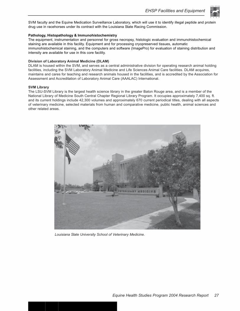
Equine Health Studies Program 2004 Research Report 27
EHSP Facilities and Equipment
SVM faculty and the Equine Medication Surveillance Laboratory, which will use it to identify illegal peptide and proteindrug use in racehorses under its contract with the Louisiana State Racing Commission.
Pathology, Histopathology & ImmunohistochemistryThe equipment, instrumentation and personnel for gross necropsy, histologic evaluation and immunohistochemicalstaining are available in this facility. Equipment and for processing cryopreserved tissues, automaticimmunohistochemical staining, and the computers and software (ImagePro) for evaluation of staining distribution andintensity are available for use in this core facility.
Division of Laboratory Animal Medicine (DLAM)DLAM is housed within the SVM, and serves as a central administrative division for operating research animal holdingfacilities, including the SVM Laboratory Animal Medicine and Life Sciences Animal Care facilities. DLAM acquires,maintains and cares for teaching and research animals housed in the facilities, and is accredited by the Association forAssessment and Accreditation of Laboratory Animal Care (AAALAC) International.
SVM LibraryThe LSU-SVM Library is the largest health science library in the greater Baton Rouge area, and is a member of theNational Library of Medicine South Central Chapter Regional Library Program. It occupies approximately 7,400 sq. ft.and its current holdings include 42,300 volumes and approximately 670 current periodical titles, dealing with all aspectsof veterinary medicine, selected materials from human and comparative medicine, public health, animal sciences andother related areas.
Louisiana State University School of Veterinary Medicine.
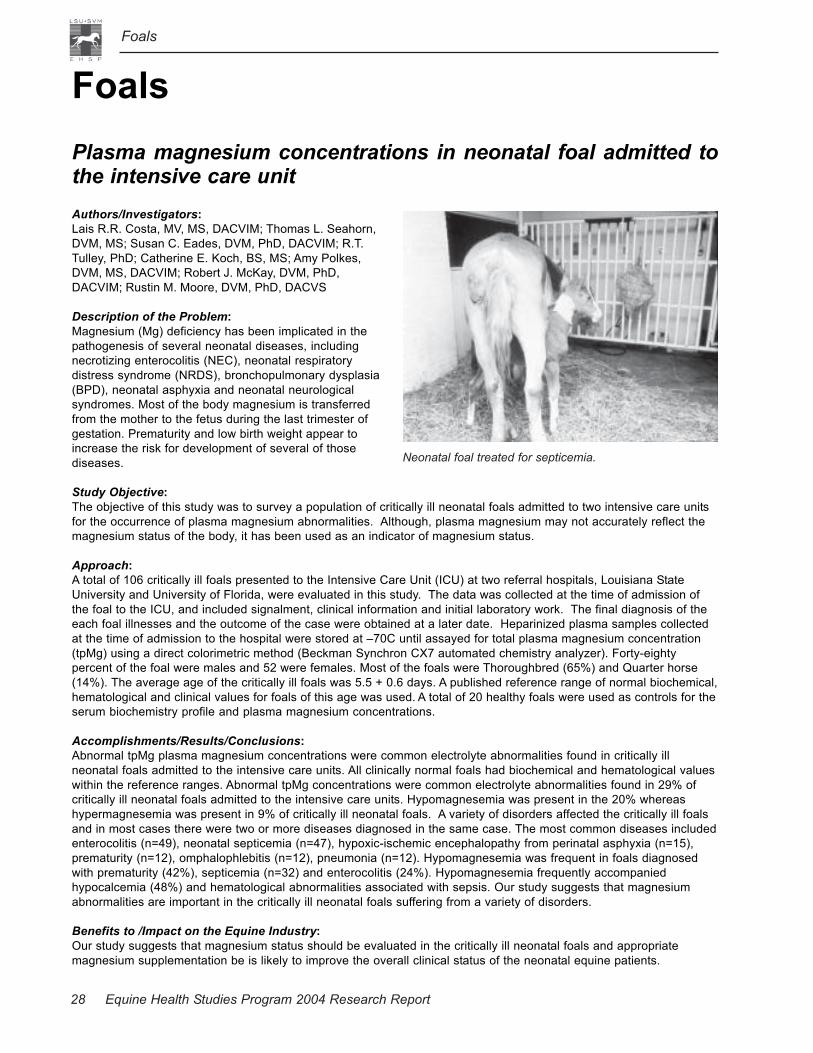
28 Equine Health Studies Program 2004 Research Report
FoalsPlasma magnesium concentrations in neonatal foal admitted tothe intensive care unitAuthors/Investigators:Lais R.R. Costa, MV, MS, DACVIM; Thomas L. Seahorn,DVM, MS; Susan C. Eades, DVM, PhD, DACVIM; R.T.Tulley, PhD; Catherine E. Koch, BS, MS; Amy Polkes,DVM, MS, DACVIM; Robert J. McKay, DVM, PhD,DACVIM; Rustin M. Moore, DVM, PhD, DACVS
Description of the Problem:Magnesium (Mg) deficiency has been implicated in thepathogenesis of several neonatal diseases, includingnecrotizing enterocolitis (NEC), neonatal respiratorydistress syndrome (NRDS), bronchopulmonary dysplasia(BPD), neonatal asphyxia and neonatal neurologicalsyndromes. Most of the body magnesium is transferredfrom the mother to the fetus during the last trimester ofgestation. Prematurity and low birth weight appear toincrease the risk for development of several of thosediseases.
Study Objective:The objective of this study was to survey a population of critically ill neonatal foals admitted to two intensive care unitsfor the occurrence of plasma magnesium abnormalities. Although, plasma magnesium may not accurately reflect themagnesium status of the body, it has been used as an indicator of magnesium status.
Approach:A total of 106 critically ill foals presented to the Intensive Care Unit (ICU) at two referral hospitals, Louisiana StateUniversity and University of Florida, were evaluated in this study. The data was collected at the time of admission ofthe foal to the ICU, and included signalment, clinical information and initial laboratory work. The final diagnosis of theeach foal illnesses and the outcome of the case were obtained at a later date. Heparinized plasma samples collectedat the time of admission to the hospital were stored at –70C until assayed for total plasma magnesium concentration(tpMg) using a direct colorimetric method (Beckman Synchron CX7 automated chemistry analyzer). Forty-eightypercent of the foal were males and 52 were females. Most of the foals were Thoroughbred (65%) and Quarter horse(14%). The average age of the critically ill foals was 5.5 + 0.6 days. A published reference range of normal biochemical,hematological and clinical values for foals of this age was used. A total of 20 healthy foals were used as controls for theserum biochemistry profile and plasma magnesium concentrations.
Accomplishments/Results/Conclusions:Abnormal tpMg plasma magnesium concentrations were common electrolyte abnormalities found in critically illneonatal foals admitted to the intensive care units. All clinically normal foals had biochemical and hematological valueswithin the reference ranges. Abnormal tpMg concentrations were common electrolyte abnormalities found in 29% ofcritically ill neonatal foals admitted to the intensive care units. Hypomagnesemia was present in the 20% whereashypermagnesemia was present in 9% of critically ill neonatal foals. A variety of disorders affected the critically ill foalsand in most cases there were two or more diseases diagnosed in the same case. The most common diseases includedenterocolitis (n=49), neonatal septicemia (n=47), hypoxic-ischemic encephalopathy from perinatal asphyxia (n=15),prematurity (n=12), omphalophlebitis (n=12), pneumonia (n=12). Hypomagnesemia was frequent in foals diagnosedwith prematurity (42%), septicemia (n=32) and enterocolitis (24%). Hypomagnesemia frequently accompaniedhypocalcemia (48%) and hematological abnormalities associated with sepsis. Our study suggests that magnesiumabnormalities are important in the critically ill neonatal foals suffering from a variety of disorders.
Benefits to /Impact on the Equine Industry:Our study suggests that magnesium status should be evaluated in the critically ill neonatal foals and appropriatemagnesium supplementation be is likely to improve the overall clinical status of the neonatal equine patients.
Foals
Neonatal foal treated for septicemia.
Equine Health Studies Program 2004 Research Report 29
Take Home Message:Magnesium abnormalities should be part of the biochemical evaluation of neonatal foals and correction of magnesiumabnormalities should be part of the treatment of these foals.
Acknowledgments:This study was supported by funds from the Equine Health Studies Program, School of Veterinary Medicine, LouisianaState University.
Year Completed: 2000
Published Manuscripts/Abstracts:Costa LRR, Eades SC, Tulley RT, Koch CE, Polkes A, Bain F, Moore RM. Plasma magnesium concentrations inneonatal foals admitted to the intensive care unit. Advances in Magnesium Research: Nutrition and Health. Eastleigh:John Libbey & Co, 2001;487-490.
Costa LRR, Eades SC, Tulley RT, Koch CE, Polkes A, Bain F, Moore RM. Plasma magnesium concentrations inneonatal foals admitted to the intensive care unit. Mag Bull 2000; 22 (4); Mag Res 2001;14 (1/2); 127-128. 9th
International Congress of Magnesium 2000.
Foals
30 Equine Health Studies Program 2004 Research Report
Gastrointestinal TractIn vitro effects of oxytocin, acepromazine, detomidine, xylazine,butorphanol, terbutaline, isoproterenol, and dantrolene onsmooth and skeletal muscles of the equine esophagusAuthors/Investigators:Anne A. Wooldridge, DVM, MS, DACVIM; Susan C. Eades, DVM, PhD, DACVIM; Giselle Hosgood, BVSc, MS, PhD,DACVS; Rustin M. Moore, DVM, PhD, DACVS
Description of the Problem:Obstruction of the esophagus (choke) is a common problem in horses that most often results from impaction of foodmaterial preventing passage of feed material and water through the esophagus to the stomach. This results inregurgitation of food, water, and saliva from the nose and pain manifest as stretching the neck and gagging. Affectedhorses may cough, develop fever, and exhibit colic. The food impaction distends the esophagus causing pain andspasm of the esophageal muscles. The primary goal of treatment is to relieve the obstruction by gentle pressure andwarm water lavage via a nasogastric tube. Esophageal spasm impedes these efforts at removing the impacted food.Medications are often administered to relax the esophagus and reduce esophageal spasm; however, the efficacy ofthese medications has not been demonstrated.
Study Objective:The objective of this study was to characterize the in vitro effects of oxytocin, acepromazine, xylazine, butorphanol,detomidine, dantrolene, isoproterenol, and terbutaline on skeletal and smooth muscle from the equine esophagus.
Approach:Circular and longitudinal strips from the skeletal and smooth muscle of the esophagus were suspended in tissue baths,connected to force-displacement transducers interfaced with a physiograph, and electrical field stimulation wasapplied. Cumulative concentration-response curves were generated for oxytocin, acepromazine, xylazine, detomidine,butorphanol, isoproterenol, terbutaline, and dantrolene. Mean maximum twitch amplitude for 3 contractions/min wasrecorded and compared with predrug-vehicle values for the skeletal muscle segments, and area under the curve (AUC)for 3 contractions/min was compared with predrug-vehicle values for the smooth muscle segments.
Accomplishments/Results/Conclusions:No drugs caused a significant change in skeletal muscle response. In smooth muscle, isoproterenol, terbutaline, andoxytocin significantly reduced AUC in a concentration-dependent manner.
Benefits to/Impact on the Equine Industry:Isoproterenol, terbutaline, and oxytocin cause relaxation of the smooth muscle portion of the esophagus. However,none of the medications affected the skeletal muscle strips.
Take Home Message:These medications cannot cause enough relaxation of the esophagus to be relied upon heavily for removal ofesophageal obstruction in horses.
Acknowledgments: This study was supported by funds from the Equine Health Studies Program, School of VeterinaryMedicine, Louisiana State University.
Year Completed: 2000
Published Manuscripts/Abstracts:Wooldridge AA, Eades SC, Moore RM. Effect of oxytocin, acepromazine, isoproterenol, xylazine, and dantrolene onelectrical field stimulated smooth and striated esophageal muscle of horses. J Vet Intern Med 1999;13(3) 229.
Wooldridge AA, Eades SC, Moore RM. The in vitro effects of oxytocin, acepromazine, detomidine, xylazine,butorphanol, terbutaline, isoproterenol, and dantrolene on smooth and skeletal muscles of the equine esophagus. AmJ Vet Res 2002;63:1732-1737.
Gastrointestinal Tract
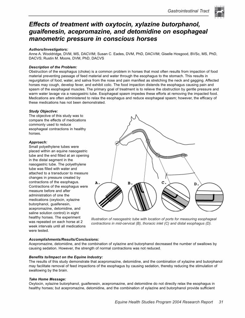
Equine Health Studies Program 2004 Research Report 31
Effects of treatment with oxytocin, xylazine butorphanol,guaifenesin, acepromazine, and detomidine on esophagealmanometric pressure in conscious horsesAuthors/Investigators:Anne A. Wooldridge, DVM, MS, DACVIM; Susan C. Eades, DVM, PhD, DACVIM; Giselle Hosgood, BVSc, MS, PhD,DACVS; Rustin M. Moore, DVM, PhD, DACVS
Description of the Problem:Obstruction of the esophagus (choke) is a common problem in horses that most often results from impaction of foodmaterial preventing passage of feed material and water through the esophagus to the stomach. This results inregurgitation of food, water, and saliva from the nose and pain manifest as stretching the neck and gagging. Affectedhorses may cough, develop fever, and exhibit colic. The food impaction distends the esophagus causing pain andspasm of the esophageal muscles. The primary goal of treatment is to relieve the obstruction by gentle pressure andwarm water lavage via a nasogastric tube. Esophageal spasm impedes these efforts at removing the impacted food.Medications are often administered to relax the esophagus and reduce esophageal spasm; however, the efficacy ofthese medications has not been demonstrated.
Study Objective:The objective of this study was tocompare the effects of medicationscommonly used to reduceesophageal contractions in healthyhorses.
Approach:Small polyethylene tubes wereplaced within an equine nasogastrictube and the end fitted at an openingin the distal segment in thenasogastric tube. The polyethylenetube was filled with water andattached to a transducer to measurechanges in pressure created bycontractions of the esophagus.Contractions of the esophagus weremeasure before and afteradministration of one themedications (oxytocin, xylazinebutorphanol, guaifenesin,acepromazine, detomidine, andsaline solution control) in eighthealthy horses. The experimentwas repeated on each horse at 2week intervals until all medicationswere tested.
Accomplishments/Results/Conclusions:Acepromazine, detomidine, and the combination of xylazine and butorphanol decreased the number of swallows bycausing sedation. However, the strength of normal contractions was not reduced.
Benefits to/Impact on the Equine Industry:The results of this study demonstrate that acepromazine, detomidine, and the combination of xylazine and butorphanolmay facilitate removal of feed impactions of the esophagus by causing sedation, thereby reducing the stimulation ofswallowing by the brain.
Take Home Message:Oxytocin, xylazine butorphanol, guaifenesin, acepromazine, and detomidine do not directly relax the esophagus inhealthy horses; but acepromazine, detomidine, and the combination of xylazine and butorphanol provide sufficient
Gastrointestinal Tract
Illustration of nasogastric tube with location of ports for measuring esophagealcontractions in mid-cervical (B), thoracic inlet (C) and distal esophagus (D).
32 Equine Health Studies Program 2004 Research Report
sedation to reduce the stimulation of swallows by the brain, thereby indirectly decreasing the contraction of theesophagus. This allows feed impactions to be flushed out with water via a nasogastric tube more easily.
Acknowledgments:This study was supported by funds from the Equine Health Studies Program, School of Veterinary Medicine, LouisianaState University.
Year Completed: 2000
Published Manuscripts/Abstracts:Wooldridge AA, Eades SC, Hosgood GL, Moore RM. Effects of treatment with oxytocin, xylazine butorphanol,guaifenesin, acepromazine, and detomidine on esophageal manometric pressure in conscious horses. Am J Vet Res2002;63:1738-44.
Wooldridge AA, Eades SC, Moore RM, Hosgood G. Effects of oxytocin, xylazine/butorphanol combination, guaifenesin,acepromazine, and detomidine on oesophageal manometry in conscious horses, in Proceedings. 7th InternationalEquine Colic Research Symposium 2002; 7:49.
Plasma magnesium concentrations in critically ill horsesaffected with gastrointestinal tract diseaseAuthors/Investigators:Lais R.R. Costa, MV, MS, DACVIM; Susan C. Eades, DVM, PhD, DACVIM; R.T. Tulley, PhD; Catherine E. Koch, BS,MS; Giselle Hosgood, DVM, PhD, DACVS; Rustin M. Moore, DVM, PhD, DACVS
Description of the Problem:Horses affected with acute gastrointestinal tract diseases are often dehydrated and have a variety of severe electrolyteabnormalities. Plasma or serum magnesium (Mg) concentration is likely affected, because its homeostasis resultsprincipally from intestinal absorption and renal excretion. However, the incidence of hypomagnesemia in critically illhorses affected with gastrointestinal tract disorders remains unreported.
Study Objective:The objective of this study was to survey a subpopulation of critically ill horses affected with gastrointestinal tractdisease to determine the incidence of plasma magnesium abnormalities and concurrent plasma electrolytederangements.
Approach:A total of 155 critically ill horses with gastrointestinal tract (GIT) disease referred to the Intensive Care Unit (ICU) atLouisiana State University Veterinary Teaching Hospital and Clinics were evaluated. The data collected at the time ofadmission to the ICU included signalment, initial clinical information and laboratory work. The final diagnosis and theoutcome of each case were obtained at a later date. The GIT disorders were categorized in 5 groups: (1) smallintestinal strangulation (n=34) which included incarcerations in the epiploic foramen and in a mesenteric rent,strangulating lipoma and volvulus; (2) large intestinal strangulation (n=22) which included large colon volvulus; (3)small intestinal inflammatory disease (n=21) which included duodenitis-proximal jejunitis and all other causes ofenteritis; (4) large intestinal inflammatory disease (n=27) which included colitis and enterocolitis; and (5) simpleobstruction of the large and small intestines (n=51) which included cases of impactions, enteroliths, fecaliths,intraluminal foreign bodies and non-strangulating large colon displacements. The biochemical variables evaluatedincluded: sodium, potassium, chloride, calcium, creatinine, BUN, and total protein. Heparinized plasma samples werecollected and stored at –70°C at the time of hospital admission, and total plasma magnesium concentration (tpMg) wasmeasured using a direct colorimetric method (Beckman Synchron CX7 automated chemistry analyzer). Twentyclinically healthy horses were used to validate the colorimetric test used for Mg quantification.
Accomplishments/Results/Conclusions:Abnormalities of plasma magnesium concentration were the most frequent electrolyte changes in plasma of critically illhorses with GIT disease. The incidence of hypomagnesemia was greatest in horses with small intestinal strangulation,colitis and simple intestinal obstructions, respectively. Several electrolyte abnormalities occurred concurrently withabnormalities in plasma Mg concentration. Hypocalcemia and hypokalemia were the most common concurrentelectrolyte abnormalities in hypomagnesemic ill horses. There were no statistical differences for the plasma Mgconcentrations among groups. Plasma concentrations of chloride, BUN, and PCV were significantly associated with
Gastrointestinal Tract

Equine Health Studies Program 2004 Research Report 33
In vitro effects of an electron transport inhibitor on resistance,permeability, and adenine nucleotide content of equine colonicmucosal tissueAuthors/Investigators:Joanne Tetens, DVM, MS, PhD; DACVS; Giselle Hosgood, BVSc, MS, PhD,DACVS; Steven A. Barker, PhD; Catherine E. Koch, MA; Marian Waguespack,BS; Priti Juneja, MS
Description of the Problem:Because gastrointestinal mucosal cells are highly metabolically active and requirelarge quantities of ATP for maintenance of normal cellular function, the mucosallayer is principally affected during ischemia. Increased cellular permeability,which occurs during ischemia and hypoxia, leads to translocation of bacteriaand endotoxin from the gastrointestinal tract lumen into the splanchnic andsystemic circulation. One mechanism that may be involved in the increase ingastrointestinal mucosal permeability during ischemia is decreased endogenoussynthesis of adenosine triphosphate (ATP). The epithelial intercellularpermeability barrier is maintained largely by the tight junction (TJ). In vitro studieshave demonstrated that a correlation exists between cellular ATP content andTJ integrity. During ascending colon volvulus in horses, mucosal ATP contentdiminishes and does not fully recover upon reperfusion. The depletion of mucosalATP content may lead to rapid opening to TJs and increased passage of bacteriaand endotoxin through the paracellular pathway into the splanchnic circulation.Early re-establishment of cellular ATP content following an intestinal ischemic
Gastrointestinal Tract
survival. There was no association between Mg concentration and survival in any of the groups, probably because theoutcome of the horses largely results from the successful treatment of the GIT disorder. Alterations in plasma sodium,potassium and chloride were significantly associated with plasma Mg concentrations. The frequency of hypocalcemiain hypomagnesemic horses was significantly greater than in normomagnesemic horses (expected frequency) within theoverall GIT disease group, simple intestinal obstruction group and colitis group. The frequency of hypokalemia inhypomagnesemic horses was significantly greater than in normomagnesemic horses within the overall GIT diseasegroup, simple intestinal obstruction group, enteritis group and colitis group.
Benefits to/Impact on the Equine Industry:The results of this study demonstrate that abnormalities in plasma Mg concentration are common in critically ill horsesaffected with GIT diseases, and that hypomagnesemia is frequently accompanied by other plasma electrolyteabnormalities, especially hypocalcemia and hypokalemia. We speculate that the treatment of hypomagnesemia islikely to improve the balance of other electrolytes and contribute to the improvement of overall clinical status of thesepatients.
Take Home Message:The results of this study indicate that abnormalities in magnesium concentration are important in the critically ill horsesaffected with gastrointestinal tract disorders.
Acknowledgments:This study was supported by funds from the Equine Health Studies Program, School of Veterinary Medicine, LouisianaState University. The authors thank Drs. M. Mirza and C. Ramaswamy for their contributions to this study.
Year Completed: 2000
Published Abstracts/Manuscripts:Costa LRR, Eades SC, Tulley RT, Koch CE, Richard SC, Moore RM: Plasma magnesium concentrations in critically illhorses affected with gastrointestinal tract disease. “In Advances in Magnesium Research: Nutrition and Heath”, 2001,(eds.) Y. Rayssinger, A. Mazur and J. Dularch, pp 487-490; Johm Libbey & Co.
Costa LRR, Eades SC, Tulley RT, Koch CE, Richard SC, Moore RM: Plasma magnesium concentrations in critically illhorses affected with gastrointestinal tract disease. Abstract Mag Bull 2000 22 (4), Mag Res 14 (1/2); 127, 2001(abstract). 9th International Congress of Magnesium, September 2000, Vichy, France.
Photomicrograph of a normal sectionof the left ventral colonic mucosa.
34 Equine Health Studies Program 2004 Research Report
Gastrointestinal Tract
event may help minimize mucosal barrier disruption with subsequent passage of toxic mediators into the splanchniccirculation, thereby decreasing morbidity and mortality.
Study Objective:The objective of the study was to evaluate the effect of antimycin A (an agent to produce chemical hypoxia) on equinecolonic mucosal resistance, permeability and adenine nucleotide content, and secondly, to determine the stability ofadenine nucleotides in an in vitro system.
Approach:Part 1: Colonic mucosal segments were harvested and placed in organ baths. The segments were bathed in oxygenatedRinger’s solution. Four different organ bath conditions were used (control with glucose, control without glucose, AA-depleted, AA-depletion/glucose-repletion. All tissues were allowed an equilibration period of 15 minutes prior to collectionof the baseline sample. The remainder of the samples were collected at 60, 120, 180 and 240 minutes. The sampleswere frozen and subsequently analyzed for adenine nucleotide content (ATP, ADP and AMP). Part 2: Colonic mucosalsegments were harvested and mounted in Ussing chambers. Tissues were bathed with Ringer’s solution and oxygenated.Tissues were continually short-circuited except at 15-minute intervals, when the spontaneous potential difference (PD)was measured. Resistance was calculated from the spontaneous PD and short-circuit current. Baseline electrical readingswere taken following a 15-minute equilibration period. In all tissues except the control tissues, AA was added. After a 60-minute incubation period, the AA was removed from all tissues except the depletion group and the Ringer’s solutionreplaced. The tissues were repleted with the following treatments: no glucose, 10 mM glucose, 10 mM glucose plus 10-
4M ATP, and 10 mM glucose plus 10-4M ATP-MgCl2. The tissues were maintained for an additional 180 minutes. Additionally,mannitol fluxes were performed on all tissue types to determine changes in permeability.
Accomplishments/Results/Conclusions:Part 1: There were no changes across time for ATP in the substrate-containing Ringer’s solution group. However, bothADP and AMP decreased across time. In the substrate-free group, ATP decreased at 240 min and ADP decreased 60-240min. There were no changes across time for AMP. Significant decreases in adenine nucleotides were observed in tissuesexposed to AA across time. In tissues depleted with AA and then repleted with 50 mM glucose, adenine nucleotidecontent followed a similar pattern as the depleted group. However, unlike the depleted group, there was no further declinein ATP content after the 60-min depletion period. Part 2: There was no change in the control group for resistance acrosstime. There was a significant decrease in resistance across time in all groups treated with AA. Improvement in resistanceupon repletion was not observed. There was no change in mannitol fluxes for any group across time with the exception ofthe AA-depleted and the AA-depleted/glucose-repleted groups.
Benefits to/Impact on the Equine Industry:Adenine nucleotides are important for maintenance of normal cellular function and they have been implicated in regulationof tight junctions. When tight junctions open, both bacteria and endotoxin can escape from the intestinal lumen and enterthe splanchnic and systemic circulations. The ability to develop an in vitro system to deplete adenine nucleotides inequine colonic tissue and evaluate the effects of ATP depletion on tight junction integrity will enable investigators todevelop therapies to replenish ATP, which may minimize/prevent transmural migration of endotoxin and bacteria. Ultimately,both morbidity and mortality may be decreased in horses with gastrointestinal tract disease.
Take Home Message:Adenine nucleotides (such as ATP) have been demonstrated to help regulate the passageways (tight junctions) that existbetween cells. When these tight junctions are open, bacteria and toxins from the intestinal tract can enter the bloodstream, which will make the horse sick. This study attempted to create a depletion followed by repletion of ATP todetermine the affects on tissue permeability. However, we were able to deplete ATP but were unsuccessful with ourrepletion technique.
Acknowledgments:This study was supported by a grant from the Comparative Gastroenterology Society.
Year Completed: 2000
Published Manuscripts/Abstracts:None at this time. Manuscript in progress.
Equine Health Studies Program 2004 Research Report 35
Gastrointestinal Tract
The effect of Black Walnut extract on equine colonichistopathology and in vitro ion transportAuthors/Investigators:Rebecca S. McConnico DVM, PhD, DACVIM; Ashley M. Stokes DVM, PhD; Susan C. Eades DVM, PhD, DACVIM;Rustin M. Moore DVM, PhD, DACVS
Description of the Problem:Laminitis is a serious crippling disease affecting the equine hoof and often occurs secondary to episodes of colic andother systemic diseases. Elucidation of the pathophysiologic mechanisms associated with colic and laminitis andgastrointestinal tract disease are major goals of the Louisiana State University Equine Health Studies program, and in arecent large-scale survey of equine practitioners these were the most important disease categories that respondentsfelt needed further research. As global research efforts continue to help explain specific theories on the pathogenesisof both of these separate causes of morbidity and mortality, the interconnection between the two diseases is well-accepted and justifies further examination.
Study Objective:The objective of this study was to examine in vitro intestinal secretory response and structural alterations of the largecolon in horses following intragastric administration of BWE to induce toxicity compared to tissues from untreatedcontrol horses in the presence and absence of prostaglandin inhibition.
Approach:Intestinal tissue samples were obtained from right ventral, left ventral, and right dorsal colons (RVC, LVC, RDC) ofseven healthy control and seven adult horses after intragastric administration of BWE. Colonic mucosal segmentswere mounted in Ussing chambers and spontaneous electrical potential differences (PD) and short circuit current (Isc)were recorded. Tissue segments were incubated in Ringer’s solution with or without prostaglandin-inhibitor. Duplicatesamples were examined by light microscopy and graded for necrosis, hemorrhage, edema, and inflammation.
Accomplishments/Results/Conclusions:Colonic tissues from BWE-treated horses had an overall greater Isc for 4 h incubation period, compared to untreatedcontrol tissues. Right ventral colonic tissue from BWE-treated horses incubated with or without prostaglandin blockadeshowed a gradual decrease in short-circuit current over 4 hrs. No significant differences in Isc and PD were observedfrom BWE-exposed colonic tissues incubated with or without prostaglandin blockade (RVC, LVC). A gradual andsteady increase in tissue Isc was observed for BWE-treated RDC (+/- prostaglandin inhibition) compared with that ofhealthy control horse tissue (+/- prostaglandin inhibition). BWE-treated colonic mucosal tissues had greaterhistological evidence of inflammation, edema, and hemorrhage compared to controls.
Benefits to/Impact on the Equine Industry:This research project provides groundwork information furthering our understanding of this crippling equinemusculoskeletal disease which will allow development of interventional preventative or treatment modalities.
Take Home Message:Equine colonic tissue exposed to BWE has increased secretory response compared with normal control tissue andprostaglandin inhibition does not appear to attenuate this response. Exposure to BWE results in substantial mucosaledema and marked inflammation.
Acknowledgments:This study was supported in part by a grant from the Equine Health Studies Program, School of Veterinary Medicine,Louisiana State University. The authors thank Dr. Joanne Tetens for surgical assistance and Mike Keowen, FrankGarza, Catherine Koch, and Priti Juneja for technical assistance.
Year Completed: 2000
Published Manuscripts/Abstracts:McConnico RS, Holm AS, Eades SC, Moore RM. The effect of Black Walnut extract on equine colonic histopathologyand in vitro ion transport. 7th International Equine Colic Symposium 2002.
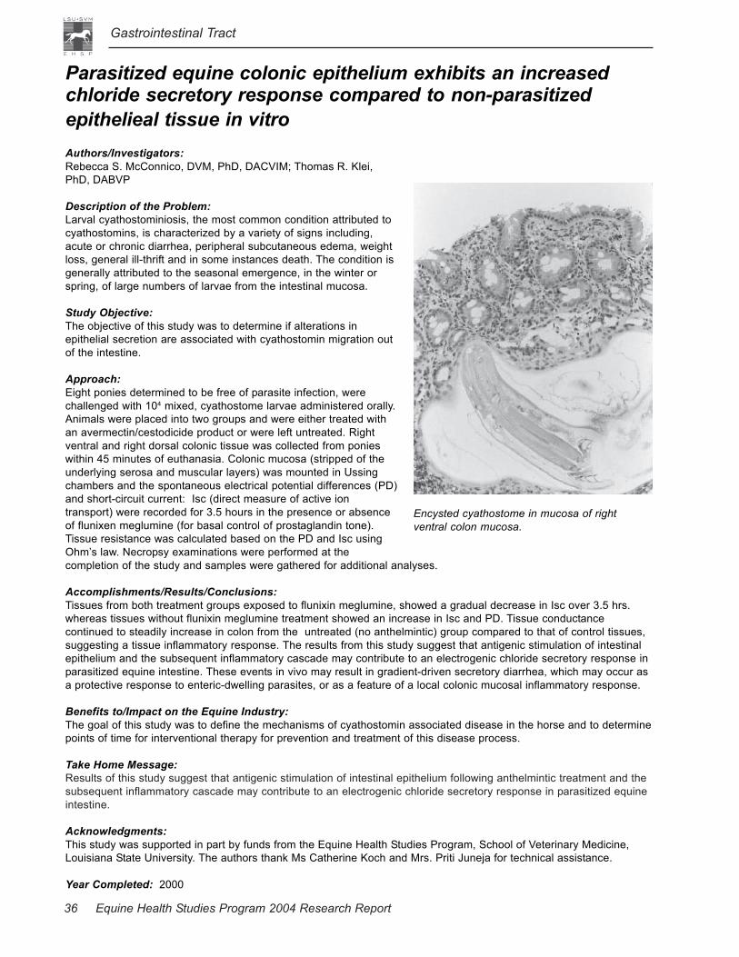
36 Equine Health Studies Program 2004 Research Report
Gastrointestinal Tract
Parasitized equine colonic epithelium exhibits an increasedchloride secretory response compared to non-parasitizedepithelieal tissue in vitroAuthors/Investigators:Rebecca S. McConnico, DVM, PhD, DACVIM; Thomas R. Klei,PhD, DABVP
Description of the Problem:Larval cyathostominiosis, the most common condition attributed tocyathostomins, is characterized by a variety of signs including,acute or chronic diarrhea, peripheral subcutaneous edema, weightloss, general ill-thrift and in some instances death. The condition isgenerally attributed to the seasonal emergence, in the winter orspring, of large numbers of larvae from the intestinal mucosa.
Study Objective:The objective of this study was to determine if alterations inepithelial secretion are associated with cyathostomin migration outof the intestine.
Approach:Eight ponies determined to be free of parasite infection, werechallenged with 104 mixed, cyathostome larvae administered orally.Animals were placed into two groups and were either treated withan avermectin/cestodicide product or were left untreated. Rightventral and right dorsal colonic tissue was collected from ponieswithin 45 minutes of euthanasia. Colonic mucosa (stripped of theunderlying serosa and muscular layers) was mounted in Ussingchambers and the spontaneous electrical potential differences (PD)and short-circuit current: Isc (direct measure of active iontransport) were recorded for 3.5 hours in the presence or absenceof flunixen meglumine (for basal control of prostaglandin tone).Tissue resistance was calculated based on the PD and Isc usingOhm’s law. Necropsy examinations were performed at thecompletion of the study and samples were gathered for additional analyses.
Accomplishments/Results/Conclusions:Tissues from both treatment groups exposed to flunixin meglumine, showed a gradual decrease in Isc over 3.5 hrs.whereas tissues without flunixin meglumine treatment showed an increase in Isc and PD. Tissue conductancecontinued to steadily increase in colon from the untreated (no anthelmintic) group compared to that of control tissues,suggesting a tissue inflammatory response. The results from this study suggest that antigenic stimulation of intestinalepithelium and the subsequent inflammatory cascade may contribute to an electrogenic chloride secretory response inparasitized equine intestine. These events in vivo may result in gradient-driven secretory diarrhea, which may occur asa protective response to enteric-dwelling parasites, or as a feature of a local colonic mucosal inflammatory response.
Benefits to/Impact on the Equine Industry:The goal of this study was to define the mechanisms of cyathostomin associated disease in the horse and to determinepoints of time for interventional therapy for prevention and treatment of this disease process.
Take Home Message:Results of this study suggest that antigenic stimulation of intestinal epithelium following anthelmintic treatment and thesubsequent inflammatory cascade may contribute to an electrogenic chloride secretory response in parasitized equineintestine.
Acknowledgments:This study was supported in part by funds from the Equine Health Studies Program, School of Veterinary Medicine,Louisiana State University. The authors thank Ms Catherine Koch and Mrs. Priti Juneja for technical assistance.
Year Completed: 2000
Encysted cyathostome in mucosa of rightventral colon mucosa.

Equine Health Studies Program 2004 Research Report 37
Gastrointestinal Tract
Published Manuscripts/Abstracts:McConnico RS, Klei TR. Parasitized equine colonic epithelium exhibits an increased chloride secretory responsecompared to non-parasitized epithelieal tissue in vitro. 7th International Equine Colic Symposium, Manchester, Eng,UK, July 2002.
Detection and comparison of nitric oxide in clinically healthyhorses and those with naturally acquired large colon and smallintestinal strangulating obstructionAuthors/Investigators:Mustajab H. Mirza, DVM, MS; Thomas L. Seahorn, DVM, MS, DACVIM; Julian L. Oliver, DVM, PhD, DACVP; GiselleHosgood, BVSc, MS, PhD, DACVS; Rustin M. Moore, DVM, PhD, DACVS
Description of the Problem:Strangulation obstruction involving the large colon and small intestine is a common cause of colic in horses. The highmortality associated with intestinal strangulating obstruction may be related to a sustained reduction of blood flow andhypoperfusion (attributable to increased vascular resistance) after surgical correction and continued ischemic injury. There is limited information available regarding the pathophysiology of intestinal ischemia and ischemia-reperfusion inhorses. It is well accepted that nitric oxide (NO) plays a role in these processes, and therefore, it is reasonable toassume that NO is involved in the pathophysiologic cascade of ischemia and ischemia-reperfusion.
Study Objective:The objective of this study was to determine whether nitric oxide (NO) is present in clinically normal horses under basalconditions, and if it increases secondary to naturally acquired large colon volvulus or small intestinal strangulation.
Approach:Adult horses with naturally acquired strangulating large colon volvulus (n=11), small intestinal strangulation (n=20) and10 clinically healthy horses with no signs of gastrointestinal tract disease were used. Jugular venous blood andabdominal fluid were collected in both heparinized and EDTA tubes for NO quantification, and CBC and abdominal fluidcytology, respectively. Urine was collected via urinary catheterization or during voiding. Creatinine concentrations weredetermined in all fluid samples. NO concentrations were standardized to the creatinine concentration in the respectivesamples. A biopsy specimen was collected from the pelvic flexure of the large colon or the affected segment of thejejunum at surgery or after euthanasia and divided into subsections for fixation in zinc formalin and cryopreservation inOCT gel. NADPH diaphorase histochemical stains were performed on cryopreserved tissues. Routine histology andinducible nitric synthase (iNOS) and nitrotyrosine (NT) immunohistochemical stains were performed on paraffin-embedded tissues.
Accomplishments/Results/Conclusions:Large colon - Based upon localization of NADPH diaphorase staining in endothelium and neuronal tissue, it appearsthat eNOS and nNOS are present under basal conditions in the vasculature and neuronal networks of the large colon,where they likely are responsible for mediating physiologic or cytoprotective effects of NO. The increased iNOSstaining in mucosal and submucosal leukocytes may represent potential detrimental effects of iNOS-derived NOproduction locally in the large colon subsequent to naturally acquired large colon volvulus. The absence of an increasein nitrotyrosine staining in affected horses may reflect an inadequate time frame for its production in these horses.Alternatively, the lack of an increase in plasma, abdominal fluid and urine NO concentrations may reflect venousocclusion of colonic vasculature and subsequent prevention of locally produced NO from entering systemic circulation,or alternatively a lack of systemic production. This study suggest that NO is present under physiologic conditions in thelarge colon and its role in disease deserves further study. Small intestine - This study demonstrated indirectly throughNADPH diaphorase staining that eNOS and nNOS are present under basal conditions in the vasculature and neuronalnetworks of the small intestine of clinically healthy horses, which have been shown in other species to be associatedwith physiologic or cytoprotective effects of NO. The increased iNOS staining in mucosal and submucosal leukocytesof affected horses may represent potential detrimental effects of iNOS-derived NO production locally in the smallintestine. The increased mucosal leukocyte nitrotyrosine staining score in affected horses likely represents increasedperoxynitrite subsequent to increased iNOS-derived NO and increased superoxide production. Although the biologicand pathophysiologic relevance of these findings could not be determined, this study provides preliminary evidencethat NO deserves further investigation in small intestinal strangulation obstruction in horses.
38 Equine Health Studies Program 2004 Research Report
Gastrointestinal Tract
Benefits to/Impact on the Equine Industry:Nitric oxide is present under basal conditions in the large and small intestine of horses and appears to be involved inthe pathophysiologic cascade of ischemia in the small intestine. Further study of the role of NO in this process may bewarranted.
Take Home Message:This study suggest that NO is present under physiologic conditions in the large colon and small intestine of horses, andprovides preliminary evidence that NO deserves further investigation in small intestinal strangulation obstruction inhorses.
Acknowledgments:This study was supported by grants from the American Association of Equine Practitioners, ComparativeGastroenterology Society, Veterinary Clinical Sciences Organized Research Fund, and the Equine Health StudiesProgram, School of Veterinary Medicine, Louisiana State University.
Year Completed: 1998
Published Manuscripts/Abstracts:Mirza MH, Oliver JL, Seahorn TL, Hosgood G, Moore RM. Detection and comparison of nitric oxide in clinically normalhorses and those with small intestinal strangulation obstruction. Can J Vet Res 1999;63: 230-240.
Mirza MH, Seahorn TL, Oliver JL, Moore RM. Detection and comparison of nitric oxide in clinically normal horses andthose with large colon volvulus, in Proceedings. Sixth Colic Research Symposium 1998;30.
Mirza MH, Moore RM, Seahorn TL, Oliver JL. Nitric oxide (NO) in horses with large colon volvulus. Vet Surg 1997;26(5) 438, 1997.
Mirza MH, Moore RM, Seahorn TL, Oliver JL. Detection of nitric oxide in horses with large colon volvulus, inProceedings. American Association of Equine Practitioners 1997;43, 403-404.
Quantitation of adenine nucleotides in equine colonic mucosaltissue using high performance liquid chromatographyAuthors/Investigators:Joanne Tetens, DVM, MS, PhD, DACVS; Steven A. Barker, PhD; Marian Waguespack, MS (?); Giselle L. Hosgood,BVSc, MS, PhD, DACVS
Description of the Problem:Gastrointestinal mucosal cells have a high metabolic and, therefore, require large quantities of adenosine triphosphate(ATP) for maintenance of normal cellular function. As a result, the mucosal layer is principally affected during ischemia.One proposed mechanism for the increase in cellular permeability observed during ischemia is decreased endogenousproduction of ATP. In an experimental model of ascending colon volvulus in ponies, mucosal ATP content diminished92% during ischemia and recovered to only 44% of control values upon reperfusion. In cultured renal and intestinalepithelial cell lines, decreasing cellular ATP content by inducing chemical hypoxia results in rapid opening of tightjunctions, which increases paracellular permeability. Therefore, mucosal ATP depletion, which occurs duringgastrointestinal ischemia, may cause rapid opening of tight junctions, leading to enhanced absorption of bacteria andendotoxin into the splanchnic circulation. These findings suggest that increased metabolic support to this chemicallyinjured colonic epithelium may be important in decreasing morbidity and improving survival.
Study Objective:The objectives of this study were to validate an established method for adenine nucleotide separation in equine colonicmucosal tissue, to determine the inherent variability in the tissue and extraction method, and to determine the stabilityof ATP, ADP, and AMP in the tissue across time.
Approach:Equine colonic mucosal tissue obtained from a single horse was immediately submersed in liquid nitrogen and storedat -70?C. Samples were lyophilized, extracted, and separated by high performance liquid chromatography (HPLC). Inexperiment 1, one tissue sample from 1 parent segment was processed for adenine nucleotide quantitation. The singlesample was loaded into the instrument and quantified 18 consecutive times to determine the inherent variability of the
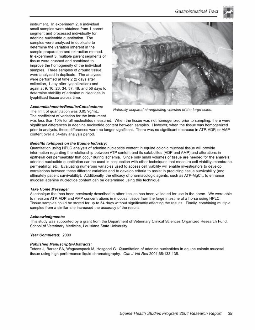
Equine Health Studies Program 2004 Research Report 39
Gastrointestinal Tract
instrument. In experiment 2, 6 individualsmall samples were obtained from 1 parentsegment and processed individually foradenine nucleotide quantitation. Thesamples were analyzed in duplicate todetermine the variation inherent in thesample preparation and extraction method.In experiment 3, multiple parent segments oftissue were crushed and combined toimprove the homogeneity of the individualsamples. Three samples of ground tissuewere analyzed in duplicate. The analyseswere performed at time 2 (2 days aftercollection, 1 day after lyophilization) andagain at 9, 16, 23, 34, 37, 48, and 56 days todetermine stability of adenine nucleotides inlyophilized tissue across time.
Accomplishments/Results/Conclusions:The limit of quantitation was 0.05 ?g/mL.The coefficient of variation for the instrumentwas less than 10% for all nucleotides measured. When the tissue was not homogenized prior to sampling, there weresignificant differences in adenine nucleotide content between samples. However, when the tissue was homogenizedprior to analysis, these differences were no longer significant. There was no significant decrease in ATP, ADP, or AMPcontent over a 54-day analysis period.
Benefits to/Impact on the Equine Industry:Quantitation using HPLC analysis of adenine nucleotide content in equine colonic mucosal tissue will provideinformation regarding the relationship between ATP content and its catabolites (ADP and AMP) and alterations inepithelial cell permeability that occur during ischemia. Since only small volumes of tissue are needed for the analysis,adenine nucleotide quantitation can be used in conjunction with other techniques that measure cell viability, membranepermeability, etc. Evaluating numerous variables used to access cell viability will enable investigators to developcorrelations between these different variables and to develop criteria to assist in predicting tissue survivability (andultimately patient survivability). Additionally, the efficacy of pharmacologic agents, such as ATP-MgCl2, to enhancemucosal adenine nucleotide content can be determined using this technique.
Take Home Message:A technique that has been previously described in other tissues has been validated for use in the horse. We were ableto measure ATP, ADP and AMP concentrations in mucosal tissue from the large intestine of a horse using HPLC.Tissue samples could be stored for up to 54 days without significantly affecting the results. Finally, combining multiplesamples from a similar site increased the accuracy of the results.
Acknowledgments:This study was supported by a grant from the Department of Veterinary Clinical Sciences Organized Research Fund,School of Veterinary Medicine, Louisiana State University.
Year Completed: 2000
Published Manuscripts/Abstracts:Tetens J, Barker SA, Wagusespack M, Hosgood G. Quantitation of adenine nucleotides in equine colonic mucosaltissue using high performance liquid chromatography. Can J Vet Res 2001;65:133-135.
Naturally acquired strangulating volvulus of the large colon.
40 Equine Health Studies Program 2004 Research Report
Gastrointestinal Tract
Pharmacological evaluation of endothelin-antagonists inblocking constriction of equine colonic vessels induced byendothelin-1Authors/Investigators:Changaram S. Venugopal, BVSc, MSc, MS, PhD; Rustin M. Moore, DVM, PhD, DACVS
Description of the Problem:Horses with strangulating large-colon volvulus die owing to severe mucosal injury despite surgical correction andintensive medical care. Mucosal injury has been attributed to sustained reduction in blood flow. Endothelium derivedsubstances such as endothelin and nitric oxide regulate vasomotor tone and blood flow. Experimental studies haveshown that altered vascular tone contributes to ischemia/perfusion injury. Endothelin is a potent vasoconstrictor agentthat could drastically increase vascular resistance resulting in reduced blood flow.
Study Objective:The objective of this study was to find a potential therapeutic agent that will block the contractile effects of ET-1 onvascular smooth muscle. This project determined the efficacy and affinity of two newly discovered endothelin-antagonists to block ET-1 induced responses and compared them as potential therapeutic agents in alleviatingendothelin induced contractile response of colonic vessels in horses.
Approach:Mesenteric colonic vessels were collected from the left ventral colon of horses under barbiturate anesthesia. From thesegment, 4 mm wide vessel rings were cut and were fixed in a tissue bath containing tyrode’s solution. The ring is alsoattached to a force transducer and measured the tension developed on the tissues induced by graded concentration ofET-1. The effectiveness of the antagonists were measured by incubating the tissues with the antagonists and thenconstructing a concentration response curves. The inhibition of the contractile response to ET-1 was considered as theefficacy of the antagonists.
Accomplishments/Results/Conclusions:Both antagonists dose-dependently inhibited the contractile responses of the vessel rings to ET-1. Upon comparingthe other properties of the compounds, B1, which was water-soluble was preferred to B2 for further studies.
Take Home Message:The contractile effects of endothelin on equine colonic vessels can be inhibited by its receptor antagonists. Furtherstudies are needed to evaluate the therapeutic use of these agents in disease conditions. Search for specific receptorantagonists are also needed.
Acknowledgments:This study was supported by a grant from the Equine Health Studies Program, School of Veterinary Medicine,Louisiana State University. The authors thank Earnestine Holmes and Catherine Koch for technical assistance.
Year Completed: 1998
Published Manuscripts/Abstracts:Venugopal CS, Holmes EP, Koch CE, Curtis LA, Holm AS, Moore RM. In vitro pharmacological evaluation of two endothelin-1 antagonists on equine colonic vessel rings. Am J Vet Res 2001;62(2):154-159.
Venugopalan CS, Moore RM, Holmes EP, Koch CE. Effect of endothelin antagonists on the in vitro contractile responsesof equine colonic vessels to endothelin-1. FASEB J 1998;12(4): A383.
Holm AS, Venugopal CS, Holmes EP, Koch CE, Moore RM. Inhibition of endothelin-1 (ET-1) induced contraction of equinecolonic vessels by potential ET-1 receptor antagonists, in Proceedings. Sixth Colic Research Symposium 1998;30.
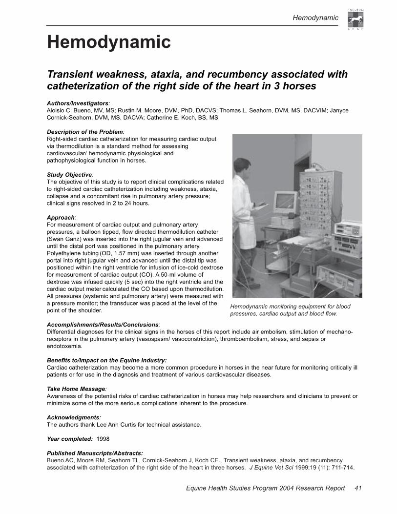
Equine Health Studies Program 2004 Research Report 41
HemodynamicTransient weakness, ataxia, and recumbency associated withcatheterization of the right side of the heart in 3 horsesAuthors/Investigators:Aloisio C. Bueno, MV, MS; Rustin M. Moore, DVM, PhD, DACVS; Thomas L. Seahorn, DVM, MS, DACVIM; JanyceCornick-Seahorn, DVM, MS, DACVA; Catherine E. Koch, BS, MS
Description of the Problem:Right-sided cardiac catheterization for measuring cardiac outputvia thermodilution is a standard method for assessingcardiovascular/ hemodynamic physiological andpathophysiological function in horses.
Study Objective:The objective of this study is to report clinical complications relatedto right-sided cardiac catheterization including weakness, ataxia,collapse and a concomitant rise in pulmonary artery pressure;clinical signs resolved in 2 to 24 hours.
Approach:For measurement of cardiac output and pulmonary arterypressures, a balloon tipped, flow directed thermodilution catheter(Swan Ganz) was inserted into the right jugular vein and advanceduntil the distal port was positioned in the pulmonary artery.Polyethylene tubing (OD, 1.57 mm) was inserted through anotherportal into right jugular vein and advanced until the distal tip waspositioned within the right ventricle for infusion of ice-cold dextrosefor measurement of cardiac output (CO). A 50-ml volume ofdextrose was infused quickly (5 sec) into the right ventricle and thecardiac output meter calculated the CO based upon thermodilution.All pressures (systemic and pulmonary artery) were measured witha pressure monitor; the transducer was placed at the level of thepoint of the shoulder.
Accomplishments/Results/Conclusions:Differential diagnoses for the clinical signs in the horses of this report include air embolism, stimulation of mechano-receptors in the pulmonary artery (vasospasm/ vasoconstriction), thromboembolism, stress, and sepsis orendotoxemia.
Benefits to/Impact on the Equine Industry:Cardiac catheterization may become a more common procedure in horses in the near future for monitoring critically illpatients or for use in the diagnosis and treatment of various cardiovascular diseases.
Take Home Message:Awareness of the potential risks of cardiac catheterization in horses may help researchers and clinicians to prevent orminimize some of the more serious complications inherent to the procedure.
Acknowledgments:The authors thank Lee Ann Curtis for technical assistance.
Year completed: 1998
Published Manuscripts/Abstracts:Bueno AC, Moore RM, Seahorn TL, Cornick-Seahorn J, Koch CE. Transient weakness, ataxia, and recumbencyassociated with catheterization of the right side of the heart in three horses. J Equine Vet Sci 1999;19 (11): 711-714.
Hemodynamic
Hemodynamic monitoring equipment for bloodpressures, cardiac output and blood flow.
42 Equine Health Studies Program 2004 Research Report
Cardiopulmonary and sedative effects of intravenousadministration of low doses of medetomidine and xylazine toadult horsesAuthors/Investigators:Aloisio C. Bueno, MV, MS; Janyce Cornick-Seahorn, DVM, MS, DACVA; Thomas L. Seahorn, DVM, MS, DACVIM;Giselle Hosgood, BVSc, MS, DACVS; Rustin M. Moore, DVM, PhD, DACVS
Description of the Problem:Alpha-2 adrenoceptor agonists are commonly used in equine practice for sedation and analgesia for diverse reasonsincluding diagnostic, dental and standing surgical procedures; preanesthesic medication, and to control abdominal pain(colic).
Study Objective:The objective of this study is to determine the cardiopulmonary and sedative effects of medetomidine hydrochloride inadult horses and to compare those effects with effects of an equipotent dose of xylazine hydrochloride.
Approach:Ten healthy adult female horses of various breeds were used. Horses were randomly assigned to 2 groups. Group-1(n=5) received medetomidine (4?g/kg IV) and group-2 (n=5) received xylazine (0.4 mg/kg IV). Heart rate (HR),respiratory rate (RR), arterial blood pressures (ABP), pulmonary arterial blood pressures (PABP) and cardiac output(CO) were recorded at baseline and every 5 minutes for a total of 1 hour. Rectal body temperature and blood gasanalyses were performed every 15 minutes, and sedation and ataxia scores were assigned every 5 minutes throughoutthe study period.
Accomplishments/Results/Conclusions:Arterial blood pressure was significantly decreased throughout the study for G-1 horses (medetomidine) and wassignificantly decreased for 40 minutes for G-2 horses (xylazine). Compared with baseline values cardiac output wassignificantly decreased in G-1 at 10, 20, and 40 minutes and significantly increased in G-2 at 40 and 60 minutes.Despite the significant decreases in RR, blood gas values did not significantly change over time. Ataxia and sedationscores were of similar magnitude for both groups, and all horses resumed eating hay 10 to 55 minutes after drugadministration. This study suggests that equipotent low-doses of medetomidine and xylazine induce comparable levelsof ataxia and sedation and similar cardiopulmonary changes in adult horses.
Benefits to/Impact on Equine Industry:Although not approved for use in horses, medetomidine could be use for a variety of reasons for more profoundsedation/analgesia with similar physiological alterations compared with xylazine.
Take Home Message:Medetomidine (sedative/analgesic) may provide more profound analgesia and sedation of longer duration for horses.
Acknowledgments:This study was supported by grants from the the Equine Veterinary Research Program and the Department ofVeterinary Clinical Sciences Organized Research Fund, School of Veterinary Medicine, Louisiana State University. Theauthors thank Catherine E. Koch and Dr. Lee Ann Fugler for technical assistance.
Year Completed: 1998
Published Manuscripts/Abstracts:Bueno AC, Cornick-Seahorn JL, Seahorn TL, Hosgood G, Moore RM. Cardiopulmonary and sedative effects ofintravenous administration of low doses of medetomidine and xylazine to adult horses. Am J Vet Res 1999;60 (11):1371-1376.
Bueno AC, Cornick-Seahorn JL, Moore RM, Seahorn TL. Cardiopulmonary effects of low-dose medetomidine andxylazine administered intravenously in endotoxin-challenged horses, in Proceedings. Sixth Equine Colic ResearchSymposium 1998;33.
Hemodynamic

Equine Health Studies Program 2004 Research Report 43
Hemodynamic and metabolic alterations associated withintravenous infusion of a combination of adenosinetriphosphate and magnesium chloride in conscious horsesAuthors/Investigators:Joanne Tetens, DVM, MS, PhD, DACVS; Aloisio C. Bueno, DVM, MS, DACVS; Janyce L. Cornick-Seahorn, DVM, MS,DACVA; Giselle Hosgood, BVSc, MS, PhD, DACVS; Susan C. Eades, DVM, PhD, DACVIM; Rustin M. Moore, DVM,PhD, DACVS
Description of the Problem:Shock can be defined as “inadequate blood flow to vital organs or the inability of the body cell mass to metabolizenutrients normally.” The predominant changes during shock develop in the microcirculation, affecting cell membranetransport and function, energy metabolism, and mitochondrial function. During hypoxic conditions, endogenousproduction of ATP is decreased. A major rate-limiting factor in shock and ischemia, and thus resuscitation, isresynthesis of ATP. Therefore, a logical therapeutic approach to increase tissue ATP concentrations is to infuse thesubstrate (ATP) directly, rather than administer agents that would lead to ATP synthesis. Low-flow conditions andorgan ischemia develop commonly in horses subsequent to intestinal strangulation, enterocolitis and proximal enteritis,laminitis, endotoxemia, sepsis, and severe dehydration and exhaustion. Administration of a combination of ATP andMgCl2 after shock and other adverse circulatory conditions in humans and laboratory animals improves mitochondrialfunction and tissue ATP content; restores organ function, blood flow and microcirculation; improves reticuloendothelialfunction, survival time, and survival rate; and down-regulates synthesis and release of inflammatory cytokines.
Study Objective:The objectives of this study were to determine the hemodynamic and metabolic effects of IV infusion of ATP-MgCl2combination in clinically normal, conscious adult horses and to determine a maximal safe IV infusion rate.
Approach:Six horses received an IV infusion of ATP-MgCl2 combination, beginning at a rate of 0.05 mg of ATP/kg of body weight/min, which was increased by 0.05 mg/kg/min increments at 10-minute intervals until a rate of 1.0 mg/kg/min wasachieved. Data were collected prior to the start of the infusion, at the end of each infusion rate, and at 15-minute
intervals for the next hour after discontinuation of the infusion.Measured or calculated hemodynamic variables includedcardiac output, cardiac index, heart rate, stroke volume,systemic and pulmonary arterial pressures, and systemic andpulmonary vascular resistances. Arterial blood gas tensions,CBC, plasma biochemical profiles, urine volume and specificgravity, and selected clinical signs of disease also wereevaluated.
Accomplishments/Results/Conclusions:Intravenous infusion of ATP-MgCl2 significantly increasedcardiac output, decreased systemic vascular resistance, andcaused mild pulmonary hypertension. Magnitude of thehemodynamic alterations was dependent on rate of infusion.Maximal safe infusion rate for these horses was 0.3 mg/kg/min.All horses became lethargic, and their appetites diminishedduring the infusion; 5 horses had mild signs of abdominaldiscomfort. Flank sweating was observed in all horses asinfusion rate increased. Urine volume and specific gravity andhematologic, biochemical, and arterial blood gas alterationswere detected during and after the infusion. Intravenousadministration of ATP-MgCl2 in healthy, conscious, adult horsescaused various metabolic and hemodynamic alterations thatwere without appreciable detrimental effects.
Benefits to/Impact on the Equine Industry:Endotoxemia and gastrointestinal tract diseases in horses canbe associated with high morbidity and mortality. During thesedisease processes, endogenous production of ATP is markedlydecreased, which leads to significant alterations in cellular
Hemodynamic
Naturally acquired strangulating volvulus of the largecolon.

44 Equine Health Studies Program 2004 Research Report
Hemodynamic
metabolic processes. Such alterations can be irreversible and lead to death of the affected animal. Since IVadministration of an ATP-MgCl2 combination in healthy, conscious, adult horses was without appreciable side effects,especially when administered at a rate of 0.3 mg of ATP/kg of body weight/min, this combination may be useful duringendotoxemia or gastrointestinal tract disease by supplying ATP to help maintain cellular metabolic functions.
Take Home Message:A combination of ATP and magnesium chloride can be safely administered to conscious, healthy, adult horses at a rateof 0.3 mg of ATP/kg of body weight/min. Since many disease processes result in decreased ATP production,administration of ATP-MgCl2 may lessen the severity of illness and/or increase survival rate of sick horses. However,since ATP-MgCl2 can have profound effects on blood pressure, further investigations will need to be performed beforethis combination can be recommended clinically as a therapeutic agent for conditions resulting in decreased blood flowto major tissues/organs.
Acknowledgments:This study was supported by funds from a USDA Section 1433 grant, School of Veterinary Medicine, Louisiana StateUniversity.
Year Completed: 1998
Published Manuscripts/Abstracts:Tetens J, Bueno AC, Cornick-Seahorn JL, Hosgood G, Eades SC, Moore RM. Hemodynamic and metabolic alterationsassociated with intravenous infusion of ATP-MgCl2 in conscious horses. Sixth Equine Colic Research Symposium1998.
Tetens J, Bueno AC, Cornick-Seahorn JL, Hosgood G, Eades SC, Moore RM. Hemodynamic and metabolic alterationsassociated with intravenous infusion of ATP-MgCl2 in conscious horses. Phi Zeta Research Day, Louisiana StateUniversity 1999.
Tetens J, Bueno AC, Cornick-Seahorn JL, Hosgood G, Eades SC, Moore RM. Intravenous infusion of ATP-MgCl2 inconscious horses. Advances in Veterinary Shock Research 1999.
Tetens J, Bueno AC, Cornick-Seahorn JL, Hosgood G, Eades SC, Moore RM. Adenosine triphosphate-magnesiumchloride infusion in conscious horses- hemodynamic and metabolic alterations. Ninth Annual American College ofVeterinary Surgeons Symposium 1999.
Tetens J, Bueno AC, Cornick-Seahorn JL, Hosgood G, Eades SC, Moore RM. Hemodynamic and metabolic alterationsassociated with intravenous infusion of a combination of adenosine triphosphate and magnesium chloride in conscioushorses. Am J Vet Res 1999;60:1140-1147.
Pharmacokinetics, clinical signs and hemodynamic effects ofan intravenous bolus dose of Nù -nitro-L-arginine methyl ester(L-NAME) in healthy conscious horsesAuthors/Investigators:Carlos R. Pinto, MedVet, PhD, DACT; Susan C. Eades, DVM, PhD, DACVIM; Steve A. Barker, PhD; Charles R. Short,DVM, PhD; Rustin M. Moore, DVM, PhD, DACVS
Description of the Problem:Nitric oxide is an endogenous mediator involved in numerous biologic function and pathophysiologic processes.Studies evaluating the role of NO have shown it plays an important role in several pathologic conditions in horses,including but not limited to diseases of joints, intestinal tract and reproduction. Because of its solubility, tissuedistribution, and effects on NOS blockade, L-NAME is a potentially useful drug for experimental or clinical use inhorses. Knowledge of its pharmacokinetics is crucial to pursue research investigating the role of NO in equinephysiological and pathophysiological processes, and potentially for therapeutic purposes.
Study Objective:The objective of this study is to determine the pharmacokinetics, clinical signs and hemodynamic effects, includingsystemic arterial blood pressure, pulmonary arterial blood pressure, right atrial pressure, heart rate and rhythm, of anintravenous bolus administration of L-NAME in clinically healthy conscious horses.

Equine Health Studies Program 2004 Research Report 45
Hemodynamic
Approach:Six clinically healthy adult horses. A bolus dose of L-NAME (40 mg/kg, iv, 10%, w/v) was infused into the left jugularvein. Blood was collected prior to injection and serially thereafter for 36 hours from the right jugular vein and thesamples were processed for measurement of L-NAME and its metabolite, Nù -nitro-L-arginine (L-NNA), using HPLC.Arterial, right atrial and pulmonary arterial pressures, heart rate and rhythm, clinical signs, and intestinal sounds weremonitored continuously 20 min before and for 6 hrs after intravenous administration of L-NAME.
Accomplishments/Results/Conclusions:The overall biologic half-life of L-NAME was 3.7 minutes whereas the half-life was 19.5 hours for L-NNA. The meanresidence time, the average time a compound remains in the body, was 14.4 minutes and 28.2 hours, respectively.There was an immediate increase in systemic arterial pressure that returned to normal baseline values by 6 hrs after L-NAME administration. Mean right atrial pressure was increased significantly between 20 and 30 min, whereas meanpulmonary arterial pressure was not significantly affected by L-NAME administration. Mean values for heart rates werenot significantly different from the baseline values.
Benefits to/Impact on the Equine Industry:Because of its solubility, tissue distribution, and effects on NOS blockade, L-NAME is a potentially useful drug forexperimental or clinical use in horses. Knowledge of its pharmacokinetics and its effect on clinical signs andhemodynamic variables is crucial to pursue research investigating the role of NO in equine physiological andpathophysiological processes. This study provides useful information for the pharmacokinetics and hemodynamiceffects of L-NAME administration in horses.
Take Home Message:L-NAME has a short half-life and L-NNA is rapidly detectable in plasma after L-NAME administration. Hydrolysis of L-NAME does not correlate with plasma L-NNA concentrations. Transient L-NAME-induced systemic hypertension, lackof significant alterations in the pulmonary vascular system, heart rate or cardiac rhythm associated with a mild changein demeanor, depression and temporary anorexia that subsided within 12 hrs after L-NAME treatment indicated that L-NAME is relatively safe when administered to conscious, resting horses. The pharmacokinetics profile of L-NAMEadministration in horses documented in this study may be beneficial to developing pharmacologic regimens forexperimental or therapeutic purposes.
Acknowledgments:This study was supported by a grant from the Equine Health Studies Program, School of Veterinary Medicine,Louisiana State University.
Year Completed: 2000
Published Manuscripts/Abstracts:Pinto CRR. Effects of nitric oxide synthase inhibitors on equine and bovine follicular dynamics and steroidogenesis, inDissertation. Louisiana State University, 2001.
In vitro responses of equine colonic arterial and venous rings toadenosine triphosphateAuthors/Investigators:Joanne Tetens, DVM, MS, PhD, DACVS; Changaram S. Venugopal, BVSc, MSc, PhD; Earnestine P. Holmes, BS;Catherine E. Koch, MS; Giselle L. Hosgood, BVSc, MS, PhD, DACVS; Rustin M. Moore, DVM, PhD, DACVS
Description of the Problem:Strangulating volvulus of the ascending colon in horses is a disease characterized by colonic luminal obstruction andvascular occlusion that results in colonic ischemia, mucosal necrosis, and vascular thrombosis. This disease isassociated with high mortality, which may be related to a sustained reduction of blood flow and hypoperfusionattributable to increased vascular resistance after surgical correction and continued ischemic injury. Endothelialdamage in the colonic vasculature develops subsequent to ischemia-reperfusion injury, and this damage can beexacerbated by endotoxin. Therefore, the sustained decrease in colonic blood flow in horses with strangulatingvolvulus may be associated with damage to the endothelium of colonic vessels. Extracellular purines have importantand diverse effects on many biological processes, including regulation of vascular tone. Adenosine triphosphate (ATP)is principally an endothelium-dependent vasodilator that is rapidly metabolized and has a brief duration of action.Because the endothelium is damaged during periods of colonic ischemia and reperfusion, the effects of ATP on
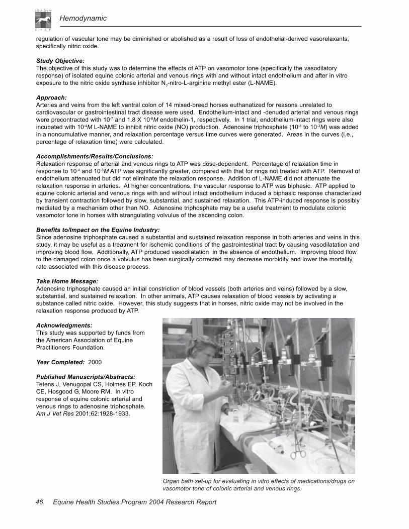
46 Equine Health Studies Program 2004 Research Report
Hemodynamic
regulation of vascular tone may be diminished or abolished as a result of loss of endothelial-derived vasorelaxants,specifically nitric oxide.
Study Objective:The objective of this study was to determine the effects of ATP on vasomotor tone (specifically the vasodilatoryresponse) of isolated equine colonic arterial and venous rings with and without intact endothelium and after in vitroexposure to the nitric oxide synthase inhibitor N?-nitro-L-arginine methyl ester (L-NAME).
Approach:Arteries and veins from the left ventral colon of 14 mixed-breed horses euthanatized for reasons unrelated tocardiovascular or gastrointestinal tract disease were used. Endothelium-intact and -denuded arterial and venous ringswere precontracted with 10-7 and 1.8 X 10⁻⁸M endothelin-1, respectively. In 1 trial, endothelium-intact rings were alsoincubated with 10-4M L-NAME to inhibit nitric oxide (NO) production. Adenosine triphosphate (10-8 to 10-3M) was addedin a noncumulative manner, and relaxation percentage versus time curves were generated. Areas in the curves (i.e.,percentage of relaxation time) were calculated.
Accomplishments/Results/Conclusions:Relaxation response of arterial and venous rings to ATP was dose-dependent. Percentage of relaxation time inresponse to 10-4 and 10-3M ATP was significantly greater, compared with that for rings not treated with ATP. Removal ofendothelium attenuated but did not eliminate the relaxation response. Addition of L-NAME did not attenuate therelaxation response in arteries. At higher concentrations, the vascular response to ATP was biphasic. ATP applied toequine colonic arterial and venous rings with and without intact endothelium induced a biphasic response characterizedby transient contraction followed by slow, substantial, and sustained relaxation. This ATP-induced response is possiblymediated by a mechanism other than NO. Adenosine triphosphate may be a useful treatment to modulate colonicvasomotor tone in horses with strangulating volvulus of the ascending colon.
Benefits to/Impact on the Equine Industry:Since adenosine triphosphate caused a substantial and sustained relaxation response in both arteries and veins in thisstudy, it may be useful as a treatment for ischemic conditions of the gastrointestinal tract by causing vasodilatation andimproving blood flow. Additionally, ATP produced vasodilatation in the absence of endothelium. Improving blood flowto the damaged colon once a volvulus has been surgically corrected may decrease morbidity and lower the mortalityrate associated with this disease process.
Take Home Message:Adenosine triphosphate caused an initial constriction of blood vessels (both arteries and veins) followed by a slow,substantial, and sustained relaxation. In other animals, ATP causes relaxation of blood vessels by activating asubstance called nitric oxide. However, this study suggests that in horses, nitric oxide may not be involved in therelaxation response produced by ATP.
Acknowledgments:This study was supported by funds fromthe American Association of EquinePractitioners Foundation.
Year Completed: 2000
Published Manuscripts/Abstracts:Tetens J, Venugopal CS, Holmes EP, KochCE, Hosgood G, Moore RM. In vitroresponse of equine colonic arterial andvenous rings to adenosine triphosphate.Am J Vet Res 2001;62:1928-1933.
Organ bath set-up for evaluating in vitro effects of medications/drugs onvasomotor tone of colonic arterial and venous rings.
Equine Health Studies Program 2004 Research Report 47
Inflammatory
InflammatoryPlasma and urine nitric oxide concentrations in horses given alow dose of endotoxinAuthors/Investigators:Aloisio C. Bueno, MV, MS; Thomas L. Seahorn, DVM, MS, DACVIM; Janyce Cornick-Seahorn, DVM, MS, DACVA;David W. Horohov, PhD; Rustin M. Moore, DVM, PhD, DACVS
Description of the Problem:Endotoxemia is often encountered in horses with a variety of diseases including those related to the gastrointestinaltract (colic). Endotoxemia develops in approximately 25% of horses with colic and high mortality rates have beenreported. An increased production of nitric oxide (NO) has been reported in various species during endotoxemia.However, literature investigating the role of NO in endotoxemia in horses is lacking. Therefore, this experiment wasconduct to determine if NO concentration in plasma and urine would be elevated in horses given a low-dose ofendotoxin.
Study Objective:The objective of this study is to determine if the induction of this low-dose model of endotoxemia would create aninflammatory response and to correlate the clinical signs, hemodynamic, metabolic, hematologic variables, andcytokine production with the measured nitric oxide concentrations in urine and plasma.
Approach:All the horses were placed in stocks and instrumented in the same fashion. Eight horses received endotoxin (E. coli, 35ng/kg, IV) over 30 min and 3 horses (sentinel group) received normal saline IV over the same time period. Clinicalsigns, hemodynamic variables were recorded, hematologic, metabolic variables, serum for cytokine bioassays, andplasma and urine were collected prior to endotoxin or saline infusion (t=0) and then serially throughout the 24 hourstudy period.
Accomplishments/Results/Conclusions:Despite the induction of an inflammatory response and characteristic alterations of endotoxemia, differences in plasmaNO concentrations were not apparent. Moreover, urine NO concentrations significantly decreased at 4 and 20 to 24hours in endotoxin treated horses. There were no differences in the sentinel group for any of the measured variables.Thus, more research is necessary to unravel and determine the complex role of NO in physiologic and pathologicprocesses especially in vivo.
Benefits to/Impact on the Equine Industry:Determination of the role of NO in endotoxemia in horses may help define potential treatment options with NO agonistor antagonist depending on the stage of endotoxic shock in horses.
Take Home Message:Endotoxemia is a complex and multifactorial condition, and it is very difficult to study in live animals owing to itsvariability. Although systemic NO concentrations were not increased in this study in horses exposed to a low-dose ofendotoxin, additional studies are needed to better define its role in the pathophysiology of endotoxemia in order to tryto increase survivability of this devastating condition.
Acknowledgments:This study was supported by grants from the Equine Health Studies Program and the Department of Veterinary ClinicalSciences Organized Research Fund, School of Veterinary Medicine, Louisiana State University. The authors thankCatherine E. Koch, Dr. Lee Ann Fugler, and Dr. Britta S. Leise for technical assistance.
Year Completed: 1998
Published Manuscripts/Abstracts:Bueno AC, Seahorn TL, Cornick-Seahorn JL, Horohov DW, Moore RM. Plasma and urine nitric oxide concentrations inhorses administered a low dose of endotoxin. Am J Vet Res 1999;60 (8): 969-976.

48 Equine Health Studies Program 2004 Research Report
Inflammatory
Bueno AC, Moore RM, Seahorn TL, Cornick-Seahorn JL, Horohov DW. Quantification of plasma and urine nitric oxide(NO) concentrations in experimentally induced endotoxemia in horses, in Proceedings. Sixth Colic ResearchSymposium 1998;16.
Bueno AC, Moore RM, Seahorn TL, Cornick-Seahorn JL, Horohov DW. Quantification of plasma and urine nitric oxideconcentrations in experimentally induced endotoxemia in horses. Vet Surg 1998;27 (5) 502.
Nitric oxide concentrations in cerebrospinal fluid of horses withor without neurologic diseaseAuthors/Investigators:Lais R.R. Costa, MV, MS, DACVIM; Thomas L. Seahorn, DVM, MS, DACVIM; Rustin M. Moore, DVM, PhD, DACVS
Description of the Problem:Nitric oxide is a labile, soluble gas synthesized from L-arginine by nitric oxide synthase (NOS); it undergoes rapidspontaneous decomposition to the stable end-products, nitrate and nitrite. Nitric oxide has a myriad of local actions, mostnotably cytoprotective as well cytotoxic effects on the host tissue depending upon the source, duration and magnitude of itsproduction.
Study Objective:The objective of this study was to evaluate whether nitric oxide (NO) concentration increases in cerebrospinal fluid(CSF) and plasma of horses with neurologic disease, in particular equine protozoal myelitis (EPM). It washypothesized that NO serves as an inflammatory mediator in the central nervous system.
Approach:Horses admitted to the Louisiana State University and Kansas State University Veterinary Teaching Hospitals with complaintof neurologic disease were evaluated using a systematic approach to neurologic examination. CSF analysis, includingcytology (erythrocytes and nucleated cell counts, and cell morphology), biochemistry (protein, creatine kinase), albuminquotient and IgG index were performed in all cases. Additional diagnostic tests, such as cervical radiographs and CSFserology, were performed as applicable in order to establish the diagnosis. Venous blood from jugular venipuncture and CSFsamples from the lumbosacral or atlanto-occipital spaces were collected in heparinized tubes, and cell-free supernatantswere stored at - 70°C until assayed for NO concentrations using an automated chemiluminescence NO analyzer. Thecontrols for nitrate standards were used according to manufacturer’s instructions. The NO concentration was standardizedto the creatinine concentration in respective samples, and expressed as µM NO/mg creatinine. The horses were groupedaccording to the final diagnosis, and the mean ± standard mean error (SEM) nitric oxide concentrations were calculatedfor each group.
Accomplishments/Results/Conclusions:A wide variety of neurologic diseases were evaluated in this study. Those included 57 horses with a presenting complaintof neurologic problem and 12 horses that were completely normal. Among the horses evaluated for neurologic deficit, 6had infectious neurologic diseases (1 verminous encephalitis, 1 bacterial meningitis, 4 viral encephalomyelitis by EEE,and 16 EPM), 13 had traumatic and degenerative diseases (8 CVM, 1 EDM, 1 EMND, 2 trauma/fracture and 2idiopathic neuropathy), 10 horses had equivocal signs, 8 horses had musculoskeletal disorders and 4 had noneurologic deficits. The NO concentration in plasma and CSF varied among and within the different groups of diseases.The variation in plasma and CSF NO concentrations were not statistically significant between groups. The heterogeneity ofthe neurologic diseases presented and the small sample size for some of the categories are likely to have influenced theresults. Moreover, the variation in duration and severity, thus accounting for different stages of the diseases, are also likely toaffect the magnitude of NO concentrations in plasma and CSF. Concentrations of NO in the CSF samples did not correlatewith protein concentration, nucleated cell count, creatine kinase or IgG concentration in CSF. Although the presence ofnitrate has been shown to closely correspond to the levels of NO secretion. The concentrations of NO in CSF samples ofhorses affected with EPM were significantly higher than those of normal horses, and those with bacterial and viralencephalomyelitis, but lower than those from horses with degenerative diseases. Interestingly, NO synthesis bymacrophages and neutrophils has been shown to markedly increase in response to protozoal infections in humans andanimal models for human diseases, such as Plasmodium, Leishmania and Trypanosoma.
Benefits to/Impact on the Equine Industry:The importance of NO as an inflammatory mediator in neurologic diseases of horses remains unclear. The presentstudy is the first report of NO concentration in CSF of horses with neurologic diseases. Future investigations evaluating the
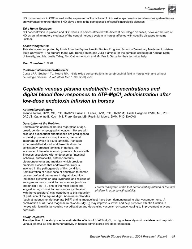
Equine Health Studies Program 2004 Research Report 49
Inflammatory
NO concentrations in CSF as well as the expression of the isoform of nitric oxide synthase in central nervous system tissuesare warranted to further define if NO plays a role in the pathogenesis of specific neurologic diseases.
Take Home Message:NO concentration in plasma and CSF varies in horses affected with different neurologic diseases, however the role ofNO as an inflammatory mediator of the central nervous system in horses affected with specific diseases remainsunclear.
Acknowledgments:This study was supported by funds from the Equine Health Studies Program, School of Veterinary Medicine, LouisianaState University. The authors thank Drs. Bonnie Rush and Julia Flaminio for the samples collected at Kansas StateUniversity, and Ms. Leslie Talley, Ms. Catherine Koch and Mr. Frank Garza for their technical help.
Year Completed: 1998
Published Manuscripts/Abstracts:Costa LRR, Seahorn TL, Moore RM. Nitric oxide concentrations in cerebrospinal fluid in horses with and withoutneurologic disease. J Vet Intern Med 1998;12 (3) 255.
Cephalic venous plasma endothelin-1 concentrations anddigital blood flow responses to ATP-MgCl2 administration afterlow-dose endotoxin infusion in horsesAuthors/Investigators:Joanne Tetens, DVM, MS, PhD, DACVS; Susan C. Eades, DVM, PhD, DACVIM; Giselle Hosgood, BVSc, MS, PhD,DACVS; Catherine E. Koch, MS; Frank Garza, MS; Rustin M. Moore, DVM, PhD, DACVS
Description of the Problem:Endotoxemia affects all horses regardless of age,breed, gender, or geographic location. Horses withcolic and subsequent endotoxemia are predisposedto develop numerous complications, the mostimportant of which is acute laminitis. Althoughexperimentally-induced endotoxemia does notconsistently produce laminitis in horses, theincidence of laminitis is much greater in horses withillnesses associated with endotoxemia (intestinalischemia, enterocolitis, anterior enteritis,pleuropneumonia and metritis), which providesempirical evidence that endotoxemia likely isinvolved in the pathogenesis of this condition.Administration of a low dose of endotoxin to horsescauses profound decreases in digital blood flow.Increased systemic or local synthesis and release ofendogenous vasoconstrictor substances (such asendothelin-1 (ET-1), one of the most potent andlongest acting constrictor substances synthesizedwith the vasculature) may contribute to this reductionof perfusion of the equine digit. Adenine nucleotides(such as adenosine triphosphate [ATP] and its metabolites) have been demonstrated to alter vasomotor tone. Acombination of ATP and magnesium chloride (MgCl2) may improve survival and help preserve athletic function inhorses with laminitis by causing vasodilatation and decreasing vascular resistance leading to improvement in tissueperfusion.
Study Objective:The objective of the study was to evaluate the effects of IV ATP-MgCl2 on digital hemodynamic variables and cephalicvenous plasma ET-like immunoreactivity in horses administered low-dose endotoxin.
Lateral radiograph of the foot demonstrating rotation of the thirdphalanx in a horse with laminitis.

50 Equine Health Studies Program 2004 Research Report
Inflammatory
Approach:Twelve adult horses were used in the study. Horses were administered endotoxin (LPS) or saline solution, IV, during a30-minute period. Immediately thereafter, horses in each group (2 groups of 6 horses) were infused IV with ATP-MgCl2or saline solution. Two weeks later, horses were administered the opposite solution (LPS or saline solution), but it wasfollowed by the same infusion as 2 weeks previously (i.e., ATP-MgCl2 or saline solution). Digital arterial pressure,digital blood flow, and cephalic venous plasma ET-1 concentrations were measured.
Accomplishments/Results/Conclusions:Digital arterial blood slow transiently increased and then decreased for 5 hours in horses administered endotoxinfollowed by ATP-MgCl2. A similar, but shorter duration, decrease was observed in horses administered endotoxinfollowed by saline solution. In horses administered saline solution followed by ATP-MgCl2, digital arterial blood flowdecreased. Mean digital arterial pressure was decreased in horses administered endotoxin followed by either ATP-MgCl2 or saline solution. In horses receiving saline solution followed by ATP-MgCl2, mean digital arterial pressuretransiently increased and then decreased until the ATP-MgCl2 solution was discontinued. In horses receiving endotoxinfollowed by either saline solution or ATP-MgCl2, cephalic venous plasma ET-like immunoreactivity increased. In horsesreceiving saline solution followed by ATP-MgCl2, there was a transient increase in ET-like immunoreactivity.
Benefits to/Impact on the Equine Industry:Low-dose endotoxin administration to clinically healthy horses resulted in a transient, significant increase in cephalicvenous plasma ET-1 concentrations. Therefore, ET-1 may be responsible (via venoconstriction) for the decrease indigital blood flow that occurs during endotoxin administration. Additionally, ET-1 may play a pivotal role in thedevelopment of laminitis by contributing to poor laminar perfusion and subsequent rotation/distal displacement of thethird phalanx. By blocking ET-1, we may be able to prevent laminitis during endotoxemic episodes. Finally,administration of ATP-MgCl2 did not prevent the decrease in digital arterial pressure and blood flow or increase incephalic venous plasma ET-1 concentrations during low-dose endotoxin administration.
Take Home Message:Administration of a low-dose endotoxin (a component of bacteria commonly found in the gastrointestinal tract)significantly increased the amount of ET-1 (a potent constrictor of blood vessels) found in blood taken from a large veinwhich drains the equine foot. Therefore, ET-1 may be responsible for the decrease in blood flow and blood pressure tothe foot of horses administered endotoxin. Since horses that suffer from endotoxemia are prone to develop laminitis(inflammation of the soft tissues of the foot) and laminitis may be caused by constriction of blood vessels to the foot,ET-1 may be involved in the development of laminitis during disease processes that cause endotoxemia. Finally, IVadministration of ATP-MgCl2 (which can dilate blood vessels) did not improve blood pressure or blood flow to the foot orlower the levels of ET-1 measured in the blood from horses given a low-dose of endotoxin.
Acknowledgments:This study was supported by funds from the American Horse Show Association and the Morris Animal Foundation.
Year Completed: 2000
Published Manuscripts/Abstracts:Tetens J, Eades SC, Hosgood G, Koch CE, Garza F, Moore RM: Cephalic venous plasma endothelin-1 concentrationsand digital hemodynamic responses to ATP-MgCl2 administration after low-dose endotoxin infusion in horses. SixthColic Research Symposium 2002.
Tetens J, Eades SC, Hosgood G, Koch CE, Garza F, Moore RM. Cephalic venous plasma ET-1 and digital blood flowresponses to ATP-MgCl2 after low-dose endotoxin infusion in horses. Seventh Equine Colic Research Symposium2002.
Temporal effects of freezing on plasma nitric oxideconcentrations in poniesAuthors/Investigators:Jeremy Hubert, BVSc, MRCVS, MS, DACVS; Thomas Seahorn, DVM, MS, DACVIM; Thomas Klei, PhD; Giselle HosgoodBVSc, MS, PhD, DACVS; Rustin Moore DVM, PhD, DACVS
Description of the Problem:The purpose of this study was to determine the effect of freezing upon plasma NO concentrations. Previous studies inother species have shown increases in NO concentrations subsequent to freezing and storage. Initial storage in
Equine Health Studies Program 2004 Research Report 51
Inflammatory
polypropylene containers revealed increased NO concentrations over time; this was also found in glass containers.These authors stated that fresh plasma samples should be analyzed. In this study nitric oxide (NO) in fresh plasma wascompared to frozen plasma in order to determine the temporal effects of freezing. Nitric oxide (NO) is a known modulatorof specific physiological roles namely NO derived from the enzyme constitutive nitric oxide synthase (NOS); this NOregulates vasomotor tone and neural pathways. However in certain situations the NO is stimulated by inducible NOS(iNOS) and released in greater concentrations and for a longer duration that may be pathophysiologic and damaging totissues.
Study Objective:The objective of this study is to compare concentrations of nitric oxide (NO) in fresh plasma versus frozen plasma and todetermine the temporal effects of freezing on jugular venous plasma NO concentrations in clinically healthy ponies.
Approach:Twenty-eight helminth-naive ponies, ranging in age from 4 to 6 months, were raised and maintained under parasite-freeconditions. Blood was collected from the jugular vein into vacutainer tubes containing sodium heparin. These sampleswere centrifuged at 1,500 x g for 10 minutes, and the plasma supernatant was collected with glass pipettes. Sampleswere analyzed fresh for plasma NO concentrations, the remainder of the samples were divided into 1-mL aliquots whichwere stored in polypropylene vials at –70 C until analyzed for NO concentration. Plasma NO concentration was measuredon fresh samples and on frozen samples at monthly intervals, using a chemiluminescent method. One aliquot from eachpony was thawed in dark conditions for 20-30 minutes at one-month intervals, for a one-year period.
Accomplishments/Results/Conclusions:There were significant differences in the plasma NO concentration across time compared with the baseline value. TheNO concentrations were significantly increased above the baseline value at 1, 3, 4, 6, 7, and 11 months. Although theincreases in plasma NO concentrations were statistically significant, compared with values in fresh samples, the valueswere not of a magnitude expected from diseased horses. Studies performed in our laboratory have indicated that if NOconcentrations are increased in diseased states via pathophysiological mechanisms involving iNOS, the expectedconcentrations are considerably greater than controls.
Despite measurable changes in NO concentrations in frozen plasma samples, there was no loss of nitrate in our samples.Although plasma NO concentrations, compared with fresh samples, were increased at some sampling periods, themagnitude of the increase was not considered substantial compared with studies where there is evidence of increasedNO production associated with disease conditions. To avoid any discrepancies, it may be preferable to analyze the freshplasma samples for NO concentration. Alternatively, samples to be analyzed for NO and compared should be processedand stored frozen under similar conditions and for similar times.
Benefits to/Impact on the Equine Industry:The roles of NO in many biological systems (gastrointestinal, respiratory, musculoskeletal, reproductive) in the horse arebeing studied by several researchers. This information is pertinent to other researchers to accurately elucidate NOconcentrations in bodily fluids.
Take Home Message:It may be pertinent to analyze fresh plasma when trying to elucidate NO concentrations.
Acknowledgments:This study was supported by grants from USDA 1433 funds and the Department of Veterinary Clinical SciencesOrganized Research Fund, School of Veterinary Medicine, Louisiana State University. The authors thank Catherine E.Koch for technical assistance.
Year Completed: 1999
Published Manuscripts/Abstracts:Hubert JD, Seahorn TL, Hosgood G, Klei T, Moore RM. Temporal effects of freezing on plasma nitric oxide concentrationsin ponies. Can J Vet Res 2003;67 (1) 72.

52 Equine Health Studies Program 2004 Research Report
Inflammatory
Effects of adenosine triphosphate-magnesium chloride oncardiopulmonary and clinicopathologic variables, cytokineactivity, and endothelin concentration in horses administered alow dose of endotoxinAuthors/Investigators:Joanne Tetens, DVM, MS, PhD, DACVS; Rustin M. Moore, DVM, PhD, DACVS; Giselle Hosgood, BVSc, MS, PhD,DACVS; Susan C. Eades, DVM, PhD, DACVIM; Michael L. Keowen, BS; David W. Horohov, PhD
Description of the Problem:Endotoxemia remains the leading cause of death in horses. Endotoxemia affects all horses regardless of age, breed,sex, or geographic location. The prevalence of endotoxemia in horses with acute gastrointestinal tract disease (i.e.,colic) admitted to referral veterinary hospitals is estimated to be approximately 25%, and it represents a major cause ofdeath in horses with colic. The most common gastrointestinal tract diseases associated with endotoxemia includeenterocolitis-enteritis and intestinal strangulation-obstruction and are often associated with complications such aslaminitis and gastrointestinal ileus. Bascteria attributable to gram-negative organisms or endotoxemia causesdecreases in cardiac output, systemic vascular resistance, and mean arterial pressure, whereas there is an increase inpulmonary vascular resistance. Some of the hemodynamic derangements during endotoxemia may be subsequent torelease of endothelin-1 (ET-1), the most potent vasoconstrictor of mammalian vessels that has been identified.Concentrations of ET-1 increase during endotoxemia secondary to endotoxin-induced stimulation of endothelial cells.Endothelin-1 may act as a local vasoconstrictor during severe endotoxic shock and contribute to pulmonaryhypertension and peripheral vasomotor disturbances (e.g., decreased blood flow to the gastrointestinal tract and digits)observed during this syndrome. Administration of ATP-MgCl2 following hemorrhagic shock and other adversecirculatory conditions in laboratory animals can improve mitochondrial function and tissue ATP content; restore organfunction, blood flow, and perfusion; improve reticuloendothelial function, survival time, and survival rate; anddownregulate the synthesis and release of inflammatory cytokines.
Study Objective:The objective of this study was to evaluate the systemic effects of IV infusion of ATP-MgCl2 after low-dose endotoxininfusion in horses.
Approach:Twelve adult horses were used in the study. Horses were administered endotoxin (LPS) or saline solution, IV, during a30-minute period. Immediately thereafter, horses in each group (2 groups of 6 horses) were infused IV with ATP-MgCl2or saline solution. Two weeks later, horses were administered the opposite solution (LPS or saline solution), but it wasfollowed by the same infusion as 2 weeks previously (i.e., ATP-MgCl2 or saline solution). Cardiopulmonary andclinicopathologic variables, cytokine activity, and ET-1 concentrations were recorded.
Accomplishments/Results/Conclusions:IV infusion of ATP-MgCl2 after administration of a low dose of endotoxin failed to attenuate the cardiopulmonary,clinicopathologic, and cytokine alterations that develop secondary to endotoxin exposure. The combination of LPS andATP-MgCl2 potentiated pulmonary hypertension, leukopenia, and neutropenia when compared with the combination ofLPS and saline solution. The combination of LPS and ATP-MgCl2 resulted in thrombocytopenia. Endothelinconcentration was increased in jugular venous and pulmonary arterial plasma in horses receiving LPS and ATP-MgCl2.Similar increases were not observed with LPS and saline solution.
Benefits to/Impact on the Equine Industry:Many biological agents are involved in the development of hemodynamic and metabolic derangements that occurduring shock. Adenine nucleotides (which include ATP and its metabolites) can potentially have adverse effects on thecardiopulmonary and metabolic systems of diseased horses. Their role in hemodynamic derangements, leukocyteadherence, and coagulopathies that occur during endotoxemic episodes warrants further investigation. Gaining abetter understanding of what roles (both positive and negative) adenine nucleotides play during shock could potentiallyresult in decreased morbidity and mortality in equine patients by inhibiting the adverse effects and potentiating thepositive effects of these substances.
Take Home Message:In this study, administration of a combination of adenosine triphosphate and magnesium chloride did not protect horsesfrom the adverse effects caused by administration of endotoxin (a toxin commonly absorbed into the blood stream fromthe intestinal tract of sick horses). The findings from this study also suggest that since adenosine triphosphate and

Equine Health Studies Program 2004 Research Report 53
Inflammatory
other compounds related to it are released from cells during shock, these agents could be partially responsible forsome of the negative effects (i.e., poor blood pressure, clotting problems, etc.) that are observed both duringexperimentally-induced and naturally-acquired disease processes.
Acknowledgments:This study was supported by funds from the Morris Animal Foundation.
Year Completed: 2000
Published Manuscripts/Abstracts:Tetens J, Moore RM, Hosgood G, Eades SC, Keowen ML, Horohov DW. Effects of ATP-MgCl2 on clinical signs,hemodynamic, metabolic, hematologic, and serum biochemical variables and TNF-?, IL-6, and endothelin-1 in horsesadministered low-dose endotoxin. Sixth Colic Research Symposium 2002.
Tetens J, Moore RM, Hosgood G, Eades SC, Keowen ML, Horohov D. Effects of ATP-MgCl2 on biological variables inhorses administered low-dose endotoxin. Seventh Equine Colic Research Symposium 2002.
Tetens J, Moore RM, Hosgood GL, Eades SC, Keowen ML, Horohov DW. Effects of adenosine triphosphate-magnesium chloride on cardiopulmonary and clinicopathologic variables, cytokine activity, and endothelin concentrationin horses administered a low dose of endotoxin. Am J Vet Res 2004;65:225-237.

54 Equine Health Studies Program 2004 Research Report
Musculoskeletal
MusculoskeletalAlterations of nitric oxide and endothelin-1 subsequent toexperimental lipopolysaccharide induced acute synovitis inhorsesAuthors/Investigators:Daniel J. Burba, DVM, DACVS; Jorge de la Calle, LVM, MS; Rustin M. Moore, DVM, PhD, DACVS; Julian Oliver, DVM,PhD; Casey J. LeBlanc DVM, PhD; Giselle Hosgood, BVSc, MS, PhD, DACVS; David W. Horohov, PhD
Description of the Problem:Joint disease is a common clinical condition arising from numerous etiologies, and is a major cause of lameness andwastage in equine athletes. It is well documented that the concentration of certain inflammatory mediators increaseslocally in joints of horses with articular disease. Numerous investigations have revealed that nitric oxide (NO) andendothelin-1 (ET-1) are increased in serum and synovial fluid of humans with joint disease. Nitric oxide is a multi-functional intercellular and intracellular messenger molecule thatplays a role in a variety of physiological processes. Someinvestigators believe that synthesis of NO in joints may play a majorrole in the pathophysiology of arthritis. Endothelin-1 is a 21-aminoacid polypeptide with two intra-molecular disulfide bonds that inducespotent and sustained vasoconstriction. Endothelin-1 may also playan important role in the pathogenesis of joint disease in horses. Amodel was used to induce acute synovitis by injectinglipopolysaccharide (LPS) into the joint cavity to measure the levels ofNO and ET-1 in the synovial fluid.
Study Objective:The objective of this study was to determine the effect oflipopolysaccharide (LPS) injection into the middle carpal joint onsynovial fluid and cephalic venous nitric oxide (NO) and endothelin-1(ET-1) concentrations and compare these to values obtained fromthe contralateral control limb, which was injected with saline solution.
Approach:Ten clinically healthy horses were used in this study. Synovitis wasinduced in one randomly chosen middle carpal joint by intra-articularinjection of E. Coli LPS. The contralateral joint was used as a salineinjected control. Synovial fluid, cephalic and jugular venous bloodsamples were collected at 0, 2, 8, 12, 24, and 48 hrs after injection.Concentrations of ET-1 and NO were assessed byradioimmunoassay and by a chemiluminescence method,respectively.
Accomplishments/Results/Conclusions:Comparisons of NO concentrations between treated and control joints did not reveal significant differences for venousplasma or synovial fluid samples. Comparisons of ET-1 concentrations between treated and control legs did not revealsignificant differences for cephalic venous plasma. However, the concentrations for ET-1 in jugular venous plasmawere significantly decreased at 8 hours after injection when compared with baseline. There was a significant increasein ET-1 concentrations of synovial fluid of treated and control joints at 8, 12, 24 and 48 hours after injection, comparedwith baseline. ET-1 appears to be locally synthesized in the joints of horses. Intra-articular injection of LPS did notsignificantly alter the NO concentrations in the synovial fluid in this study.
Benefits to/Impact on the Equine Industry:Information gained from this study revealed that ET-1 (an enzyme) may play a part in degenerative joint disease inhorses. Thus the next step is to determine if by blocking ET-1 will help with treating degenerative joint disease inhorses.
Lateral radiographic projection demonstratingdegenerative joint disease (osteoarthritis) inthe antebrachiocarpal and middle carpal joints.
Equine Health Studies Program 2004 Research Report 55
Musculoskeletal
Take Home Message:ET-1 may play a role in the pathophysiology of joint disease in horses.
Acknowledgments:This study was supported by a grant from the Houston Equine Research Organization. The authors thank CatherineKoch and Frank Garza for technical assistance.
Year Completed: 1999
Published Manuscripts/Abstracts:DelaCalle J, Burba DJ, Moore RM, Hosgood G, Oliver JL, LeBlanc C. Alterations of nitric oxide and endothelin-1subsequent to experimental lipopolysaccharide-induced synovitis in horses. Vet Surg 2001;30 (5) 492.
DelaCalle J, Burba DJ, Moore RM, Hosgooc G, Horohov DW, Oliver JL. Alterations of nitric oxide and endothelin-1subsequent to experimental lipopolysaccharide induced acute synovitis in horses, in Proceedings. British EquineVeterinary Association Congress 2001;40:6.
Synovial fluid and plasma nitric oxide and endothelin-1concentrations in horses with and without joint diseaseAuthors/Investigators:Daniel J. Burba, DVM, DACVS; Jorge de la Calle, LVM, MS, DACVS; Rustin M. Moore, DVM, PhD, DACVS; JamieWilliams, DVM, MS; Casey J LeBlanc, DVM, PhD; Giselle Hosgood, BVSc, MS, PhD, DACVS
Description of the Problem:Joint disease is a common clinical condition arising from numerous etiologies, and is a major cause of lameness andwastage in equine athletes. It is well documented that the concentration of certain inflammatory mediators increaseslocally in joints of horses with articular disease. Numerous investigations have revealed that nitric oxide (NO) andendothelin-1 (ET-1) are increased in serum and synovial fluid of humans with joint disease. Nitric oxide is a multi-functional intercellular and intracellular messenger molecule that plays a role in a variety of physiological processes.Some investigators believe that synthesis of NO in joints may play a major role in the pathophysiology of arthritis.Endothelin-1 is a 21-amino acid polypeptide with two intra-molecular disulfide bonds that induces potent and sustainedvasoconstriction. Endothelin-1 may also play an important role in the pathogenesis of joint disease in horses.
Study Objective:The objective of this study was to determine and compare the plasma and synovial fluid endothelin-1 (ET-1) and nitricoxide (NO) concentrations in normal joints from clinically healthy horses with values obtained from horses with jointdisease affected with acute synovitis, degenerative joint disease (DJD) and joint sepsis.
Approach:Thirty-six horses with joint disease and 15 clinically healthy horses with no evidence of joint disease were included inthe study. Horses with joint disease were assigned to one of the three groups, including those with synovitis, DJD, orsepsis based upon clinical and radiographic exams and synovial fluid analysis. ET-1 and NO concentrations weremeasured in plasma from whole blood, collected from the jugular vein and cephalic/saphenous vein, respectively, ofthe limb with an affected or non-affected (normal) joint, as well as synovial fluid via arthrocentesis from the involvedjoint.
Accomplishments/Results/Conclusions:Comparisons of ET-1 concentrations between affected and non affected groups did not reveal significant differences invenous plasma. However, concentrations of ET-1 in synovial fluid obtained from the joint sepsis group weresignificantly greater, compared with values from the synovitis, DJD and non-affected groups. Comparisons of NOconcentrations between affected and non-affected groups did not reveal significant differences in venous plasma orsynovial fluid. It appears from this study ET-1 is locally synthesized in the joints of horses with varying types of jointdisease.
Benefits to/Impact on the Equine Industry:Information gained from this study revealed that ET-1 (an enzyme) may play a part in degenerative joint disease inhorses. Concentrations varied among different types of joint disease with concentrations significantly increased insynovial fluid of horses with joint sepsis. Thus the next step is to determine if by blocking ET-1 will help with treatingdegenerative joint disease in horses.

56 Equine Health Studies Program 2004 Research Report
Take Home Message:ET-1 may play a role in the pathophysiology of joint disease in horses.
Acknowledgments:This study was supported by a grant from the Houston Equine Research Organization. The authors thank CatherineKoch and Frank Garza for technical assistance.
Year Completed: 1999
Published Manuscripts/Abstracts:De la Calle J, Burba DJ, Ramaswamy CM, Hosgood, G, Williams J, LeBlanc C, Moore RM: Plasma and synovial fluidendothelin-1 and nitric oxide concentrations in horses with normal and diseased joints. Am J Vet Res 2002;63 (12)1648-1654.
DelaCalle J, Burba DJ, Moore RM, Hosgood G, Williams J, LeBlanc C. Plasma and synovial fluid nitric oxide andendothelin-1 concentrations in horses with normal and diseased joints. Vet Surg 2001;30 (5) 491.
DelaCalle J, Burba DJ, Moore RM, Hosgood G, LeBlanc C, Williams J. Plasma and synovial nitric oxide and endothelin-1 concentrations in horses with normal and diseased joints, in Proceedings. 40th British Equine Veterinary AssociationCongress 2001;40:6.
Pharmacological control of digital vasculature in the preventionand treatment of laminitis in horsesAuthors/Investigators:Ashley M. Stokes, DVM, PhD; Changaram S. Venugopal, BVSc, PhD; Giselle Hosgood, BVSc, MS, PhD, DACVS;Susan C. Eades, DVM, PhD, DACVIM; Rustin M. Moore, DVM, PhD, DACVS
Description of the Problem:Laminitis is a common, excruciatingly painful disease which forces the horse to lay in lateral recumbency for hoursresulting in pressure sores,anorexia, and often leads toeuthanasia. Although theetiology of laminitis is not fullyunderstood, it has beenhypothesized that ischemia,owing to vasoconstriction,plays a causative role inlaminitis. During acutelaminitis, vascular resistanceincreases 3.5 times abovenormal resulting in asignificant reduction in bloodflow to the foot. This period ofreduced blood flow causeslaminar inflammation, edema,and necrosis, which oftenleads to separation of thedistal phalanx from the hoofwall. Endothelin (ET), a potentvasoconstrictor, is released bythe endothelium and increasesvascular resistance. ETreceptors are found onvascular smooth muscle andendothelial cells. Stimulationof ETA and ETB2 receptorsresult in sustainedvasoconstriction and ETB1
The synthesis of endothelin (ET-1) is stimulated by many inflammatory mediatorsand changes in blood flow. ET-1 is primarily synthesized and released fromendothelial cells. The two important receptor types are ETA and ETB. ET-1 effectson vascular smooth muscle are primarily through the ETA receptor, resulting inprofound contraction.
Musculoskeletal
Equine Health Studies Program 2004 Research Report 57
Musculoskeletal
receptors trigger the release of nitric oxide. Since the onset of laminitis is characterized by digital venoconstriction andchanges in systemic arterial and venous pressures, determining the role of ET in the pathogenesis of this disease mayprovide further important knowledge for the prevention and treatment of laminitis in horses. Availability of ET receptorantagonists has made it possible to study the individual effects of ET on equine palmar digital vessels.
Study Objective:The objectives of this study were to determine the concentration-response relationship of endothelin-1 (ET-1), comparetwo ET receptor antagonists and determine the concentrations that effectively block the vasomotor effects of ET-1, andcompare the effectiveness of ET-1 to previously studied vasoconstrictors in equine palmar digital arterial and venousrings in vitro.
Approach:Palmar digital arterial and venous rings from 8 non-laminitic horses were used. Vessel rings were placed in Tyrode’ssolution, one side fixed to the floor of an organ bath and the other side to a force-displacement transducer. Twoseparate studies were conducted: (I) incubation with one ET receptor antagonist (PD142893 or PD145065) followed byan ET-1 concentration-response curve on medial vessel rings; and (II) comparison of ET-1 to norepinephrine (NE) andhistamine (HST) and comparison of contractile responses of medial vs. lateral vessel rings.
Accomplishments/Results/Conclusions:In study I, ET-1 administration caused pronounced and sustained concentration-dependent contraction of vessel rings.These contractile responses were effectively inhibited by the two ET antagonists in a concentration-dependent manner.Venous rings were significantly more sensitive to the contractile effects of ET-1 than arterial rings. In study II, ET-1 andNE were found to be significantly more potent vasoconstrictors than HST in both arteries and veins. No significantdifferences were found between the effects of ET and NE in arteries and veins.
Benefits to/Impact on the Equine Industry:The pathophysiology of equine laminitis remains unclear; however, vascular alterations are known to be an importantaspect of the earliest stages of the disease. The identification of a key vascular mediator, and the determination of aneffective inhibitor of this mediator, may lead to the development of preventatives and therapeutics for this devastatingdisease.
Take Home Message:Identification of ET-1 as a very potent vasoconstrictor of the equine digit is an important step in understanding thepathophysiology of equine acute laminitis, a disease characterized by altered digital hemodynamics. The receptorantagonist PD 145065 demonstrated potential therapeutic use in treating acute laminitis and may prove to be useful inpreventing these known hemodynamic alterations.
Acknowledgments:This study was supported by a grant from the Geraldine R. Dodge Foundation, the Kenneth A. Scott Trust, the BerniceBarbour Foundation, the Marilyn M. Simpson Charitable Trust, the Humane Society of the United States, and theMassachusetts Society for the Prevention of Cruelty to Animals. The authors would like to thank Earnestine P. Holmes,Catherine E. Koch, Dr. Lee Ann Curtis, and Dr. Chidambaram Ramaswamy for technical assistance.
Year Completed: 1998
Published Manuscripts/Abstracts:Stokes AM, Venugopal CS, Hosgood G, Eades SC, Moore RM. Comparison of two endothelin antagonists on in vitroresponses of equine palmar digital arterial and venous rings to endothelin-1. Can J Vet Res. In press.
Stokes Holm AM, Venugopal CS, Hosgood G, Eades SC, Moore RM. Comparison of effect of two endothelin antagonistson in vitro responses of equine palmar digital arterial and venous rings to endothelin-1 in normal horses and those withnaturally acquired laminitis, in Proceedings. Seventh Colic Research Symposium 2002;141.
Holm AS, Venugopal CS, Eades SC, Moore RM. In vitro and in vivo effects of endothelin-1 and endothelin antagonistson the digital vasculature in clinically healthy horses. Vet Surg 2001;30 (5) 495.
Holm AS, Eades SC, Venugopal CS, Oliver JL, Moore RM. Pathophysiologic role of endothelin-1 in equine acutelaminitis, in Proceedings. Havermeyer Foundation Workshop: Equine Immunology 2001;40.
58 Equine Health Studies Program 2004 Research Report
Musculoskeletal
The effects of endothelin-1 and nitric oxide on digitalhemodynamics of normal horsesAuthors/Investigators:Ashley M. Stokes, DVM, PhD; Susan C. Eades, DVM, PhD, DACVIM; Changaram S. Venugopal, BVSc, MSc, PhD;Catherine E. Koch, MS; Rustin M. Moore, DVM, PhD, DACVS
Description of the Problem:Acute laminitis is a severely debilitating and painful disease of the sensitive and insensitive laminae of the equine digitcharacterized by increased venous tone, increased pressure within the capillary bed, and edema formation leading toreduced delivery of oxygen and nutrients to the soft tissues of the digit. Ultimately, this leads to separation of theinterdigitating soft tissue structures that suspend the distal phalanx within the hoof capsule. This separation leads torotation and/or sinking of the distal phalanx, a very painful condition from which approximately 75% of horses requireeuthanasia. Endothelin-1 (ET-1), a potent vasoconstrictor, is released by the endothelial cells that line the bloodvessels. ET-1 production is increased by many of the inflammatory mediators known to be elevated during acutelaminitis. Nitric oxide (NO) is also a substance produced by the endothelium, but its production leads to dilation of thevasculature and opposes the actions of ET-1. Knowledge of the effects of these substances on the digital vasculaturewould contribute to our understanding of the vascular pathophysiology of acute laminitis.
Study Objective:Our global hypothesis is that the alterations in digital hemodynamics associated with the onset of acute laminitis arecaused by the local imbalance of the endothelium-derived substances ET-1 (increased) and nitric oxide (decreased)which ultimately lead to laminar ischemia and necrosis. Our study hypothesis was the administration of an ETantagonist would prevent or reverse the decreased digital blood flow caused by ET-1 infusion into the digital artery. Thepurpose of these studies was to evaluate the effect of ET-1, an ET antagonist (PD145065), and nitroglycerine (NG) ondigital hemodynamics in clinically healthy, conscious horses.
Approach:Palmar digital blood flow, mean digital arterial (MDAP) and venous (MDVP) pressures, laminar capillary perfusion(CPU), and systemic mean arterial (MAP) pressure were evaluated in 3 studies. Study I - digital intra-arterial ET-1infusion. Studies II and III - dose of ET-1 required to decrease digital blood flow by 75% (ET75) administered followed byPD145065 or saline, and then NG administration.
Accomplishments/Results/Conclusions:Endothelin-1 infused into the digital arterial vasculature resulted in a concentration-dependant reduction in blood flow,which could be prevented or reversed with administration of the ET antagonist. After administration of higher doses ofET-1, horses demonstrated lameness similar to that observed in laminitis that resolved as blood flow was restored tothe digit. These results support a role for ET-1 as a vasoconstrictor of the equine digit, which indicates it may beinvolved in the pathogenesis of acute laminitis. NG administration improved ET-induced blood flow reduction in boththe ET antagonist and saline-treated groups. The endothelial-derived substances ET-1 and nitric oxide have opposingand profound actions in the vasculature. Digital intra-arterial infusion of ET-1 reduced blood flow, which was reversiblewith ET antagonist administration and further improved with NG administration.
Benefits to/Impact on the Equine Industry:Alterations in blood flow during the acute stages of laminitis have been well documented. Endothelin has beenidentified as an important mediator of atherosclerosis and hypertension. This study demonstrates the effectiveness ofthe ET antagonist and NG in restoring digital vasoconstriction due to the potent actions of ET-1. The identification of akey vascular mediator, and the determination of an effective inhibitor of this mediator, may lead to the development ofpreventatives and therapeutics for this devastating disease.
Take Home Message:Future studies may demonstrate that an ET receptor antagonist and NG may prove useful for the correction of digitalhemodynamic alterations characteristic of acute laminitis in horses.
Acknowledgments:This study was supported by a grant from the Louisiana State University School of Veterinary Medicine USDA 1433Formula Funds.
Year Completed: 1999
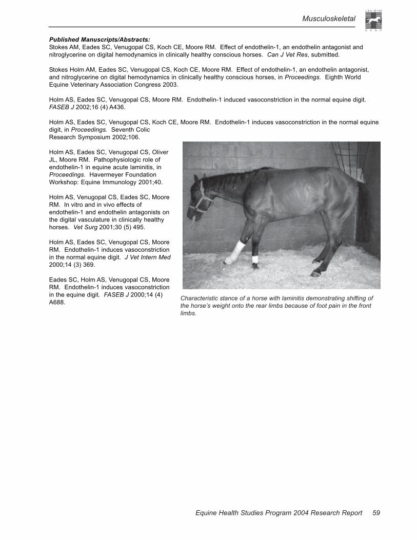
Equine Health Studies Program 2004 Research Report 59
Musculoskeletal
Published Manuscripts/Abstracts:Stokes AM, Eades SC, Venugopal CS, Koch CE, Moore RM. Effect of endothelin-1, an endothelin antagonist andnitroglycerine on digital hemodynamics in clinically healthy conscious horses. Can J Vet Res, submitted.
Stokes Holm AM, Eades SC, Venugopal CS, Koch CE, Moore RM. Effect of endothelin-1, an endothelin antagonist,and nitroglycerine on digital hemodynamics in clinically healthy conscious horses, in Proceedings. Eighth WorldEquine Veterinary Association Congress 2003.
Holm AS, Eades SC, Venugopal CS, Moore RM. Endothelin-1 induced vasoconstriction in the normal equine digit.FASEB J 2002;16 (4) A436.
Holm AS, Eades SC, Venugopal CS, Koch CE, Moore RM. Endothelin-1 induces vasoconstriction in the normal equinedigit, in Proceedings. Seventh ColicResearch Symposium 2002;106.
Holm AS, Eades SC, Venugopal CS, OliverJL, Moore RM. Pathophysiologic role ofendothelin-1 in equine acute laminitis, inProceedings. Havermeyer FoundationWorkshop: Equine Immunology 2001;40.
Holm AS, Venugopal CS, Eades SC, MooreRM. In vitro and in vivo effects ofendothelin-1 and endothelin antagonists onthe digital vasculature in clinically healthyhorses. Vet Surg 2001;30 (5) 495.
Holm AS, Eades SC, Venugopal CS, MooreRM. Endothelin-1 induces vasoconstrictionin the normal equine digit. J Vet Intern Med2000;14 (3) 369.
Eades SC, Holm AS, Venugopal CS, MooreRM. Endothelin-1 induces vasoconstrictionin the equine digit. FASEB J 2000;14 (4)A688. Characteristic stance of a horse with laminitis demonstrating shifting of
the horse’s weight onto the rear limbs because of foot pain in the frontlimbs.

60 Equine Health Studies Program 2004 Research Report
Parasitology
ParasitologyAlterations in histologic and immunohistochemical staining fornitric oxide alterations in parasite free ponies infected withStrongylus vulgarisAuthors/Investigators:Jeremy D. Hubert, BVSc, MRCVS, MS, DACVS; Thomas L. Seahorn, DVM, MS; Thomas R. Klei, PhD; Julian L. Oliver,DVM, PhD; Giselle Hosgood BVSc, MS, PhD, DACVS; Rustin M. Moore DVM, PhD, DACVS
Description of the Problem:This study utilizes a model of gastrointestinal inflammation created by the parasite Strongylus vulgaris to evaluate therole of nitric oxide in GI inflammation. Nitric oxide (NO) is a known modulator of specific physiological roles namely NOderived from the enzyme constitutive nitric oxide synthase (NOS); this NO regulates vasomotor tone and neuralpathways. However in certain situations the NO is stimulated by inducible NOS (iNOS) and released in greaterconcentrations and for a longer duration that may be pathophysiologic and damaging to tissues. In this study thedegree of NO release is determined by evaluating the tissues with special stains to determine if the parasite causesinduction of iNOS and thus increased NO concentrations.
Study Objective:The objective of this study is to determine the effect of infection with Strongylus vulgaris on helminth-naïve ponies andto determine the presence of tissue NO synthase (NOS) using NADPH diaphorase histochemical and inducible NOSand nitrotyrosine immunohistochemical staining.
Approach:Twenty-eight ponies reared helminth-naïve were used. Group 1 ponies (n=7) were administered 500 third stage S.vulgaris larvae orally and given a placebo on day 56 postinoculation. Group 2 ponies (n=16) considered the controlgroup, consisted of 13 ponies who were administered 500 S. vulgaris larvae orally on day 0 and were treated with aproprietary anthelminthic drug on day 56, and 3 ponies that did not receive any S. vulgaris larvae. The ponies wereeuthanatized on day 95. All group 2 ponies were determined to be free of helminths at necropsy. Specimens werecollected after euthanasia for histologic evaluation, tissue NO synthase (NOS) as determined by NADPH diaphorasehistochemical, and iNOS nitrotyrosine (NT) immunohistochemical staining.
Accomplishments/Results/Conclusions:Evidence of increased NO synthesis in the intestinal tract of parasitized ponies is suggested by increased NADPH,iNOS and increased nitrotyrosine staining. These findings provide some evidence of the role of NO in parasite-inducedintestinal inflammation in ponies.
Benefits to/Impact on the Equine Industry:Gastrointestinal problems are the second most common problem after lameness or musculoskeletal related healthissues due to the athletic nature of the horse. However; a significant component of gastrointestinal problems or colicare directly related to parasite burden and pathology due to parasites. Experimental infection with S. vulgaris larvaehave been shown to cause significant inflammation and the mechanism of this inflammation needs to be elucidated soas to develop and institute relevant treatments methods and protocols.
Take Home Message:This study reveals that there is a role of NO in gastrointestinal inflammation. Further studies are required to elucidatethe exact role in the pathophysiological observed.
Acknowledgments:This study was supported by grants from USDA 1433 funds and the Department of Veterinary Clinical SciencesOrganized Research Fund, School of Veterinary Medicine, Louisiana State University. The authors thank Mae Lopezand Catherine Koch for technical assistance.
Year Completed: 1999
Equine Health Studies Program 2004 Research Report 61
Parasitology
Published Manuscripts/Abstracts:Hubert JD, Seahorn TL, Oliver J, Hosgood G, Klei T, Moore RM. Alterations in histologic and immunohistochemicalstaining for nitric oxide alterations in parasite free ponies infected with Strongylus vulgaris. Can J of Vet Res,submitted.
Hubert JD, Seahorn TL, Oliver JL, Klei TR, Hosgood G, Moore RM. Alterations in histologic and immunohistochemicalstaining for nitric oxide in helminth-naive ponies infected with Strongylus vulgaris, in Proceedings. Seventh ColicResearch Symposium 2002.
Clinical signs, hematologic, cytokine, and plasma nitric oxidealterations in response to Strongylus vulgaris infection inponiesAuthors/Investigators:Jeremy D. Hubert, BVSc, MRCVS, MS, DACVS; Thomas L. Seahorn, DVM, MS; Thomas R. Klei, PhD; Giselle HosgoodBVSc, MS, PhD, DACVS; Rustin M. Moore DVM, PhD, DACVS
Description of the Problem:This study utilizes a model of gastrointestinal inflammation created by the parasite Strongylus vulgaris to evaluate therole of nitric oxide in GI inflammation. Nitric oxide (NO) is a known modulator of specific physiological roles namely NOderived from the enzyme constitutive nitric oxide synthase (NOS); this NO regulates vasomotor tone and neuralpathways. However in certain situations the NO is stimulated by inducible NOS (iNOS) and released in greaterconcentrations and for a longer duration that may be pathophysiologic and damaging to tissues. In this study theclinical signs of the ponies are observed as well as relevant hematological parameters measures to ascertain that thereis inflammation caused by ingestion of the parasite and then the plasma levels of NO are measured to see if there is acorrelation between NO and inflammation.
Study Objective:The objective of this study is to illustrate the inflammatory response created by the parasite and correlate it with plasmanitric oxide concentrations.
Approach:Twenty-eight ponies were reared parasite free. Group 1 (n=21) was administered 500 S. vulgaris L3 larvae vianasogastric tube and group 2 (n=7) received a saline control. Ponies were monitored for clinical signs daily, and bloodwas collected for CBC, serum cytokines (TNF, IL-1, IL-6), and plasma NO on day 0 and serially on days 3, 6, 9, 14, 21and 45 days after infection.
Accomplishments/Results/Conclusions:Group 1 ponies were depressed, anorexic and febrile for a variable period, but group 2 ponies remained free of thesesigns. Plasma NO was increased on day 21 in group-1 and on days 9 and 21 in group-2. Total WBC counts wereincreased on day 6, 9, 14, and 21 in group-1, but there were no changes in group-2. Segmented neutrophils wereincreased on day 6 in group-1 and were decreased on days 14, 21, and 45 in group-2; there were no differences inband neutrophils in either group. Lymphocytes were decreased on days 6 and 9 in group-1 and increased on days 14,21, and 45 in group-2. The RBC count and PCV were decreased in group-1 on days 9, 14, 21, and 45 and in group-2on day 45. Total plasma protein concentration was increased on days 14 and 45 in group-1; there were no differencesacross time in group-2. Fibrinogen concentration was increased on days 6, 9, 14, 21, and 45 in group-1, and on day21 in group-2. There were no differences in serum cytokines across time in group 1 ponies.
Benefits to/Impact on the Equine Industry:Gastrointestinal problems are the second most common problem after lameness or musculoskeletal related healthissues due to the athletic nature of the horse. However; a significant component of gastrointestinal problems or colicare directly related to parasite burden and pathology due to parasites. Experimental infection with S. vulgaris larvaehave been shown to cause significant inflammation and the mechanism of this inflammation needs to be elucidated soas to develop and institute relevant treatments methods and protocols.
Take Home Message:The significance of the role of NO in GI inflammation in this model could not be determined. Further studies arerequired.
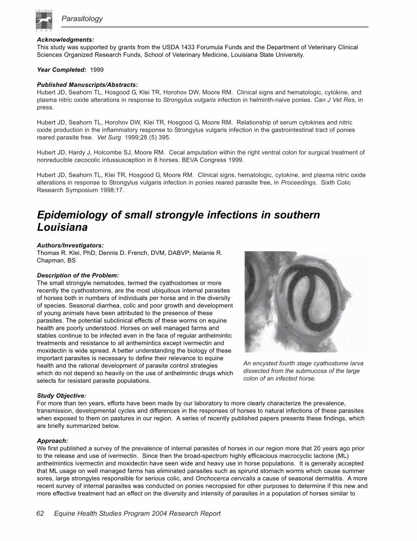
62 Equine Health Studies Program 2004 Research Report
Parasitology
Acknowledgments:This study was supported by grants from the USDA 1433 Forumula Funds and the Department of Veterinary ClinicalSciences Organized Research Funds, School of Veterinary Medicine, Louisiana State University.
Year Completed: 1999
Published Manuscripts/Abstracts:Hubert JD, Seahorn TL, Hosgood G, Klei TR, Horohov DW, Moore RM. Clinical signs and hematologic, cytokine, andplasma nitric oxide alterations in response to Strongylus vulgaris infection in helminth-naive ponies. Can J Vet Res, inpress.
Hubert JD, Seahorn TL, Horohov DW, Klei TR, Hosgood G, Moore RM. Relationship of serum cytokines and nitricoxide production in the inflammatory response to Strongylus vulgaris infection in the gastrointestinal tract of poniesreared parasite free. Vet Surg 1999;28 (5) 395.
Hubert JD, Hardy J, Holcombe SJ, Moore RM. Cecal amputation within the right ventral colon for surgical treatment ofnonreducible cecocolic intussusception in 8 horses. BEVA Congress 1999.
Hubert JD, Seahorn TL, Klei TR, Hosgood G, Moore RM. Clinical signs, hematologic, cytokine, and plasma nitric oxidealterations in response to Strongylus vulgaris infection in ponies reared parasite free, in Proceedings. Sixth ColicResearch Symposium 1998;17.
Epidemiology of small strongyle infections in southernLouisianaAuthors/Investigators:Thomas R. Klei, PhD; Dennis D. French, DVM, DABVP; Melanie R.Chapman, BS
Description of the Problem:The small strongyle nematodes, termed the cyathostomes or morerecently the cyathostomins, are the most ubiquitous internal parasitesof horses both in numbers of individuals per horse and in the diversityof species. Seasonal diarrhea, colic and poor growth and developmentof young animals have been attributed to the presence of theseparasites. The potential subclinical effects of these worms on equinehealth are poorly understood. Horses on well managed farms andstables continue to be infected even in the face of regular anthelmintictreatments and resistance to all anthemintics except ivermectin andmoxidectin is wide spread. A better understanding the biology of theseimportant parasites is necessary to define their relevance to equinehealth and the rational development of parasite control strategieswhich do not depend so heavily on the use of anthelmintic drugs whichselects for resistant parasite populations.
Study Objective:For more than ten years, efforts have been made by our laboratory to more clearly characterize the prevalence,transmission, developmental cycles and differences in the responses of horses to natural infections of these parasiteswhen exposed to them on pastures in our region. A series of recently published papers presents these findings, whichare briefly summarized below.
Approach:We first published a survey of the prevalence of internal parasites of horses in our region more that 20 years ago priorto the release and use of ivermectin. Since then the broad-spectrum highly efficacious macrocyclic lactone (ML)anthelmintics ivermectin and moxidectin have seen wide and heavy use in horse populations. It is generally acceptedthat ML usage on well managed farms has eliminated parasites such as spirurid stomach worms which cause summersores, large strongyles responsible for serious colic, and Onchocerca cervcalis a cause of seasonal dermatitis. A morerecent survey of internal parasites was conducted on ponies necropsied for other purposes to determine if this new andmore effective treatment had an effect on the diversity and intensity of parasites in a population of horses similar to
An encysted fourth stage cyathostome larvadissected from the submucosa of the largecolon of an infected horse.
Equine Health Studies Program 2004 Research Report 63
Parasitology
those examined prior to the advent of ML use. The overall prevalence and intensity of strongyle parasites was reducedin the more recent survey. However, with the exception of the stomach worms, the diversity of parasites recovered wasnot altered. This suggests that while some species such as the very pathogenic large strongyles are less common andnot found in the numbers per horse seen prior to ML use they still occur in horses not held in closed herds under heavyanthelmintic pressure. Twenty-six species of cyathostome including two species not found in the earlier survey werealso found. The prevalence of round worms (Parascaris equorum) pinworms (Oxyuris equi) and tapeworms(Anoplocephala perfoliata) have not changed. Clearly while having a major positive impact on equine health, the use ofML anthelmintics has not modified the diversity of internal parasites in the horse population at large.
Accomplishments/Results/Conclusions:The life cycle of the equine cyathostomins is typical of strongyle nematodes and is direct. Eggs released by femaleworms living in the large intestine and cecum pass on to pasture in the feces of the hoarse. These develop to infectivethird stage larvae (L3) which are ingested. These larvae enter the mucosa of the large intestine. At this point thedevelopmental pattern can take one of two paths. L3 may encyst and go into an arrested phase of development (fig).These hypobiotic larvae or early L3 (EL3) live in this state for an undetermined period completing their development tothe fourth stage when stimulated by unknown host factors. When this development occurs in a synchronous fashionclinical signs occur which are associated with the disruption of intestinal tissues and emergence of larvae into thelumen of the large intestine and cecum.
Benefits to/Impact on the Equine Industry:Knowledge of the seasonal aspects of small strongyle parasitism is important in determining periods of maximaltransmission and development of the parasites within the horse. While difficult, counting the numbers of L3 on pastureherbage has traditionally been used to indirectly determine seasonal transmission. Studies of this nature wereconducted over a three-year period from 1986-1989 and again for another year in 1996-1997 in East Baton Rougeparish. The pastures used held a pony herd of mares and their foals. Fecal egg counts (FEC) from mares and foalsand pasture L3/kg dry herbage were used to measure changes in parasite populations within the ponies and on thepasture. A FEC rise occurred during the late summer and early autumn, which preceded a peak of L3 on pasture duringthe winter season. The numbers of L3 were reduced during the hottest months of the year due mainly to minimumdaily temperature above 18C and in the winter due to brief periods of freezing (fig). However, L3 numbers quicklyreturned to peak levels when temperatures returned to normal winter ranges.
An alternate developmental path occurs when the entering L3 do not arrest but develop through the mucosal L4 stageto the luminal L4 and adult stages. A more accurate picture of the seasonal nature of cyathostomin transmission anddevelopment was seen in studies that counted parasites in ponies removed from pasture at different times of the yearor others which utilized parasite free pony yearlings as sentinels. This allowed for the determination of the proportionof parasites in the different stages of development as well as seasonal transmission cycles. In studies of sentinelsmaximal transmission was seen during the winter and the lowest occurred during the summer, supporting theobservations of L3 on pasture herbage. Adult parasite numbers were highest in the spring and mucosal parasitenumbers were the lowest at this time as expected. This suggests that a seasonal development of EL3 and emergenceof larvae occur in the winter-spring, which corresponds, to periods of larval cyatostominosis. Nonetheless, in thesestudies EL3 were the major portion of the parasite population within the horse at all seasons. The total parasiteburdens, a sum of all stages, was constant in young ponies, <5yrs of age, removed from pasture. However, peakpercentages of adult parasites occurred during the fall accounting for the pasture contamination and high L3 numbersseen on pasture during the winter. EL3 numbers increase during this period reaching a peak percentage during thesummer. Increased age of ponies and presumably the development of acquired immunity resulting from increasedparasite exposure resulted in an increased proportion of EL3 in the parasite population within the horse. Thisphenomenon appears to be separate from protective immunity in that increased induction of arrested developmentoccurs rapidly in yearlings.
Take Home Message:These experimental and field observations are the most extensive series of studies reported on seasonal and hostfactors effecting cyathostomin transmission and the accumulation of EL3 in a single region to date. The data collectedindicate that treatment of horses in this region with anthelmintics which eliminate EL3 during the late summer or earlyfall will reduce the number of adults and concomitantly the contamination of fall and winter pastures with infectivelarvae. These treatments will also reduce the accumulation of potentially dangerous levels of EL3. This reduction inreinfection should make possible the development of parasite control programs less dependent on the extensive use ofanthelmintics in mature horses. Other factors such as age, housing conditions and anthelmintic efficacy within a givenpopulation must also be taken into consideration in designing specific parasite control strategies.
Acknowledgments:These studies were supported in part by the Louisiana Agricultural Experiment Station and contracts from Merial Ltd.,Pfizer Inc., Virbac, and Fort Dodge Animal Health.

64 Equine Health Studies Program 2004 Research Report
Parasitology
Year Completed: 2000
Published Manuscripts/Abstracts:Chapman MR, Kearney MT, Klei TR. Equine cyathostome populations: Accuracy of species composition estimations.Vet Parasitol 2003;116: 15-21.
Chapman MR, French DD, Klei TR. Prevalence of strongyle nematodes in naturally infected ponies of different agesand during different seasons of the year in Louisiana. J Parasitol 2003;89: 309-314.
Chapman MR, French DD, Klei TR. Gastrointestinal helminths of ponies in Louisiana: a comparison of speciescurrently prevalent with those present 20 years ago. J Parasitol 2002;88:1130-4.
Chapman MR, French DD, Taylor HW, Klei TR. One season of pasture exposure fails to induce a protective resistanceto cyathostomes but increases numbers of hypobiotic third stage larvae. J Parasitol 2002;88: 687-683.
Chapman MR, French DD, Klei TR. Seasonal transmission of gastrointestinal parasites of equids in southernLouisiana. J Parasitol 2001;87:1371-1378.
Klei TR, Rehbein S, Visser M, Langholff WK, Chapman MR, French DD, Hanson P. Re-evaluation of ivermectin againstequine strongyles after 20 years of use. Vet Parasitol 2001;98: 315-320.
Baudena MA, Chapman MR, Larsen M, Klei TR. Efficacy of the nematophagous fungus Duddingtonia flagrans inreducing equine cyathostome larvae on pasture in south Louisiana. Vet Parasitol 2000;28;89(3):219 230.
Baudena MA, Chapman MR, French DD, Klei TR. Seasonal development and survival of equine cyathostome larvaeon pasture in south Louisiana. Vet Parasitol 2000;29;88(1 2):51 60.
Chapman MR, Kearney M T, Klei TR. An experimental evaluation of methods used to enumerate mucosal cyathostomelarvae in ponies. Vet. Parasitol 1999;86: 191-202.
Immunity to strongyle nematodes in poniesAuthors/Investigators:Thomas R. Klei, PhD; Melanie R. Chapman, MS; David W. Horohov, PhD; Rustin M. Moore, DVM, PhD, DACVS;Jenifer Edmonds, DVM, Ph.D.; Maria Alexandra Baudena, DVM, MS, Ph.D.; Dennis D. French, DVM, DABVP; H.Wayne Taylor, DVM, PhD, DACVP
Description of the Problem:Understanding the horse’s immune response to pathogens is an essential part of understanding diseases they cause.Detailed studies on acquired resistance to nematodes of horses are limited and pertain almost exclusively to strongyleinfections. Based on their biology it is not surprising that theacquisition and character of immunity to infections of the largestrongyle, specifically Strongylus vulgaris and the smallstrongyles or cyathostomes differ markedly. We have investigatedboth over the past 25 years and summarize our most recentstudies below.
While Strongylus vulgaris is a rare parasite on well managedfarms, infections of parasite free reared ponies with knownnumbers infective larvae (L3) has been a useful model to studyequine immune responses to nematode parasites. The mostdetailed information on equine immunity to nematodes has comefrom this model system. In this system we have recentlydemonstrated that immunization of naive ponies with radiationattenuated L3 (IRRL3) induces a distinct Th2 immune responsewhich is protective against reinfection. However, immunizationwith soluble adult worm antigen in Ribi adjuvant induces amarked Th1 response and is not protective. Cytokine, antibodyand cellular responses were characterized to determine whetherprior immunization with Ribi adjuvant would down regulate the
A scanning electron micrograph of Strongylusvulgaris third stage infective larvae with attachedeosinophils. Antibodies mediate the adherence ofthe cells and lead to killing in vitro.

Equine Health Studies Program 2004 Research Report 65
Parasitology
Th2 response induced by a subsequent IRRL3 immunization as well as the protection it induces. Similar to poniesvaccinated only with IRRL3, animals immunized with both Ribi and IRRL3 were still protected against challengeinfections even though they demonstrated alterations in the factors associated with the protective immune responses.Protection against infection was associated with increases in IL-5 and IL-13 mRNA in colonic lymph nodes, as well asan eosinophilia. A prior immunization with Ribi and it Th1 response reduced levels of Il-5 and eosinophils it was notsufficient to reduce protective immunity. Unlike immune responses to small strongyles describe below elevate levels ofIl-4 and mastocytosis were not associated with protection against this large strongly parasite.
Unlike the large strongyles the small strongyles (cyathostomes or more recently cyatostomins) continue to be found inhorses on well managed farms and have developed resistance to all classes of anthelmintic except the macrocycliclactones. Data on field observations suggest that horses acquire immunity to cyathostome infections with age. Whenexamining this resistance using strongyle fecal egg counts it is clear and not surprising that horses respond tostrongyle nematode infections like other hosts. The frequencies of EPGs are evenly dispersed within the yearlingpopulation but over dispersed in the mature horse population. In this situation very few mature animals are infectedwith large numbers of parasites. It is likely this difference in acquired resistance is genetically regulated. Twoexperiments have been conduced recently to define more carefully the immunity to these most important worms.Parasite recovery data following challenge in these experiments showed that ponies with acquired resistance showed areduction in the total numbers of parasites, including adults with in the lumen of the intestine, and developing larvaewith in intestinal wall. The acquisition of resistance was also accompanied by an increase in hypobiotic larvae,intestinal mast cells, eosinophils and parasite specific antibody responses. These types of immune responses are theconstant increases in Th2 cytokine mRNA seen in colonic lymph nodes, principally IL-4.
We have recently participated in studies with scientists at the University of Liverpool in their efforts to identify newdiagnostic methods to determine the risk of larval cyathostomiasis. These studies utilize a unique antigen identified bythe group from developing larvae within the mucosa of the horse. The titer of IgG(T) to these antigens corresponds tothe number of larvae with in the intestinal wall of the horse. This assay when further developed will allow for the clinicalassessment of cyathostomiasis and advance antemotrtum epidemiological studies.
The data recovered to date provide important information on immunity to nematode parasites. Further, these studiesalso help characterize basic aspects of equine immunology important to studies of pathogenesis and vaccination.
Because it is apparent that nematode parasites can significantly alter the horse’s cytokine profile, the question “doparasite infections alter the outcome of vaccinations with other proteins?” was asked. Antibody, lymphoproliferationand cytokine responses to single intra muscular inoculations of KLH (a large foreign protein) were measured in ponieswith high, medium and low parasite burdens. The data indicated that heavily parasitized ponies had a uniformly markedreduction in immune responses directed toward the vaccination. This response was not polarized in a Th1/Th2fashion. While necessary to confirm, the data suggest that nematode parasitism may detrimentally affect vaccinationagainst other agents.
Acknowledgments:These studies were supported in part by grants from the USDA-NRI-CGP, Fort-Dodge Animal Health, and theAgricultural Experiment Station, Louisiana State University.
Published Manuscripts/Abstracts:Dowdall SM, Proudman CJ, Klei TR, Mair T, Matthews JB. Characterization of IgG(T) serum antibody responses to twolarval antigen complexes in horses naturally- or experimentally-infected with cyathostomins. Int J Parasitol 2004;34:101-108.
Dowdall SMJ, Proudman CJ, Love S, Klei TR, Mathews JB. Purification and analyses of the specificity of two putativediagnostic antigens for larval cyathostomin infections in horses. Res Vet Sci 2003;75:223-229.
Chapman, MR, French DD, Taylor HW, Klei TR. One season of pasture exposure fails to induce a protective resistanceto cyathostomes but increases numbers of hypobiotic third stage larvae. J Parasitol 2002;88: 687-683.
Edmonds JD, Horohov DW, Chapman MR, Pourciau SS, Antoku K, Snedden K, Klei TR. Altered immune responses toa heterologous protein in ponies with heavy gastrointestinal parasite burdens. Equine Vet J 2001;33:658-63.
Klei TR. Equine immunity to parasites. Vet Clin North Am Equine Pract 2000;16(1):69 78, vi.
Le Jambre LF, Gray GD, Klei TR. Workshop on irradiated larval vaccines. Int J Parasitol 1999;29(1):193 8.
Klei TR, Chapman MR. Immunity in equine cyathostome infections. Vet Parasitol 1999;86: 191-202.

66 Equine Health Studies Program 2004 Research Report
Parasitology
Swiderski CE, Klei TR, Folsom RW, Pourciau SS, Chapman A, Chapman MR, Moore RM, McClure JR, Taylor HW,Horohov DW. Vaccination against Strongylus vulgaris in ponies: Comparison of the humoral and cytokine responsesof vaccinates and nonvaccinates. Advances in Veterinary Medicine v 41, Veterinary Vaccines and Diagnostics. NewYork: Academic Press, 1999;389-404.
Swiderski CE, Klei TR, Horohov DW. Quantitative measurement of equine cytokine mRNA expression by polymerasechain reaction using target-specific standard curves. J Immunol Method 1999; 222: 155-169.
Monahan CM, Chapman MR, French DD, Taylor HW, Klei TR. Experimental cyathostome challenge of poniesmaintained with or without benefit of daily pyrantel tartrate feed additive: Comparisons of parasite burdens, immunityand colonic pathology. Vet Parasit 1998;74: 229-241.
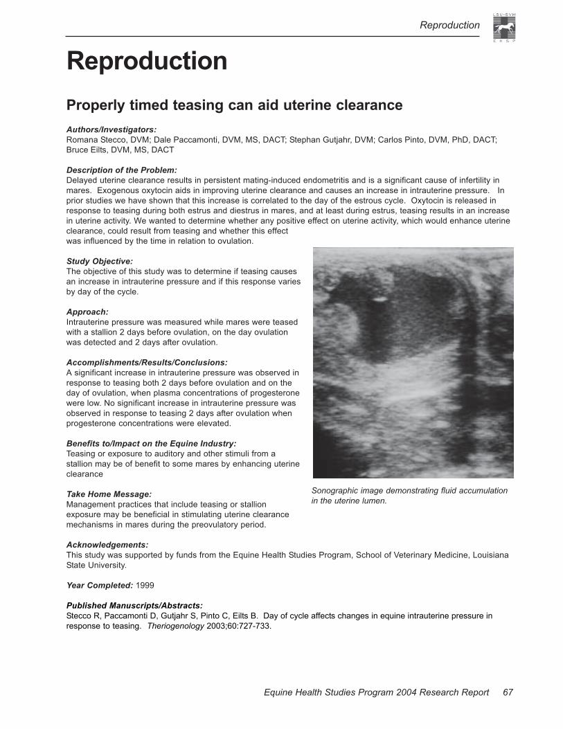
Equine Health Studies Program 2004 Research Report 67
Reproduction
ReproductionProperly timed teasing can aid uterine clearanceAuthors/Investigators:Romana Stecco, DVM; Dale Paccamonti, DVM, MS, DACT; Stephan Gutjahr, DVM; Carlos Pinto, DVM, PhD, DACT;Bruce Eilts, DVM, MS, DACT
Description of the Problem:Delayed uterine clearance results in persistent mating-induced endometritis and is a significant cause of infertility inmares. Exogenous oxytocin aids in improving uterine clearance and causes an increase in intrauterine pressure. Inprior studies we have shown that this increase is correlated to the day of the estrous cycle. Oxytocin is released inresponse to teasing during both estrus and diestrus in mares, and at least during estrus, teasing results in an increasein uterine activity. We wanted to determine whether any positive effect on uterine activity, which would enhance uterineclearance, could result from teasing and whether this effectwas influenced by the time in relation to ovulation.
Study Objective:The objective of this study was to determine if teasing causesan increase in intrauterine pressure and if this response variesby day of the cycle.
Approach:Intrauterine pressure was measured while mares were teasedwith a stallion 2 days before ovulation, on the day ovulationwas detected and 2 days after ovulation.
Accomplishments/Results/Conclusions:A significant increase in intrauterine pressure was observed inresponse to teasing both 2 days before ovulation and on theday of ovulation, when plasma concentrations of progesteronewere low. No significant increase in intrauterine pressure wasobserved in response to teasing 2 days after ovulation whenprogesterone concentrations were elevated.
Benefits to/Impact on the Equine Industry:Teasing or exposure to auditory and other stimuli from astallion may be of benefit to some mares by enhancing uterineclearance
Take Home Message:Management practices that include teasing or stallionexposure may be beneficial in stimulating uterine clearancemechanisms in mares during the preovulatory period.
Acknowledgements:This study was supported by funds from the Equine Health Studies Program, School of Veterinary Medicine, LouisianaState University.
Year Completed: 1999
Published Manuscripts/Abstracts:Stecco R, Paccamonti D, Gutjahr S, Pinto C, Eilts B. Day of cycle affects changes in equine intrauterine pressure inresponse to teasing. Theriogenology 2003;60:727-733.
Sonographic image demonstrating fluid accumulationin the uterine lumen.
68 Equine Health Studies Program 2004 Research Report
Reproduction
Effect of dose and day of treatment on uterine response tooxytocin in maresAuthors/Investigators:Stephan Gutjahr, DVM; Dale L. Paccamonti, DVM, MS, DACT; Jonathan Pycock, DVM; Marcel Taverne, PhD; Stef J.Dieleman, PhD; G.C. van der Weijden, DVM
Description of the Problem:Delayed uterine clearance results in persistent mating-induced endometritis and is a significant cause of infertility inmares. Oxytocin is commonly used to treat this condition, however little is known about the effect elicited by differentdoses of oxytocin nor is there any information about the effect of treatment in relation to the time of ovulation.
Study Objective:The objective of this study was to determine the effect of dose and day of oxytocin treatment on intrauterine pressure.
Approach:Mares were treated with either a low (10 IU) or high (25 IU) dose of oxytocin 2 days before ovulation, on the day ofovulation and 2 days after ovulation.
Accomplishments/Results/Conclusions:The administration of both 10 and 25 IU oxytocin induced a response. The intensity of response depended on the dayof administration and the dose of oxytocin. The variation of response was significantly greater after 10 IU oxytocincompared with 25 IU oxytocin. The uterine response was greatest on Day 2 prior to ovulation and lowest on Day 2 afterovulation. The response was negatively correlated to increasing plasma progesterone.
Benefits to/Impact on the Equine Industry:The results of this study show that oxytocin administration to mares before ovulation provides a greater response thanafter ovulation. A decline in the intensity of response after ovulation can be compensated for with a higher dose ofoxytocin.
Take Home Message:It may be beneficial to breed an infertile mare that has a uterine clearance problem earlier in relation to ovulation. Wewould then have a longer period before she ovulates when oxytocin treatment is most effective to treat the problem.
Acknowledgements: University of Utecht, Netherlands.
Year Completed: 1999
Published Manuscripts/Abstracts:Gutjahr S, Paccamonti DL, Pycock JF, Taverne MA, Dieleman SJ, van der Weijden GC. Effect of dose and day oftreatment on uterine response to oxytocin in mares. Theriogenology 2000;54, 447-456.
Prostaglandin F2á metabolite (PGFM) response to exogenousoxytocin and determination of the half-life of oxytocin innonpregnant maresAuthors/Investigators:Dale L. Paccamonti, DVM, MS, DACT; Jonathan Pycock, DVM; Marcel Taverne, Ph.D.; Mart Bevers, Ph.D., G.C. vander Weijden, DVM; Stephan Gutjahr, DVM; D. Schams, Ph.D.; David Blouin, Ph.D.
Description of the Problem:Prostaglandin and oxytocin are commonly used after breeding to treat infertility, specifically the problem of delayeduterine clearance. Both cause an increase in uterine contractions and improve the uterus’ ability to clear itself of debrisand infection. Some reports seemed to indicate that oxytocin caused a release of endogenous (the mare’s own)prostaglandin yet the intensity of this effect was unclear. Furthermore, the half-life of oxytocin, an indication of howlong it would remain active, was unknown.
Equine Health Studies Program 2004 Research Report 69
Study Objective:The objective of this study was to determine the half-life of oxytocin and to evaluate the release of endogenousprostaglandin after the administration of oxytocin to mares.
Approach:Oxytocin was given intravenously and blood was then obtained at intervals to determine the amount of oxytocin andprostaglandin in the bloodstream.
Accomplishments/Results/Conclusions:The half-life of oxytocin in the mare is 7 minutes. Administration of oxytocin results in a significant release ofendogenous prostaglandin, further increasing uterine motility and enhancing uterine clearance.
Benefits to/Impact on the Equine Industry:This study will help to design better protocols for the treatment of delayed uterine clearance.
Take Home Message:Because of the short half-life of oxytocin, repeated small doses may be more beneficial than a single large dose.
Year Completed: 1998
Published Manuscripts/Abstracts:Paccamonti DL, Pycock JF, Taverne MA, Bevers M, van der Weijden GC, Gutjahr S, Schams D, Blouin D. PGFMresponse to exogenous oxytocin and determination of the half-life of oxytocin in nonpregnant mares. Eq Vet J1999;31:285-288.
Role of nitric oxide in ovulation in the mareAuthors/Investigators:Carlos Pinto, DVM, Ph.D., DACT; Dale L. Paccamonti, DVM, MS, DACT; Bruce E. Eilts, DVM, MS, DACT; C. S.Venugopal, DVM, Ph.D.; Charles R. Short, DVM, Ph.D.; Laura R. Gentry,Ph.D.; Donald L. Thompson, Ph.D.; Robert A.Godke,Ph.D.
Description of the Problem:The process of ovulation in horses and other species is not completely understood. More detailed knowledge willenable us to better control ovulation and aid us in the breeding management of horses. Recent studies suggest thatnitric oxide (NO) may have a role in regulating ovarian physiology.
Study Objective:The objective of this study was to investigate the role of nitric oxide in ovulation in the mare.
Approach:Nitric oxide synthesis in the pre-ovulatory follicle was inhibited to see if ovulation was delayed. Nitric oxide inhibitors(L-NAME or aminoguanidine) were injected into the pre-ovulatory follicle during estrus by a transvaginal ultrasoundguided approach and follicular development and the time to ovulation were monitored. In another experiment, the levelof nitric oxide in the follicle was evaluated before and after the administration of hCG (human chorionic gonadotropin),which stimulates follicular maturation and ovulation. Follicular fluid was obtained by transvaginal ultrasound guidedaspiration for analysis. In a third experiment, the role that nitric oxide plays in the production of steroid hormones bythe follicle was investigated in vitro. Granulosa cells were obtained from follicles and cultured in vitro. Nitric oxidedonors and inhibitors were added to determine the effect on production of progesterone and estrogen.
Accomplishments/Results/Conclusions:In the first experiment, the administration of nitric oxide inhibitors delayed ovulation, suggesting a role for nitric oxide infollicular growth and ovulation in horses. In the second experiment, increased intrafollicular concentrations of nitricoxide found after administration of hCG provide evidence for the presence of an nitric oxide-generating system in theequine preovulatory follicle. In the third experiment, addition of a nitric oxide donor to granulosa cells in vitro reducedestrogen production, while addition of a nitric oxide inhibitor increased estrogen production.
Benefits to/Impact on the Equine Industry:These studies indicate a role of nitric oxide in the process of ovulation in the mare. Further studies may enable us tobetter control ovulation, either by inducing, or alternatively delaying, ovulation in a tightly controlled fashion.
Reproduction

70 Equine Health Studies Program 2004 Research Report
Take Home Message:Better methods to induce or delay ovulation, possibly with the aid of nitric oxide mediators may enable us to bettercontrol ovulation and enhance breeding management.
Published Manuscripts/Abstracts:Pinto CR, Paccamonti DL, Eilts BE, Short CR, Godke RA. Effect of nitric oxide synthase inhibitors on ovulation in hCG-stimulated mares. Theriogenology 2002;58:1017-26.
Pinto CR, Paccamonti DL, Eilts BE, VenugopalCS, Short CR, GentryLR, Thompson DL, Godke RA. Concentrations ofnitric oxide in equine preovulatory follicles before and after administration of human chorionic gonadotropin.Theriogenology 2003;60:819-827.
Pinto CR, Paccamonti DL, Eilts BE, Short CR, Gentry LR, Thompson DL, Godke RA. Evidence for a nitric oxide-mediated modulation of equine granulosa cell steroidogenesis. Theriogenology 2002;58:579-583.
“In-vitro” produced foalsAuthors/Investigators:Richard Cochran, Ph.D.; Marius Meintjes, DVM, Ph.D.; Brett Reggio, Ph.D.; Darin Hylan, MS; Joel Carter; Carlos Pinto,DVM, Ph.D., DACT; Dale Paccamonti, DVM, MS, DACT; Robertr Godke, Ph.D.
Description of the Problem:In vitro fertilization is commonplace in other species. It has great economic benefits in the cattle industry and providessignificant advantages in the treatment of infertility in humans. Successful in vitro fertilization in horses could providesimilar benefits. However, success has proven elusive.
Study Objective:The objective of this study was to develop a successful method for in vitro fertilization in horses and produce live foals.
Approach:Evaluation of various methods including sub-zonal injection of sperm (SUZI) and eventually succeeded withintracytoplasmic sperm injection (ICSI).
Accomplishments/Results/Conclusions:We were the first to report the birth of foals from oocytesobtained from live mares. Our first pregnancy failed togo to term. That same year, another laboratoryannounced the birth of a foal derived by the ICSImethod, but the oocytes (‘eggs’) were obtained in thelaboratory from ovaries acquired from a slaughterhouse.Foals developed from oocytes from live mares were stillan elusive goal. The following year, our first live foal (afilly named Sweet Pea) was born, followed by anotherfoal (another filly, Trixie) and then a third foal (this oneonly received a number). (Note: A group in Australiareported similar results the same year. We havedeveloped a relationship with this group and currentlyhave a joint equine practice residency program withthem.)
Benefits to/Impact on the Equine Industry:With these early achievements reported by ourlaboratory, other laboratories have been able to achieveimprovements in the success rate of ICSI. This mayprove to provide valuable benefits to the treatment ofinfertility in stallions and mares.
Reproduction
Intracytoplasmic sperm injection into oocyte to yield in vitro-derived embryos.

Equine Health Studies Program 2004 Research Report 71
Take Home Message:Research at LSU in assisted reproductive techniques continues to achieve important breakthroughs in a number ofspecies. Continued research in this area will undoubtedly benefit the equine industry in numerous ways.
Acknowledgements:This work was performed in collaboration with the Embryo Biotechnology Laboratory, LSU AgCenter.
Published Manuscripts/Abstracts:Cochran R, Meintjes M, Reggio B, Hylan D, Carter J, Pinto C, Paccamonti D, Graff K, Godke R. Production of live foalsfrom sperm-injected oocytes harvested from pregnant mares. J Reprod Fertil Suppl 2000;56, 503-512.
Cochran R, Meintjes M, Reggio B, Hylan D, Carter J, Pinto C, Paccamonti D, Godke RA. Live foals produced fromsperm-injected oocytes derived from pregnant mares. J Eq Vet Sci 1998;18:736-740.
Reproductive parameters of Miniature horse stallionsAuthors/Investigators:Dale L. Paccamonti, DVM, MS, DACT; Ab Buiten, Ph.D.; Joyce Parlevliet, DVM, Ph.D.; Ben Colenbrander, DVM, Ph.D.
Description of the Problem:Standards for breeding soundness evaluation, including parameters used for semen evaluation, of ‘standard’ sizedhorses, such as Quarter Horses, Arabians and Thoroughbreds are well established. However, no such standardsexisted for Miniature Horses. The increasing popularity of Miniature horses, and the resulting increase in theireconomic value, resulted in an increased demand for the evaluation of breeding stallions. Veterinarians needed toknow what could be considered normal and acceptable for a Miniature stallion.
Study Objective:The objective of this study was to determine normalvalues for testicular measurements and semenparameters for Miniature stallions
Approach:The results of 216 breeding soundness evaluationsperformed on Miniature stallions were analyzed.
Accomplishments/Results/Conclusions:Miniature stallions differ significantly from ‘standardsized’ stallions in testicular size and numbers ofsperm produced.
Benefits to/Impact on the Equine Industry:Veterinarians now have reference values whenevaluating a Miniature Horse stallion for breedingsoundness.
Take Home Message:Normal values have been established and can beused to assess the reproductive potential of Miniaturehorse stallions.
Acknowledgements: University of Utrecht, Netherlands.
Year Completed: 1998
Published Manuscripts/Abstracts:Paccamonti DL, Buiten AV, Parlevliet J, Colenbrander B. Reproductive parameters of miniature stallions.Theriogenology 1999;51:1343-1349.
Reproduction
Collection of a Miniature horse stallion using an artificial vaginafor evaluating breeding soundness.
72 Equine Health Studies Program 2004 Research Report
The effects of pH, osmolarity and urine contamination on equinespermatozoal motilityAuthors/Investigators:Stefanie Griggers, DVM; Dale L. Paccamonti, DVM, MS, DACT; R. Alex Thompson, DVM, Ph.D.; Bruce E. Eilts, DVM,MS, DACT
Description of the Problem:Urospermia, the contamination of semen with urine, results in infertility in stallions. Development of techniques to treatsemen of stallions experiencing urospermia thereby improving fertility would be beneficial.
Study Objective:To determine whether it was the change in pH or the osmolarity resulting from urine contamination that was causing themost harm to the sperm, and to determine whether the addition of semen extender, either alone or in conjunction withcentrifugation to remove the urine, could improve semen quality.
Approach:Semen was collected and subjected to conditions of varying pH, of varying osmolarity, and various quantities of urineand effects on motility were recorded. Then semen contaminated with urine was extended with either of 2 semenextenders, followed by centrifugation or not centrifuged, in an attempt to alleviate the detrimental effect of urine onmotility.
Accomplishments/Results/Conclusions:The results of these experiments showed that alterations in pH and osmolarity negatively affected stallion spermmotility. Optimal pH and osmolarity appeared to be approximately 7.7 and 315, respectively. Contamination of theejaculate with urine significantly decreased sperm motility. Smaller quantities of dilute urine were less detrimental thanlarger quantities of dilute urine, and dilute urine was less detrimental than more concentrated urine. The addition ofsemen extender restored the motility of urine contaminated semen to that of the uncontaminated control, howevercentrifugation to remove urine provided no significant advantage.
Benefit to/Impact on the Equine Industry:Semen samples from stallions experiencing urospermia may be processed to restore fertility.
Take Home Message:Addition of semen extender may improve fertility of semen contaminated with urine. Centrifuging the semen samplewill not provide any additional benefit.
Year Completed: 1998
Published Manuscripts/Abstracts:Griggers S, Paccamonti DL, Thompson RA, Eilts BE. The effects of pH, osmolarity and urine contamination on equinespermatozoal motility. Theriogenology 2001;56:613-622.
Inducing earlier cyclicity in maresAuthors/Investigators:Angela Klump, MS; Abdul Aljarrah, DVM; Marina Sansinena, Ph.D.; Darin Hylan, MS; Carlos Pinto, DVM, Ph.D., DACT;Bruce Eilts, DVM, MS, DACT; Robert Godke, Ph.D. ; Dale Paccamonti, DVM, MS, DACT
Description of the Problem:Because many horse breed associations arbitrarily impose an artificial birthday of January 1 in the Northernhemisphere, horse breeders desire to get mares pregnant early in the year. However, mares are seasonal long-daybreeders, and are usually in anestrus during the winter months. Before resuming normal estrous cycles, mares gothrough transition, a period of anovulatory estrual activity. During transition, estrous periods are irregular and ovulationunpredictable, frustrating reproductive management on the stud farm. Researchers have investigated many ways toadvance the onset of cyclicity and shorten transition in order to expedite getting mares pregnant earlier in the year.
Reproduction

Equine Health Studies Program 2004 Research Report 73
Study Objective:We hypothesized that by aspirating the dominant follicle during the late transitional period, we could induce formationof luteal tissue and thereby hasten the onset of cyclicity. Our specific objective was to perform follicular aspiration andcompare the date of first rise in progesterone between treated and control mares.
Approach:In an attempt to shorten the period of transition after seasonal anestrus and hasten the onset of cyclicity in mares,ultrasound guided transvaginal follicular aspiration was performed on mares when a follicle >35 mm was present.
Accomplishments/Results/Conclusions:Mares that underwent follicular aspiration had serum progesterone > 1 ng/ml sooner than untreated controls (80.5 +/-7.3 d after January 1 vs. 104.3 +/- 8.8 d, mean +/- SE; P = 0.024). Nine of 11 treatment mares formed luteal tissuewithin 8 d after the first aspiration. The remaining two mares formed luteal tissue after a second aspiration procedure.When an aspiration procedure resulted in the formation of luteal tissue, progesterone rose to >1 ng/ml within one week(6.7 +/- 0.9 d).
Benefits to/Impact on the Equine Industry:This technique, used to induce earlier cyclicity, resulted in mares cycling almost 3 weeks earlier than the control mares.
Take Home Message:Follicular aspiration may be a useful tool to induce earlier cycling in mares.
Acknowledgements:This work was performed in collaboration with the Embryo Biotechnology Laboratory, LSU Agricultural Center.
Year Completed: 2000
Published Manuscripts/Abstracts:Klump A, Aljarrah A, Sansinena M, Hylan D, Pinto C, Eilts B, Godke R, Paccamonti D. Follicular aspiration duringtransition to advance the onset of cyclicity. Pferdeheilkunde 2003;19:609-610.
Using acupuncture to advance the onset of cylicityAuthors/Investigators:Alexandra Niemantsverdriet Murton, DVM, DACT; Dale Paccamonti, DVM, MS, DACT; Bruce Eilts, DVM, MS, DACT;Carlos Pinto, DVM, Ph.D., DACT; Elena Garcia, DVM; Glenn Pettifer, DVM, DACVA
Description of the Problem:Because many horse breed associationsarbitrarily impose an artificial birthday of January1 in the Northern hemisphere, horse breedersdesire to get mares pregnant early in the year.However, mares are seasonal long-day breeders,and are usually in anestrus during the wintermonths. Before resuming normal estrous cycles,mares go through transition, a period ofanovulatory estrual activity. During transition,estrous periods are irregular and ovulationunpredictable, frustrating reproductivemanagement on the stud farm. Researchers haveinvestigated many ways to advance the onset ofcyclicity and shorten transition in order to expeditegetting mares pregnant earlier in the year.Because a non-invasive method to advancecyclicity would be desirable, and there is currentlyinterest in non-traditional medicine, acupuncturewas investigated as a means to get mares to cycleearlier in the year.
Reproduction
Advancing the onset of cyclicity in mares will result in earlierpregnancy and subsequent foaling.
74 Equine Health Studies Program 2004 Research Report
Study Objective:The objective of this study was to use acupuncture to get mares cycling earlier in the year.
Approach:In Year 1, traditional standard acupuncture at various sites (Rusty – can give you specific sites if you want them) wasused beginning in January on a group of anesstrus mares. In Year 2, modifications were made to include moxibustionand electro-acupuncture to increase the stimulation at the acupuncture sites.
Accomplishments/Results/Conclusions:Treated mares and control mares began cycling at the same time each year. Acupuncture failed to induce earliercylicity.
Benefits to/Impact on the Equine Industry:The use of acupuncture in equine medicine may have a place, for example in pain management. However, it is unlikelythat acupuncture will be of use in inducing earlier cylicity in mares.
Take Home Message:Other methods, such as lighting schemes or follicular aspiration (see above) are more useful for inducing cyclicity iinmares.
Acknowledgements: This study was funded by a grant from the American Association of Equine Practitioners.
Year Completed: 2000
Published Manuscripts/Abstracts: Manuscript in progress.
Using acupuncture to aid uterine clearanceAuthors/Investigators:Alexandra Niemantsverdriet Murton, DVM, Dip ACT; Paccamonti Dale L Dale DVM, MS, Dip ACT; Eilts Bruce E DVM,MS, Dip ACT; Carlos RF Pinto, DVM, PhD, Dip ACT; Glenn Pettifer, DVM, Dip ACVA
Description of the Problem:Delayed uterine clearance has been identified as a significant cause of infertility in mares. Many treatment modalitieshave been investigated to aid in enhancing uterine clearance. Because of an interest in non-traditional methods andreports of the use of acupuncture to treat various reproductive problems in other species, yet a lack of scientificallycontrolled studies, we wanted to determine if acupuncture treatment could cause a change in intrauterine pressure. Anincrease in intrauterine pressure would indicate in increase in uterine motility and a positive effect on uterine clearance.
Study Objective:The objective of this study was to determine if acupuncture at selected sites would stimulate uterine contractions andresult in an increase in intrauterine pressure.
Approach:When mares were in estrus, with a follicle > 35 mm, intrauterine pressure was measured for a period of time (as acontrol) and then acupuncture was performed while intrauterine pressure recordings were continued.
Accomplishments/Results/Conclusions:Although final analysis of the data is still underway, preliminary analysis has not shown an effect of acupuncture onintrauterine pressure.
Year Completed: 2000
Published Manuscripts/Abstracts: Manuscript in progress.
Reproduction
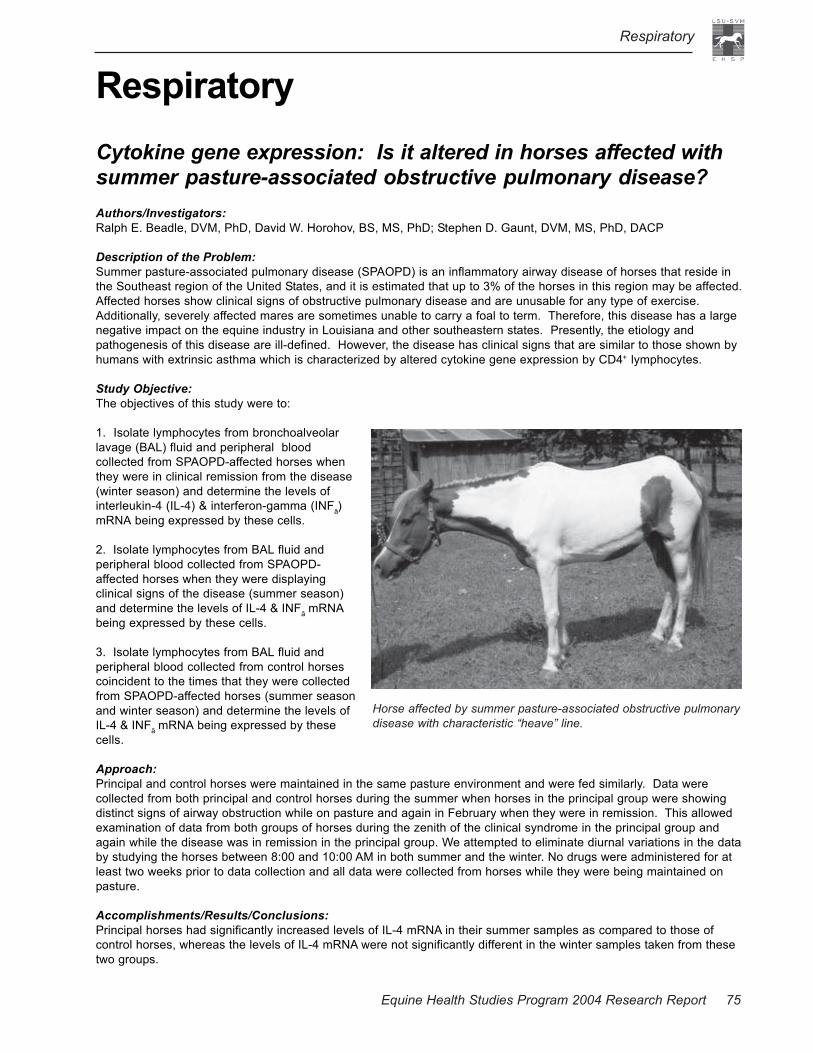
Equine Health Studies Program 2004 Research Report 75
Respiratory
RespiratoryCytokine gene expression: Is it altered in horses affected withsummer pasture-associated obstructive pulmonary disease?Authors/Investigators:Ralph E. Beadle, DVM, PhD, David W. Horohov, BS, MS, PhD; Stephen D. Gaunt, DVM, MS, PhD, DACP
Description of the Problem:Summer pasture-associated pulmonary disease (SPAOPD) is an inflammatory airway disease of horses that reside inthe Southeast region of the United States, and it is estimated that up to 3% of the horses in this region may be affected.Affected horses show clinical signs of obstructive pulmonary disease and are unusable for any type of exercise.Additionally, severely affected mares are sometimes unable to carry a foal to term. Therefore, this disease has a largenegative impact on the equine industry in Louisiana and other southeastern states. Presently, the etiology andpathogenesis of this disease are ill-defined. However, the disease has clinical signs that are similar to those shown byhumans with extrinsic asthma which is characterized by altered cytokine gene expression by CD4+ lymphocytes.
Study Objective:The objectives of this study were to:
1. Isolate lymphocytes from bronchoalveolarlavage (BAL) fluid and peripheral bloodcollected from SPAOPD-affected horses whenthey were in clinical remission from the disease(winter season) and determine the levels ofinterleukin-4 (IL-4) & interferon-gamma (INFã)mRNA being expressed by these cells.
2. Isolate lymphocytes from BAL fluid andperipheral blood collected from SPAOPD-affected horses when they were displayingclinical signs of the disease (summer season)and determine the levels of IL-4 & INFã mRNAbeing expressed by these cells.
3. Isolate lymphocytes from BAL fluid andperipheral blood collected from control horsescoincident to the times that they were collectedfrom SPAOPD-affected horses (summer seasonand winter season) and determine the levels ofIL-4 & INFã mRNA being expressed by thesecells.
Approach:Principal and control horses were maintained in the same pasture environment and were fed similarly. Data werecollected from both principal and control horses during the summer when horses in the principal group were showingdistinct signs of airway obstruction while on pasture and again in February when they were in remission. This allowedexamination of data from both groups of horses during the zenith of the clinical syndrome in the principal group andagain while the disease was in remission in the principal group. We attempted to eliminate diurnal variations in the databy studying the horses between 8:00 and 10:00 AM in both summer and the winter. No drugs were administered for atleast two weeks prior to data collection and all data were collected from horses while they were being maintained onpasture.
Accomplishments/Results/Conclusions:Principal horses had significantly increased levels of IL-4 mRNA in their summer samples as compared to those ofcontrol horses, whereas the levels of IL-4 mRNA were not significantly different in the winter samples taken from thesetwo groups.
Horse affected by summer pasture-associated obstructive pulmonarydisease with characteristic “heave” line.
76 Equine Health Studies Program 2004 Research Report
Respiratory
The levels of INFã mRNA were significantly higher in principal horses than they were in control horses in the summersamples. Additionally, principal horses had significantly increased levels of INFã mRNA in their summer samples ascompared to their winter samples, whereas control horses did not.
These results are consistent with the hypothesis that SPAOPD horses are reacting to an antigen in their summerenvironment that is not producing a similar response in control horses. This finding indicates that there was not aTH1:TH2 isotype switching occurring in these horses but rather that both TH1 and TH2 lymphocyte populations werebeing simultaneously stimulated.
Benefits to/Impact on the Equine Industry:The findings from this study that affected horses express higher levels of IL-4 mRNA and INFã mRNA during diseaseexacerbations have important clinical implications. Such results indicate that it may be possible to manipulate theimmune systems of affected horses to prevent them from expressing the higher levels of these cytokines or to preventthe resultant cytokines from interacting with their receptors. If such therapy were successful, a major step forwardwould have accomplished in the prevention and treatment of SPAOPD in horses.
Take Home Message:The results of this study have provided insight into the pathogenesis of SPAOPD in horses. Further studies are neededto determine if it is possible to alter the cytokine gene expression of SPAOPD-affected horses with specific drugtherapy and/or other management schemes. If appropriate management schemes can be devised, then it should bepossible to more successfully treat or prevent this disease in the future.
Acknowledgments:This research was supported by a grant from the Equine Health Studies Program, School of Veterinary Medicine,Louisiana State University, and a research grant from the Grayson-Jockey Club Research Foundation, Inc.
The investigators gratefully acknowledge and thank Keiko Antoku, Kim Snedden, Chad Vanderheyden, Dr. Britta Leise,Susan Porciau, Paul Hollier and Frank Garza for their technical assistance.
Year Completed: 1998
Published Manuscripts/Abstracts:Beadle RE, Horohov DW and Gaunt SD. Interleukin-4 and interferon-gamma gene expression in summer pasture-associated obstructive pulmonary disease affected horses. Equine Vet J 2002;34:389-94.
Correlation of clinical score, pleural pressure, bronchoalveolarlavage fluid cytology, and pulmonary histology in horses withsummer pasture-associated obstructive pulmonary diseaseAuthors/Investigators:Lais R.R. Costa, MV, MS, DACVIM; Thomas L. Seahorn, DVM, MS, DACVIML; Rustin M. Moore, DVM, PhD, DACVS;H. Wayne Taylor, DVM, PhD, DACVP; Stephen D. Gaunt, DVM, MS, PhD, DACP; Ralph E. Beadle, DVM, PhD
Description of the Problem:Summer pasture-associated obstructive pulmonary disease (SPAOPD) is a recurrent seasonal form of chronicobstructive pulmonary disease (COPD) observed in horses residing in pasture in the southeastern region of the UnitedStates. It is a naturally-acquired, progressive, obstructive disease of the small airways characterized by coughing andpronounced abdominal lift at the end of exhalation manifested during late spring, summer and early autumn, firstdescribed by R. Beadle in 1983. Nonetheless, very little has been published with respect to clinical and pathologicalfeatures of this disease.
Study Objective:The objectives of this study were: (1) to describe the clinical presentation (clinical score of respiratory effort) andpulmonary function test (pleural pressure), bronchoalveolar lavage fluid (BALF) cytology, pulmonary histopathology ofpercutaneous biopsy and of postmortem specimens from horses affected with SPAOPD, (2) to correlate the clinical andpathological changes, and (3) compare these findings with those in clinically normal horses.
Equine Health Studies Program 2004 Research Report 77
Respiratory
Approach:A total of 14 horses were evaluated in the study, 8 adult horses affected with SPAOPD and 6 adult horses withoutevidence of respiratory disease. Clinical scores of respiratory effort (CSRE), changes in pleural pressure during tidalbreathing, cytology and bacterial culture of BALF and light microscopy of pulmonary parenchyma collected viapercutaneous biopsy and at postmortem were evaluated.
Accomplishments/Results/Conclusions:The CSRE for SPAOPD-affected horses (median 5.75; range 4.0-7.5) was significantly greater compared with normalhorses (median 2.0; range 2.0-3.0). Cytology of BALF from SPAOPD-affected horses yielded predominantly non-degenerate neutrophils. Histopathologic lesions were identified throughout the lung lobes, and included markedaccumulation of basophilic mucus and neutrophils within the small airways, bronchiolar goblet cell metaplasia and mildperibronchial inflammatory infiltrate. Histopathology of specimens collected via percutaneous biopsy was predictive ofdisease and corresponded to findings at postmortem. The CSRE was highly correlated to pulmonary function inSPAOPD-affected horses. The CSRE and pulmonary function were highly correlated with mucus accumulation in theairways in affected horses. Peribronchial inflammatory infiltrate correlated with percentage of neutrophils in BALF ofaffected horses. Both CRSE and pulmonary function provided valid estimation of disease severity. BALF cytology ofSPAOPD-affected horses was variable, although in most cases diagnostically useful. Severe mucus accumulation inthe airways was the most remarkable histopathologic finding in SPAOPD. Lung biopsy specimens were consistentlyusefulness in the diagnosis of SPAOPD. The lung biopsy procedure was safe as no complications were observedduring a 24-hour period.
Benefits to /Impact on the Equine Industry:This study reports in detail the clinical and pathologic findings in horses affected with SPAOPD providing a landmarkdescription of the disease. The correlation of the clinical findings and severity of the clinical signs with the pathologiclesions indicated that the disease is characterized by reversible airway remodeling primarily associated with mucushypersecretion.
Take Home Message:The clinical and pathologic findings in horses affected with SPAOPD suggest that signs are likely to be reversible afterinstituting therapy that effectively yields clearance of airway mucus and bronchodilation.
Acknowledgments:This study was supported by a grant from the USDA 1433 Formula Funds, School of Veterinary Medicine, LouisianaState University.
Year completed: 1998
Published Manuscripts/Abstracts:Costa LRR, Seahorn TL, Moore RM, Taylor HW, Gaunt SD, Beadle RE. Correlation of clinical score, intrapleuralpressure, cytologic findings of bronchoalveolar fluid, and histopathologic lesions of pulmonary tissue in horses withsummer pasture-associated obstructive pulmonary disease. Am J Vet Res 2000; 61 (2) 167-173.
Costa LRR, Seahorn TL, Moore RM, Taylor HW, Gaunt SD, Beadle RE. Correlation of clinical score, bronchoalveolarlavage fluid analysis, and pulmonary histology in horses with summer-pasture-associated pulmonary obstructivedisease residing in Louisiana. J Vet Intern Med 1999;13 (3) 271.
Plasma and bronchoalveolar fluid concentrations of nitric oxideand histochemical and immunohistochemical localization ofnitric oxide synthesis in lungs of horses with summer pasture-associated obstructive pulmonary diseaseAuthors/Investigators:Lais R.R. Costa, MV, MS, DACVIM; Thomas L. Seahorn, DVM, MS, DACVIM; Rustin M. Moore, DVM, PhD, DACVS;Julian L. Oliver, DVM, PhD, DACVP; Giselle Hosgood, BVSc, MS, PhD, DACVS
78 Equine Health Studies Program 2004 Research Report
Respiratory
Description of the Problem:Summer pasture-associated obstructive pulmonary disease (SPAOPD) is a naturally-acquired, seasonal, obstructivedisease of the small airways of horses residing in pasture. There are a number of potential inflammatory mediatorsincriminated in the pathogenesis of SPAOPD, which clinically resembles human asthma. Nitric oxide has beendemonstrated to be involved in the pathogenesis of airway inflammation and hypersensitivity of asthma.
Study Objective:The objective of this study was to determine the concentration of nitric oxide (NO) in plasma and bronchoalveolarlavage fluid and localization of nitric oxide synthesis in lungs of horses affected with summer pasture-associatedobstructive pulmonary disease (SPAOPD), compared with non-affected horses.
Approach:Seven adult horses affected with SPAOPD and 6 clinically normal (non-affected) adult horses were evaluated clinicallyand the severity of SPAOPD was determined by clinical scores, change in intrapleural pressure during tidal breathing,cytologic analysis of bronchoalveolar lavage fluid (BALF) and histopathologic evaluation by H&E staining. Nitric oxideconcentrations in plasma, BALF and epithelial lining fluid (ELF) were determined via a chemiluminescent method.Immunohistochemical staining of formalin-fixed specimens of pulmonary tissue for inducible nitric oxide synthase(iNOS) and nitrotyrosine (NT), and histochemical staining for nicotinamide adenine dinucleotide phosphate diaphorase(NADPHd) activity in cryopreserved specimens were performed and evaluated with light microscopy. Sections fromeach lung lobe were evaluated and compared.
Accomplishments/Results/Conclusions:There was a trend of higher plasma NO concentrations in SPAOPD-affected compared with clinically normal horses.Nitric oxide concentrations in BALF or ELF did not differ between SPAOPD-affected and non-affected horses. TheiNOS staining of bronchial epithelial cells was greater in affected than non-affected horses in 3/5 lung sections. The NTstaining and NADPHd activity of bronchial epithelial cells, as well as the NT and iNOS staining and NADPHd activity ofperibronchial leukocytes in the pulmonary tissue did not differ between SPAOPD-affected and non-affected horses.
Benefits to/Impact on the Equine Industry:We demonstrated that immunoreactivity to iNOS is increased in bronchial epithelial cells of SPAOPD-affectedcompared with non-affected horses, suggesting that NO plays a role in the airway inflammation and hypersensitivityseen in horses with this disease. Clinical Relevance is that NO may have cytotoxic effects in the airway of horses withSPAOPD.
Take Home Message:Nitric oxide may play a role in the airway inflammation and hypersensitivity seen in horses with SPAOPD.
Acknowledgments:This study was supported by a grant from the United States Department of Agriculture 1433 Formula Funds, School ofVeterinary Medicine, Louisiana State University. The authors thank Mae Lopez for performing theimmunohistochemical and histochemical staining, and Catherine Koch, Dr. Lee Ann Curtis and Frank Garza for theirvaluable technical assistance.
Year completed: 1999
Published Manuscripts/Abstracts:Costa LRR, Seahorn TL, Moore RM, Oliver JL, Hosgood GL. Plasma and bronchoalveolar fluid concentrations of nitricoxide and localization of nitric oxide synthase in the lungs of horses with summer pasture-associated obstructivepulmonary disease. Am J Vet Res 2001;62 (9) 1381-1386.
Costa LRR, Seahorn TL, Moore RM, Oliver J, Beadle RE. Nitric oxide as an inflammatory mediatory in horses withsummer pasture-associated obstructive pulmonary disease. J Vet Intern Med 2000;14 (3) 371.
Costa LRR, Seahorn TL, Moore RM, Oliver J, Beadle RE. Nitric oxide as an inflammatory mediatory in horses withsummer pasture-associated obstructive pulmonary disease, in Proceedings. 17th Symposium of the ComparativeRespiratory Society 1999.

Equine Health Studies Program 2004 Research Report 79
Respiratory
Comparative responses of bronchial rings to mediators of airwayhyper reactivity in healthy horses and those affected with summerpasture-associated obstructive pulmonary diseaseAuthors/Investigators: Changaram S. Venugopal, BVSc, MSc, MS, PhD; Rustin M. Moore, DVM, PhD, DACVS; EarnestineP. Holmes, BS; Catherine E. Koch MS; Thomas L. Seahorn, DVM, MS; Ralph E. Beadle, DVM, PhD
Description of the Problem:Summer pasture-associated obstructive pulmonary disease (SPAOPD) is an inflammatory, obstructive airway diseasesimilar to heaves (COPD) that occurs in subtropical regions of the US during late summer and early fall and is associatedwith grazing in lush pastures. It is a severely debilitating, devastating and often career ending or life threatening pulmonarydisease of horses leading to morbidity and loss of performance. During periods of airway obstruction, horses developairway hyper reactivity resulting in severe bronchoconstriction to inflammatory mediators released locally into the airwaylumen and toward the bronchial smooth muscle. These mediators are responsible for bronchial smooth muscle contraction,increased vascular permeability, increased mucus secretion, and damage to the airway epithelium resulting in airwayremodeling leading to airway hyper reactivity. Airway hyper reactivity is a condition characterized by increased sensitivity(hyper sensitivity) and increased maximal responsiveness (hyper responsiveness). Commonly accepted airwayinflammatory mediators involved in airway disease include histamine, bradykinin, prostaglandins F2", serotonin, leukotrienes(LTD), substance P and platelet activating factor (PAF).
Study Objective:The objective of this study was to compare responses of bronchial rings obtained from clinically healthy horses and thoseaffected with SPAOPD to selected mediators known to cause airway hyper reactivity.
Approach:4mm wide bronchial rings were cut from the 4th -7th generation branches of airways from clinically healthy and SPAOPDaffected horses. They were mounted in organ baths containing Tyrode’s solution and were attached to force transducersinterfaced with a polygraph. Initial tension of 2 g was applied to the rings and an equilibration time of 45 minutes weregiven. Then concentration response relationships of the rings to graded concentrations of selected mediators (10-8 to 10-
4M) were determined and analyzed for statistical significance at each concentration of the agents.
Accomplishments/Results/Conclusions:Acetylcholine, histamine, serotonin and leukotriene D4 (LTD) induced consistent concentration-dependent contractileresponses in bronchial rings. PGF2a induced week and inconsistent contractile response. The other two agents, norepinephrine and substance P, did not induce concentration–dependent responses. Considering the overall group-drugeffect, acetylcholine, histamine serotonin and LTD were effective in causing consistent concentration-dependent contractileresponses in both groups of rings. Only serotonin and histamine induced significant responses between the groups. Theresponses to serotonin on SPAOPD rings were significantly greater that those of the clinically healthy horses whereas,histamine produced a significantly lower response in SPAOPD horses. Significant responses were evident at concentrationsranging from 10-6 to 10-4M.
Take Home Message:Serotonin causes airway hyper reactivity in SPAOPD horses. Therefore, treatment modalities using serotonin antagonistsshould be investigated in SPAOPD horses. Antihistamines may not be useful.
Acknowledgments:This study was supported by a grant from the Equine Health Studies Program, School of Veterinary Medicine, LouisianaState University.
Year Completed: 1999
Published Manuscripts/Abstracts:Venugopal CS, Moore RM, Holmes EP, Koch CE, Seahorn TL, Beadle RE. Comparative responses of bronchial rings tomediators of airway hyper reactivity in healthy horses and those affected with summer pasture-associated obstructivepulmonary disease. Am J Vet Res 2001;62: 259-263.
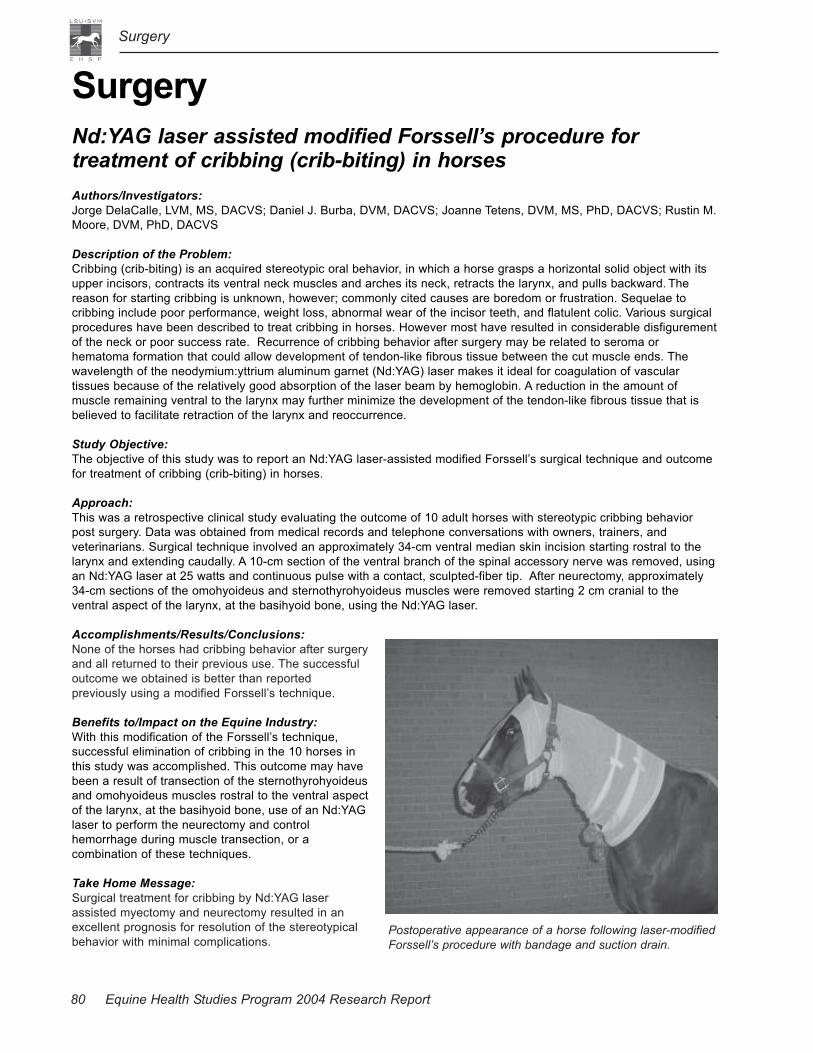
80 Equine Health Studies Program 2004 Research Report
Surgery
SurgeryNd:YAG laser assisted modified Forssell’s procedure fortreatment of cribbing (crib-biting) in horsesAuthors/Investigators:Jorge DelaCalle, LVM, MS, DACVS; Daniel J. Burba, DVM, DACVS; Joanne Tetens, DVM, MS, PhD, DACVS; Rustin M.Moore, DVM, PhD, DACVS
Description of the Problem:Cribbing (crib-biting) is an acquired stereotypic oral behavior, in which a horse grasps a horizontal solid object with itsupper incisors, contracts its ventral neck muscles and arches its neck, retracts the larynx, and pulls backward. Thereason for starting cribbing is unknown, however; commonly cited causes are boredom or frustration. Sequelae tocribbing include poor performance, weight loss, abnormal wear of the incisor teeth, and flatulent colic. Various surgicalprocedures have been described to treat cribbing in horses. However most have resulted in considerable disfigurementof the neck or poor success rate. Recurrence of cribbing behavior after surgery may be related to seroma orhematoma formation that could allow development of tendon-like fibrous tissue between the cut muscle ends. Thewavelength of the neodymium:yttrium aluminum garnet (Nd:YAG) laser makes it ideal for coagulation of vasculartissues because of the relatively good absorption of the laser beam by hemoglobin. A reduction in the amount ofmuscle remaining ventral to the larynx may further minimize the development of the tendon-like fibrous tissue that isbelieved to facilitate retraction of the larynx and reoccurrence.
Study Objective:The objective of this study was to report an Nd:YAG laser-assisted modified Forssell’s surgical technique and outcomefor treatment of cribbing (crib-biting) in horses.
Approach:This was a retrospective clinical study evaluating the outcome of 10 adult horses with stereotypic cribbing behaviorpost surgery. Data was obtained from medical records and telephone conversations with owners, trainers, andveterinarians. Surgical technique involved an approximately 34-cm ventral median skin incision starting rostral to thelarynx and extending caudally. A 10-cm section of the ventral branch of the spinal accessory nerve was removed, usingan Nd:YAG laser at 25 watts and continuous pulse with a contact, sculpted-fiber tip. After neurectomy, approximately34-cm sections of the omohyoideus and sternothyrohyoideus muscles were removed starting 2 cm cranial to theventral aspect of the larynx, at the basihyoid bone, using the Nd:YAG laser.
Accomplishments/Results/Conclusions:None of the horses had cribbing behavior after surgeryand all returned to their previous use. The successfuloutcome we obtained is better than reportedpreviously using a modified Forssell’s technique.
Benefits to/Impact on the Equine Industry:With this modification of the Forssell’s technique,successful elimination of cribbing in the 10 horses inthis study was accomplished. This outcome may havebeen a result of transection of the sternothyrohyoideusand omohyoideus muscles rostral to the ventral aspectof the larynx, at the basihyoid bone, use of an Nd:YAGlaser to perform the neurectomy and controlhemorrhage during muscle transection, or acombination of these techniques.
Take Home Message:Surgical treatment for cribbing by Nd:YAG laserassisted myectomy and neurectomy resulted in anexcellent prognosis for resolution of the stereotypicalbehavior with minimal complications.
Postoperative appearance of a horse following laser-modifiedForssell’s procedure with bandage and suction drain.
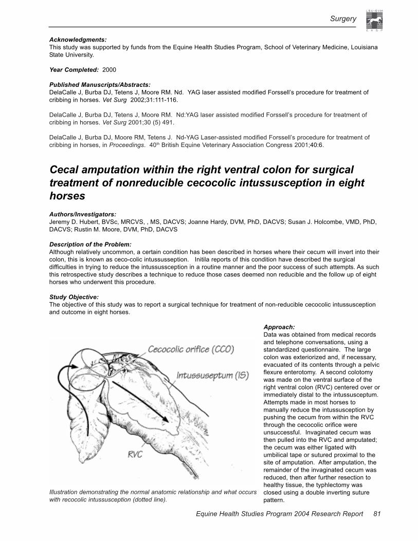
Equine Health Studies Program 2004 Research Report 81
Surgery
Acknowledgments:This study was supported by funds from the Equine Health Studies Program, School of Veterinary Medicine, LouisianaState University.
Year Completed: 2000
Published Manuscripts/Abstracts:DelaCalle J, Burba DJ, Tetens J, Moore RM. Nd. YAG laser assisted modified Forssell’s procedure for treatment ofcribbing in horses. Vet Surg 2002;31:111-116.
DelaCalle J, Burba DJ, Tetens J, Moore RM. Nd:YAG laser assisted modified Forssell’s procedure for treatment ofcribbing in horses. Vet Surg 2001;30 (5) 491.
DelaCalle J, Burba DJ, Moore RM, Tetens J. Nd-YAG Laser-assisted modified Forssell’s procedure for treatment ofcribbing in horses, in Proceedings. 40th British Equine Veterinary Association Congress 2001;40:6.
Cecal amputation within the right ventral colon for surgicaltreatment of nonreducible cecocolic intussusception in eighthorsesAuthors/Investigators:Jeremy D. Hubert, BVSc, MRCVS, , MS, DACVS; Joanne Hardy, DVM, PhD, DACVS; Susan J. Holcombe, VMD, PhD,DACVS; Rustin M. Moore, DVM, PhD, DACVS
Description of the Problem:Although relatively uncommon, a certain condition has been described in horses where their cecum will invert into theircolon, this is known as ceco-colic intussusseption. Initila reports of this condition have described the surgicaldifficulties in trying to reduce the intussussception in a routine manner and the poor success of such attempts. As suchthis retrospective study describes a technique to reduce those cases deemed non reducible and the follow up of eighthorses who underwent this procedure.
Study Objective:The objective of this study was to report a surgical technique for treatment of non-reducible cecocolic intussusceptionand outcome in eight horses.
Approach:Data was obtained from medical recordsand telephone conversations, using astandardized questionnaire. The largecolon was exteriorized and, if necessary,evacuated of its contents through a pelvicflexure enterotomy. A second colotomywas made on the ventral surface of theright ventral colon (RVC) centered over orimmediately distal to the intussusceptum.Attempts made in most horses tomanually reduce the intussusception bypushing the cecum from within the RVCthrough the cecocolic orifice wereunsuccessful. Invaginated cecum wasthen pulled into the RVC and amputated;the cecum was either ligated withumbilical tape or sutured proximal to thesite of amputation. After amputation, theremainder of the invaginated cecum wasreduced, then after further resection tohealthy tissue, the typhlectomy wasclosed using a double inverting suturepattern.
Illustration demonstrating the normal anatomic relationship and what occurswith recocolic intussusception (dotted line).

82 Equine Health Studies Program 2004 Research Report
Surgery
Accomplishments/Results/Conclusions:All horses survived to hospital discharge. One horse died 3 months postoperatively; however, the remainder survived(median survival 30 months; range, 6-96 months) and returned to or exceeded previous function. Despite somecontamination during surgery, this method of surgical treatment for horses with nonreducible cecocolic intussusceptionhad a good prognosis for survival.
Benefits to/Impact on the Equine Industry:This reported technique will provide the equine surgeon with the ability to resolve what was initially considered acondition with an extremely poor prognosis.
Take Home Message:Cecocolic intussussceptions do not have the extremely poor prognosis they were considered to have and surgicalrepair should be attempted in the manner described in this report.
Acknowledgments:This study was supported by grants from USDA 1433 funds and the Department of Veterinary Clinical SciencesOrganized Research Fund, School of Veterinary Medicine, Louisiana State University.
Year Completed: 1998
Published Manuscripts/Abstracts:Hubert JD, Hardy J, Holcombe SJ, Moore RM. Cecal amputation within the right ventral colon for surgical treatment ofnonreducible cecocolic intussusception in eight horses. Vet Surg 2000;29(4) 317-325.
Hubert JD, Hardy J, Holcombe SJ, Moore RM. Cecal amputation within the right ventral colon for surgical treatment ofnonreducible cecocolic intussusception in 8 horses. BEVA Congress 1999.
Hubert JD, Hardy J, Holcombe SJ, Moore RM. Cecal amputation within the right ventral colon for surgical treatment ofnonreducible cecocolic intussusception in 8 horses. Vet Surg 1999;28 (5) 395.
Hubert JD, Hardy J, Holcombe SJ, Moore RM. Cecal amputation via a right ventral colon enterotomy for correction ofnonreducible cecocolic intussusception in 8 horses, in Proceedings. Sixth Colic Research Symposium 1998;62.
Factors associated with racing performance of Thoroughbredsundergoing lag screw repair of condylar fractures of the thirdmetacarpal or metatarsal boneAuthors/Investigators: George S. Martin, DVM, MS, MBA, DACVS
Description of the Problem: Condylar fractures of the third metacarpal and metatarsal bones occur relativelycommonly in racing Thoroughbreds. The prognosis for horses with these injuries depends upon the fracture type andconfiguration. The gender of the horse also affects decision regarding treatment of these fractures. There areconflicting opinions from surgeons regarding the best surgical treatment of horses with condylar fractures.
Study Objective: The objective of this study was to evaluate effects of sex, fracture configuration, affected limb, andscrew placement on outcome of Thoroughbreds with condylar fractures involving the third metacarpal or metatarsalbone.
Approach: This cohort study involved 56 Thoroughbred racehorses that sustained a condylar fracture of the thirdmetacarpal or metatarsal bone and underwent surgical repair. The age, sex, affected limb, fracture configuration,fracture length, fracture fragment width, and distance of the most distal screw from the articular surface were analyzed,using logistic regression models.
Accomplishments/Results/Conclusions: Females were more likely to have displaced fractures and race in fewerraces after surgery than males. The sex and fracture configuration were associated with the number of postoperativeraces. Among horses that returned to racing, those with thicker fracture fragments were 11 times more likely as horseswith thinner fracture fragments to win a race after surgery. Horses with longer fractures and older horses had fewerpostoperative races. Horses in which the most distal screw had been placed further from the joint surface had moreraces.
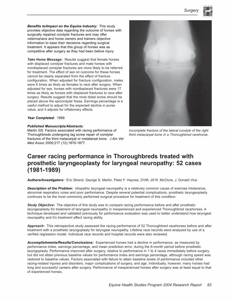
Equine Health Studies Program 2004 Research Report 83
Benefits to/Impact on the Equine Industry: This studyprovides objective data regarding the outcome of horses withsurgically repaired condylar fractures and may offerveterinarians and horse owners and trainers objectiveinformation to base their decisions regarding surgicaltreatment. It appears that this group of horses was ascompetitive after surgery as they had been before injury.
Take Home Message: Results suggest that female horseswith displaced condylar fractures and male horses withnondisplaced condylar fractures are more likely to be referredfor treatment. The effect of sex on outcome for these horsescannot be clearly separated from the effect of fractureconfiguration. When adjusted for fracture configuration, maleswere 6 times as likely as females to race after surgery. Whenadjusted for sex, horses with nondisplaced fractures were 17times as likely as horses with displaced fractures to race aftersurgery. Results suggest that the most distal screw should beplaced above the epicondylar fossa. Earnings percentage is auseful method to adjust for the expected decline in pursevalue, and it adjusts for inflationary effects.
Year Completed: 1999
Published Manuscripts/Abstracts:Martin GS: Factors associated with racing performance ofThoroughbreds undergoing lag screw repair of condylarfractures of the third metacarpal or metatarsal bone. J Am VetMed Assoc 2000;217 (12):1870-1877.
Career racing performance in Thoroughbreds treated withprosthetic laryngeoplasty for laryngeal neuropathy: 52 cases(1981-1989)Authors/Investigators: Eric Strand, George S. Martin, Peter F. Haynes, DVM; Jill R. McClure, J. Donald Vice
Description of the Problem: Idiopathic laryngeal neuropathy is a relatively common cause of exercise intolerance,abnormal respiratory noise and poor performance. Despite several potential complications, prosthetic laryngeoplastycontinues to be the most commonly performed surgical procedure for treatment of this condition.
Study Objective: The objective of this study was to compare racing performance before and after prostheticlaryngeoplasty for treatment of laryngeal neuropathy in inexperienced and experienced Thoroughbred racehorses. Atechnique developed and validated previously for performance evaluation was used to better understand how laryngealneuropathy and it’s treatment affect racing ability.
Approach: This retrospective study assessed the racing performance of 52 Thoroughbred racehorses before and aftertreatment with a prosthetic laryngoplasty for laryngeal neuropathy. Lifetime race records were analyzed by use of averified regression model. Individual race records and hospital records were also reviewed.
Accomplishments/Results/Conclusions: Experienced horses had a decline in performance, as measured byperformance index, earnings percentage, and mean prediction error, during the 6-month period before prostheticlaryngoplasty. Performance improved after surgery, relative to performance in 1 to 4 races immediately before surgery,but did not attain previous baseline values for performance index and earnings percentage, although racing speed wasrestored to baseline values. Factors associated with failure to attain baseline levels of performance included otherracing-related injuries and disorders, major complications of surgery, and age. Individually, however, many horses hadlong and successful careers after surgery. Performance of inexperienced horses after surgery was at least equal to thatof experienced horses.
Surgery
Incomplete fracture of the lateral condyle of the rightthird metacarpal bone in a Thoroughbred racehorse.
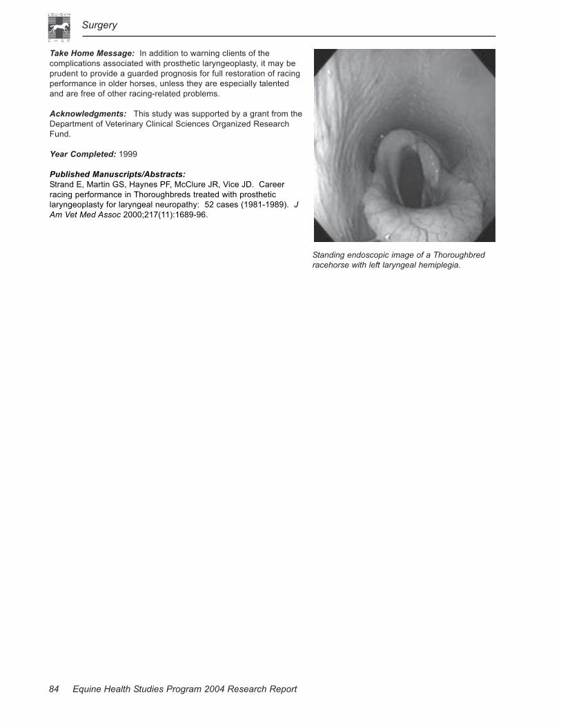
84 Equine Health Studies Program 2004 Research Report
Surgery
Take Home Message: In addition to warning clients of thecomplications associated with prosthetic laryngeoplasty, it may beprudent to provide a guarded prognosis for full restoration of racingperformance in older horses, unless they are especially talentedand are free of other racing-related problems.
Acknowledgments: This study was supported by a grant from theDepartment of Veterinary Clinical Sciences Organized ResearchFund.
Year Completed: 1999
Published Manuscripts/Abstracts:Strand E, Martin GS, Haynes PF, McClure JR, Vice JD. Careerracing performance in Thoroughbreds treated with prostheticlaryngeoplasty for laryngeal neuropathy: 52 cases (1981-1989). JAm Vet Med Assoc 2000;217(11):1689-96.
Standing endoscopic image of a Thoroughbredracehorse with left laryngeal hemiplegia.
2004 Research ReportEquine Health Studies ProgramSchool of Veterinary Medicine
Louisiana State UniversityBaton Rouge, Louisiana 70803
Phone: (225) 578-9500Fax: (225) 578-9605
E-mail: [email protected]: www.equine.vetmed.lsu.edu

LOUISIANA STATE UNIVERSITY
School of Veterinary MedicineBaton Rouge, LA 70803
Non-Profit Org.U.S. Postage
PAIDPermit No. 733
Baton Rouge, LA