Embed Size (px)
Citation preview
Introduction to
NUCLEAR MEDICINE
Introduction to
NUCLEAR MEDICINE
Department of Nuclear Medicine
University of Debrecen
József Varga
2011.2
Nuclear Medicine: ManualsNuclear Medicine: Manuals
• Link to English reference manual:
http://www.auntminnie.com/index.asp?sec=ref&sub=ncm
• Lectures in English:http://www.nmc.dote.hu/nmt_eng/oktatas_e.htm
• In Hungarian:
http://www.nmc.dote.hu/nmtk/index.html
• Book: A Nukleáris Medicina Tankönyve(Szerk. Szilvási I.; B+V Kiadó, 2002, 2010)
• Required reading:Taylor A., Alazraki N., and Schuster D.M.:A Clinician's Guide to Nuclear Medicine (2nd Edition)
The Society of Nuclear Medicine, Reston, 2006
ISBN: 0972647872
Varga J, 2011 Introduction to Nuclear Medicine
3
1924: Principle of radiotracer
applications:
Changing an atom in a molecule for its radioisotope will not change its
chemical and biological behaviour
significantly.
Consequence: the movement, distribution,
concentration of the molecule can be
measured with radiation detectors.
György HEVESY (1885-1966)
1943 Nobel Laureate in
Chemistry
„for his work on the use of
isotopes as tracers in the study of
chemical processes”
NUCLEAR MEDICINENUCLEAR MEDICINE
Varga J, 2011 Introduction to Nuclear Medicine 4
Medical & biological applications of radionuclidesMedical & biological applications of radionuclides
Medical applications
(nuclear medicine)
Research applications
(nuclear medicine)
Diagnostics
Therapy
„In vivo”
imaging
„In vitro”
Application of diagnostic
methods for research
„Molecular imaging”
Combination of analitical
laboratory methods with
radiotracer technique
„In vivo”
non-imaging
Varga J, 2011 Introduction to Nuclear Medicine
5
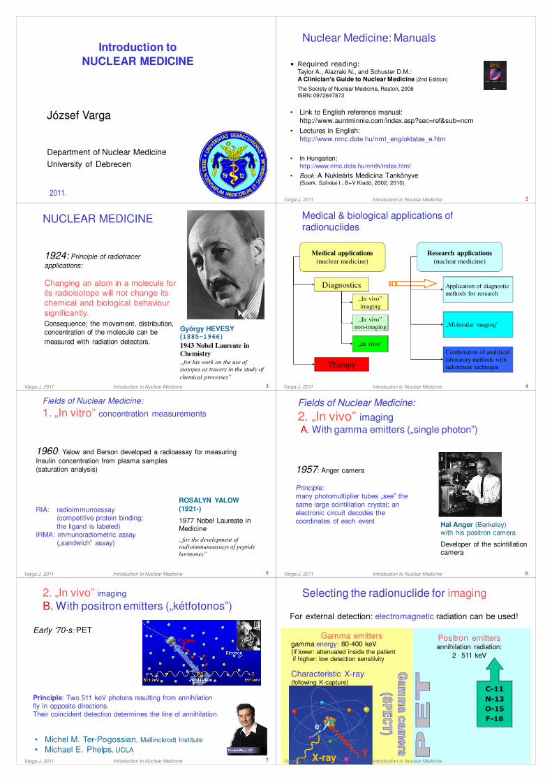
ROSALYN YALOW
(1921-)
1977 Nobel Laureate in Medicine
„for the development of
radioimmunoassays of peptide
hormones”
1960: Yalow and Berson developed a radioassay for measuring
Insulin concentration from plasma samples
(saturation analysis)
RIA: radioimmunoassay(competitive protein binding;
the ligand is labeled)IRMA: immunoradiometric assay
(„sandwich” assay)
Fields of Nuclear Medicine:
1. „In vitro” concentration measurements
Fields of Nuclear Medicine:
1. „In vitro” concentration measurements
Varga J, 2011 Introduction to Nuclear Medicine 6
Hal Anger (Berkeley)with his positron camera
Developer of the scintillation camera
1957: Anger camera
Principle: many photomultiplier tubes „see” the
same large scintillation crystal; an electronic circuit decodes the coordinates of each event
Fields of Nuclear Medicine:
2. „In vivo” imaging
A. With gamma emitters („single photon”)
Fields of Nuclear Medicine:
2. „In vivo” imaging
A. With gamma emitters („single photon”)
Varga J, 2011 Introduction to Nuclear Medicine
7
2. „In vivo” imaging
B. With positron emitters („kétfotonos”)
2. „In vivo” imaging
B. With positron emitters („kétfotonos”)
Early ‘70-s: PET
Principle: Two 511 keV photons resulting from annihilation fly in opposite directions.Their coincident detection determines the line of annihilation.
• Michel M. Ter-Pogossian, Mallinckrodt Institute
• Michael E. Phelps, UCLA
Varga J, 2011 Introduction to Nuclear Medicine 8
Selecting the radionuclide for imagingSelecting the radionuclide for imaging
C-11
N-13
O-15
F-18
e-
γγγγX-ray
Characteristic X-ray(following K-capture)
For external detection: electromagnetic radiation can be used!
Gamma emittersgamma energy: 80-400 keV(if lower: attenuated inside the patientif higher: low detection sensitivity)
Positron emittersannihilation radiation:
2 ⋅ 511 keV
Varga J, 2011 Introduction to Nuclear Medicine

9
Producing artificial radioactive materialProducing artificial radioactive material
Stanley Livingstone and Ernest Lawrence with their 8 MeV cyclotron
(1935)
• In nuclear reactors(high neutron flux)
• Using accelerators(circular: cyclotron)
expensive!
Ernest Lawrence(Berkeley)
inventor of the cyclotron
Varga J, 2011 Introduction to Nuclear Medicine 10
Radionuclides in Nuclear Medicine,
UK 2003/04
Tc-99m; 79.5%
Kr-81m; 6.1%
Cr-51; 3.8%
Tl-201; 2.4%
I-131; 2.3%
F-18; 1.5%
C-14; 1.2%
Xe-133; 0.8%
I-123; 0.7%
In-111; 0.4%
Egyéb; 1.3%
Gamma imaging
RN therapy
PET imaging
Varga J, 2011 Introduction to Nuclear Medicine
11
99Mo-99mTc generator99Mo-99mTc generator
Evacuated vial
Eluent
Filter
Air filter
Alumina column
Lead shielding
Varga J, 2011 Introduction to Nuclear Medicine 12
Anger (gamma) camera
1. Collimator
2. Crystal: NaI (Tl)
3. Photomultipliertubes
4. Impulses
5. Anger circuit
6. X, Y coordinates
7. „Good” events
8. Memory scope
9. Analog-digitalconverters
10. Computer
Matrix
circuitDifferential
discriminator
Varga J, 2011 Introduction to Nuclear Medicine
13
• To form an image from the detected photons, the direction of movements
should be known
• The collimator lets through only the photons that move perpendicularly to its
plane
Detector
IIIIIIIIIIIIIIIIIIIIIIIIIIIIIIIIIIIIIIIIIIII ⇐⇐⇐⇐ Lead
collimator
Photomultipliers
+ preamplifiers
Source: Freek Beekman et al., Utrecht
The role of a
collimator
The role of a
collimator
Crystal
Varga J, 2011 Introduction to Nuclear Medicine
PET: ConceptPET: Concept
Varga J, 2011 14Introduction to Nuclear Medicine
15
Principle:
Beta-emitting radiopharmaceuticals go directly to the cells or tissue to be destroyed or deactivated
Very specific radiopharmaceuticals are needed
Unsealed preparation: One that mixes in the patients’ body on a molecular level(e.g. after intravenous injection)
Fields of Nuclear Medicine:
3. Therapy with unsealed radiactive preparations
Fields of Nuclear Medicine:
3. Therapy with unsealed radiactive preparations
Varga J, 2011 Introduction to Nuclear Medicine 16
Source: „What is Nuclear Medicine?” (SNM)
Medical imagingMedical imaging
Varga J, 2011 Introduction to Nuclear Medicine

17
Targets and tools of medical imagingTargets and tools of medical imaging
Varga J, 2011 Introduction to Nuclear Medicine 18
Average of health-care level I , 1991-96 (UNSCEAR)
1 10 100 1 000 10 000 100 000 1 000 000
X-ray, dental
X-ray, medical
CT
Angiography
Interventional
Nuclear imaging
Radionuclide therapy
Teletherapy
Brachytherapy
Number of procedures / million population
Therapy
Diagnosis
United Nations Scientific Committee on the Effects of Atomic Radiations
Varga J, 2011 Introduction to Nuclear Medicine
19
Functional vs. structural imaging:Low-grade recidive glioma (FDG)
PET Center,
Debrecen
Varga J, 2011 Introduction to Nuclear Medicine 20Varga J, 2011 Introduction to Nuclear Medicine
21
Detection sensitivity of imaging techniquesDetection sensitivity of imaging techniques
Imaging Concentration of tracer / „contrast”
technique material (mol/kg body mass)
UH 10-3
CT 10-3
Gamma camera 10-9- 10-12
PET 10-9- 10-12
MRI 10-5
MRS 10-5
Source: G. von Schulthess, University Hospital, Zürich
Varga J, 2011 Introduction to Nuclear Medicine 22
Static:Imaging an equilibrium distribution
Dynamic:Series of images following the accumulation /metabolic pathways / secretion of a radiopharmaceutical
Whole body:Static images connected
Tomographic:Single Photon Emission Computed Tomography (SPECT)Positron Emission Tomography (PET)
Emission imaging: Study typesEmission imaging: Study types
Varga J, 2011 Introduction to Nuclear Medicine
23
Whole body
bone scintigram
Spot images
Varga J, 2011 Introduction to Nuclear Medicine 24
RAW image Metz-filtered
Example: Static imageThyroid scintigram without and with filtering
Example: Static imageThyroid scintigram without and with filtering
Varga J, 2011 Introduction to Nuclear Medicine
25
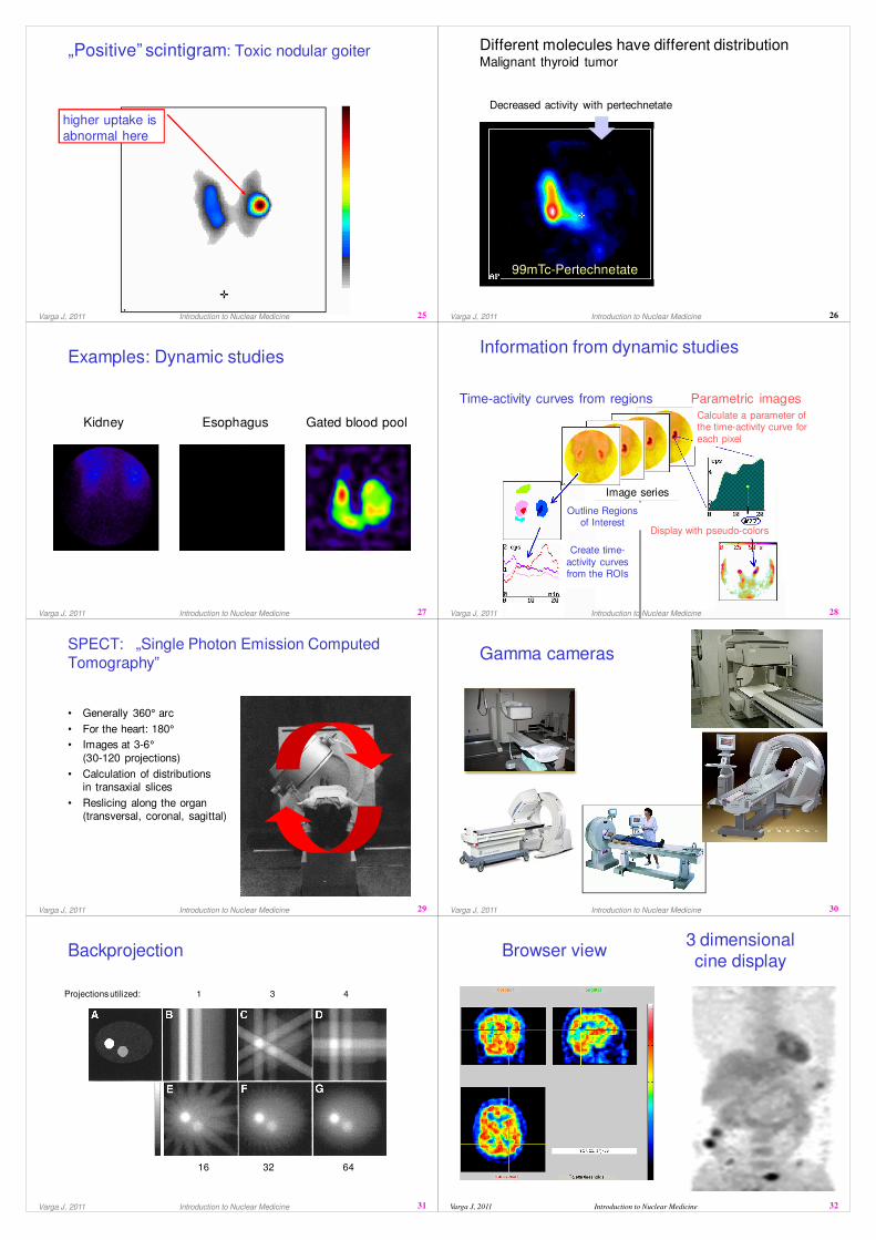
higher uptake is
abnormal here
„Positive” scintigram: Toxic nodular goiter„Positive” scintigram: Toxic nodular goiter
Varga J, 2011 Introduction to Nuclear Medicine Varga J, 2011 Introduction to Nuclear Medicine 26
Different molecules have different distribution Malignant thyroid tumor
Different molecules have different distribution Malignant thyroid tumor
99mTc-Pertechnetate
Decreased activity with pertechnetate
Increased MIBI accumulation
99mTc-MIBI
27
Examples: Dynamic studiesExamples: Dynamic studies
Kidney Esophagus Gated blood pool
Varga J, 2011 Introduction to Nuclear Medicine 28
Time-activity curves from regions Parametric images
Image series
Calculate a parameter of the time-activity curve for
each pixel
Display with pseudo-colors
Outline Regions of Interest
Create time-
activity curves from the ROIs
Information from dynamic studiesInformation from dynamic studies
Varga J, 2011 Introduction to Nuclear Medicine
29
SPECT: „Single Photon Emission Computed
Tomography”
SPECT: „Single Photon Emission Computed
Tomography”
• Generally 360° arc
• For the heart: 180°
• Images at 3-6°
(30-120 projections)
• Calculation of distributionsin transaxial slices
• Reslicing along the organ(transversal, coronal, sagittal)
Varga J, 2011 Introduction to Nuclear Medicine 30
Gamma camerasGamma cameras
Varga J, 2011 Introduction to Nuclear Medicine
31
BackprojectionBackprojection
Projections utilized: 1 3 4
16 32 64
Varga J, 2011 Introduction to Nuclear Medicine Varga J, 2011 Introduction to Nuclear Medicine 32
3 dimensional
cine display
3 dimensional
cine displayBrowser viewBrowser view

33
PET - advantagesPET - advantages
• Coincidence detection at 180°:- higher sensitivity- better signal/noise ratio
• Easier attenuation correction(sum of the two paths inside =body thickness)
• More physiologic radiopharmaceuticals(C-11, N-13, O-15, F-18)
• Dynamic tomography is possible(simultaneous acquisition from all projections)
*
Varga J, 2011 Introduction to Nuclear Medicine Varga J, 2011 Introduction to Nuclear Medicine 34
Brain receptor imagingPET ligands for imaging various receptor systems
Brain receptor imagingPET ligands for imaging various receptor systems
[F-18]-memantin
(NMDA-receptor)
[F-18] fluoro-2-
deoxy-glucose
O
OH
F
HO
OH
HO
N
SCH3NH2
FH2C
CH3
Cl
OH
Cl
OCH3
NHN
O CH2CH3
N
N
N
F
O
OCH2CH
3
O
CH3
NH3C
H
H Cl
NO
Ph
[C-11]-raclopride
(dopamine D2
receptor)
[C-11]-McN 5652
(serotonin
transporter)
[C-11]-b-CPPIT
(dopamine
transporter)
[C-11]-flumazenil
(benzodiazepine-
receptor)
Source: G. von Schulthess, University Hospital, Zürich
35
Our PET is
growing up
From P. Vernon, GE
Varga J, 2011 Introduction to Nuclear Medicine Varga J, 2011 Introduction to Nuclear Medicine 36
„Structure without function is a corpse;function without structure is a ghost”
37
Functional and morphological imaging:
Complementary rolesFunctional and morphological imaging:
Complementary roles
PET & SPECT CT, MR
functional information structural / morphological
information
significant partial volume effect better resolution
higher noise CT: high patient dose
attenuation and scatter degrades
images
MR: inhomogeneous image,
geometric distortion (due to
magnetic field inhomogeneity)
Varga J, 2011 Introduction to Nuclear Medicine 38
Hybrid devicesHybrid devices
• PET & CT or SPECT &CT on the same gantry
• Subsequent imaging,
while the patient lies in the same position
Varga J, 2011 Introduction to Nuclear Medicine
• SPECT/CT
39
Hybrid imaging, step 1: CTHybrid imaging, step 1: CT
Varga J, 2011 Introduction to Nuclear Medicine 40
Hybrid imaging, step 2: EmissionHybrid imaging, step 2: Emission
Varga J, 2011 Introduction to Nuclear Medicine

Varga J, 2011 Introduction to Nuclear Medicine 41
History: tomographyHistory: tomography
1895: X-ray (Röntgen)
1958: Gamma camera (Hal Anger)
1962: Emission reconstruction tomography (David Kuhl)
1971: CT (Godfrey Hounsfield)
CT image reconstruction (Allan M. Cormack)
197~: PET (Michel Ter-Pogossian)
1976: SPECT camera (John Keyes)Brain SPECT camera (Ronald Jaszczak)
1992: SPECT/CT, attenuation correction with CT(T. F. Lang, Bruce H. Hasegawa)
2000: PET-CT (Ron Nutt, David Townsend)
2008: Human PET/MRI
Why to use hybrid devices?Why to use hybrid devices?
1. To integrate anatomical with functional information:
• Localization
• Correction for partial volume effect
2. Attenuation correction
• Faster(shorter imaging time)
• More acurate (less noisy)
Varga J, 2011 Introduction to Nuclear Medicine 42
CT !!!
43
Attenuation correction of SPECTAttenuation correction of SPECT
Uncorrected(filtered backprojection)
Attenuation map Corrected(OS-EM)
Source: M. King et al.Varga J, 2011 Introduction to Nuclear Medicine 44
Tumor localization for radiation therapyTumor localization for radiation therapy
Varga J, 2011 Introduction to Nuclear Medicine
45
Effective doses (mSv)Effective doses (mSv)
0
5
10
15
20
25
Th
yro
id
Sta
t. k
idn
ey
Dy
n.
kid
ne
y (
DT
PA
)
Dy
n.
kid
ney
(M
AG
3)
Myo
card
.pe
rf.
(str
es
s+
res
t)
Bra
in p
erf
usio
n
Bo
ne
Lun
g p
erf
.
He
pa
tob
ilia
ry
Infl
am
ma
tion
(G
a)
My
oc
ard
. S
PE
CT
/CT
(s
tre
ss
+re
st)
FD
G+
LR
-CT
FD
G+
HR
CT
Intr
av
en
ous
Pye
log
ram
Bari
um
sw
allo
w
Bari
um
me
al
CT
he
ad
CT
che
st
CT
abd
om
en
CT
pelv
is
CT
(h
ea
d o
r c
he
st)
PT
CA
(h
eart
stu
dy)
Coro
na
ry a
ngio
gra
m
Ma
mm
ogra
m
Lum
bar
spin
e s
eri
es
Tho
rac
ic s
pin
e s
eri
es
Cerv
ica
l s
pin
e s
erie
s
Sk
ull (
PA
or
AP
)
Ch
es
t (P
A a
nd l
ate
ral)
Th
ora
cic
sp
ine
(A
P)
Lu
mb
ar
spin
e (
AP
)
Abd
om
en
Pe
lvis
or
hip
s
(mS
v) Scintigraphy Hybrid Complicated radiological Simple X-ray
Varga J, 2011 Introduction to Nuclear Medicine 46
NM, UK 2003/04
Myocardial perfusion ; 15.0%
Brain perfusion HMPAO; 0.7%
Bone Phosphates; 29.0%
Lung perfusion MAA; 14.0%
Lung ventillation ; 11.3%
Kidney, dynamic ; 5.3%
Kidney, static DMSA; 4.3%
Inflammation HMPAO; 1.2%
Thyroid Pertechnetate; 1.6%
Cardiac wall motion Tc-rbc;
1.5%
Thyroid therapy I-131; 1.5%
Tumor metabolism FDG; 1.3%
Other PET ; 0.4%
GFR Cr-51 EDTA; 3.4%
Helicobacter Pylori C-14 urea; 1.0%
0.0% 5.0% 10.0% 15.0% 20.0% 25.0% 30.0% 35.0%
SPECT
Planar
gamma
camera
Therapy
PET
Non-
imaging
Varga J, 2011 Introduction to Nuclear Medicine