Embed Size (px)
Citation preview
Unite a States atent [19] Simon et al.
4,457,919 Jul. 3, 1984
Patent Number:
Date of Patent:
[11]
[45]
[54]
[75]
[73]
[21]
[22]
[62]
[51] [52]
[58]
[56]
METHOD OF IMPARTING IMMUNOMODULATING, ANTIVIRAL OR ANTITUMOR ACI‘IVITY
Inventors: Lionel N. Simon, Santa Ana, Calif., Alfredo Giner-Sorolla; Riverside, Conn., and Alvin Guttag; Bethesda, Md.
Assignees: Newport Pharmaceutical International, Inc., Newport Beach, Calif., Sloan-Kettering Institute, New York, NY.
Appl. No.: 379,066
Filed: May 17, 1982
Related US. Application Data
Division of Ser. No. 130,334, Mar. 14, 1980, Pat. No. 4,340,726. ‘ ‘
Int. Cl.3 ................... .. A61K 31/52; C07D 473/30 US. Cl. .................................. .. 424/180; 424/253;
544/265 Field of Search .............. .. 424/180, 253; 544/277,
544/ 265
References Cited
U.S. PATENT DOCUMENTS
4,151,277 4,221,794 4,221,910 4,321,376
4/1979 Albrecht ............................... ., 536/4
9/1980 Simon et a1. . . . . . . . . . . .. 424/253
9/1980 Giner-Sorolla .... .. 544/265
3/1982 Otani et a1. ....................... .. 544/277
OTHER PUBLICATIONS
Sinkula, Annual Reports in Medicinal Chemistry, vol. 10, pp. 306416, 1975. Yoshikawa, Bull. of The Chem. Soc. of Japan, vol. 42, pp. 3505-3508, (1969). '
Primary Examiner—Nicholas S. Rizzo Attorney, Agent, or Firm—Cushman, Darby & Cushman
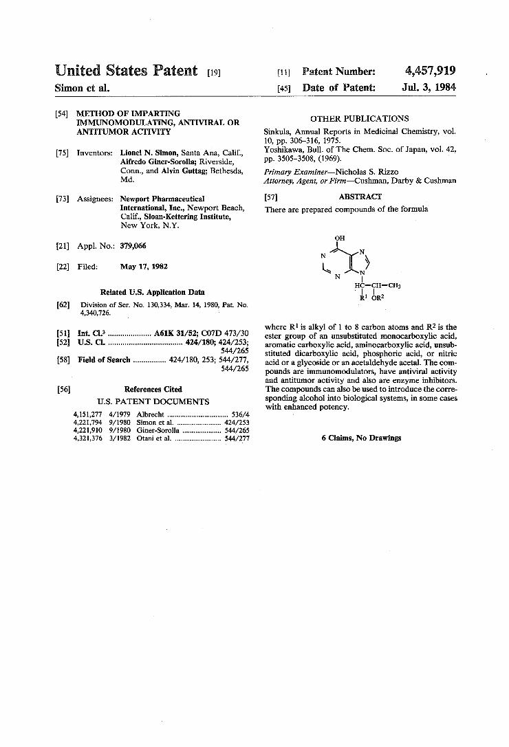
[57] ABSTRACT There are prepared compounds of the formula
1J1,» N |
nc-cn-cn; R1 on2
where R1 is alkyl of 1 to 8 carbon atoms and R2 is the ester group of an unsubstituted monocarboxylic acid, aromatic carboxylic acid, aminocarboxylic acid, unsub stituted dicarboxylic acid, phosphoric acid, or nitric acid or a glycoside or an acetaldehyde acetal. The com pounds are immunomodulators, have antiviral activity and antitumor activity and also are enzyme inhibitors. The compounds can also be used to introduce the corre sponding alcohol into biological systems, in some cases with enhanced potency.
6 Claims, No Drawings
4,457,919 1
METHOD OF IMPARTING IMMUNOMODULATING, ANTIVIRAL OR
ANTITUMOR ACTIVITY
The invention described herein was made in the course of work under a grant or award from the De partment of Health, Education and Welfare.
CROSS REFERENCE TO RELATED APPLICATIONS
This is a division of application Ser. No. 130,334 ?led Mar. 14, 1980 now US. Pat.'No. 4,340,726. There are disclosed and claimed in Giner-Sorolla
~ application Ser. No. 942,804, ?led Sept. 15, 1978 and now US. Pat. No. 4,221,910, certain 9-(hydroxyalkyl) purines. These same 9-(hydroxyalkyl) purines and cer tain acid addition salts thereof are also disclosed in Simon et al application Ser. No. 942,802, ?led Sept. 15, 1978 and now US. Pat. No. 4,221,909. The Simon et a1 application claims the acid addition salts and certain uses of the purines and the acid addition salts. The entire disclosure of the Giner-Sorolla application and the Simon et al' application are hereby incorporated by reference and relied upon.
SUMMARY OF THE INVENTION
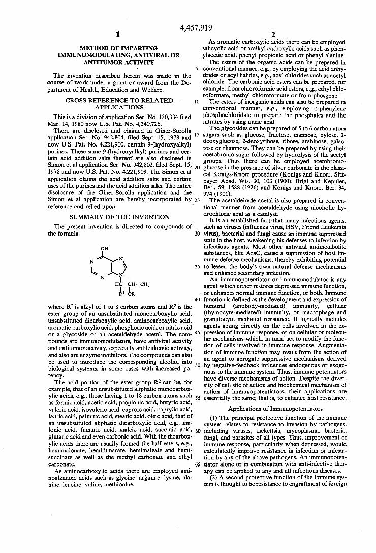
The present invention is directed to compounds of the formula
OH
N ’ N
\ :NIQ l-lC-CH-CH;
R1 OR
where R1 is alkyl of 1 to 8 carbon atoms and R2 is the ester group of an unsubstituted monocarboxylic acid, unsubstituted dicarboxylic acid, aminocarboxylic acid, aromatic carboxylic acid, phosphoric acid, or nitric acid or a glycoside or an acetaldehyde acetal. The com pounds are immunomodulators, have antiviral activity and antitumor activity, especially antileukemic activity, and also are enzyme inhibitors. The compounds can also be used to introduce the corresponding alcohol into biological systems, in some cases with increased po tency. The acid portion of the ester group R2 can be, for
example, that of an unsubstituted aliphatic monocarbox ylic acids, e.g., those having 1 to 18 carbon atoms such as formic acid, acetic acid, propionic acid, butyric acid, valeric acid, isovaleric acid, caproic acid, caprylic acid, lauric acid, palmitic acid, stearic acid, oleic acid, that of an unsubstituted aliphatic dicarboxylic acid, e.g., ma lonic acid, fumaric acid, maleic acid, succinic acid, glutaric acid and even carbonic acid. With the dicarbox ylic acids there are usually formed the half esters, e.g., hemimalonate, hemifumarate, hemimaleate and hemi succinate as well as the methyl carbonate and ethyl carbonate. As aminocarboxylic acids there are employed ami
noalkanoic acids such as glycine, arginine, lysine, ala nine, leucine, valine, methionine.
5
25.
30
2 As aromatic carboxylic acids there can be employed
salicyclic acid or aralkyl carboxylic acids such as phen ylacetic acid, phenyl propionic acid or phenyl alanine. The esters of the organic acids can be prepared in
conventional manner, e.g., by employing the acid anhy drides or acyl halides, e.g., acyl chlorides such as acetyl chloride.‘ The carbonic acid esters can be prepared, for example, from chloroformic acid esters, e.g., ethyl chlo roformate, methyl chloroformate or from phosgene. The esters of inorganic acids can also be prepared in
conventional manner, e.g., employing o-phenylene phosphochloridate to prepare the phosphates and the nitrates by using nitric acid. The glycosides can be prepared of 5 to 6 carbon atom
sugars such as glucose, fructose, mannose, xylose, 2 deoxyglucose, Z-deoxyribose, ribose, arabinose, galac tose or rhamnose. They can be prepared by using their acetobromo sugar followed by hydrolysis of the acetyl groups. Thus there can be employed acetobromo glucose in the presence of silver carbonate in the classi cal Konigs-Knorr procedure (Konigs and Knorr, Sitz bayer Acad. Wis. 30, 103 (1900); Brigl and Keppler, Ber., 59, 1588 (1926) and Konigs and Knorr, Ber. 34, 974 (1901). The acetaldehyde acetal is also prepared in conven
tional manner from acetaldehyde using alcoholic hy drochloric acid as a catalyst.
It is an established fact that many infectious agents, such as viruses (in?uenza virus, HSV, Friend Leukemia virus), bacterial and fungi cause an immune suppressed state in the host, weakening his defenses to infection by infectious agents. Most other antiviral antimetabolite substances, like AraC, cause a suppression of host im mune defense mechanisms, thereby exhibiting potential to lessen the body’s own natural defense mechanisms and enhance secondary infection. An immunopotentiator or immunomodulator is any
agent which either restores depressed immune function, I or enhances normal immune function, or both. Immune
65
function is de?ned as the development and expression of humoral (antibody-mediated) immunity, cellular (thymocyte-mediated) immunity, or macrophage and granulocyte mediated resistance. It logically includes agents acting directly on the cells involved in the ex pression of immune response, or on cellular or molecu lar mechanisms which, in turn, act to modify the func tion of cells involved in immune response. Augmenta tion of immune function may result from the action of an agent to abrogate suppressive mechanisms derived by negative-feedback in?uences endogenous or exoge nous to the immune system. Thus, immune potentiators have diverse mechanisms of action. Despite the diver sity of cell site of action and biochemical mechanism of action of immunopotentiators, their applications are essentially the same; that is, to enhance host resistance.
Applications of Immunopotentiators (1) The principal protective function of the immune
system relates to resistance to invasion by pathogens, including viruses, rickettsia, mycoplasma, bacteria, fungi, and parasites of all types. Thus, improvement of immune response, particularly when depressed, would calculatedly improve resistance in infection or infesta tion by any of the above pathogens. An immunopoten tiator alone or in combination with anti-infective ther apy can be applied to any and all infectious diseases.
(2) A second protective‘function of the immune sys tem is thought to be resistance to engraftment of foreign
4,457,919 3
tissue, either natural as in the fetal-maternal relation ship; or unnatural as performed by the transplant physi cian. Immunopotentiators can also be used to facilitate rejection of fetal or placental tissues or to modify or induce tolerance to grafts.
(3) A third protective function of the immune system is thought to be resistance to malignant cell develop ment as in cancer. The use of immunopotentiators can be used in cancer treatment to enhance tumor rejection and to inhibit tumor recurrences following other forms of therapy.
(4)>A fourth protective function involves the capacity to recognize foreignness and to maintain nonreactivity to self by positive suppressor mechanisms. In auto immune and related disorders, immune reactivity di rected at self antigens or exaggerated, elevated re sponses are apparent which are self-destructive. Im munopotentiators can be used to restore normal sup pressor mechanisms, induce tolerance, or otherwise promote a normal immune response. Each of the protective functions of the immune sys
tem can be modi?ed by non-speci?c therapy with im munopotentiators alone or in combination with other agents employed to improve resistance or to kill the invading pathogen. In addition, speci?c resistance can be augmented by use of immunopotentiators in conjunc tion with some form of antigen as in a vaccine employ ing, for example, virus, tumor cells, etc. This use can be to induce either speci?c immunity or tolerance. The latter might be exempli?ed by use with antigen in al lergy or auto-immune diseases. Use of immunopotentia tors may be either therapeutic or prophylactic; the lat ter particularly in aging, where infection, auto
5
25
immunity, and cancer are more common. The timing of 35 administration and routes are variable and may be criti cal in determining whether a positive or negative re sponse results. Any agent capable of augmenting im mune response may inhibit it depending on timing and dose; thus, under certain circumstances an im munopotentiator could be used as an immunosuppres sive agent for use in allergy, auto-immunity and trans
. plantation.
The parent alcohol when R1 is hexyl is erythro-9-(2 hydroxy-3-nonyl)-hypoxanthine which has been tested under the identifying number NPT 15392. Among the compounds within the present invention are erythro-9 (2-acetoxy-3-nonyl)-hypoxanthine (identi?ed as NPT 15458, see Example 1), erythro-9-(2-succinoxy-3 nonyl)-hypoxanthine (identi?ed as NPT 15457, see Ex ample 2), and erythro-9-(2-phosphate-3¢nonyl)-hypox anthine. '
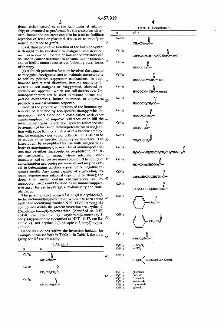
Other compounds within the invention include, for example, those set forth in Table 1. In Table l, the alkyl group for R1 are all n-alkyl.
40
55
60
65
4 TABLE l-continued
R1 R2 '
C6Hl3 "
CH3(CH2)1eC
can; " CH3(CH2)7CH=CH(CH2)7C— (cis)
Cam; I ‘H
HOOCCHZC
Cams _ “
HOOCCH=CHC— (cis)
CeHu n
nooccn=crice (trans) Cal-I13 "
HOOCCH2CH2CO—
Cal-I13 "
HOOC(CH2)3C
C6Hl3 "
CH2(NH2)C
C6H13 " cmcnmngc
C6H13 "
H2NC(=NIDNHCH2CH2CH2CH(NH2)C—
C6H13 "
H2N(CH2)4Cl-l(Nl-I2)C—
C6313 I "
cr-nscmcmcnmngc
Cal-I13 _ ‘A ll
' (CH3)ZCHCI1ZCYH(NH2)C—
Cams i
" CH2C'—
C6H13 : ll
CH2CH2C_
Cal-I13 “ ,
O-HOC5H5C- '
C6Hl3 "PC3142 C6H13 '-N02
Cal-I13 /
CH3CH (acetaldehyde acetal) . \.
C5H13 glucoside C6H13 riboside C6l-l13 fructoside C6H13 mannoside C6H13 rhamnoside C6H|3 xyloside
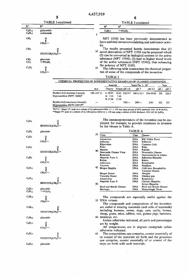
4,457,919 5 6
TABLE l-continued TABLE l-continued R1 R2‘ R1 R2
C6H13 galactoside Cal-I17 —PO3H2 C6H13 arabinoside 5 I
CH3 0 NPT 15392 has been previously demonstrated to cH3g_ have antiviral immunomodulating and anti-tumor activ
ity. CH3 The results presented herein demonstrate that (1)
|| 10 novel derivatives of NPT 15392 can be prepared which HOOCCHgCHzC- . . . .
(2) can be converted in biological systems to the act1ve CH3 glucoside substance ‘(NPT 15392), (3) lead to higher blood levels CH3 -P03H2 of the active substances (NPT 15392), thus enhancing
the potency of NPT 15392. CZHS fl) 15 The following table summarizes the chemical proper
CH3C—- ties of some of the compounds of the invention.
TABLE 2
CHEMICAL PROPERTIES OF REPRESENTATIVE EXAMPLES OF CLAIMED COMPOUNDS
Analysis )‘max/Am X 103 Amin rn.p. Theory Found pH 1.0 pH 7 pH 9.5 pH 1 pH 7
Erythro-9-(2-Acetoxy-3-nonyl)- ISO-151° C. C 59.97 59.83 250/9.7 249.5/ 10.1 254/ 10.06 220 222.5 Hypoxanthine (NPT 15458)’ H 7.55 7.42
N 17.48 17.39 Erythro-9-(2-succinoxy-3-nonyl)- —- — 250/— 249/-— 254/ 222 223
Hypoxanthine (NPT-15457)" 'HPLC - Single UV peak on a column of 5p. spherosorb ODS. 2.1 X 250 mm using solvent of 65% methanol: 35% .05 M H3PO4 "Single UV peak on a column of 5p. Ultmpliere ODS 4.6 X 250 mm using a solvent of 65% Methanol: 35% 0.05 M H3PO4
C2H5 The immunopotentiators of the invention can be em " ployed, for example, to provide resistance to invasion
HOOCCHZCHZC“ by the viruses in Table A.
C2115 glucoside TABLE A 35 Virus Class Disease
C3H7 O . || Arenavirus RNA Rift Valley Fever
CH3C— In?uenza RNA In?uenza Rhinovirus RNA Common Cold
can, Polio RNA Polio || . 40 Measles RNA Rubella
HOOCCHZCHZC" Newcastle Disease Virus RNA Newcastles Disease Rotavirus RNA Gastroenteritis in infants
C4H9 O Hepatitis Type A RNA Infectious Hepatitis ii Rabies RNA Rabies
CH3C- Arbovirus RNA Encephalitis Vaccinia I DNA Smallpox
C5H11 4'5 Herpes Simplex DNA Cold sore, Encephalitis, ii ' Venereal Disease
CH3C- Herpes Zoster DNA Shingles Varicella Zoster DNA Chicken pox
CSHH 0 Adenovirus DNA Respiratory ll Hepatitis Type B DNA Chronic Hepatitis,
HOOCCHZCHZC- 5O Severe Hepatitis Hoof and Mouth Disease DNA Hoof and Mouth Disease
CSHII glucoside Machupo DNA Hemorrhagic Fever C5H11 —PO3H2
C7H15 The compounds are especially useful against the "_ 55 RNA viruses.
CH3C . . . .
The compounds and compositions of the lnvention C7H15 " are useful in treating mammals (and cells of mammals)
' ‘ ' l h ses HOOCCHZCH2C_ including humans, swine, dogs, cats, catt e, or , sheep, goats, mice, rabbits, rats, guinea pigs, hamsters,
C7H15 glucoside 6o monkeys, etc- _ . _
Unless otherwlse lndlcated, all parts and percentages CsHn “ are by weight.
CH3C- All temperatures are in degrees Centigrade unless otherwlse mdicated.
CsHn ‘'3 65 The compositions can comprise, consist essentially of HOOCCHZCH2C_ or consist of the materials set forth and the processes
can comprise, consist essentially of or consist of the 08H" glucoside steps set forth with such materials.
4,457,919 7
The compositions can be administered to the mam mals by conventional techniques, e.g., orally, nasally, rectally, vaginally or parenterally. They can be em ployed as injectable solutions, e. g., in water, or as tables, pills, capsules, etc.
DESCRIPTION OF THE PREFERRED EMBODIMENTS EXAMPLE 1
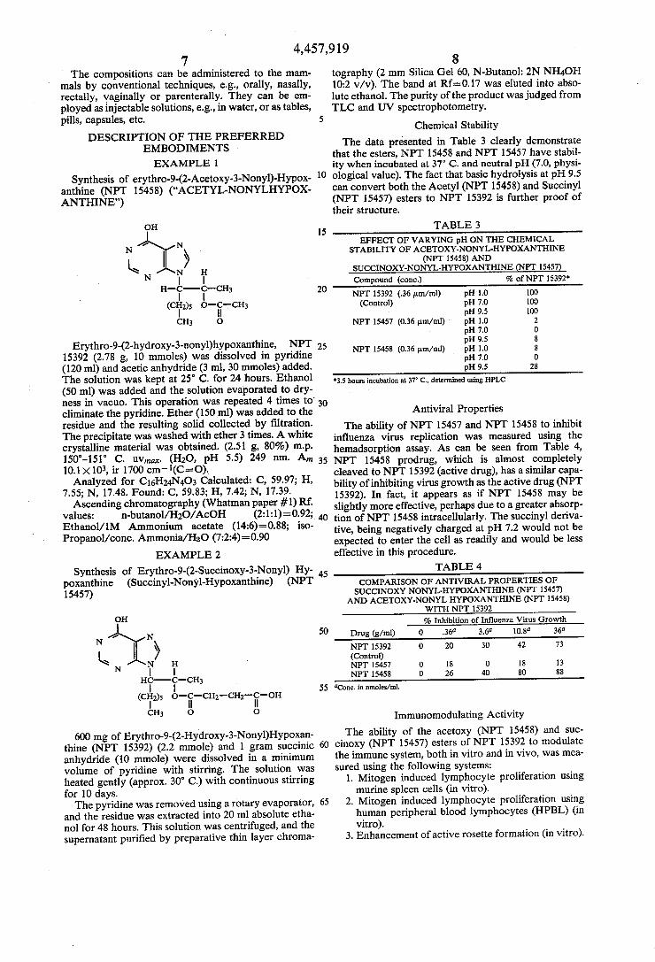
Synthesis of erythro-9-(2-Acetoxy-3-Nonyl)-Hypox anthine (NPT 15458) (“ACETYL-NONYLHYPOX ANTHINE”)
on
N’ N
k I) \ N N
O
Erythro-9-(2-hydroxy-3-nonyl)hypoxanthine, NPT 15392 (2.78 g, 10 mmoles) was dissolved in pyridine (120 ml) and acetic anhydride (3 ml, 30 mmoles) added. The solution was kept at 25° C. for 24 hours. Ethanol (50 ml) was added and the solution evaporated to dry ness in vacuo. This operation was repeated 4 times to‘ eliminate the pyridine. Ether (150 ml) was added to the residue and the resulting solid collected by ?ltration. The precipitate was washed with ether 3 times. A white crystalline material was obtained. (2.51 g, 80%) mp. l50°-l5l° C. uvmax. (H2O, pH 5.5) 249 nm. A,,,
Y 10.1 X 10³, it 1700 cm'*1(C=O). Analyzed for C16H24N4O3 Calculated: C, 59.97; H,
7.55; N, 17.48. Found: C, 59.83; H, 7.42; N, 17.39. Ascending chromatography (Whatman paper #1) Rf.
values: n-butanol/HzO/AcOH (2:1:l)=0.92; Ethanol/ 1M Ammonium acetate (14:6)=0.88; iso Propanol/conc. Ammonia/H2O (7:2:4)=0.90
EXAMPLE 2
Synthesis of Erythro-9-(2-Succinoxy-3-Nonyl) Hy poxanthine (Succinyl-Nonyl-Hypoxanthine) (NPT 15457)
on
N’N
(ii \
Ni‘ H
(CIiI2)s CH3 0
600 mg of Erythro-9-(2-Hydroxy-3-Nonyl)Hypoxan thine (NPT 15392) (2.2 mmole) and 1 gram succinic anhydride (l0 mmole) were dissolved in a minimum volume of pyridine with stirring. The solution was heated gently (approx. 30° C.) with continuous stirring for 10 days. The pyridine was removed using a rotary evaporator,
and the residue was extracted into 20 ml absolute etha nol for 48 hours. This solution was centrifuged, and the supernatant puri?ed by preparative thin layer chroma
20
30
35
45
50
55
60
65
8 tography (2 mm Silica Gel 60, N-Butanol: 2N NH4OH 10:2 v/v). The band at Rf=0.l7 was eluted into abso lute ethanol. The purity of the product was judged from TLC and UV spectrophotometry.
Chemical Stability The data presented in Table 3 clearly demonstrate
that the esters, NPT 15458 and NPT 15457 have stabil ity when incubated at 37° C. and neutral pH (7.0, physi ological value). The fact that basic hydrolysis at pH 9.5 can convert both the Acetyl (NPT 15458) and Succinyl (NPT 15457) esters to NPT 15392 is further proof of their structure.
TABLE 3 EFFECT OF VARYING pH ON THE CHEMICAL
STABILITY OF ACETOXY-NONYL-HYPOXANTHINE (NPT 15458) AND
SUCCINOXY-NONYL-I-IYPOXANTHINE (NPT 15457) Compound (cone) % of NPT 15392‘
NPT 15392 (.36 um/ml) pH 1.0 100 (Control) pH 7.0 100
pH 9.5 100 NPT 15457 (0.36 um/ml) pH 1.0 2
pH 7.0 0 pH 9.5 8
NPT 15458 (0.36 um/ml) pH 1.0 8 pH 7.0 0 pH 9.5 28
‘3.5 hours incubation at 37° C., determined using HPLC
Antiviral Properties The ability of NPT 15457 and NPT 15458 to inhibit
influenza virus replication was measured using the hemadsorption assay. As can be seen from Table 4, NPT 15458 prodrug, which is almost completely cleaved to NPT 15392 (active drug), has a similar capa bility of inhibiting virus growth as the active drug (NPT 15392). In fact, it appears as if NPT 15458 may be slightly more effective, perhaps due to a greater absorp tion of NPT 15458 intracellularly. The succinyl deriva tive, being negatively charged at pH 7.2 would not be expected to enter the cell as readily and would be less effective in this procedure.
TABLE 4 COMPARISON OF ANTIVIRAL PROPERTIES OF SUCCINOXY NONYL-HYPOXANTHINE (NPT 15457)
AND ACETOXY-NONYL HYPOXANTHINE (NPT 15458) WITH NPT 15392 % Inhibition of Influenza Virus Growth
Drug (g/ml) 0 .36‘1 3.6“ 10.8” 36”
NPT 15392 0 20 30 42 73
(Control) NPT 15457 0 l8 0 18 13 NPT 15458 0 26 40 80 88
I7Conc. in nmoles/ml.
Immunomodulating Activity The ability of the acetoxy (NPT 15458) and suc
cinoxy (NPT 15457) esters of NPT 15392 to modulate the immune system, both in vitro and in vivo, was mea sured using the following systems:
1. Mitogen induced lymphocyte proliferation using murine spleen cells (in vitro).
2. Mitogen induced lymphocyte proliferation using human peripheral blood lymphocytes (HPBL) (in vitro).
3. Enhancement of active rosette formation (in vitro).
4,457,919 9
4. Enhancement of IgM antibody formation in mice immunized with sheep red blood cells (SRBC) (in vivo).
1. The data presented in Table 5 clearly demonstrates the ability of NPT 15458 to augment PHA induced lymphocyte proliferation as measured by incorporation of tritiated thymidine. Approximately 25% increase was observed with a concentration of 50 ug/ ml of NPT 15458. A similar increase was observed with 10 ug/ml of NPT 15392.
2. The data presented in Table 6 clearly demonstrates that equimolar concentrations of NPT 15457 and NPT 15458 are effectve in augmenting PHA induced lym phocyte proliferation using HPBL. This is interesting in view of the data given in Table 9, which shows that HPBL will cleave both esters to NPT 15392. NPT 15458, which is cleaved to a greater extent than NPT 15457 (see Table 9) is a more effective potentiator (Table 6, 1.48 versus 1.28) of the PHA induced transfor mation.
3. As can be noted from Table 7, both NPT 15457 and NPT 15458 are effective in modulating active rosette formation in HPBL. It is interesting to note that in Experiment #1, where the placebo-treated lympho cytes have a very low level of active rosettes (im munode?cient) NPT 15457 and NPT 15458 are as ac tive as NPT 15392 in restoring the depressed immunity to normal levels. In Experiment #2, placebo-treated‘ controls have abnormally high values for active ro settes. In this instance, NPT 15392 and NPT 15458 are able to decrease the high levels of normal values; thus, demonstrating the true immunomodulatory properties of these drugs.
4. NPT 15457 and NPT 15458 and the “active” drug, NPT 15392, were given to Balb/c mice who had been immunized with sheep red blood cells SRBC. Antibody production to SRBC was measured. It is interesting to note that NPT 15458 and NPT 15392 were very effec tive in modulating the production of IgM antibodies. NPT 15392, at the levels studied, produce a 46% in crease in IgM production. Higher doses of NPT 15392 will cause less of an increase or decrease over control values. It is interesting to note that NPT 15458, which is converted to NPT 15392 and leads to at least 10 fold higher blood values of NPT 15392 (Table 10), produces an inhibition of IgM formation (Table 8). This is to be expected since NPT 15458 produces very high levels of NPT 15392 which are most likely inhibitory to IgM formation. The data presented in Table 8 demonstrates the immunomodulatory activity of NPT 15457 and NPT 15458. It should be noted that the effect of NPT 15457 is less than that of NPT 15458 and that less NPT 15457 is converted to NPT 15392 than NPT 15458 (Table 10).
TABLE. .5 ,
IMMUNQMQIPUILAITING.PRQliERTIE? 0F ACETOXY-NONYL-BXPOXANIIaNE(NPT 1545s» STIMULATIQNQFI’N viv‘o IM‘ITQGEN-I‘NDUCED
LYMPHQCY'I‘E PROLIFERATION iisiiviouse s'PLEENbELLs
_ .. .. , .__.._e‘m Iiicor .
ujf‘ompound (Cone. ‘rig/m1)‘ —PII_A 5FPIIAQ§JF D/C v0 (c) 137 621,647 471 ---7
NPT 15392 10 ng/ml (D) 117 68,864 588 1.25 NPT 1545s 10 ng/ml (D) 153 66,800 436 .93 NPT 1545s so ng/rnl (D) 138 81,297 5159 “1.25am
"8.1. equals stimulation index
10
-20
35
55
60
65
10 TABLE 6
IMMUNOMODULATING PROPERTIES OF SUCCINOXY-NONYL-I-IYPOXANTI-IINE (NPT 15457)
AND ACETOXY-NONYL-HYPOXANTHINE (NPT 15458); ENHANCEMENT OF MITOGEN-INDUCED
LYMPI-IOCYTE PROLIFERATION IN HUMAN PERIPHERAL BLOOD LYMPHOCYTES
Com M Compound n moles/ml ~PHA +PI~IA S.I. D/C
NPT 15392‘ 0 (C) 379 59,404 156 — 0.036 (D) 297 59,167 199 1.27 0.36 (D) - 577 69,057 120 .76 3.6 (D) 381 67,328 176 1.128
NPT 15457 0 (C) 277 54,015 195 — 0.036 (D) 240 59,569 250 1.28 0.36 (D) 228 59,689 261 1.33 3.6 (D) 232 52,149 224 1.14
NPT 15458 0 (C) 277 54,015 195 —— 0.036 (D) 214 59,898 280“ 1.43 0.36 (D) 221 63,858 289“ 1.48 3.6 (D) 238 69,055 290“ 1.48
‘values are means of three experiments "p 2 0.05
TABLE 7
IMMUNOMODULATING PROPERTIES OF ACETOXY-NONYL-I-IYPOXANTHINE (NPT 15458) AND SUCCINOXY-NONYL-I-IYPOXANTHINE (NPT 15457):
ENHANCEMENT OF ACTIVE E-ROSETTE FORMATION % Active _
(Conc. E-Rosettes (x i S.E.) Compound 11 moles/ml) #1 #2
Placebo (0) 9.23 i 2.9 34.2 i 6.6 (Control) NPT 15392 (3.6) 16.2 i 1.0 — (Control) (0.36) — 15.4 i 2.9“ NPT 15457 (3.6) 24.8 i 6“ —
(0.36) — 33.6 i 2.4 NPT 15458 (3.6) 19.4 i 1.0“ —
(0.36) — 23.25 i 1.24“
"p § 0.05 versus placebo control.
TABLE 8 IMMUNOMODULATING PROPERTIES OF
ACETOXY-NONYL HYPOXANTI-IINE (NPT 15458) AND SUCCINOXY NONYL-HYPOXANTI-IINE (NPT 15457): INHIBITION OF IgM ANTIBODY PRODUCTION
IN SRBC IMMUNIZED BALD/C MICE
1 # IgM Expt _Plaques % of # Substance (Dose i.p.) (x t SE.) Control
1 NPT 15392 (1.80 n moles/kg X 4) 41 i 4 146“ 2 (.18 n moles/kg X 4) 68 i 2 147“ 1 NPT 15457 (1.8 n moles/kg X 4) 19 i 3 68” 2 (.18 n moles/kg X 4) 47 i 3 102 1 NPT 15458 (1.8 n moles/kg X 4) 8 i 1 28.5“ 2 (.18 n moles/g X 4) 11 i 1 23.9“ 1 CON- (placebo X 4) 28 i 2 100
TROL 2 (placebo X 4) 46 i 2 100
"p § 0.01 compared to placebo control
Metabolic Conversion
As can be seen from Table 9, incubation of the npro drug‘s”, NPT 15457, and NPT 15458wit11 Vé’ro Cells (African Green Monkey Kidney Cellé), Liver Homoge hate, and Human Peripheral B1006 Lymphocytes (HPBL) leads t6 the formation @f the “aetive” drug, NPT 15392. The conversion ‘of NPT 15458 to NPT 15392 appears in occur to a greater extent ‘than the eenversio'n of NPT 15457 to NPT 15392. 7 The data ‘in Table 10, drama-names that Beth NPT
‘15457 and NPT 1545s can be absorbed me the blood
4,457,919 11
after i.p. administration and that administration of NPT 15458 leads to almost 12 fold higher levels of NPT 15392, than if NPT 15392 is given by itself. This clearly establishes the fact that NPT 15458 and NPT 15457 can produce NPT 15392 in vivo and further at least one of these derivatives gives much higher blood levels allow ing for greater activity.
TABLE 9 FORMATION OF NPT 15392 FROM
ACETOXY-NONYL-HYPOXANTI-IINE (NPT 15458) AND SUCCINOXY-NONYL-HYPOXANTI-IINE (NPT 15457)
BY BIOLOGICAL MEANS % of NPT 15392‘1 at time
(hrs) Compound 0 0.5 24
NPT 15392 Vero Cells 100 — 100 Liver Homogenate 100 100 — HPBL 100 — 100
NPT 15457 Vero Cells 0 -- 0 Liver Homogenate 0 l4 — l-IPBL 0 — 18.2
NPT 15458 Vero Cells 0 — 50 Liver Homogenate 0 7O — HPBL 0 — 100
"Determined using HPLC
TABLE 10 FORMATION OF NPT 15392 AFTER LP.
ADMINISTRATION TO MICE OF ACETOXY-NONYL-HYPOXANTI-IINE (NPT 15458) AND SUCCINOXY-NONYL-I-IYPOXANTHINE (NPT 15457)
Compound Blood Level (um/ml x 103) Administered‘ animal# NPT15392 NPT 15457 NPT15458
NPT15392 1 0.22 _ _
2 0.68 _ _
NPT 15457 1 0.47 1.4 - 2 0.22 0.57 _
NPT15458 1 8.2 _ o 2 2.7 - o
‘(360 P-m/ks)
Effect of Erythro-9-(2-Acetoxy-3-Nonyl)-Hypoxan thine on Growth of Tumor (Leukemia) Cells In Vivo
% Inhibition of Growth
Compound (Conc.) L12_l0 (mouse) 8068 (Human) NPT 15458 (10 pg/ml) 40 32
. TABLE 11
SUMMARY OF BIOLOGICAL PROPERTIES PROPERTY NPT 15457 NPT 15458 NPT 15392
Convertible to NPT 15392 Chemical '
pH 9.5 Yes Yes Not Applicable pH 7.0 No No Not Applicable Biological l-IPBL 18 100 Not Applicable Liver 14 70 Not Applicable Vero 0 50 Not Applicable Mouse 25 100 Not Applicable Biological Activity Antiviral Very Yes (88%) Yes (73%) (in vitro) slight Immunomodulating Yes Yes Yes (+12—27%) (in vitro) (+ 14-28%) (+43—48%) Lymph. Prolif. lmmunomodulating Yes Yes (+11%) Yes (+78%) (in vitro) (+ 166%) Rosette
5
15
20
25
30
35
40
45
55
60
65
12 TABLE ll-continued
SUMMARY OF BIOLOGICAL PROPERTIES
PROPERTY NPT 15457 NPT 15458 NPT 15392
Immunomodulating Moderate Yes (-72%) Yes (+46%) (in vivo) (0-60) SRPC-lgM
The following procedures were employed in order to make the determination of properties discussed above.
A. Cell Culture Methods
A. Hela or Vero Cell Propagation
1. Cell cultures in 120 cm2 ?asks are subcultured in monolayers in the following manner:
2. The media are poured off, and the monolayer washed two times with approximately 50 ml per wash of calcium and magnesium free phosphate buffered saline (PBS), at a pH of 7.2.
3. One ml of trypsin-EDTA solution containing 0.5 g trypsin (1:250) and 2.0 gEDTA/liter of Hanks balanced salt solution (HBSS) without Ca+ + and Mg+ + is added at 37° C. to each ?ask and dispersed over the monolayer with gentle shaking.
4. The flasks are then placed in an incubator at 37° C. for approximately 3-5 minutes, depending on the time required to dislodge the cells. Shaking by hand is re quired.
5. Ten ml of planting medium is added to each ?ask and the cells are dispersed by aspirating and expelling the suspension from the pipette. This is done ten times.
6. The contents of a series of ?asks were pooled and the cells in the suspension were diluted with planting medium to 7—8.5>< 104 cells/ml.
7. The planting medium consisted of the following composition: Minimum Essential Medium Eagles (MEM) with Earle’s salts and HEPES buffer supple mented by adding the following substances as speci?ed to 100 ml of MEM:
10 ml of fetal calf serum (?nal concentration: 10%) 1 ml of l-glutamine (200 Molar) 1 ml of 10,000 units pencillin, 10,000 pg streptomycin and 10,000 neomycin mixture
8. The cells are subcultured into Costar tissue culture trays consisting of 24 ?at bottom wells each with a 3 ml capacity per well; the cell culture suspension (1 ml) is added to each well.
9. The monolayers are used for experimentation when they reach almost con?uent growth (approxi mately 1-2 days).
10. Separate ?asks are used for maintaining the cell lines. Maintenance media consists of MEM plus supple~ ments (see step 7) with FSC reduced to 5% ?nal con
centration. '
B. Red Blood Cells
1. Whole blood is obtained by cardiac puncture from male Hartly strain guinea pigs.
2. About 10 cc of whole blood is mixed with 25 ml of Alsever’s solution, and may be stored at 4° C. for up to 1 week.
3. Just prior to use, the RBC’s are washed 3 times in phosphate-buffered saline (PBS), pH 7.2.
4. Each wash is accomplished by centrifuging the RBC suspension for 10 minutes at 450 g at room temper ature.
4,457,919 13
5. A 0.4% v/v RBC suspension is made with Hanks balanced salt solution.
C. Egg Propagation of Viruses
A. Infection '
1. Nine to ten day fertile chicken eggs are candled for viability and the placement of the air sac marked on the shell with a pencil. Questionable eggs (lack of move ment or discoloration) are discarded.
2. The shell surfaces are disinfected with 70% ethanol and allowed to dry.
3. A small hole is punched through the shell with a sterile egg punch, approximately $1" within the pencilled circle marking the air sac of each egg.
4. One-tenth ml of various virus suspension, 102 to 103 EID50/ ml, is inoculated through the hole with a 1 cc Tuberculine syringe at a 45° angle into the allantoic cavity. Care is taken not to injure the embryo or yolk sac.
6. One inch pieces of plastic tape are used to seal the hole and each egg is labeled with the appropriate virus, egg passage number and date.
7. Eggs are incubated at 35°-37° for 2 to 5 days, de pending on the rapidity of viral growth. Incubation time and temperature are recorded along with other pertinent data for each passage.
B. Harvest of ~Allantoic Fluids 1. At the end of the incubation period, the eggs are
chilled for 3-4 hours at 4° C. to minimize bleeding into the allantoic cavity during the virus harvesting proce dure.
2. Shell surfaces are again disinfected with 70% etha nol and allowed to dry.
3. Sterile scissors are used to cut away the top of the shell along the pencilled line, and the membranes are teased off with sterile forceps.
4. The forceps are inserted gently between the em bryo and shell membrane and the embryo pushed to one side, forming a “pocke ” of allantoic ?uid. Care is taken not to puncture the amnion (except in cases where the amnionic fluid is also to be harvested) and to minimize tearing of placental arteries.
5. A sterile, disposable 10 ml pipette with mechanized vacuum bulb is used to collect the allantoic ?uid and transfer it to sterile, disposal centrifuge tube. Approxi mately 5-8 ml can be harvested from each egg.
6. The harvested material is centrifuged at 1200 g for 10 minutes at 4° C. Supernates are transferred to sterile tubes.
7. Samples are taken from each pool for titration and sterility testing. The remaining suspension is aliquotted into sterile 2 cc serum tubes and labeled with strain, passage number and date.
8. Aliquots are immediately frozen in liquid nitrogen and placed in storage at —70° C. in an ultrafreezer or liquid N; cryofreezer. '
D. Virus Propagation in Tissue Culture
A. Infection
1. Twenty-four to 48 hour monolayer cultures of the appropriate cell type, usually in 250 cm2 disposable tissue culture ?asks are chosen for use when barely con?uent.
2. All infection and harvesting are performed in a biological safety cabinet and only mechanical pipetting devices used. Sterile techniques are observed.
15
20
25
35
45
50
55
60
65
14 3. Twenty-?ve ml of maintenance medium is used to
replace growth medium in cultures to be infected. Con trol cultures also receive 25 ml of maintenance medium.
4. The seed virus is diluted with serumfree MEM according to information supplied by the source, or from titration of previous passages, and 0.5 ml is added to each culture (except controls).
5. Cultures are incubated at 37° C. in a moist atmo sphere of 5% CO2, 95% air for 48-72 hours, with daily observation for the development of cytopathic effects (CPE) characteristic of each virus.
B. Harvesting l. Cultures are removed for harvesting when the
CPE reaches a score of 3 to 4+. 0—No apparent cytopathic effects l+—25% of cells showing cytopathic effects 2+—50% of cells showing cytopathic effects 3+—75% of cells showing cytopathic effects 4+—l00% of cells showing cytopathic effects 2. Cultures are frozen and thawed three times to lyse
and dislodge the cell layer. After thawing for the third time, the culture ?uid is transferred to a sterile dispos able centrifuge tube and centrifuged at 300 g for 30 minutes to remove cell debris. This virus suspension is checked for bacterial contamination.
3. Supernatants are immediately aliquotted (0.5-1 ml) into 2 cc sterile serum tubes and frozen in liquid nitro gen.
4. For titration, monolayer cultures in 24-well ?at bottom tissue culture plates are prepared and infected as follows:
(a) Serial ten-fold dilutions of the virus are made in cold, 5% Fetal Calf Serum (FCS) maintenance medium (10-1 to l0—7).
(b) One ml of each dilution is added to individual wells in triplicate.
(c) Control cultures containing only maintenance medium are included.
5. Cultures are incubated for 48 hours at 37° C. in moist 5% CO2, 95° air, and the cell layers scored as previously described. TCID50’s titer are calculated ac cording to the Reed and Munch method of titration.
E. Hemadsorption Assay 1. Just prior to carrying out HAd assays, media are
decanted from cell culture trays. 2. One mililiter of maintenance medium is added to
each culture well. 3. Two series of control cultures receive maintenance
medium alone. 4. Test cultures and one of the control series are im
mediately innoculated with 0.1 ml of diluted virus prep aration. (Input titers are speci?ed as to previous HAdFFU assays). HAdFFU is hemadsorption foci forming units.
5. The other series of controls are maintained unin fected, with MEM alone added as a blank innoculation.
6. Cultures are incubated at 37° C. for 16 to 18 hours, unless indicated otherwise.
7. After the innoculation period the media are de canted and the cells washed once with PBS at pH 7.2.
8. Five-tenths ml of the RBC suspension is added per well and the cultures maintained for 30 minutes at room temperature.
9. The RBC suspension is then decanted and the cul tures are washed two to three times with PBS to re move all but speci?cally bound RBC’s.
4,457,919 15
10. Finally, 1.0 ml of Hanks Balanced Salt Solution (HBSS) is added to each well.
11. Counting of the foci of hemadsorbed RBC’s ini tially is performed using a Nikon inverted phase-con trast microscope with a 4X objective. _
12. In all cases, a minimum of ?ve random ?elds are selected per well for counting.
13. The counting of HAd fool is performed on a Bausch & Lomb Omnicon Alpha Image Analyzer.
14. The image of the microscope ?eld is projected onto a Vidicon scanner. It is also displayed on a televi sion screen so that the operator could detect and dis count errors due to debris. Foci are detected by gray ness level and image size.
15. To eliminate counting of residual unadsorbed RBC’s, an oversized count module is programmed to screen out individual RBC’s.
16. The statistical analysis involves analyzing the data using an analysis of variance nested design model. Source of variation may be due to treatments, wells nested within treatments, and an experimental error term due to ?elds within wells. Means infected cells per ?eld and standard errors of the mean will be calculated for each well. Means and standard errors will also be calculated for each treatment by pooling the wells nested within each ‘treatment together. The mean in fected cells per ?eld‘ for-v each treatment will be com pared with control using Dunnets’ multiple comparison test regardless of overall F-test for treatment. (See be low).
Control Treatment 1
Well 1 Well 2 Well 3 Well 1 Well 2 Well
X] i SE1 52 t SE; is; i s53 x1 -_l- SE1 32" i SE2, x3’ i SE3 Pooled x6 i SEC xT t SET
‘F 11 represents ?eld #
F. Preparation of Human Peripheral Blood Lymphocytes (HPBL)
A. Preparation of Ficoll-Hypaque Separation Medium 1. Twenty-two and a half grams of Ficoll 400 (Phar
macia, M.W. z400,000) are dissolved in 200 ml of dis tilled H2O. When most of the material is in solution, the volume is then adjusted to 250 ml with distilled H20.
2. Thirty-four grams of Hypaque (sodium diatrizoate, Sterling Organics, M.W. :636) are dissolved in 100 ml distilled H20.
3. Solutions are then sterile ?ltered thorugh a 0.45;]. millipore ?lter and stored in sterile containers at 4° C. away from light.
4. Working solutions are prepared by mixing 10 ml Hypaque solution with 24 ml Ficoll solution. The mix ture is warmed to 25° C. with constant stirring.
B. Separation of PBL’s from Whole Blood
1. Human blood samples are obtained by veinipunc ture into heparinized 20 ml-vacutainer tubes. Fifteen to twenty ml of the undiluted blood is gently layered over an equal volume of Ficoll-Hypaque solution in sterile 50 m1 polycarbonate centrifuge tubes using aseptic tech niques.
2. Prepared samples are centrifuged at 25° C., 400>< g for 30 mins. The fuzzy white band at the interface is
15
20
25
30
35
45
55
60
65
16 aseptically aspirated into a 50 ml sterile centrifuge tube with 10-20 ml RPMI-1640. The cells are washed once at 25° C. 400><g for 10 mins. at 25° C.
3. Pelleted PBL’s are resuspended in 1 ml RPMI for every 8 ml whole blood originally used and counted with a Coulter counter. Concentration is adjusted with RPMI-1640 supplemented with glutamine and antibiot ics. The cells are held at room temperatue until used.
G. Stimulation of HPBL with FHA and LPS 1. Ten to 40 ml of whole blood is drawn from healthy
volunteers in heparinized tubes. 2. Lymphocytes are separated from the above blood
using Ficoll-hypaque separation techniques. 3. Various concentration of test compound are pre
pared in RPMI-1640. 4. The concentration of lymphocytes are adjusted to
2X l06/ml of RPMI-1640. 5. Lymphocytes are incubated with test compound
(for 90 minutes at 37° C. 6. Sheep Red Blood Cells (SRBC.s) (one to two
weeks old) are washed 3 times with PBS and diluted to a ?nal concentration of 0.5% inj RPMI-1640.
7. Reaction tubes are set up containing the following: 0.2 ml lymphocytes (2X 10⁶) 0.2 ml 9% Ficoll in RPMI-1640 0.2 ml SRBC’s (0.5% in RPMI-1640) 8. The above reactants are centrifuged at 200x g for
5 minutes at room temperature. 9. The sediment is then gently resuspended and a 101
sample placed in a hemocytometer. 10. The number of rosettes are then counted using a
microscope with any lymphocyte having 3 or more attached red cells being counted as a rosette.
H. E-rosette Forming Cells
A. Preparation of Mitogens 1. Lipopolysaccharide B or Phytohemagglutinin P
powder is‘weighed and dissolved in RPMI-1640 at a concentration of 1000 pig/ml (1 mg/ml) and ?lter steril ized through a 0.45p. millipore ?lter.
2. Solutions are aliquotted (1 ml/tube) asceptically into labelled sterile cryotubes and frozen. Samples are not refrozen after partial use, but may be stored for a day or two at 4° C. Lyophilized powder is kept refriger ated at 4° C. .
B. Preparation of Cultures
l. PBL’s are prepared as in SOP #I-004 in serum-free RPMI-1640 supplemented with glutamine and antibiot ic-antimycotic solution (GIBCO). One tenth ml per well is added to each well of 96-well microtest culture plates (CoStar Plastics) with 8-channel automatic mi cropipettors (Flow Labs.) ?tted with sterile tips.
2. Mitogen solutions are thawed rapidly and diluted to 4 times the desired concentration with RPMI-1640.
3. Test compounds are prepared for use as per SOP #C-002, using RPMI-1640 as diluent, and diluted to 4 times the ?nal desired concentration.
4. Fifty pl of each dilution of mitogen and/or test compound is added to 6 replicate cultures. Plain RPMI 1640 is used in the same amounts in control cultures for a total volume of 0.2 ml/well. '
5. Cultures are incubated at 37° C. in a humidi?ed 95% air, 5% CO2 atmosphere (pH =6.0- 7.0) for 48 hours. The cells are then labelled with 0.5p.Ci/Well
4,457,919 17
3H-TdR (Thymidine) for an additional 18 hours. the cells are harvested with a multiple automatic sample harvester (M.A.S.H.) machine. Nonadherent cells are aspirated onto glass ?ber ?lter strips with 0.9% saline solution (10 well ?ushes) and the'cells lysed with dis tilled water (20 well ?ushes). The strips are thoroughly air dried, residual cell deposits cut from the strips and each sample placed in 1 dram glass scintillation vials. Scintillation ?uid concentrate (Scintiprep 1, Fisher Chemicals) is diluted 1:50 with scintillation grade tolu ene (Packard) and 1 ml added to each vial. Samples are evaluated by liquid scintillation spectroscopy and the data expressed as the means counts per minute per test group.
I. Immunoplacque-‘Assay A. Preparation of NPT 5392
1. A solution containing 500 ug/ml NPT 15392 is prepared by adding a preweighed amount of the drug powder to sterile PBS and sonicating the solution for 30 minutes.
2. The wavelength of GCA/McPherson double beam spectrophotometer is set to 250 nm.
3. The NPT 15392 is diluted 1:10 to 0.1 normal HCl. 4. Fifteen ml of 0.1 normal HCl is poured into beaker.
This solution is used to rinse cuvettes. Both cuvettes are ?lled with 0.1 normal HCl and the wavelength is read.’ Absorbance should be within 0001-0005 from the ze roed value.
5. The sample cuvette is emptied and the diluted drug solution is added.
6. Absorbance is recorded and concentration is calcu lated as follows:
‘ A (absorbance! Formula. 1131 X
Dilution factor X Atomic weight (278) = g/ml
B. Prior to Day 0
1. NPT 15392 drug solution is made by dissolving 500 g NPT 15392 per ml PBS,
2. The concentration is checked according to section A of this SOP.
3. The stock is aliquotted into 1 ml samples and stored at —20° C. for several months.
4. Solutions are thawed and diluted to desired con centration.
C. Agarose Base Plates are Made as Follows:
l. A 1.4% suspension of agarose in PBS (7 g in 500 ml) is autoclaved for 15 min. _
2. Using a Cornwell syringe, 3 ml volumes of molten agarose are aseptically dispensed into Falcon #1006 petri dishes and swirled to form an even layer.
3. These plates may be stored up to one week at 4° C. prior to ‘use. The plates are stored in an inverted posi tion (agar on top of plate).
D. Preparation of Guinea Pig Serum
1. Ten ml of blood is removed by cardiac puncture from two to six guinea pigs and placed in a 50 ml Falcon #2070 centrifuge tube without anticoagulants.
2. These tubes are incubated for 45 min. at 37° C. for clot formation.
3. Tubes are then removed from incubator and put on ice for 30 min. to retract the clot.
15
20
25
30
35
50
55
60
65
18 4. The serum is aseptically poured off each tube,
pooled, dispensed into 1 ml aliquots and stored at —70° C. until used. '
E. Immunization, Day 0 1. Mice are immunized as follows: sheep blood (sheep
#23) is received weekly from Hyland Labs. It is asepti cally collected in two volumes of Alseviers to one vol ume of blood.
2. Five-tenths ml of the sheep blood in Alseviers is washed three times in sterile PBS in an IEC clinical centrifuge, setting #4 (2800 rpm) for 10 min. at room temperature.
3. The pellet is resuspended in 1:10 in sterile PBS. 4. A model Z Coulter counter is calibrated according
to Coulter Instruction Manual. 5. A l:l0,000 dilution is needed for a Coulter counter
cell count and is obtained by ?rst making a 1:100 dilu tion in PBS and then diluting this solution 1:10 in isoton. The Coulter counter threshold is adjusted to setting #5.
6. The cell count is determined and the stock solution is diluted to 4><l07 cells/ml. This ?nal suspension is used for immunization.
7. Each mouse is immunized by iv injection into the lateral tail vein (warmed in a 50° C. water bath for veinous dilation) with 0.1 ml SRBC suspension; the final concentration of SRBC’s is 4X 10⁶ per mouse.
F. Treatment
1. Mice are treated by i.p. injection on days 0, l, 2 and 3. A syringe (1 cc) is ?tted with a one-half inch, 26 gauge needle. The needle is introduced at a 45° angle along the right side of the linea alba. A 0.2 m1 volume is given to both drug and control treated groups.
2. Drug groups are given 0.2 ml of a desired concen tration NPT 15392 solution which, for a 20 g mouse, is 5 ug/ml.
G. Spleen Preparation, Day 4
l. Spleens are removed‘[ aseptically and placed indi vidually in Falcon #2025 tissue culture tubes containing 3 ml MEM. The tubes are then stored on ice.
2. Spleens are homogenized in the same tube with a te?on pestle attached to a G. K. Heller variable speed reversible motor connected with a G. K. Heller GT-21 motor controller setting #6.
3. The homogenization time and action should be uniform from sample to sample.
4. Samples are then ?ltered through a 100 mesh 40 micron stainless screen into a standard tissue culture tube. The screen is rinsed with 3 ml MEM and the cell suspension is stored on ice.
5. A 1:1000 dilution is made for a Coulter counter cell count and is‘obtained by first making a 1:100 dilution in PBS followed by a 1:10 dilution in isoton.
6. The cell count is obtained and the stock suspension is diluted to 1X 10⁷ cells/ml. The Coulter counter threshold is set at 10. Red cells are lysed with three drops of Zap isoton.
H. Preparation of Top Agar (0.7%) l. Thirty-?ve hundredths gram agarose and 0.53
gram MEM powder are placed into an Erlenmeyer ?ask. Fifty ml of distilled H2O is then added to the ?ask.
2. The solution is autoclaved for 15 min. at 250° F. and 15 psi. It is then placed in a 45° water bath for 5 min. The pH is adjusted to approximately 7.2 by the addition of sodium bicarbonate (0.1 ml Na2CO3).
4,457,919 19
3. One ml aliquots are dispensed into 5 ml tissue cul ture tubes previously placed in a water bath. Allow several extra tubes for replacement in case of plating error.
I. Preparation of 10% SRBC Solution
1. Five ml sheep blood (same batch as used for immu nization) is washed three times in PBS using an IEC Clinical Standard Centrifuge, speed #4 (2800 rpm) for 10 min.
2. After the third wash, the cells are resuspended in a volume of PBS ten times the volume of the packed cells, i.e., 0.5 ml of packed SRBC’s, Q.S to 5 ml with PBS.
J. Plating
Agar plates are removed from regrigerator and al lowed to warm to room temperature. They are labeled in triplicate for each experimental group.
2. Agar-?lled tubes are removed from the water bath. One-tenth of the 10% SRBC’s and 0.1 ml spleen cell suspension are added to each agar tube. The tubes are agitated on a Vortex mixer.
3. The contents of the tubes are poured immediately into agar base plates and swirled until a smooth layer is formed. The plates are ‘placed on a level surface until the agar solidi?es.
4. The plates are incubated at 37° C. in a humidi?ed atmosphere of 5% CO2 and 95% air for 90 min.
5. Guinea pig complement (see preparation, section D) is removed from freezer, thawed at room tempera ture and diluted 1:10 in PBS.
6. Plates are removed from incubator and 1 ml of the diluted complement is added to each plate.
7. Plates are then incubated another 30-45 minutes at 37° C. Plates are then removed from incubator and counted using oblique light.
8. Plates can be stored at 4° C. in an inverted position and counted up to 24 hours later.
_ Summary of Therapeutic Uses in the Working Examples
The subject compounds of this invention have been shown to inhibit the replication of a representative sam ple of RNA viruses using standard tissue culture tech niques. In the case of the RNA viruses, in?uenza virus belonging to the A sub-type was shown to be inhibited, using the hemadsorption technique. Several members of the Series NPT 15457 and NPT 15458 were shown to inhibit the replication of in?uenza virus at concentra tions ranging from 3.6-360 n moles/ml. Other members of the RNA class of viruses are
shown in Table 6 and are responsible for the diseases speci?ed. Of all the diseases in the world, at least 25% are known to be caused by viruses. In addition, a num ber of viruses have been isolated that are shown to produce tumors. Thus, antiviral agents may be expected to, themselves, have some antitumor, e. g., antileukemic properties. The activity of one of these agents, NPT 15458 as
inhibitors of the growth of abnormal lymphocytes has been determined. Notably, NPT 15458 is capable of inhibiting the proliferation of mouse leukemic lymp nocytes (an L-l2l0 cell line) in tissue culture. A 40% inhibition of L-l2l0 cells was effected by NPT 15458 at 10 pg/ml.
Finally, the data presented in Table 3 demonstrates that at normal body pH’s (7.2) the compounds have good chemical stability but are cleaved to the “active”
15
20
25
30
35
45
55
65
20 substance NPT 15392 at alkaline pH. As noted in Table 9 the esters, specially NPT 15458, are cleved by incuba tion with animal tissues to NPT 15392. This presumably is why the subject compounds, “pro-drugs” have their indicated biological activity. In addition, the data pres ented in Table 10 clearly demonstrates that at least 12 fold higher blood levels of NPT 15392, the biologically “active” substance, are produced when NPT 15458 is injected i.p. than when NPT 15392 itself, is adminis tered to mice. Further as can be noted in Table 9 Vero Cells (kidney), Liver, and HPBL all cleave the “pro host” to the biologically active NPT 15392. The class of substances of the invention speci?cally
inhibit the replication of viruses, modulate (potentiate or inhibit) the immune response and inhibit the growth of leukemic lymphocytes. Based on in vitro experiments and the higher blood levels achieved with these com pounds compared to NPT 15392, which demonstrate activity over a concentration range of 0.01-100 ug/ml, expected dose ranges effective in mammals are 0.0005-50 mg/kg.
Formulations
The compounds of the present invention can be fed to a mammal at a dosage of l-l000 mg/kg of body weight and can be anticipated to be active at levels as low as 0.0005 mg/kg. They may be administered in tablet or capsule form
to humans and animals and where solubility permits in the form an aqueous syrups, or as solutions in oil, or where insoluble as a suspension. Typical pharmaceuti cal formulation are described below:
Capsule NPT 15458: 0.1-500 mg Avicel pH l0l (microcrystalline cellulose): to make
800 mg.v
Suspension Aqueous suspensions can be made with a number of
suspending agents incorporated with the active drug substances. Included as suspending agents are such sub stances as sodium carboxymethylcellulose, Na alginate, tragacanth, Avicel RC-59l (microcrystalline cellulose), methylcellulose, Veegum, Xanthan gum. In addition to a suspending agent such substances as sweeteners, ?a vors, colorants, preservatives, protective colloids, and dispersants may be added.
SYRUP FORMULATION
NPT 15458 0.05—250 mg (or at a maximum level of solubility)
Com Sugar 3.25 g Y Distilled Water I .05 g FD and C Red 40 .00175 g Sodium Saccharin .00250 g Alcohol U.S.P. .08 g Methyl Paraben U.S.P .005 g Glycerin .001 g Cherry Flavor .3l225 g Fruit Flavor .00825 g Distilled Water g.s. ad 5 ml
TABLET FORMULATION
NPT 15458 0.1-500 mg Avicel pH 101 B0 mg Starch, modi?ed 20 mg
4,457,919 21
-continued
TABLET FORMULATION
Magnesium stearate U.S.P. 5.5 mg
Polyvinylpyrrolidone 22 mg
Stearic acid U.S.P. 30 mg
What is claimed is:
1. A method of imparting immunomodulating activ
ity, antiviral activity or antitumor activity comprising
adminstering to a mannal an effective amount for such
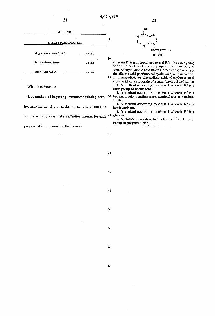
purpose of a compound of the formula:
10
15
20
30
35
45
55
65
22
on
N ’ N
k | > \
N l‘ nc-cn-cn;
R‘ on2
wherein R1 is an n-hexyl group and R2 is the ester group of formic acid, acetic acid, propionic acid or butyric acid, phenylalkanoic acid having 2 to 3 carbon atoms in the alkonic acid portions, salicyclic acid, a hemi ester of an alkaneodioic or alkenedioic acid, phosphoric acid, nitric acid, or a glycoside of a sugar having 5 to 6 atoms.
2. A method according to claim 1 wherein R2 is a ester group of acetic acid.
3. A method according to claim 1 wherein R2 is a hemimalonate, hemifumarate, hemimaleate or hemisuc cinate.
4. A method according to claim 1 wherein R2 is a hemisuccinate.
5. A method according to claim 1 wherein R2 is a glucoside.
6. A method according to 1 wherein R2 is the ester group of propionic acid.
it I? * * 1k