Embed Size (px)
Citation preview
1
Learning objectives:
1. Understand where fertilization as well as the initial divisions of the fertilized egg take place
within the mother’s reproductive.
2. Understand the significance of compaction, the segregation of the blastomeres and formation
of the blastocyst.
3. Distinguish between the descendents of the trophoblasts and the inner cell mass.
4. Understand the concepts of fate, potency, commitment and differentiation.
Summary:
This lecture begins with the events that occur immediately following fertilization within the
oviduct and the reestablishment of the diploid state. Fertilization takes place in a section of the oviduct
(FALLOPIAN TUBE) called the ampulla. It takes the embryo 5 days to reach the lumen of the uterus.
During its journey, the zygote undergoes mitotic divisions called CLEAVAGE DIVISIONS; these occur
without growth of daughter cells. These cells are now called BLASTOMERES. At the ~ the 8-cell stage
the embryo undergoes COMPACTION, leaving a portion of the blastomeres still facing the external
environment.
Compaction results in two cellular lineages: the outer TROPHOBLASTS which form the fetal
component of the placenta (Lecture 23); and the INNER CELL MASS which forms the embryo proper
and the extra embryonic membranes. These latter include the AMNIOTIC MEMBRANE of the amniotic
cavity and the extra embryonic mesoderm [EEM] (see Lecture 23). Once in the uterus, the embryo and
the uterine lining recognize each other in a biochemical sense, permitting ATTACHMENT of the em-
bryo followed by a carefully controlled IMPLANTATION.
At implantation the inner cell mass reorganizes into a two layered embryo; the epithelial EPI-
BLAST, which will form the embryo and amniotic membrane, and the HYPOBLAST, which has an
important role in the orientation of the embryonic axes.
Glossary:
Blastomeres: cells produced by cleavage divisions of the zygote.
Blastocyst: Formed from the blastomeres. Has a central fluid filled cavity (blastocoel) and is divided
into outer trophoblasts and an inner cell mass.
Cleavage divisions: Non-synchronous mitotic divisions following fertilization. No growth between cell
division cycles. This results in cells (blastomeres) of approximately equal size.
Epiblast: The inner cell mass forms a two layered embryo. The epiblast is the top layer (facing the
placenta) and forms the embryo proper and the amniotic membrane.
Fallopian tube: oviduct of the human, site of fertilization and initial cleavage divisions.
INTRODUCTION TO EMBRYOLOGY I
1A. From Cleavage To Gastrulation
Dr. Ann-Judith Silverman
Department of Anatomy & Cell Biology
Telephone: 5-3450
Email: [email protected]
2
Hypoblast: Bottom layer (facing the blastocoel) of the 2-layered embryo. Plays a role in establishing
polarity but does not contribute cells to the embryo.
Inner cell mass: will give rise to the epiblast and hypoblast.
Trophoblasts: derived from the outer cells of the blastocyst, forms the embryonic/fetal component of
the placenta.
Zygote: fertilized egg.
Lecture Notes:
Fertilization and Cleavage
The female reproductive tract is discontinuous. The ovary is not connected to the oviduct. The
ovulated ovum must be caught by the fingers (fimbria) of the oviduct and then enter the infundibulum
(funnel). Fertilization will take place in the distal 1/3 of the next compartment (ampulla) where the
zygote will remain for approximately 72 hours. This halt may prevent further interaction with the reser-
voir of sperm still in the cervical region of the uterus. The length of the transit time through the oviduct
to the uterus is important to ensure synchrony between the developmental stage of the embryo and the
uterine lining (endometrium). It takes the endometrium (lining of the uterus) several days to be made
ready to receive the embryo. Knowledge about the timing requirements for synchronization of the
embryo and mother are very important for the success of in vitro fertilization (see Fig 1-1).
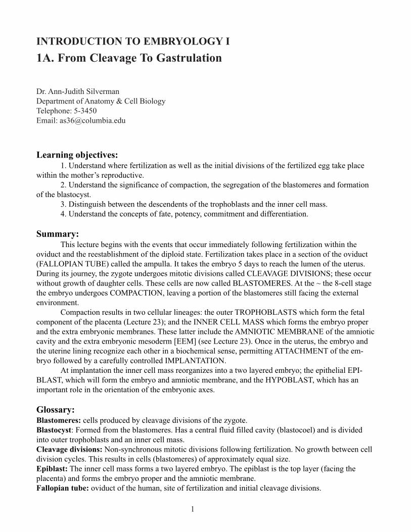
During transit, the embryo remains within its zona pellucida (extracellular material) until after its
entry into the uterine cavity (Fig.1-1). The traveling embryo goes through several mitotic divisions
called CLEAVAGE DIVISIONS (Fig 1-1). Compared to other classes of vertebrates, cleavage divisions
in mammals are very slow with ~1 per day for the first 3- 4days. These divisions increase the number of
cells (blastomeres) in the embryo, without any increase in the overall size of the embryo. Cleavage in
mammals is asynchronous so there need not be an even number of cells in the embryo. The zygotic
genome is turned on in humans between the 4 to 8 cell stage and maternal mRNA rapidly degraded. The
timing and positional relationships are important variables in determining developmental destinies.
Fig. 1-1. Cleavage and transport down the oviduct. Fertilization occurs in the ampulla of th oviduct. During the first five
days, the zygote undergoes cleavage as it travels down the oviduct and enters the uterus. On day 5, the blastocyst hatches
from the zona pellucida and is then able to implant in the uterine endometrium.
3
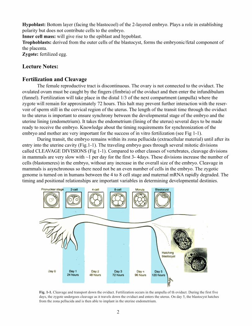
Compaction: Commitment to 2 Cell Lineages (Fig. 1-2). At ~ the 8 cell stage the embryo is
transformed from a loosely organized ball of cells into a compact closely adherent cluster (Fig. 1-2).
Fig. 1-2. Compaction. (A) Scanning electron micrograph of 10-cell human embryo before compaction. Note intercellular
clefts. (B) Scanning electron micrograph of 10-cell human embryo during process of compaction. Note the absence of
intercellular clefts between some of the blastomeres. The zona pellucida was mechanically removed from both embryos.
This process is called COMPACTION. Compaction is an extremely important event as the fates of the
cells begin to diverge radically from each other.
Developmental biologists have specific definitions for the progression of establishing final
phenotypes.
Fate: normal developmental pathway of an unperturbed cell or cell group.
Potency: description of the range of cell types that can arise from an individual cell.
Totipotent or pluripotent: The former describes a cell which is capable of making the whole
embryo. During cleavage divisions there is a loss of potency with time but some stem cells retain high
levels of potency. For example, the pluripotential stem cell in the bone marrow gives rise to all of the
different kinds of blood cells.
Commitment: Cells and tissues of the embryo receive inducing (decision making) signals that
guide their fate. A cell or tissue is said to be competent if it can respond to such a signal. A cell or tissue
is subsequently committed to a developmental fate even though no overt morphological change has
occurred.
Differentiation: overt morphological change that accompanies or follows commitment. This too
may occur in several steps.
Final differentiation: the last step in the development of a cell, resulting in a unipotential cell
that will follow the same fate for the rest of its life.
Before compaction the inner faces of the blastomeres contact other blastomeres; the outer faces
are exposed to the oviduct. Cells are therefore equally polarized vis-à-vis their environment and are
essentially identical and replaceable. For genotyping of embryos for in vitro fertilization, single cells are
removed prior to compaction and chromosomal analysis and/or PCR technology used to determine if
genetic anomalies exist. The remaining cells will compensate for what is removed.
After compaction cells are divided into inner and outer sets that have different fates. The inner
cells have surfaces that touch only other blastomeres and outer cells have one of their surfaces facing the
outer world. The process of compaction is mediated, in part, by the expression of E-cadherin, a Ca++
dependent cell adhesion molecule. Treatment of embryos with antibodies to E-cadherin will prevent
compaction or, if it has already taken place, will cause decompaction.
4
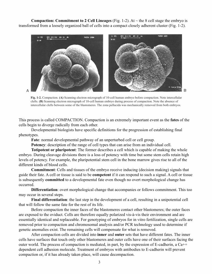
Between the 8 – 16 cell stage, deposition of extracellular matrix (ECM) occurs. ECM doesn’t
just act as glue between cells but can bind many signaling molecules and hence mediate signaling.
Blastocyst Formation: Differentiation of 2 Cell Lineages
By the 16-32-cell stage, the embryo is called a morula (Latin, mulberry) (Fig. 1-3). The outer
cells develop tight junctions which are fluid impermient. The outer cells secrete fluid (using the energy
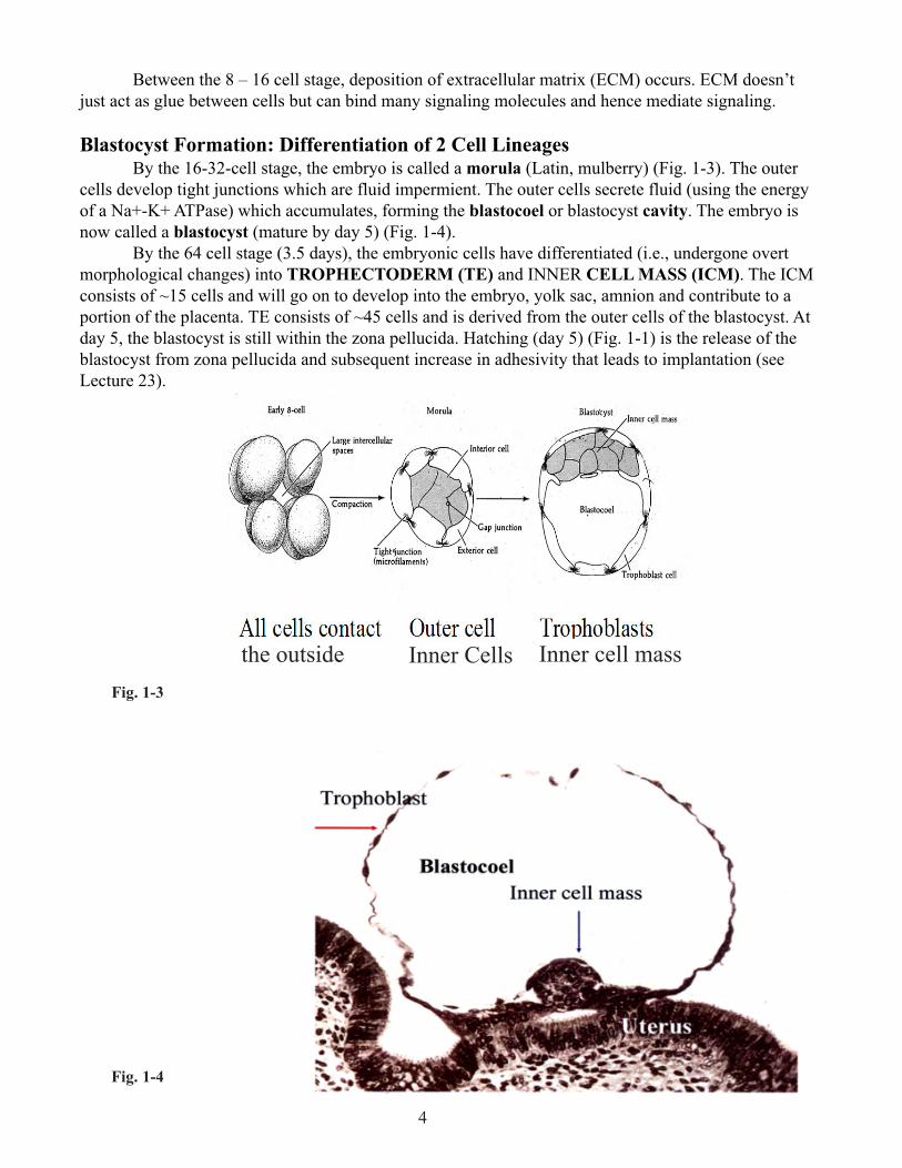
of a Na+-K+ ATPase) which accumulates, forming the blastocoel or blastocyst cavity. The embryo is
now called a blastocyst (mature by day 5) (Fig. 1-4).
By the 64 cell stage (3.5 days), the embryonic cells have differentiated (i.e., undergone overt
morphological changes) into TROPHECTODERM (TE) and INNER CELL MASS (ICM). The ICM
consists of ~15 cells and will go on to develop into the embryo, yolk sac, amnion and contribute to a
portion of the placenta. TE consists of ~45 cells and is derived from the outer cells of the blastocyst. At
day 5, the blastocyst is still within the zona pellucida. Hatching (day 5) (Fig. 1-1) is the release of the
blastocyst from zona pellucida and subsequent increase in adhesivity that leads to implantation (see
Lecture 23).
Fig. 1-4
Inner cell mass
the outside
Inner Cells
Fig. 1-3
5
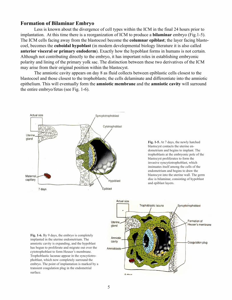
Formation of Bilaminar Embryo
Less is known about the divergence of cell types within the ICM in the final 24 hours prior to
implantation. At this time there is a reorganization of ICM to produce a bilaminar embryo (Fig.1-5).
The ICM cells facing away from the blastocoel become the columnar epiblast; the layer facing blasto-
coel, becomes the cuboidal hypoblast (in modern developmental biology literature it is also called
anterior visceral or primary endoderm). Exactly how the hypoblast forms in humans is not certain.
Although not contributing directly to the embryo, it has important roles in establishing embryonic
polarity and lining of the primary yolk sac. The distinction between these two derivatives of the ICM
may arise from their original position within the blastocyst.
The amniotic cavity appears on day 8 as fluid collects between epiblastic cells closest to the
blastocoel and those closest to the trophoblasts; the cells delaminate and differentiate into the amniotic
epithelium. This will eventually form the amniotic membrane and the amniotic cavity will surround
the entire embryo/fetus (see Fig. 1-6).
Fig. 1-5. At 7 days, the newly hatched
blastocyst contacts the uterine en-
dometrium and begins to implant. The
trophoblasts at the embryonic pole of the
blastocyst proliferates to form the
invasive syncytiotrophoblast, which
insinuates itself among the cells of the
endometrium and begins to draw the
blastocyst into the uterine wall. The germ
disc is bilaminar, consisting of hypoblast
and epiblast layers.
Fig. 1-6. By 9 days, the embryo is completely
implanted in the uterine endometrium. The
amniotic cavity is expanding, and the hypoblast
has begun to proliferate and migrate out over the
cytotrophoblast to form Heuser’s membrane.
Trophoblastic lacunae appear in the syncytiotro-
phoblast, which now completely surround the
embryo. The point of implantation is marked by a
transient coagulation plug in the endometrial
surface.
6
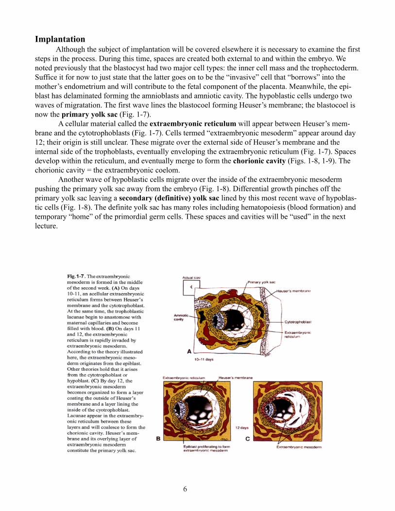
Implantation
Although the subject of implantation will be covered elsewhere it is necessary to examine the first
steps in the process. During this time, spaces are created both external to and within the embryo. We
noted previously that the blastocyst had two major cell types: the inner cell mass and the trophectoderm.
Suffice it for now to just state that the latter goes on to be the “invasive” cell that “borrows” into the
mother’s endometrium and will contribute to the fetal component of the placenta. Meanwhile, the epi-
blast has delaminated forming the amnioblasts and amniotic cavity. The hypoblastic cells undergo two
waves of migratation. The first wave lines the blastocoel forming Heuser’s membrane; the blastocoel is
now the primary yolk sac (Fig. 1-7).
A cellular material called the extraembryonic reticulum will appear between Heuser’s mem-
brane and the cytotrophoblasts (Fig. 1-7). Cells termed “extraembryonic mesoderm” appear around day
12; their origin is still unclear. These migrate over the external side of Heuser’s membrane and the
internal side of the trophoblasts, eventually enveloping the extraembryonic reticulum (Fig. 1-7). Spaces
develop within the reticulum, and eventually merge to form the chorionic cavity (Figs. 1-8, 1-9). The
chorionic cavity = the extraembryonic coelom.
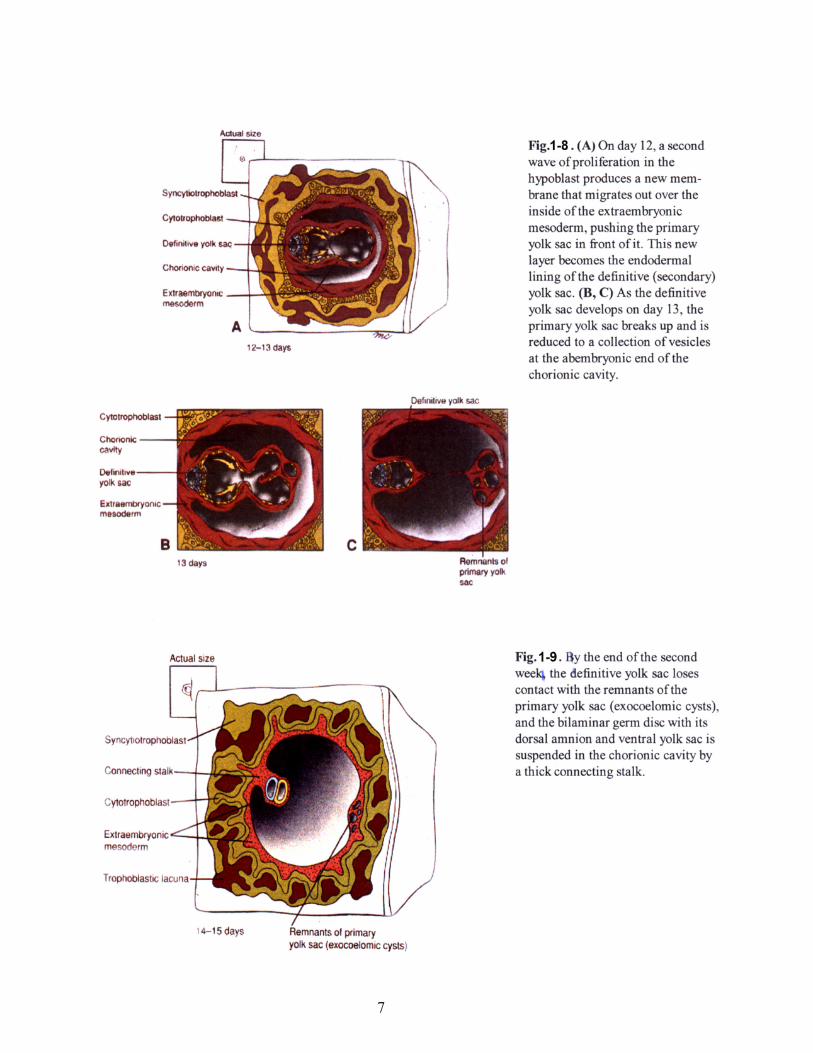
Another wave of hypoblastic cells migrate over the inside of the extraembryonic mesoderm
pushing the primary yolk sac away from the embryo (Fig. 1-8). Differential growth pinches off the
primary yolk sac leaving a secondary (definitive) yolk sac lined by this most recent wave of hypoblas-
tic cells (Fig. 1-8). The definite yolk sac has many roles including hematopoiesis (blood formation) and
temporary “home” of the primordial germ cells. These spaces and cavities will be “used” in the next
lecture.
7
8
1B. Germ Layers and Gastrulation
Learning objectives:
1. To understand the role played by hypoblast and the primitive node (Hensen’s node in chicks,
dorsal lip of the blastopore in amphibia) in producing signals that establish the axes of the embryo.
2. To understand how the three germ layers are established by cellular movements through the
primitive streak and primitive node.
3. To understand the concepts of induction and competence.
Summary:
We focus here on the events taking place in the epiblast and on its interaction with the hypoblast.
The process of cellular movement, termed gastrulation, establishes the three primary germ
layers. This occurs between days 14 and 19 post-conception. It is a series of rapid, complicated, but
coordinated movements of cells from the surface of the bilaminar embryo into the interior. Because of
the complexity of this process, many embryos do not gastrulate correctly. It is estimated that improper
gastrulation occurs in one-third of all human embryos. When this happens, a miscarriage usually takes
place, even before the woman realizes that she is pregnant.
Gastrulation movements form the three germ layers:
(1) The ectoderm, which will develop into the skin and nervous system;
(2) The mesoderm, which will develop into muscles, skeleton, connective tissue, blood, gonads,
and kidneys;
(3) The definitive endoderm, which will develop into the lining of the gut tube and respiratory
system.
Glossary:
germ layers: ectoderm, mesoderm and endoderm (see summary).
chordamesoderm: axial (midline) mesoderm which gives rise to the notochord.
competence: the ability to respond to an inductive signal. Once a competent cell responds to an induc-
tive signal, it becomes specified.
committed: the time point when a cell’s fate to a particular lineage is fixed. This does not imply final
phenotypic differentiation.
hypoblast (anterior visceral endoderm): signaling center for inducing anterior structures.
induction: the change in a cell or tissues fate due to a signal from another tissue or cell.
notochord: midline (axial) mesoderm.
prechordal plate: a portion of axial mesoderm just cranial to the notochord, will give rise to mesoderm
of the head and is also an important signaling center.
primitive node: most anterior (cranial) aspect of the primitive streak.
primitive streak: site of cell movements from epiblast to form other germ layers.
Lecture notes
Formation of the Primitive Streak
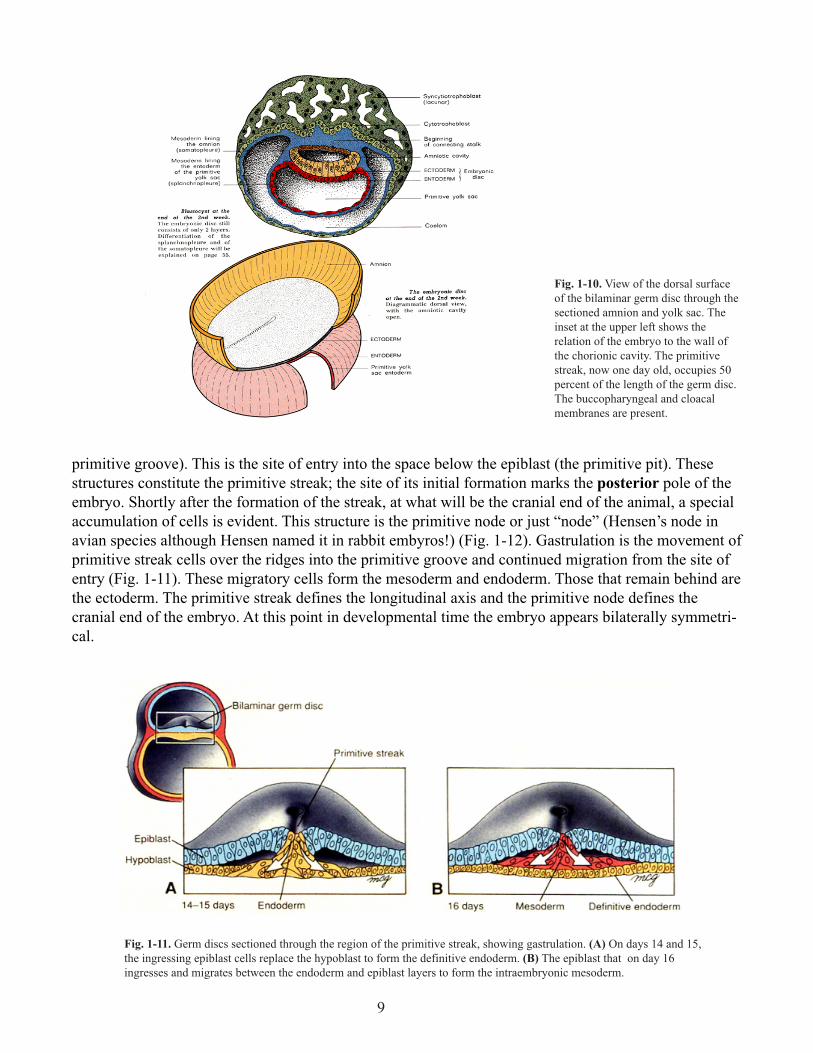
Gastrulation begins on day 14 (Fig. 1-10). Cells move from the lateral aspect of the epiblast
toward the midline, where they accumulate to form bilateral ridges with an indentation in the center (the
9
primitive groove). This is the site of entry into the space below the epiblast (the primitive pit). These
structures constitute the primitive streak; the site of its initial formation marks the posterior pole of the
embryo. Shortly after the formation of the streak, at what will be the cranial end of the animal, a special
accumulation of cells is evident. This structure is the primitive node or just “node” (Hensen’s node in
avian species although Hensen named it in rabbit embyros!) (Fig. 1-12). Gastrulation is the movement of
primitive streak cells over the ridges into the primitive groove and continued migration from the site of
entry (Fig. 1-11). These migratory cells form the mesoderm and endoderm. Those that remain behind are
the ectoderm. The primitive streak defines the longitudinal axis and the primitive node defines the
cranial end of the embryo. At this point in developmental time the embryo appears bilaterally symmetri-
cal.
Fig. 1-10. View of the dorsal surface
of the bilaminar germ disc through the
sectioned amnion and yolk sac. The
inset at the upper left shows the
relation of the embryo to the wall of
the chorionic cavity. The primitive
streak, now one day old, occupies 50
percent of the length of the germ disc.
The buccopharyngeal and cloacal
membranes are present.
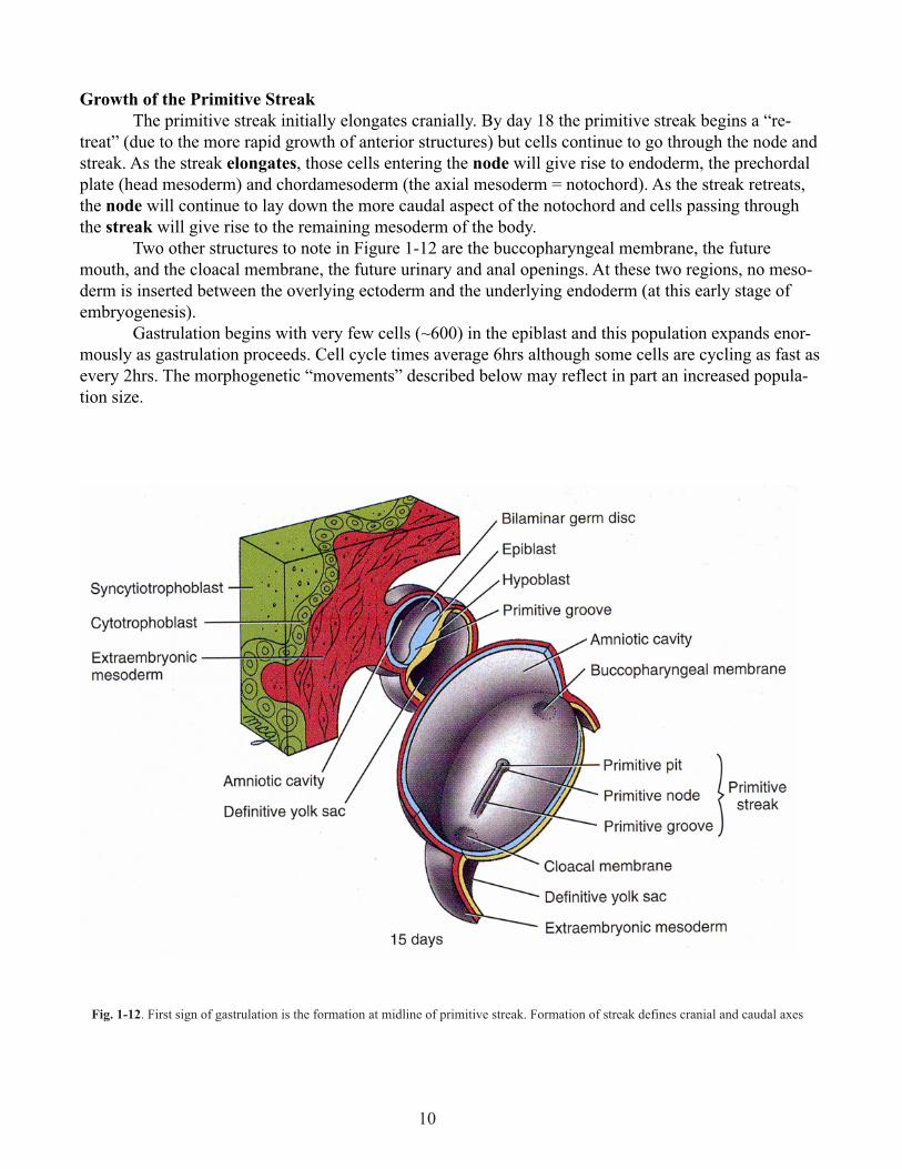
Fig. 1-11. Germ discs sectioned through the region of the primitive streak, showing gastrulation. (A) On days 14 and 15,
the ingressing epiblast cells replace the hypoblast to form the definitive endoderm. (B) The epiblast that on day 16
ingresses and migrates between the endoderm and epiblast layers to form the intraembryonic mesoderm.
10
Growth of the Primitive Streak
The primitive streak initially elongates cranially. By day 18 the primitive streak begins a “re-
treat” (due to the more rapid growth of anterior structures) but cells continue to go through the node and
streak. As the streak elongates, those cells entering the node will give rise to endoderm, the prechordal
plate (head mesoderm) and chordamesoderm (the axial mesoderm = notochord). As the streak retreats,
the node will continue to lay down the more caudal aspect of the notochord and cells passing through
the streak will give rise to the remaining mesoderm of the body.
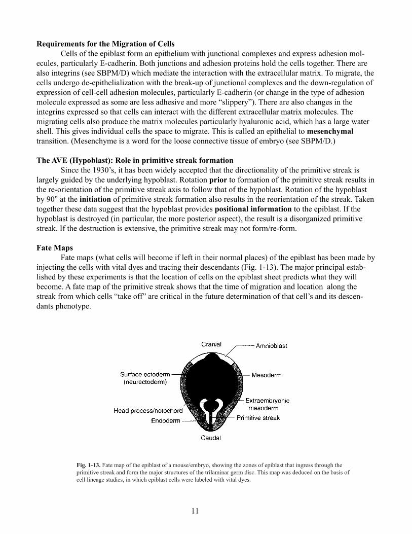
Two other structures to note in Figure 1-12 are the buccopharyngeal membrane, the future
mouth, and the cloacal membrane, the future urinary and anal openings. At these two regions, no meso-
derm is inserted between the overlying ectoderm and the underlying endoderm (at this early stage of
embryogenesis).
Gastrulation begins with very few cells (~600) in the epiblast and this population expands enor-
mously as gastrulation proceeds. Cell cycle times average 6hrs although some cells are cycling as fast as
every 2hrs. The morphogenetic “movements” described below may reflect in part an increased popula-
tion size.
Fig. 1-12. First sign of gastrulation is the formation at midline of primitive streak. Formation of streak defines cranial and caudal axes
11
Requirements for the Migration of Cells
Cells of the epiblast form an epithelium with junctional complexes and express adhesion mol-
ecules, particularly E-cadherin. Both junctions and adhesion proteins hold the cells together. There are
also integrins (see SBPM/D) which mediate the interaction with the extracellular matrix. To migrate, the
cells undergo de-epithelialization with the break-up of junctional complexes and the down-regulation of
expression of cell-cell adhesion molecules, particularly E-cadherin (or change in the type of adhesion
molecule expressed as some are less adhesive and more “slippery”). There are also changes in the
integrins expressed so that cells can interact with the different extracellular matrix molecules. The
migrating cells also produce the matrix molecules particularly hyaluronic acid, which has a large water
shell. This gives individual cells the space to migrate. This is called an epithelial to mesenchymal
transition. (Mesenchyme is a word for the loose connective tissue of embryo (see SBPM/D.)
The AVE (Hypoblast): Role in primitive streak formation
Since the 1930’s, it has been widely accepted that the directionality of the primitive streak is
largely guided by the underlying hypoblast. Rotation prior to formation of the primitive streak results in
the re-orientation of the primitive streak axis to follow that of the hypoblast. Rotation of the hypoblast
by 90° at the initiation of primitive streak formation also results in the reorientation of the streak. Taken
together these data suggest that the hypoblast provides positional information to the epiblast. If the
hypoblast is destroyed (in particular, the more posterior aspect), the result is a disorganized primitive
streak. If the destruction is extensive, the primitive streak may not form/re-form.
Fate Maps
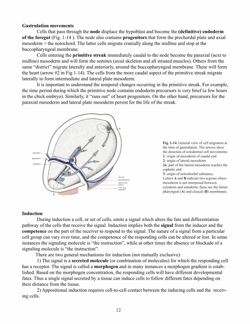
Fate maps (what cells will become if left in their normal places) of the epiblast has been made by
injecting the cells with vital dyes and tracing their descendants (Fig. 1-13). The major principal estab-
lished by these experiments is that the location of cells on the epiblast sheet predicts what they will
become. A fate map of the primitive streak shows that the time of migration and location along the
streak from which cells “take off” are critical in the future determination of that cell’s and its descen-
dants phenotype.
Fig. 1-13. Fate map of the epiblast of a mouse/embryo, showing the zones of epiblast that ingress through the
primitive streak and form the major structures of the trilaminar germ disc. This map was deduced on the basis of
cell lineage studies, in which epiblast cells were labeled with vital dyes.
12
Gastrulation movements
Cells that pass through the node displace the hypoblast and become the (definitive) endoderm
of the foregut (Fig. 1-14 ). The node also contains progenitors that form the prechordal plate and axial
mesoderm = the notochord. The latter cells migrate cranially along the midline and stop at the
buccopharyngeal membrane.
Cells entering the primitive streak immediately caudal to the node become the paraxial (next to
midline) mesoderm and will form the somites (axial skeleton and all striated muscles). Others from the
same “district” migrate laterally and anteriorly, around the buccopharyngeal membrane. These will form
the heart (arrow #2 in Fig 1-14). The cells from the more caudal aspect of the primitive streak migrate
laterally to form intermediate and lateral plate mesoderm.
It is important to understand the temporal changes occurring in the primitive streak. For example,
the time period during which the primitive node contains endoderm precursors is very brief (a few hours
in the chick embryo). Similarly, it “runs out” of heart progenitors. On the other hand, precursors for the
paraxial mesoderm and lateral plate mesoderm persist for the life of the streak.
Fig. 1-14. General view of cell migration at
the time of gastrulation. The arrows show
the direction of ectodermal cell movements:
1: origin of mesoderm of caudal end
2: origin of lateral mesoderm
2a: part of the lateral mesoderm reaches the
cephalic end
3: origin of notochordal substance
Letters A and B indicate two regions where
mesoderm is not interposed between
ectoderm and entoderm: these are the future
pharyngeal (A) and cloacal (B) membranes.
Induction
During induction a cell, or set of cells, emits a signal which alters the fate and differentiation
pathway of the cells that receive the signal. Induction implies both the signal from the inducer and the
competence on the part of the receiver to respond to the signal. The nature of a signal from a particular
cell group can vary over time, and the competence of the responding cells can be altered or lost. In some
instances the signaling molecule is “the instruction”, while at other times the absence or blockade of a
signaling molecule is “the instruction”.
There are two general mechanisms for induction (not mutually exclusive):
1) The signal is a secreted molecule (or combination of molecules) for which the responding cell
has a receptor. The signal is called a morphogen and in many instances a morphogen gradient is estab-
lished. Based on the morphogen concentration, the responding cells will have different developmental
fates. Thus a single signal secreted by a tissue can induce cells to follow different fates depending on
their distance from the tissue.
2) Appositional induction requires cell-to-cell contact between the inducing cells and the receiv-
ing cells.
13
Axial Patterning
In experiments on amphibia, Spemann first delineated the concept of induction in 1918. Using an
amphibian model he showed that ectodermal cells fated to become epidermis could take on a new fate
(become neuronal) if they are transplanted early in gastrulation to an appropriate site. They had the
competence to respond to “neuralizing” signals. If however the same experiment is performed but at a
later stage of gastrulation, the transplanted cells are no longer competent to become “neural”; they are
already committed to become “epidermal”.
In further studies on induction were conducted in the 1920’s by Spemann and Mangold
(Spemann later received the Nobel Prize for this work). They studied the role of the dorsal lip of the
blastopore (DLB) (the amphibian homologue of the node) in axis formation (Fig. 1-15).
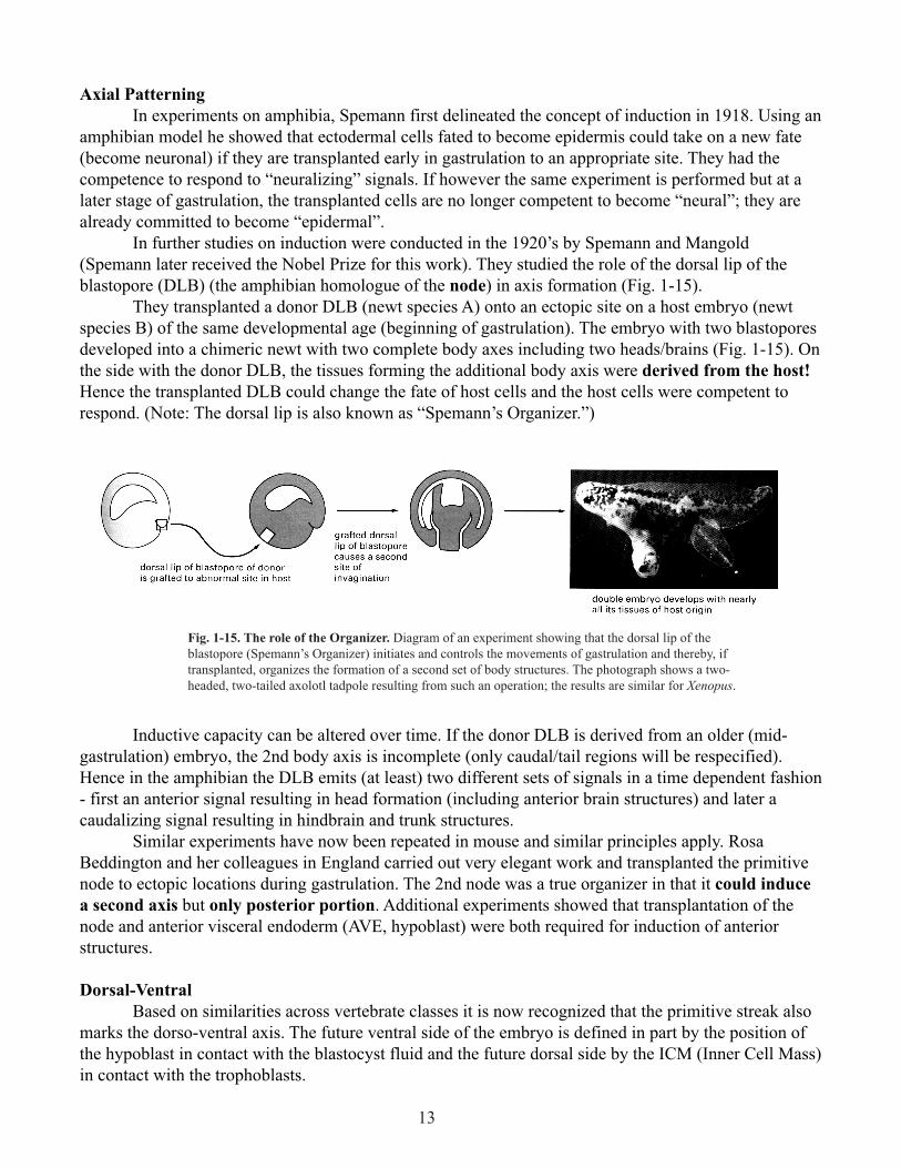
They transplanted a donor DLB (newt species A) onto an ectopic site on a host embryo (newt
species B) of the same developmental age (beginning of gastrulation). The embryo with two blastopores
developed into a chimeric newt with two complete body axes including two heads/brains (Fig. 1-15). On
the side with the donor DLB, the tissues forming the additional body axis were derived from the host!
Hence the transplanted DLB could change the fate of host cells and the host cells were competent to
respond. (Note: The dorsal lip is also known as “Spemann’s Organizer.”)
Fig. 1-15. The role of the Organizer. Diagram of an experiment showing that the dorsal lip of the
blastopore (Spemann’s Organizer) initiates and controls the movements of gastrulation and thereby, if
transplanted, organizes the formation of a second set of body structures. The photograph shows a two-
headed, two-tailed axolotl tadpole resulting from such an operation; the results are similar for Xenopus.
Inductive capacity can be altered over time. If the donor DLB is derived from an older (mid-
gastrulation) embryo, the 2nd body axis is incomplete (only caudal/tail regions will be respecified).
Hence in the amphibian the DLB emits (at least) two different sets of signals in a time dependent fashion
- first an anterior signal resulting in head formation (including anterior brain structures) and later a
caudalizing signal resulting in hindbrain and trunk structures.
Similar experiments have now been repeated in mouse and similar principles apply. Rosa
Beddington and her colleagues in England carried out very elegant work and transplanted the primitive
node to ectopic locations during gastrulation. The 2nd node was a true organizer in that it could induce
a second axis but only posterior portion. Additional experiments showed that transplantation of the
node and anterior visceral endoderm (AVE, hypoblast) were both required for induction of anterior
structures.
Dorsal-Ventral
Based on similarities across vertebrate classes it is now recognized that the primitive streak also
marks the dorso-ventral axis. The future ventral side of the embryo is defined in part by the position of
the hypoblast in contact with the blastocyst fluid and the future dorsal side by the ICM (Inner Cell Mass)
in contact with the trophoblasts.
14
Right-left axis
During early organogenesis, the laterality of the body is revealed by the looping of the heart and
rotation of the body axis as well as the asymmetric expression of genes in the in the left side of the
embryo. Without the node, expression of specific “left” genes, distribution of organs/looping of the heart
are randomized (as it is in situs inversus).
In mammals (the mouse!) the initial establishment of handedness depends on the formation of
motile cilia in cells at the node. The cilia beat counter-clockwise and cause the flow of fluid in the yolk
sac to move from right to left. A mutation in the dynein motor of these cilia results in randomization of
organ placement (e.g., heart on wrong side) (gene is called situs inversus viscerum, iv) versus the normal
condition (situs solitus). This leftward movement restricts the expression of the gene Nodal, a secreted
signaling molecular made in the lateral plate mesoderm, to the left side (Nodal is expressed asymmetri-
cally in all vertebrate classes). Nodal is further constrained to the left by the action of Lefty-1 which is
secreted from the ventral left side of the neural tube. The downstream mechanisms leading to sidedness
in humans is still an active area of investigation. Errors in right-left patterning occur in ~ 1 in 10,000
human births (See Supp et al 1998 Cell and Developmental Biology 9:77-87 if interested in more infor-
mation on genetics of human handedness mutations and clinical outcomes.) The downstream mecha-
nisms leading to sidedness in humans is still an active area of investigation.
Overview of the Embryo at the End of Gastrulation
There are now three layers:
1) Ectoderm: Its midline portion will become the nervous system, and the rest of it will become
the epidermis.
2) Mesoderm: which is subdivided into four zones: the midline notochord, paraxial somites, the
intermediate mesoderm, and the body wall/lateral plate mesoderm.
3) Definitive Endoderm: There is no gut yet. Formation of the gut occurs by the folding of the
lateral plate mesoderm (see Lecture 3 and 6).
4) The embryonic tissue is still in contact with extra-embryonic tissue. In the next lecture, we
will fold the trilaminar disc, thereby creating the body cavities and reducing contact to the connecting
stalk = the future umbilical cord.