Embed Size (px)
Citation preview

18 Narasin
O
O O OH OH H
O
OH
H
OOHO
HO
H
C43H72O11 MW: 765.0 CAS No.: 55134-13-9
[Summary of narasin] Narasin (NR) is a polyether antibiotic obtained by the incubation of an NR-producing strain of
Streptomyces aureofaciens. It is a derivative of salinomycin having an additional methyl group with the
chemical structure shown above, alternatively called “methyl salinomycin”.
For physicochemical properties, feed-grade NR technical occurs as grayish brown to dark brown
powder or particles. It is very soluble in ethyl acetate, in chloroform, in acetone, in benzene and in
dimethylsulfoxide, sparingly soluble in hexane and in petroleum ether, and practically insoluble in water.
Formulations with NR content exceeding 10% are designated as poisonous substances and those
with NR content exceeding 1%, as deleterious substances, under the Cabinet Order for the Designation of
the Poisonous and Deleterious Substances (Cabnet Order No.2, 1965). For the handling of these substances,
make sure to conform to the procesures specified in the Poisonous and Deleterious Substances Control Act
(Act No.303, 1950).
NR has an antibacterial effect on part of the Gram-positive bacteria, an anticoccidial effect, and a
growth promoting effect on chickens (including broilers).
«Standards and specifications in the Act on Safety Assurance and Quality Improvement of Feeds»
NR is a feed-grade antibiotic that was designated as a feed additive as of December 19, 2001. The
specifications for feeds containing this ingredient are specified in Appended Table No.1, 1-(1)-C of the
Standards and Specifications in the Act on Safety Assurance and Quality Improvement of Feeds.
Added amount 80 80
Feed ofinterest
For chickens(except forbroilers)
Starting chicksGrowing chicks
(in g(potency)/t)
For broilers
Starting period broilersFinishing period broilers
Like SL, MN, and LS, excessive consumption of NR can cause growth disturbance in chickens. It is

therefore necessary to strictly conform to the specified amounts of addition (80 g (potency)/1 ton of the
feed of interest) and to achive homogeneous mixture to secure the safety.
For this reason, feed manufacturers are required to control the feeds containing NR according to the
separately described control test methods (13 SeiChiku No.4573, notified by the Head of the Livestock
Division of the Production Bureau as of December 19, 2001).
[Methods listed in the Feed Analysis Standards] 1 Quantitative test methods - Plate method (premix) 1.1 Premix [Feed Analysis Standards, Chapter 9, Section 2, 18.1.1]
A. Reagent preparation
1) Sodium hydrogen carbonate-methanol solution. Dissolve 50 mg of sodium hydrogen carbonate in 200
mL of methanol [1], and filter through filter paper (No.5A).
2) Diution solvent: A mixture of water and methanol (7:3)
3) Narasin standard solution. Weigh accurately not less than 40 mg of narasin working standard[2],
accurately add sodium hydrogen carbonate-methanol solution[3] and dissolve to prepare a narasin
standard stock solution with a concentration of 1 mg (potency)/mL[4].
At the time of use, Accurately dilute a quantity of the standard stock solution with the dilution
solvent to prepare high- and low-concentration standard solutions with concentrations of 2 and 0.5 µg
(potency)/mL[5].
4) Culture medium: Medium F-22
5) Spore suspension and amount of addition. Use Bacillus subtilis ATCC 6633 as the test organism. Add
about 0.1 mL of the spore suspension with a concentration of 1×10⁹ spores/mL per 100 mL of the culture
medium.
6) Agar plate. Proceed by the cylinder plate method, respectively[6].
7) Extracting solvent. A mixture of methanol and water (9:1)
B. Preparation of sample solution
Weigh accurately 2 to 4 g of the analysis sample (equivalent to not more than 240 mg (potency)[7] as
NR), place in a 200-mL stoppered Erlenmeyer flask, add 100 mL of the extracting solvent, extract with
stirring for 20 minutes, and filter the extract through filter paper (No.5A).
Accurately dilute a quantity of the filtrate with the dilution solvent to prepare high- and low-
concentration sample solutions with concentrations of 2 and 0.5 µg (potency)/mL, respectively[8].
C. Quantification[9]
Proceed by the 2-2 dose method[10].
«Summary of analysis method»
This method is intended to determine the amount of NR in a premix by microbiological assay
using a sample solution prepared by extracting with a mixture of methabol and water (9:1) and diluting
with a mixture of water and methanol (7:3).
None of the antibacterial substances approved for combined use with NR interfere with the

quantification of NR.
The flow sheet of this method is shown in Figure 9.2.18-1.
Sample (2.0-4.0 g, equivalent to not more than 240 mg (potency) as NR)
Filter (through filter paper No.5A).
Dispense to agar plates (allow to stand at 10-20°C for 2 hr).
Incubate (at 35-37°C for 16-24 hr).
Measure the inhibition zone diameter.
Calculate the potency by the 2-2 dose method.
Dilute a quantity of filtrate with water-methanol (7:3) to prepare high- and low-concentration sample solutions (2 and 0.5 µg (potency)/mL, respectively).
Extract with 100 mL of methanol-water (9:1)(with a magnetic stirrer for 20 min).
Figure 9.2.18-1 Quantitative test method for narasin (premix)
References: Tetsuo Chihara, Katsumi Yamamoto: Research Report of Animal Feed, 28, 82 (2003)
History in the Feed Analysis Standards [26] New
«Validation of analysis method» ・Spike recovery and repeatability
Chicken premix 1 8~80 3 98.9~100.4 2.6
Chicken premix 2 8~80 3 98.1~101.4 2.5
Chicken premix 3 8~80 3 96.6~100.7 2.4
Sample type RepeatSpike
concentration(g(potency)/kg)
Spike recovery(%)
RepeatabilityRSD(% or less)
«Notes and precautions»
[1] As sodium hydrogen carbonate is difficult to dissolve in methanol, it is recommended to apply
ultrasonic waves for about 10 minutes after stirring for 30 minutes with a stirrer.
[2] For the definition etc. of narasin working standard, refer to «Notes and precautions» [9] in Section 1,
1 of this Chapter.
[3] Used to neutralize methanol. When the narasin working standard has been dissolved only with
methanol, white suspended solids can result from diluting the solution with a dilution solvent with
water content not less than 60%.
[4] For the method of preparation for the standard stock solution, refer to «Notes and precautions» [10]
in Section 1, 1 of this Chapter.
Method of preparation: Example (when the weighed amount is 50 mg)
When the labeled potency of the working standard is 963 µg (potency)/mg, 50 mg of the
working standard contains 48,150 µg (potency) (i.e., 50 mg × 963 µg (potency)/mg). To prepare a
standard stock solution with a concentration of 1,000 µg (potency)/mL, the required amount of
solvent is thus calculated to be 48.15 mL (i.e., 48,150 µg (potency) / 1,000 µg (potency)/mL).

Therefore, completely transfer 50 mg of the working standard to an Erlenmeyer flask containing
48.15 mL of methanol and dissolve to prepare a standard stock solution with a concentration of 1,000
µg (potency)/mL.
[5] For the method of preparation for the standard solution, refer to «Notes and precautions» [8] in
Section 1, 1 of this Chapter.
An example method of preparation for narasin standard solution is shown inTable 9.2.18-1.
[6] When using Medium F-22, the cylinder plate method is less sensitive to NR than the agar plate
method. However, taking into consideration the slope of the standard response line, the concentration
of the spore suspension, the spiked amount of NR, and the amount of the sample to be collected, the
cylinder plate is recommended for preparing agar plates.
[7] Good recoveries resulted in most of the cases except when an analysis sample with a spiked
concentration of 80 g (potency)/kg is collected in an amount of 5.0 g (equivalent ot 400 mg (potency)
as NR), which resulted in a poor recovery. Accordingly, the amount of the sample to be collected shall
be in the range from 2.0 to 4.0 g, equivalent to not more than 240 mg (potency) as NR.
[8] For the method of preparation for the sample solution, refer to «Notes and precautions» [8] in
Section 1, 1 of this Chapter.
An example method of preparation is shown in Table 9.2.18-1.
Table 9.2.18-1 Method of preparation for narasin standard solution and sample solution
1) Method of preparation for narasin standard solution (premix, example)
Test tube No. 1 2 3 4Amount (mL) of standard solution 2 2 4 5
Amount (mL) of water-methanol (7:3) 18 18 16 15Concentration (µg(potency)/mL) 100 10 2 0.5
Note: "2 mL" means "2 mL of standard stock solution (1 mg(potency)/mL)". 2) Method of preparation for sample solution (premix, example)
When the analysis sample is weighed in an amount equivalent to NR 200,000 µg
(potency) of NR, the concentration of NR in the filtrate is calculated to be 2,000 µg
(potency)/mL.
Test tube No. 1 2 3 4Amount (mL) of standard solution 2 2 2 5
Amount (mL) of water-methanol (7:3) 18 18 18 15Concentration (µg(potency)/mL) 200 20 2 0.5
Note: "2 mL" means "2 mL of the filtrate (2,000 µg(potency)/mL)". [9] An example standard response line for NR is shown in Figure 9.2.18-2.
Liniearity is observed in the quantification range for NR (NR concentrations between 0.5 and
2.0 µg (potency)/mL).
[10] Refer to «Notes and precautions» [53] to [60] in Section 1, 1 of this Chapter.

1.2 Feed [Feed Analysis Standards, Chapter 9, Section 2, 18.2.1]
A. Reagent preparation
1) Sodium hydrogen carbonate-methanol solution. Dissolve 50 mg of sodium hydrogen carbonate in 200
mL of methanol[1], and filter through filter paper (No.5A).
2) Diution solvent: A mixture of water and methanol (7:3)
3) Narasin standard solution. Weigh accurately not less than 40 mg of narasin working standard, accurately
add the sodium hydrogen carbonate-methanol solution and dissolve to prepare a narasin standard stock
solution with a concentration of 1 mg (potency)/mL.
At the time of use, accurately dilute a quantity of the standard stock solution with the dilution
solvent to prepare high- and low-concentration standard solution with concentrations of 2 and 0.5 µg
(potency)/mL.
4) Culture medium: Medium F-22
5) Spore suspension and amount of addition. Use Bacillus subtilis ATCC 6633 as the test organism. Add
about 0.1 mL of the spore suspension with a concentration of 1×10⁹ spores/mL per 100 mL of the culture
medium.
6) Agar plate. Proceed by the cylinder plate method[2].
7) Extracting solvent: A mixture of methanol and water (9:1)
B. Preparation of sample solution
Weigh accurately a quantity of the analysis sample (equivalent to 0.8 mg (potency)[3] as NR), place
in a 200-mL stoppered Erlenmeyer flask, add 100 mL of the extracting solvent, extract with stirring for 20
minutes, and filter the extract through filter paper (No.5A).
Load the filtrate onto a column (column tube (14 mm in internal diameter) dry-packed with 12 g[4]
of basic alumina for column chromatography (particle size: 74 to 177 µm (200 to 80 mesh)) [5]), and
Figure 9.2.18-2 Standard response line for NR (premix, example)
(Bacillus subtilis ATCC 6633, Medium F-22, Cylinder method)
14
16
18
20
22
24
26
28
0.25 0.5 1.0 2.0 4.0
阻止円直径の修正値
(mm
)
ナラシンの濃度 (μg(力価)/mL)Concentration of narasin (µg (potency)/mL)
28
26
24
22
20
18
16
14
0.25 0.5 1.0 2.0 4.0 C
orr
ecte
d in
hib
itio
n zo
ne d
iam
ete
r (m
m)

discard the first 5 mL of the filtrate[6].
Accuratey dilute a quantity of the subsequent filtrate[7] with a mixture of water and methanol (9:1)
to prepare a high-concentration sample solution with a concentration of 2 µg (potency)/mL[8], and
accurately dilute this solution with the dilution solvent to prepare a low-concentration sample solution with
a concentration of 0.5 µg (potency)/mL[9].
C. Quantification
Proceed by the 2-2 dose method[10].
«Summary of analysis method»
This method is intended to determine the amount of NR in a chicken feed by microbiological
assay using a sample solution prepared by extracting with a mixture of methanol and water (9:1) and
filtering through a column packed with basic alumina.
None of the antibacterial substances approved for combined use with NR interfere with the
quantification of NR.
The flow sheet of this method is shown in Figure 9.2.18-3.
Sample (10.0 g, equivalent to 0.8 mg (potency) as NR)
Extract with 100 mL of methanol-water (9:1) (with a magnetic stirrer for 20 min).
Filter (through filter paper No.5A).
Discard the first 5 mL of the filtrate and use the subsequent filtrate.
Dispense to agar plates (allow to stand at 10-20°C for 2 hr).
Incubate (at 35-37°C for 16-24 hr).
Measure the inhibition zone diameter.
Calculate the potency by the 2-2 dose method.
Dilute a quantity of the high-concentration sample solution with water-methanol (7:3)to prepare a low-concentration sample solution (0.5 µg (potency)/mL).
Filther through a column (12 g of basic alumina (Aluminum oxide Type F-20 (Sigma-Aldrich))
Dilute a quantity of the filtrate with water-ethanol (9:1) to prepare a high-concentrationsample solution (2 µg (potency)/mL)
Figure 9.2.18-3 Quantitative test method for narasin (chicken feed)
References: Tetsuo Chihara: Research Report of Animal Feed, 27, 80 (2002)
History in the Feed Analysis Standards [24] New

«Validation of analysis method» ・Spike recovery and repeatability
Starting chick formula feed 40~120 3 98.9~101.2 2.4Fattening broiler starter formula feed 40~120 3 99.5~101.7 3.4Fattening broiler finisher formula feed 40~120 3 97.9~99.1 4.3
Sample type RepeatSpike
concentration(g(potency)/t)
Spike recovery(%)
RepeatabilityRSD(% or lower)
・Collaborative study
Starting chick formula feed 7 80 102.3 2.4 2.2
No. oflabs
Sample typeSpike
concentration(g(potency)/t)
Spike recovery(%)
Intra-labrepeatability
RSDr(%)
Intra-labreproducibility
RSDR(%)
«Notes and precautions»
[1] As sodium hydrogen carbonate is difficult to dissolve in methanol, it is recommended to stir for 30
minutes with a stirrer and apply ultrasonic waves for about 10 minutes.
[2] For the method of preparation for the standard solution, refer to «Notes and precautions» [8] Section
1, 1 of this Chapter.
An example method of preparation for the narasin standard solution is shown in Table 9.2.18-2.
Table 9.2.18-2 Method of preparation for narasin standard solution (chicken feed, example) Test tube No. 1 2 3 4
Amount (mL) of standard solution 2 2 4 5
Amount (mL) of water-methanol (7 18 18 16 15
Concentration (µg(potency)/mL) 100 10 2 0.5
Note: "2 mL" means "2 mL of standard stock solution (1 mg(potency)/mL)". [3] When using Medium F-22, the cylinder plate method is less sensitive to NR than the agar plate
method. However, taking into consideration the slope of the standard response line, the concentration
of the spore suspension, the spiked amount of narasin, and the amount of the sample to be collected,
the cylinder plate method is recommended for preparing agar plates.
[4] Usually corresponds to 10 g.
[5] It is recommended to use a vibrator etc. to compact the packing material tightly.
[6] As the fiest portion of the filtrate is turbid, discard the first 5 mL and use the subsequent filtrate.
[7] The quantified value is constant in the 5- to 30-mL fraction.
[8] An example method of preparation for the high-concentration sample solution is shown below.
Filtrate 5 mL
Water-methanol (9:1) 15 mL [9] An example method of preparation for the low-concentration sample solution is shown below.
High-concentration samp 5 mL
Water-methanol (7:3) 15 mL [10] An example standard response line for narasin is shown in Figure 9.2.18-4.

Figure 9.2.18-4 Standard response line for narasin (chicken feed, example)
(Bacillus subtilis ATCC 6633, Medium F-22, Cylinder plate method)
2 Quantitative test method - Quantitative test method for polyether antibiotics by liquid chromatography 2.1 Premix [Feed Analysis Standards, Chapter 9, Section 2, 18.1.2]
Antibiotics of interest: SL, SD, NR and MN (4 components)
A. Reagent preparation
1) Salinomycin sodium standard solution. Dry a suitable amount of salinomycin working standard[1] under
reduced pressure (not exceeding 0.67 kPa) at 60°C for 3 hours, weigh accurately a quantity equivalent to
20 mg (potency), place in a 100-mL volumetric flask, add methanol to dissolve, and further add
methanol up to the marked line to prepare a salinomycin sodium standard stock solution (1 mL of this
solution contains an amount equivalent to 0.2 mg (potency) as salinomycin sodium).
At the time of use, accurately dilute a quantity of standard stock solution with a mixture of
methanol and water (9:1) to prepare several salinomycin sodium standard solutions containing
salinomycin sodium in amounts equivalent to 2.5 to 20 µg (potency) in 1 mL.
2) Semduramicin sodium standard solution. Weigh accurately a quantity of semduramicin working
standard equivalent to 20 mg (potency)[1], place in a 100-mL volumetric flask, add methanol to dissolve,
and further add methanol up to the marked line to prepare a standard stock solution (1 mL of this
solution contains an amount equivalent to 0.2 mg (potency) as semduramicin sodium).
At the time of use, accurately dilute a quantity of the standard stock solution with methanol to
prepare several semduramicin sodium standard solutions containing semduramicin sodium in amounts
equivalent to 2.5 to 20 µg (potency) in 1 mL.
3) Narasin standard solution. Weigh accurately a quantity of narasin working standard equivalent to 20 mg
(potency)[1], place in a 100-mL volumetric flask, add methanol to dissolve, and further add methanol up
to the marked line to prepare a narasin standard stock solution (1 mL of this solution contains narasin in
an amount equivalent to 0.2 mg (potency)).
14
16
18
20
22
24
26
28
0.25 0.5 1.0 2.0 4.0
阻止円直径の修正値
(mm
)
ナラシンの濃度 (μg(力価)/mL)Concentration of narasin (µg (potency)/mL)
28
26
24
22
20
18
16
14 0.25 0.5 1.0 2.0 4.0
Co
rrec
ted
inhi
bitio
n z
one
diam
ete
r (m
m)

At the time of use, accurately dilute a quantity of the standard stock solution with a mixture of
methanol and water (9:1) to prepare several narasin standard solutions containing narasin in amounts
equivalent to 0.5 to 20 µg (potency) in 1 mL.
4) Monensin sodium standard solution. Weigh accurately a quantity of monensin working standard
equivalent to 20 mg (potency)[1], place in a 100-mL volumetric flask, add methanol to dissolve, and
further add methanol up to the marked line to prepare a monensin sodium standard stock solution (1 mL
of this solution contains an amount equivalent to 0.2 mg (potency) as monensin sodium).
At the time of use, accurately dilute a quantity of the standard stock solution with a mixture of
methanol and water (9:1) to prepare several monensin sodium standard solutions containing monensin
sodium in amounts equivalent to 2.5 to 20 µg (potency) in 1 mL.
B. Quantification
Extraction. Weigh accurately 2 to 5 g of the analysis sample, place in a 200-mL stoppered Erlenmeyer
flask, add 100 mL of a mixture of methanol and water (9:1), extract with stirring for 20 minutes, and
filter the extract through filter paper (No.5A). Accurately dilute a quantity of the filtrate with a mixture
of methanol and water (9:1), filter through membrane filter (pore size not exceeding 0.5 µm), and use
the filtrate as the sample solution subject to liquid chromatography.
Liquid chromatography. Inject 20 µL each of the sample solution and antibiotic standard solutions into a
liquid chromatograph to obtain chromatograms.
Example operating conditions
Detector: Ultraviolet-visible absorption detector (measured wavelength: 520 nm)
Column: Octadecylsilanized silica gel column (4.6 mm in internal diameter, 150 mm in length, 5
µm in particle size)Note 1 [2]
Eluent: A mixture of methanol, water and acetic acid (940:60:1)
Reaction solutionNote 2: Gradually add 10 mL of sulfuric acid to 475 mL of methanol while stirring,
add 15 g of vanillin and dissolve (prepare at the time of use).
Flow rate: 0.6 mL/min for the eluent; 0.6 mL/min for the reaction solution
Reaction vessel temperature: 95°C
Calculation. Calculate the peak height or peak area from the obtained chromatogram[3] to prepare the
calibration curve, and estimate the amount of each antibioticNote 3, 4.
Note 1. Use a Mightysil RP-18 GP (Kanto Chemical Co., Inc.) or an equivalent.
2. Develop by allowing the eluate from the column to react with the reaction solution through the
reaction coil (0.5 mm in internal diameter, 5 mm in length (10 m )) in the reaction vessel, and
immediately transfer to the ultraviolet-visible absorption detector. The reaction solution shall be
used in a light-resistant container.
3. For monensin sodium, the calculated amount of monensin A shall be regarded as the amount of
monensin sodium. The peak of monensin A appears as the main peak on the chromatogram from
each monensin sodium standard solution. On the chromatogram of the sample solution, the peak
of monensin A appears at the same retention time as the peak of monensin A from the standard
solution.

4. For narasin, the calculated amount of narasin A shall be regarded as the amount of narasin. The
peak of narasin A appears as the main peak on the chromatogram of each narasin standard
solution. On the chromatogram from the sample solution, the peak of narasin A appears at the
same retention time as the peak of narasin A from the standard solution.
«Summary of analysis method»
This method is intended to determine the amount of salinomycin, semduramicin, narasin A, and
monensin A in a premix by determining the absorbances of their derivatives produced by extracting with
a mixture of methanol and water (9:1), separating by liquid chromatography using an octadecylsilanized
silica gel (ODS) column, and allowing to react with vanillin. It is also called the post-column
derivatization method.
The principle of this derivatization (chromogenic) reaction depends on the so-called Komarowsky
reaction, which involves aldol condensation of the alcoholic hydroxyl groups of salinomycin,
semduramicin, narasin A and monensin A with the benzaldehyde group of vanillin, in an acidic solution
containing sulfuric acid, to produce derivatives of these antibiotics that have wavelengths of maximal
absorption of about 520 nm.
This method allows for simultaneous quantification of salinomycin sodium (SL), semduramicin
sodium (SD), narasin (NR), and monensin sodium (MN). Care should be taken that, of the peaks of
monensin sodium, the peak of monensin B can appear at the same retention time as the peak of
semduramicin sodium, and thus interfere with the quantification.
For reference, the nature of separation of the mixed standard solution is shown in Figure 9.3.1-1.
Because of the above-mentioned possibility of interference from peaks other than those of interest, it is
preferable to use a single-component standard solution rather than a mixed standard solution for the
preparation of the calibration curve.
Figure 9.3.1-1 Chromatogram for a mixed standard solution
(SL: 100 ng, SD: 200 ng, NR-A: 100 ng, MN-A: 50 ng)
Narasin is a mixture of narasin A, narasin B, narasin D and narasin I, and the “narasin” designated
as a feed additive is defined as containing narasin A as the main ingredient. Monensin is a mixture of
monensin A, monensin B, monensin C and monensin D, and the “monensin” designated as a feed
additive is defined as containing monensin A as the main ingredient. In the test method described here,
SDMN
SLNR10
SD derivative
NR-A derivative
SL derivative
MN-A derivative
10
Re
ten
tion
tim
e (m
in)
Absorbance

the quantified amounts of narasin A and of monensin A are regarded as the amounts of narasin and of
monensin, respectively, based on the premises that commercial narasin and monensin formulations
contain narasin and monensin at a concentration of not less than 95%, respectively. It should be borne in
mind, therefore, that the “narasin” and “monensin” quantified by this method are not exactly the same as
the “narasin” and “monensin” quantified by microbiological assay.
For more details, refer to «Notes and precautions» [1] of General Notice 13 in Chapter 1.
The flow sheet of this method is shown in Figure 9.3.1-2.
2.0-5.0 g of sample
Filter (through filter paper No.5A).
LC-UV (520 nm)
Extract with 100 mL of methanol-water (9:1) (witha magnetic stirrer for 20 min).
Dilute a quantity of the filtrate with methanol-water (9:1).
Filter through membrane filter (pore size notexceeding 0.5 µm).
Figure 9.3.1-2 Quantitation test method for salinomycin sodium, semduramicin sodium, narasin,
and monensin sodium by liquid chromatography (premix)
References: Daisaku Makino: Research Report of Animal Feed, 27, 57 (2002)
Daisaku Makino: Research Report of Animal Feed, 27, 64 (2002)
Mayumi Nishimura: Research Report of Animal Feed, 28, 69 (2003)
Katsumi Yamamoto, Tetsuo Chihara: Research Report of Animal Feed, 28, 82 (2003)
History in the Feed Analysis Standards [25] New, [26] Component addition (semduramicin sodium,
narasin)
«Validation of analysis method» ・Spike recovery and repeatability
Starting chick grower premix 12.5~85.0 3 99.3~102.1 3.8
Broiler fattener finisher premix 12.5~85.0 3 96.3~102.7 3.0
Meat cattle fattener premix 12.5~85.0 3 98.0~100.8 2.8
Chicken premix 1 8~42 3 99.8~101.8 2.4
Chicken premix 2 8~42 3 98.5~102.4 2.8
Chicken premix 3 8~42 3 98.2~100.7 2.7
Chicken premix 1 8~80 3 98.7~103.8 0.9
Chicken premix 2 8~80 3 96.0~ 99.4 0.8
Chicken premix 3 8~80 3 96.6~ 99.8 0.5
Starting chick grower premix 5~80 3 98.2~102.4 2.1
Broiler fattener finisher premix 5~80 3 101.4~102.5 2.0
Meat cattle fattener premix 5~80 3 96.6~99.5 4.7
Spike recovery(%)
RepeatabilityRSD(% or less)
Monensin sodium
Sample type RepeatSpiked
component
Salinomycinsodium
Narasin
Semduramicinsodium
Spikeconcentration
(g(potency)/kg)
«Notes and precautions» [1] For the definition etc. of the working standards for salinomycin, semduramicin, narasin, and
monensin, refer to «Notes and precautions» [9] in Section 1, 1 of this Chapter.
[2] Any column is applicable as long as it is packed with an equivalent end-capped material. The
columns used for the validation of this method were Shodex C18M 4D for narasin and Mightysil RP-
18 GP for salinomycin, semduramicin, and monensin.
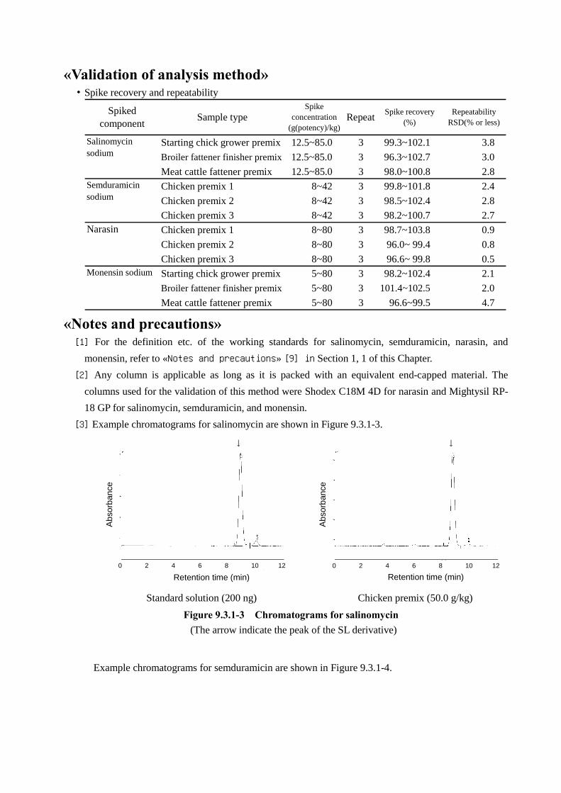
[3] Example chromatograms for salinomycin are shown in Figure 9.3.1-3.
Standard solution (200 ng) Chicken premix (50.0 g/kg)
Figure 9.3.1-3 Chromatograms for salinomycin
(The arrow indicate the peak of the SL derivative)
Example chromatograms for semduramicin are shown in Figure 9.3.1-4.
Retention time (min)
↓ ↓
Retention time (min)
0 2 4 6 8 10 12 0 2 4 6 8 10 12
Ab
sorb
ance
Ab
sorb
ance

Standard solution (equivalent to 200 ng
(potency) as SD)
Chicken premix (equivalent to 200 ng
(potency) as SD)
Figure 9.3.1-4 Chromatograms for semduramicin
(The arrow indicate the peak of the SD derivative)
Example chromatograms for narasin are shown in Figure 9.3.1-5.
Standard solution (200 ng) Chicken premix (80 g (potency)/kg)
Figure 9.3.1-5 Chromatograms for narasin
(The arrow indicate the peak of the NR-A derivative)
Example chromatograms for monensin are shown in Figure 9.3.1-6.
Standard solution (200 ng) Chicken premix (33.3 g/kg)
Figure 9.3.1-6 Chromatograms for monensin
(The arrows indicate the peaks of the MN-A derivative (main peak) and MN-B derivative)
0
1000
2000
3000
0 2 4 6 8 10 12 14
0
1000
2000
3000
0 2 4 6 8 10 12 14
Retention time (min)Retention time (min)
MN-B↓
Ab
sorb
ance
0 2 4 6 8
MN-B ↓
↓MN-A
MN-A ↓
Ab
sorb
ance
0 2 4 6 8
Retention time (min)
0 10
Retention time (min)
0 10
↓ ↓
Ab
sorb
ance
Ab
sorb
ance
Retention time (min) Retention time (min)
0 2 4 6 8 10 12 0 2 4 6 8 10 12
↓ ↓
Ab
sorb
ance
Ab
sorb
ance

2.2 Feed [Feed Analysis Standards, Chapter 9, Section 2, 18.2.2]
Antibiotics of interest: SL, SD, NR and MN (4 components)
A. Reagent preparation
1) Salinomycin sodium standard solution. Prepare a salinomycin sodium standard stock solution as
directed in 1.1-A.
At the time of use, accurately dilute a quantity of the standard stock solution with a mixture of
methanol and water (9:1) to prepare several salinomycin sodium standard solutions containing
salinomycin sodium in amounts equivalent to 0.5 to 8 µg (potency) in 1 mL.
2) Semduramicin sodium standard solution[1]. Prepare a semduramicin standard stock solution as directed
in 1.1-A.
At the time of use, accurately dilute a quantity of standard stock solution with methanol to prepare
several semduramicin sodium standard solutions containing semduramicin sodium in amounts
equivalent to 0.5 to 10 µg (potency) in 1 mL.
3) Narasin standard solution. Proceed as directed in 1.1-A.
4) Monensin sodium standard solution. Prepare a monensin standard stock solution as directed in 1.1-A.
At the time of use, accurately dilute a quantity of the standard stock solution with a mixture of
methanol and water (9:1) to prepare several monensin sodium standard solutions containing monensin
sodium in amounts equivalent to 0.5 to 15 µg (potency) in 1 mL.
B. Quantification
Extraction. Weigh 10.0 g of the analysis sample, place in a 200-mL stoppered Erlenmeyer flask, add 100
mL of a mixture of methanol and water (9:1), extract with stirring for 20 minutes, and filter the extract
through filter paper (No.5A). Filter the filtrate through membrane filter (pore size not exceeding 0.5 µm)
and use as a sample solution subject to liquid chromatography.
Liquid chromatography. Proceed as directed in 1.1-B Liquid chromatography[2].
Calculation. Proceed as directed in 1.1-B Calculation[3].
«Summary of analysis method»
This method is intended to determine the amount of salinomycin, semduramicin, narasin A and
monensin A in a chicken feed or cattle feed by post-column derivatization liquid chromatography using a
sample solution prepared by extracting with a mixture of methanol and water (9:1) as described in 1.1.
Quantification test method for polyether antibiotics by liquid chromatography (premix) in this Sectoin.
For the principle of the measurement etc., refer to 1.1 «Summary of analysis method».
In this method, none of the antibacterial substances approved for combined use with
semduramicin sodium interfere with the quantification of semduramicin sodium. Of the monensin
sodium that are not approved for combined use, however, monensin B was found to interfere with the
quantification of semduramicin sodium.
For the nature of separation of the mixed standard solution, refer to Figure 9.3.1-1.
The flow sheet of this method is shown in Figure 9.3.1-7.
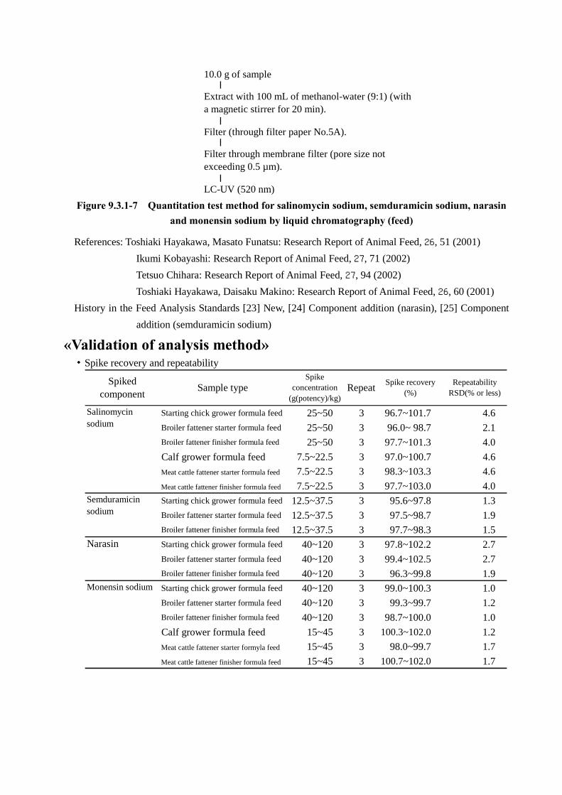
10.0 g of sample
Filter (through filter paper No.5A).
LC-UV (520 nm)
Extract with 100 mL of methanol-water (9:1) (witha magnetic stirrer for 20 min).
Filter through membrane filter (pore size notexceeding 0.5 µm).
Figure 9.3.1-7 Quantitation test method for salinomycin sodium, semduramicin sodium, narasin
and monensin sodium by liquid chromatography (feed)
References: Toshiaki Hayakawa, Masato Funatsu: Research Report of Animal Feed, 26, 51 (2001)
Ikumi Kobayashi: Research Report of Animal Feed, 27, 71 (2002)
Tetsuo Chihara: Research Report of Animal Feed, 27, 94 (2002)
Toshiaki Hayakawa, Daisaku Makino: Research Report of Animal Feed, 26, 60 (2001)
History in the Feed Analysis Standards [23] New, [24] Component addition (narasin), [25] Component
addition (semduramicin sodium)
«Validation of analysis method» ・Spike recovery and repeatability
Starting chick grower formula feed 25~50 3 96.7~101.7 4.6Broiler fattener starter formula feed 25~50 3 96.0~ 98.7 2.1Broiler fattener finisher formula feed 25~50 3 97.7~101.3 4.0
Calf grower formula feed 7.5~22.5 3 97.0~100.7 4.6
Meat cattle fattener starter formula feed 7.5~22.5 3 98.3~103.3 4.6
Meat cattle fattener finisher formula feed 7.5~22.5 3 97.7~103.0 4.0Starting chick grower formula feed 12.5~37.5 3 95.6~97.8 1.3Broiler fattener starter formula feed 12.5~37.5 3 97.5~98.7 1.9Broiler fattener finisher formula feed 12.5~37.5 3 97.7~98.3 1.5
Narasin Starting chick grower formula feed 40~120 3 97.8~102.2 2.7Broiler fattener starter formula feed 40~120 3 99.4~102.5 2.7Broiler fattener finisher formula feed 40~120 3 96.3~99.8 1.9Starting chick grower formula feed 40~120 3 99.0~100.3 1.0Broiler fattener starter formula feed 40~120 3 99.3~99.7 1.2Broiler fattener finisher formula feed 40~120 3 98.7~100.0 1.0
Calf grower formula feed 15~45 3 100.3~102.0 1.2
Meat cattle fattener starter formyla feed 15~45 3 98.0~99.7 1.7
Meat cattle fattener finisher formula feed 15~45 3 100.7~102.0 1.7
Spike recovery(%)
RepeatabilityRSD(% or less)
Sample type
Salinomycinsodium
Monensin sodium
RepeatSpiked
component
Semduramicinsodium
Spikeconcentration
(g(potency)/kg)

・Collaborative study
Salinomycin sodium Chicken formula feed 7 50 94.4 2.7 2.0 0.22
Semduramicin sodium Broiler finisher formula feed 7 25 97.9 1.8 1.8 0.18
Narasin Starting chick grower formula feed 7 80 99.7 2.9 2.1 0.25
Monensin sodium Cattle formula feed 6 30 98.0 2.0 2.6 0.27
No. oflabs
Spiked component HorRatSample typeSpike
concentration(g(potency)/t)
Spikerecovery
(%)
Intra-labrepeatability
RSDr(%)
Inter-labreproducibility
RSDR(%)
«Notes and precautions»
[1] As low concentrations of the standard solution are likely to change over time, make sure to prepare
immediately before analysis. The peak that appears at a retention time approximately 1.5 times greater
than the main peak is that of hydroxyl semduramicin, a degraded substance of the standard substance.
[2] The columns used for validation of this method are Shodex C18M4D for narasin and Mightysil RP-
18 GP for salinomycin, semduramicin, and monensin.
[3] Example chromatograms for salinomycin are shown in Figure 9.3.1-8.
Standard solution (40 ng) Starting chick grower formula feed (50 g
(potency)/t)
Figure 9.3.1-8 Chromatograms for salinomycin
(The arrow indicate the peak of the SL derivative)
Example chromatograms for semduramicin are shown in Figure 9.3.1-9.
2.0 4.0 6.0 8.0 10.0 12.0 min
500
1000
1500
2000 μV
2.0 4.0 6.0 8.0 10.0 12.0 min
500
1000
1500
2000 μV
Standard solution (50 ng) Chicken formula feed (25 g (potency)/t)
Figure 9.3.1-9 Chromatograms for semduramicin
(The arrow indicate the peak of an SD derivative)
1000
2000
3000
4000
5000 µV
2.0 4.0 6.0 10.0 8.0 0 min
1000
2000
3000
4000
5000 µV
2.0 4.0 6.0 10.0 8.0 0 min
Retention time (min)
↓
0 2 4 6 8 10
Ab
sorp
tion
Ab
sorp
tion
↓
Retention time (min)
0 2 4 6 8 10
Retention time (min)0 2 4 6 8 10 12
Ab
sorp
tion
↓ Ab
sorp
tion
↓
Retention time (min)0 2 4 6 8 10 12

Example chromatograms for narasin are shown in Figure 9.3.1-10.
Standard solution (160 ng) Starting chick grower formula feed (80 g
(potency)/t)
Figure 9.3.1-10 Chromatograms for narasin
(The arrow indicate the peak of NR-A derivative)
Example chromatograms for monensin are shown in Figure 9.3.1-11.
Standard solution (30 ng) Meat calf grower formula feed (30 g
(potency)/t)
Figure 9.3.1-11 Chromatograms for monensin
(The arrow indicate the peak of the MN-A derivative)
3 Trace quantitative test method - Trace quantitative test method for polyether antibiotics by liquid chromatography mass spectroscopy (Feed)
[Feed Analysis Standards, Chapter 9, Section 2, 18.3.1]
Antibiotics of interest: SL, SD, NR, MN and LS (5 components)
Scope of application: Formula feed
A. Reagent preparation
1) Standard stock solution of each antibiotic[1]. Weigh accurately a quantity equivalent to 20 mg (potency)
each of salinomycin working standardNote 1, semduramicin working standard, narasin working standard,
monensin working standard, and lasalocid working standard, place each in a 100-mL volumetric flask,
10 10
1000
2000
3000
4000
5000 µV
2.0 4.0 6.0 10.0 8.0 0 min
1000
2000
3000
4000
5000 µV
2.0 4.0 6.0 10.0 8.0 0 min
Retention time (min)0 2 4 6 8 10
Ab
sorp
tion
↓
Ab
sorp
tion
Retention time (min)
↓
0 2 4 6 8 10
Retention time (min)
0 10
Ab
sorp
tion
↓
Ab
sorp
tion
0 10
Retention time (min)
↓

add methanol to dissolve, and further add methanol up to the marked line to prepare respective standard
stock solutions (1 mL each of these solutions contains an amount equivalent to 0.2 mg (potency) as
salinomycin sodium, semduramicin sodium, narasin, monensin sodium, and lasalocid sodium,
respectively).
2) Mixed standard solution. At the time of use, mix quantities of the standard stock solutions of
salinomycin sodium, semduramicin sodium, narasin, monensin sodium, and lasalocid sodium.
Accurately dilute the mixture with methanol to prepare several mixed standard solutions containing
amounts equivalent to 0.1 to 2 µg (potency) as each antibiotic in 1 mL.
B. Quantification
Extraction. Weigh 10.0 g of the analysis sample, place in a 200-mL stoppered Erlenmeyer flask, add 100
mL of acetonitrile, extract with stirring for 30 minutes, and filter the extract through filter paper
(No.5A). Transfer exactly 25 mL of the filtrate to a 100-mL recovery flask, condense under reduced
pressure almost into dryness in a water bath at 40°C, and evaporate into dryness by introducing nitrogen
gas.
Add 10 mL of a mixture of hexane and ethyl acetate (9:1) to dissolve the residue, and use as the
sample solution subject to column treatment.
Column treatment. Wash a silica gel minicolumn (690 mg) with 10 mL of hexane, and on the minicolumn
reservoir place a funnel previously loaded with approximately 20 g of sodium sulfate (anhydrous)[2].
Pour the sample solution into the funnel, and allow to flow down until the liquid level reaches the
top of the column packing material. Wash the recovery flask that contained the sample solution 3 times
with 5 mL of a mixture of hexane and ethyl acetate (9:1), transfer the washings each time to the funnel ,
and allow to flow down in the same manner. Further, wash the sodium sulfate in the funnel with 5 mL of
a mixture of hexane and ethyl acetate (9:1), allow to flow down in the same manner, remove the funnel,
and add 10 mL of a mixture of hexane and ethyl acetate (9:1) to wash the minicolumn.
Place a 50-mL recovery flask under the minicolumn, and add 15 mL of a mixture of hexane and
ethanol (4:1) to the minicolumn to elute each antibiotic. Condense the eluate almost into dryness under
reduced pressure in a water bath at 40°C, and evaporate into dryness by introducing nitrogen gas.
Add exactly 10 mL of methanol to dissolve the residue, centrifuge at 5,000×g for 5 minutes, and
use the supernatant liquid as the sample solution subject to liquid chromatography-mass spectrometry.
Measurement by liquid chromatography-mass spectrometry. Inject 5 µL each of the sample solution and
mixed standard solutions into a liquid chromatograph-mass spectrometer to obtain selected ion detection
chromatograms.
Example operating conditions
Column: Octadecylsilanized silica gel column (2 mm in internal diameter, 50 mm in length, 5 µm
in particle size)Note 2
Eluent: A mixture of 5 mmol/L ammonium acetate solution and acetonitrile (1:4)
Flow rate: 0.2 mL/min
Column temperature: 40°C
Detector: Quadrupole mass spectrometerNote3

Ionization method: Electrospray ionization (ESI) (positive ion mode)
Nebulizer gas: N2 (1.5 L/min)
CDL temperature: 250°C
Heat block temperature: 200°C
Monitored ions[3]: m/z 769 (salinomycin)
m/z 891 (semduramicin)
m/z 783 (narasin A)
m/z 688 (monensin A)
m/z 608 (lasalocid)
Calculation. Calculate the peak height or peak area from the obtained selected ion detection
chromatogram[4] to prepare a calibration curve, and estimate the amount of each antibiotic in the sample
solutionNote 4.
Note 1. Prepared by drying a suitable amount under reduced pressure (not exceeding 0.67 kPa) at 60°C
for 3 hours
2. Gemini 5µ C18 110A (Phenomenex; the retention times of salinomycin, semduramicin, narasin
A, monensin A and lasalocid are approximately 9, 6, 13, 8 and 4 minutes, respectively, under the
operating conditions of this method) or an equivalent
3. Operating conditions for LCMS-2010EV (Shimadzu)
4. For narasin, the calculated amount of narasin A shall be regarded as the amount of narasin. For
monensin, the calculated amount of monensin A shall be regarded as the amount of monensin
sodium.
«Summary of analysis method»
This method is intended to determine the amounts of SL, SD, NR, MN and LS in a feed at the
same time by liquid chromatography-mass spectrometry using electrospray ionization (ESI) (positive ion
mode) using a sample solution prepared by extracting with acetonitrile, purifying through a silica gel
minicolumn, and dissolving in methanol.
The flow sheet of this method is shown in Figure 9.3.4-1.

10.0 g of the sample
Sep-Pak Plus Silica cartridge (previously washed with 10 mL of hexane).
LC-MS
Add 100 mL of acetonitrile and stir for 30 min.
Filter (through filter paper No.5A).
Wash the sodium sulfate (anhydrous) with 5 mL of hexane-ethylacetate (9:1).
Place on the minicolumn a funnel containing approximately 20 g ofsodium sulfate (anhydrous).
Add 10 mL of hexane-ethyl acetate (9:1).
Load the sample solution.
Wash with 5 mL of hexane-ethyl acetate (9:1) (3 times).
Collect 25 mL of the filtrate.
Centrifuge at a high speed (at 5,000×g for 5 min).
Add 10 mL of methanol.
Condense under reduced pressure (at 40°C or lower) and evaporateinto dryness (with nitrogen gas).
Wash the silica gel minicolumn with 10 mL of hexane-ethyl acetate(9:1).
Elute with 15 mL of hexane-ethanol (4:1).
Condense under reduced pressure (at 40°C or lower) and evaporateinto dryness (with nitrogen gas).
Figure 9.3.4-1 Method of collective trace quantitation for polyether antibiotics by liquid
chromatography-mass spectrometry
References: Daisaku Makino, Miho Yamada: Research Report of Animal Feed, 33, 62 (2008)
History in the Feed Analysis Standards [31] New
«Validation of analysis method» ・Spike recovery and repeatability
Spiked component Sample typeSpike concentration
(g(potency)/t)Repeat
Spike recovery(%)
RepeatabilityRSD(% or less)
Adult chicken grower formula feed 0.5~5 3 95.0~96.2 2.4Meat pig fattener formula feed 0.5~5 3 95.5~98.4 2.3Meat cattle fattener formula feed 0.5~5 3 89.7~98.8 2.9Adult chicken grower formula feed 0.5~5 3 89.4~89.5 1.2Meat pig fattener formula feed 0.5~5 3 80.0~84.6 10Meat cattle fattener formula feed 0.5~5 3 88.7~90.0 3.9Adult chicken grower formula feed 0.5~5 3 86.8~88.9 7.6Meat pig fattener formula feed 0.5~5 3 83.0~88.3 6.6Meat cattle fattener formula feed 0.5~5 3 83.4~89.7 13Adult chicken grower formula feed 0.5~5 3 104.3~108.7 1.5Meat pig fattener formula feed 0.5~5 3 104.1~104.5 0.9Meat cattle fattener formula feed 0.5~5 3 103.7~107.5 1.1Adult chicken grower formula feed 0.5~5 3 91.6~94.5 2.8Meat pig fattener formula feed 0.5~5 3 86.0~91.4 4.5Meat cattle fattener formula feed 0.5~5 3 85.2~89.4 3.8
Lasarosid sodium
Salinomycin sodium
Semduramicin sodium
Narasin
Monensin sodium

・Collaborative study
Salinomycinsodium
Adult chickengrower formula feed 8 0.5 95.0 2.7 6.4 0.36
Semduramicinsodium
Adult chickengrower formula feed
8 0.5 98.6 2.6 8.0 0.45
Narasin Adult chickengrower formula feed
8 0.5 88.5 3.5 5.7 0.31
Monensin sodiumAdult chicken
grower formula feed8 0.5 101.0 3.6 5.0 0.28
Lasarosid sodiumAdult chicken
grower formula feed8 0.5 93.3 3.8 8.2 0.46
Inter-labreproducibility
RSDR(%)Sample type
No. oflabs
HorRatSpiked
component
Spikeconcentration(g(potency)/t)
Spikerecovery
(%)
Intra-labrepeatability
RSDr(%)
・Lower detection limit*: 0.5 g (potency)/t for each component
«Notes and precautions»
[1] For the definition etc. of each working standard, refer to «Notes and precautions» [9] in Section 1, 1
of this Chapter.
[2] It is recommended to stuff a small amount of absorbent cotton at the top of the funnel stem on which
to place sodium sulfate (anhydrous). Alternatively, a reservoir with an appropriate frit packed with
sodium sulfate (anhydrous) is applicable.
[3] Ammonium adduct ion [M+NH4]+ of each antibiotic shall be used as monitored ions.
The mass spectra for salinomycin, semduramicin, narasin A, monensin A and lasalocid are shown in
Figure 9.3.4-2.
Under the example operating conditions mentioned above, fragment ions were detected other
than the monitored ions of interest for each antibiotic. It is therefore necessary to confirm in advance
the possible production of these fragment ions and their charge/mass ratios, as they can differ
depending on the operating conditions and the type of the liquid chromatograph-mass spectrometer.
Typical fragment ions produced under the operating conditions of this test include m/z 734, 629, 748,
635 (or 618) and 573 (or 555) for salinomycin, semduramicin, narasin A, monensin A and lasalocid,
respectively.
When these antibiotics are detected by this test method, it is recommended not only to quantify
by monitoring the ions of interest but to confirm that the same fragment ions are detected in the
sample solution as in the standard solutions under the operating conditions employed.

SL
SD
NR
MN
LS
Figure 9.3.4-2 Mass spectrum for each antibiotic
[4] Example selected ion detection (SIM) chromatograms obtained from a mixed standard solution and
sample solution are shown in Figure 9.3.4-3.
Mixed standard solution (equivalent to 0.6 ng
(potency))
Adult chicken grower formula feed (equivalent to
0.5 g (potency)/t)
Figure 9.3.4-3 SIM chromatograms for the mixed standard solution and sample solution
m/z
m/z
m/z m/z
m/z 500 700 900
500 700 900 500 700 900
500 700 900
500 700 900
Re
lativ
e io
n in
tens
ity
Re
lativ
e io
n in
tens
ity
Re
lativ
e io
n in
tens
ity
Re
lativ
e io
n in
tens
ity
Re
lativ
e io
n in
tens
ity
0 5 10 15 0 5 10 15
Retention time (min) Retention time (min)
Sig
nal
inte
nsity
Sig
nal
inte
nsity
←LS
←S
D
←M
N
←M
N
←S
D
←LS
←S
L
←S
L
←N
R
←N
R

(The arrow indicates the peak of each antibiotic)
4 Control test method - Rapid quantitative method for chicken feed [13 SeiChiku No.4573, notified by the Head of Production Bureau, Ministry of Agriculture,
Forestry and Fisheries, as of December 19, 2001] 1 Instruments and equipments
(1) Stoppered Erlenmeyer flask
(2) Volumetric flask
(3) Volumetric cylinder
(4) Test tube
(5) Pipette
(6) Thermostat
(7) Magnetic stirrer
(8) Spectrophotometer
(9) Chemical balance
2 Reagents
(1) Anhydrous ethanol (guaranteed grade)
(2) p-Dimethylaminobenzaldehyde (guaranteed grade)
(3) Sulfuric acid (guaranteed grade)
(4) Narasin working standard
3 Preparation of reagents
(1) Standard narasin solution. Place accurately 8 mg (potency) of narasin working standard in a 100-
mL volumetric flask, add anhydrous ethanol[1] and dissolve to make 100 mL, and use as the narasin
standard stock solution[2] (1 mL of this solution contains an amount equivalent to 80 µg (potency) as
narasin).
At the time of use, dilute a quantity of of this stock solution accurately 10-fold with anhydrous
ethanol (1 mL of this solution contains an amount equivalent to 8 µg (potency) as narasin).
(2) p-Dimethylaminobenzaldehyde solution[3]. Dissolve 600 mg of p-dimethylaminobenzaldehyde in
approximately 50 mL of anhydrous ethanol, gradually add 1 mL of sulfuric acid, and add anhydrous
ethanol to make 100 mL (prepare at the time of use).
(3) Sulfuric acid-anhydrous ethanol solution. Gradually add 1 mL of sulfuric acid to approximately 30
mL of anhydrous ethanol, and add anhydrous ethanol to make 100 mL (prepare at the time of use).
4 QuantificationNote 1
Place 5 g of the analysis sampleNote 2 in a 200-mL stoppered Erlenmeyer flask, add 100 mL of
anhydrous ethanol, stir with a magnetic stirrer for 10 minutes to extract narasin, immediately filter, and
use the filtrate as the sample solution.
Dispense accurately 10 mL of the sample solution to each of 50 mL test tubes A, B and C[4]. Add
accurately 5 mL of anhydrous ethanol to each of test tubes A and B and accurately 5 mL of the narasin
standard solution to test tube C. Further, add accurately 5 mL of the sulfuric acid-anhydrous ethanol

solution to test tube A and accurately 5 mL of the p-dimethylaminobenzaldehyde solution to each of test
tubes B and CNote 3, mix, and develop by heating in a thermostat at 70±1°C for 20 minutes[5]. After
allowing to cool[6], determine absorbances a, b and c of solutions A, B and C, using anhydrous ethanol as
the blank, at a wavelength of about 600 nm[7]. Separately, proceed in the same manner with an unspiked
control sample (with the same composition as the analysis sample except for the absence of narasin), and
determine absorbances a', b' and c' Note 4.
Calculate the content of narasin in the sample according to the following equation.
Content of narasin in the sample (g (potency)/t) 8080
bc
ab
bc
ab
Note 1. Make sure to avoid direct sunlight and water contamination during the process of quantification.
2. Pulverize and mix the whole amount of the collected sample before being used as the analysis
sample.
3. The sulfuric acid-ethanol solution and p-dimethylaminobenzaldehyde solution shall be added last,
and the subsequent procedure performed promptly.
4. For the blank value of the unspiked control sample[8], a previously measured one can be used as
long as the analysis sample has the same composition as the control sample.
A flow injector is applicable to the quantification procedure[9].
«Summary of analysis method»
This method is intended to determine the amount of narasin in a feed by extracting the sample
with ethanol, adding sulfuric acid- p-dimethylaminobenzaldehyde (chromogenic agent), developing by
heating in a water bath at 70°C for 20 minutes, and measuring absorbance at the wavelength of about
600 nm.
The flow sheet of this method is shown in Figure 9.2.18-5.
5 g of analysis sample
Test tube A Test tube B Test tube C
Mix. Mix. Mix.
Heat at 70°C for 20 min. Heat at 70°C for 20 min. Heat at 70°C for 20 min.
Determine absorbance (at 600 nm). Determine absorbance (at 600 nm). Determine absorbance (at 600 nm).
Allow to stand at room temperaturefor 30 min.
Allow to stand at room temperaturefor 30 min.
Allow to stand at room temperaturefor 30 min.
100 mL of anhydrous ethanol
5 mL of NR standard solution(equivalent to 8 µg/mL)
10 mL of filtrate
5 mL of anhydrous ethanol
10 mL of filtrate10 mL of filtrate
5 mL of anhydrous ethanol
Stir for 10 min (with a magnetic stirrer).
Filter (through filter paper No.5A).
5 mL of sulfuric acid-anhydrous ehtanol solution
5 mL of p -dimethylaminobenzaldehyde solution
5 mL of p -dimethylaminobenzaldehyde solution
Figure 9.2.18-5 Rapid quantitative method for narasin (chicken feed)
References: Tetsuo Chihara, Haruko Suefuji, Yukie Ishida: Livestock research, 57, 373 (2003)

Yamaguchi, Suzuki, Kawabata, Takebayashi, Tsuchihashi: Test of Feed, 540, 24 (2008)
«Notes and precautions» [1] The anhydrous ethanol shall be of a guaranteed grade. If the purity of the ethanol is low, the
contained water can interfere with the chromogenic raction with p-dimethylaminobenzaldehyde and
thus affect the quantifed results.
By the same token, make sure to avoid water contamination during the process of
quantification.
[2] The standard stock solution shall be stored in a cool, dark place and used within 2 weeks after
preparation. Alternatively, the standard stock solution can be used for about 6 months as long as it has
been prepared at a high concentration about 400 µg (potency)/mL and stored in 2-mL portions in
hermetic containers at a tempreture not more than -20°C. At the time of use, accurately dilute this
solution with anhydrous ethanol to prepare a standard stock solution at a specified concentration.
[3] The principle of this chromogenic reaction depends on so-called Komarowsky reation, which
involves aldol condensation between the alcoholic hydroxyl group of narasin and the benzaldehyde
group of p-dimethylaminobenzaldehyde under acidic conditions with sulfuric acid, producing a
narasin derivative having a maximum absorption at about 600 nm.
[4] The use of stoppered test tubes is intended to prevent ethanol volatilization and moisture
contamination in the subsequent process of heating in a water bath for development.
Care should be taken that the stopper can pop out when the tube is heated. It is recommended
to use rubber bands etc. to hold the stopper in place.
[5] The chromogenic substrate develops a blue-purple color when reacted with narasin. It can develop a
light yellow to light red color when reacted with the blank for the unspiked control sample.
The absorbance of narasin was stabilized when developed at 70°C for 15 minutes or longer.
[6] The absorbance of narasin was stabilized when allowed to cool at room temperature for 25 minites or
longer. It is permissible to cool to room temperature in a water bath etc. before determining the
absorbance.
[7] The absorptioin spectrum of the chromogenic substrate is shown in Figure 9.2.18-6. The wavelength
of maximum absorption is 598.5 nm.
[8] In the spike recovery study, the average blank value for the narasin-unspiked control sample was
found to be equivalent to about 4.2 to 4.8 g (potency)/t as narasin. It should be noted, however, that
higher values can be observed for some types of raw materials for feed (milo, fish meal, alfalfa, etc.).
[9] For the summary of the flow injection analysis, refer to «Notes and precautions» [9] in 13.
Salinomycin sodium 6.1 of this Section.

Figure 9.2.18-6 Absorption spectrum of a narasin
derivative
Wavelength (nm)
0.25
0
500 600 700 A
bso
rban
ce


![Purkinje inc. c. Famic Technologie inc. 2009 QCCA 549 COUR ......[11] Le 17 juillet 2003, Famic s’en plaint à Purkinje et lui demande d’honorer ses engagements. [12] Insatisfaite,](https://img.pdfslide.us/doc/110x75/6124a72ced44c55ba20a5d66/purkinje-inc-c-famic-technologie-inc-2009-qcca-549-cour-11-le-17-juillet.jpg)