Embed Size (px)
Citation preview
Barium Studies For GITBarium Studies For GIT Radiographic Anatomy & PathologyRadiographic Anatomy & Pathology
ByByDr Ma’moon Al-omariDr Ma’moon Al-omari
Interventional & Diagnostic Interventional & Diagnostic RadiologistRadiologist
JUSTJUST
Note: Added notes will be displayed in this Font
Single
Double (Single + Air)

Cricopharyngeus Muscle
At level of C5-C6,Part of upper esophageal
sphincter (UES)
Esophagus
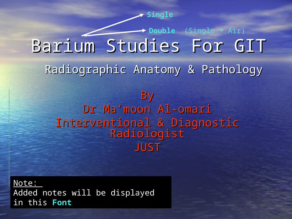
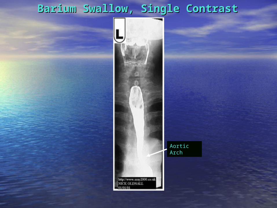
Barium Swallow, Single ContrastBarium Swallow, Single Contrast

Barium Swallow, Single ContrastBarium Swallow, Single Contrast
Main Indication:
Dyshagia

Identation of A.A
Single Contrast
Indentation of L.main
bronchus
Double Contrast
Barium Swallow, Single ContrastBarium Swallow, Single Contrast
Double Contrast
Heart
L.V.L.V.
L.A.L.A.

Barium Swallow, Double ContrastBarium Swallow, Double Contrast
Indentation of L.main
bronchus
Single Contrast
Double Contrast
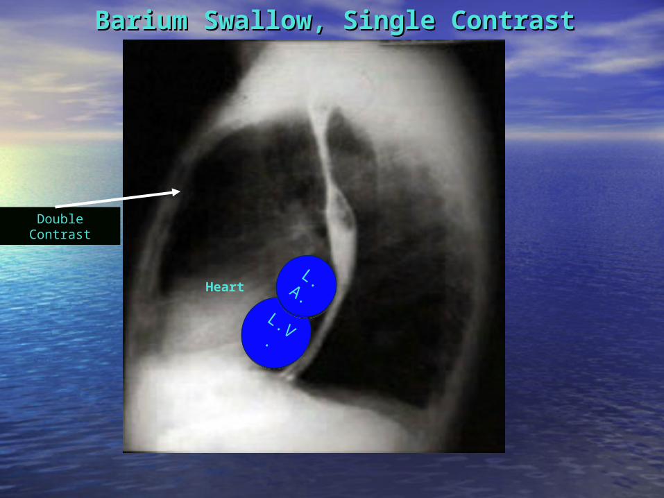
Barium Swallow, Single ContrastBarium Swallow, Single Contrast
Ampulla Normal Varient
Fundus
Body
Barium Swallow, Single ContrastBarium Swallow, Single Contrast
Aortic Arch

Barium Swallow, Double ContrastBarium Swallow, Double Contrast
Narrowing:Could be peristalsis
So other shot is advised

Angular NotchIncisura
Angularis
Barium Meal, Double ContrastBarium Meal, Double Contrast(Supine Position)(Supine Position)
Body
Antrum
Supine Position:Note Barium
Distribution in the Fundus due to gravity

Barium Meal + Follow-ThroughBarium Meal + Follow-Through(Erect Position)(Erect Position)
Barium Meal
Barium Follow-Through
Duodenal Cap
Pyloric Canal
2nd Part of Duodenum
3rd Part of Duodenum
Body
Antrum
DJJ:Normal Position= Left
side
Angular NotchIncisura Angularis
Jejunum:Plica Circularis on the
outer border
Ileum
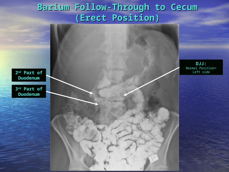
Barium Follow-Through to CecumBarium Follow-Through to Cecum(Erect Position)(Erect Position)
2nd Part of Duodenum
3rd Part of Duodenum
DJJ:Normal Position= Left
side

Small Bowel EnemaSmall Bowel Enema
A Modified Follow-Through which is called Small Bowel Enema note that the bowel is more distended here
This procedure involves inserting a thin tube through the mouth, esophagus and past the stomach to inject barium, methylcellulose and water into the small bowel. This allows for better visualization of the small bowel
than can be seen during a small bowel follow-through
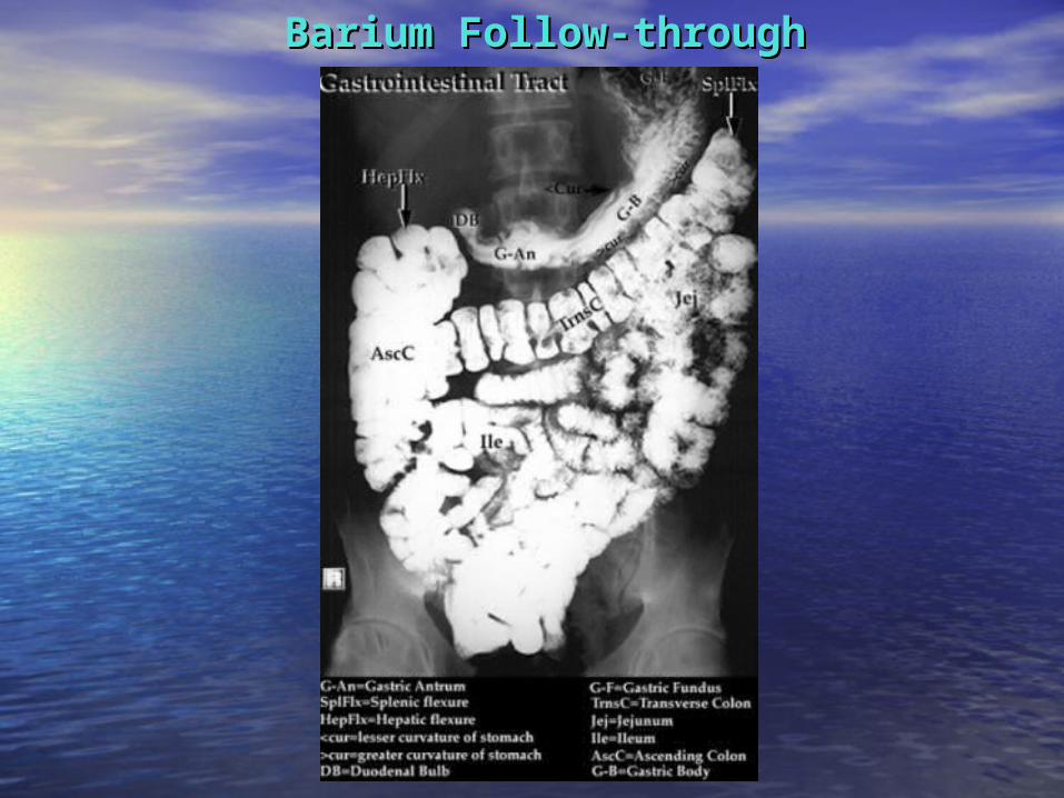
Barium Follow-throughBarium Follow-through

Barium Enema, Single ContrastBarium Enema, Single Contrast
Cecum
Terminal Ileum
Transverse Colon
Descending Colon
Sigmoid
Ascending Colon

Barium Enema, Double ContrastBarium Enema, Double Contrast(Right Lateral Decubitus)(Right Lateral Decubitus)
Hepatic Flexure
Note the effect of gravity
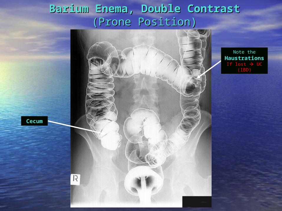
Barium Enema, Double ContrastBarium Enema, Double Contrast(Prone Position)(Prone Position)
Note the HaustrationsIf lost UC (IBD)
Cecum
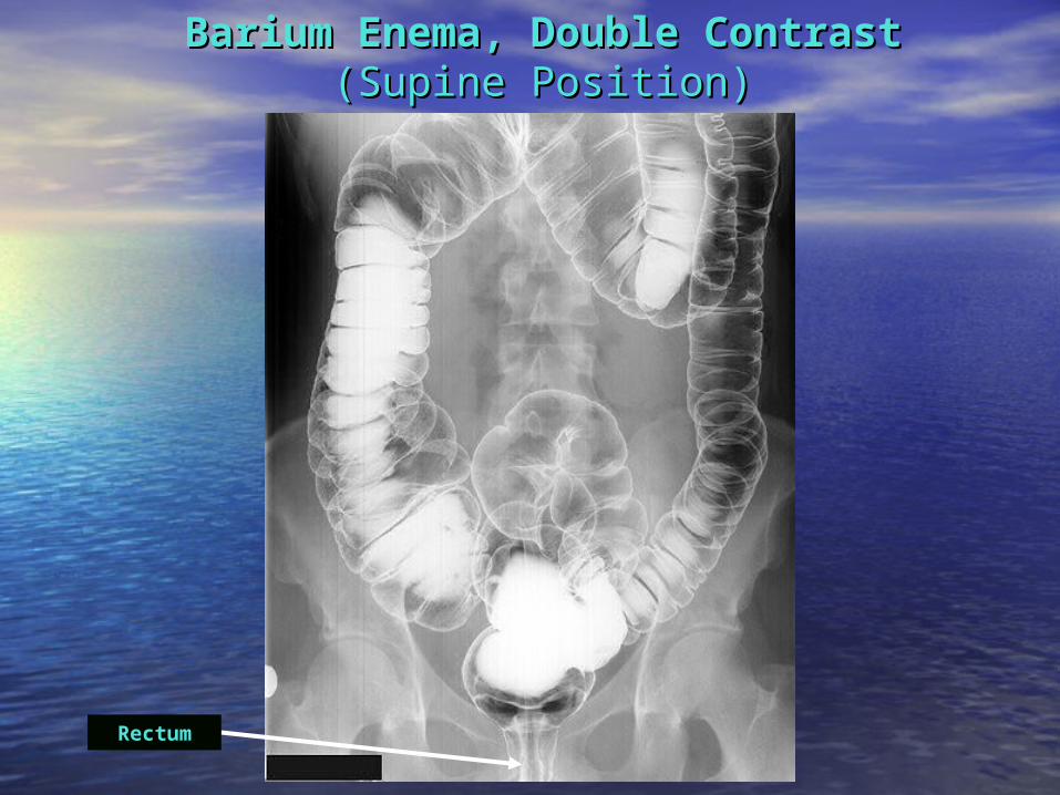
Barium Enema, Double ContrastBarium Enema, Double Contrast(Supine Position)(Supine Position)
Rectum
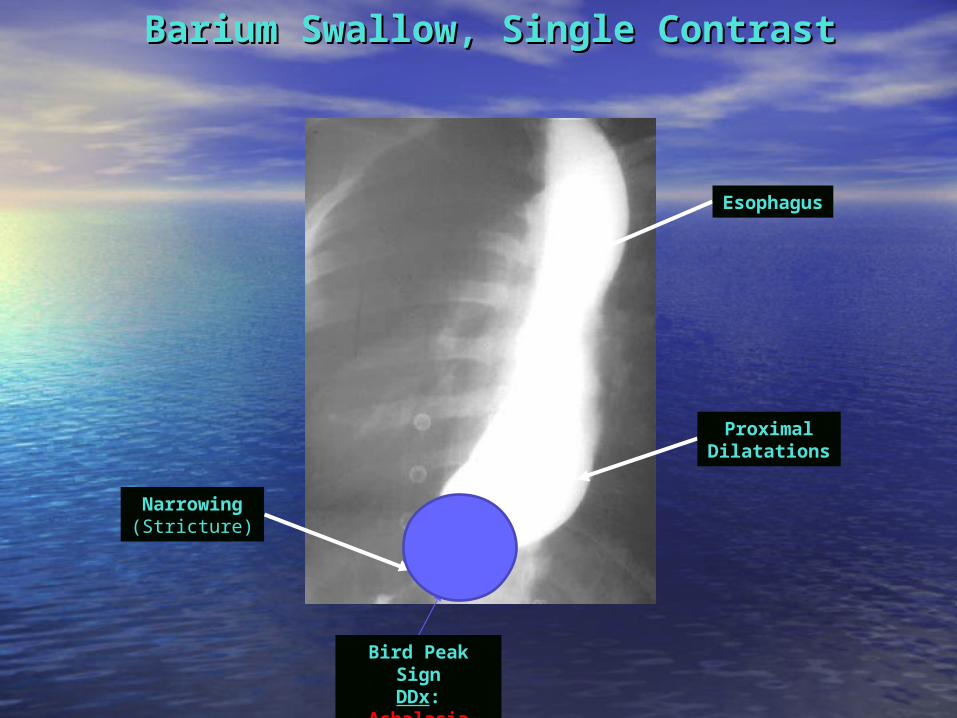
Esophagus
Barium Swallow, Single ContrastBarium Swallow, Single Contrast
ProximalDilatations
Narrowing (Stricture)
Bird Peak SignDDx:
Achalasia
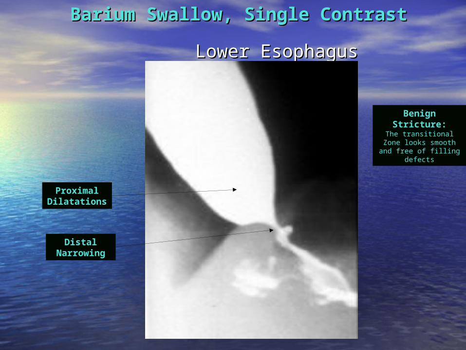
Lower EsophagusLower Esophagus
Barium Swallow, Single ContrastBarium Swallow, Single Contrast
ProximalDilatations
Distal Narrowing
Benign Stricture:
The transitional Zone looks smooth and free
of filling defects
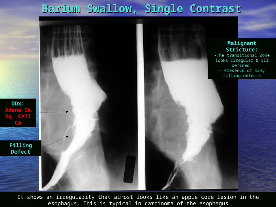
Barium Swallow, Single ContrastBarium Swallow, Single Contrast
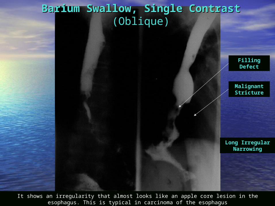
Malignant Stricture:-The transitional Zone looks
Irregular & ill defined - Presence of many filling
defects
DDx:Adeno CASq. Cell
CA
Filling Defect
It shows an irregularity that almost looks like an apple core lesion in the esophagus. This is typical in carcinoma of the esophagus
It shows an irregularity that almost looks like an apple core lesion in the esophagus. This is typical in carcinoma of the esophagus
Filling Defect
Malignant Stricture
Long Irregular Narrowing
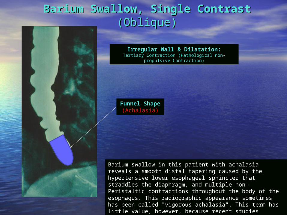
Barium Swallow, Single ContrastBarium Swallow, Single Contrast(Oblique)(Oblique)
Barium Swallow, Single ContrastBarium Swallow, Single Contrast(Oblique)(Oblique)
Barium swallow in this patient with achalasia reveals a smooth distal tapering caused by the hypertensive lower esophageal sphincter that straddles the diaphragm, and multiple non-Peristaltic contractions throughout the body of the esophagus. This radiographic appearance sometimes has been called "vigorous achalasia". This term has little value, however, because recent studies suggest that patients with so-called vigorous achalasia cannot be distinguished clinically from non-vigorous achalasia.
Irregular Wall & Dilatation:Tertiary Contraction (Pathological non-propulsive
Contraction)
Funnel Shape
(Achalasia)

Barium Swallow, Single ContrastBarium Swallow, Single Contrast(Oblique)(Oblique)Well Defined
Contrast Filled left cervical level
sac
Pharyngeal Pouch(Zenker's
Diverticulum):occurs in an area of anatomic weakness known as Killian's
dehiscence

Varices Barium swallow examination: AP view: Numerous rounded and elongated smooth-contoured filling defects are present in the inferior two thirds of the esophagus. The contour of the esophagus is irregular and spiculated.
Barium Swallow, Single ContrastBarium Swallow, Single Contrast
Irregular Multiple Filling
Defects
Differential Diagnosis Multiple Esophageal Filling Defects:1.Fungal Infx2.Polyps3.Esophageal Varices (irregular)4.Food Particles
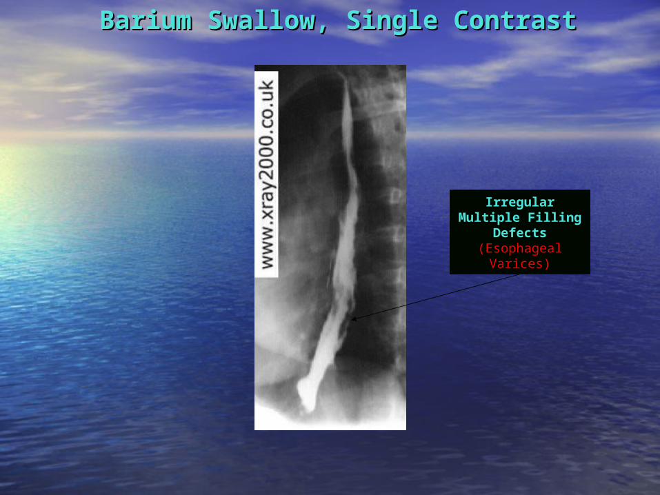
Barium Swallow, Single ContrastBarium Swallow, Single Contrast
Irregular Multiple Filling
Defects(Esophageal
Varices)

Barium Meal, Double ContrastBarium Meal, Double Contrast
Contrast Filled Speculated
Lesion(Gastric Ulcer)

Barium Meal, Double ContrastBarium Meal, Double Contrast
Rugae
Contrast Filled Outpouching at
the Greater Curviture
(Malignant Gastric Ulcer)
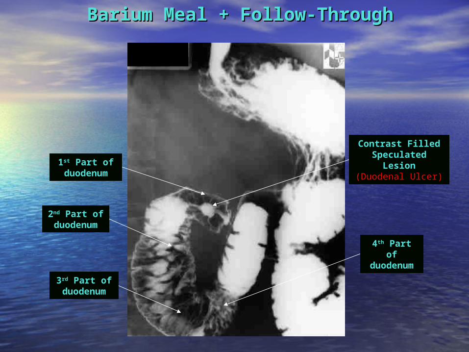
Barium Meal + Follow-ThroughBarium Meal + Follow-Through
Contrast Filled Speculated
Lesion(Duodenal Ulcer)
4th Part of duodenu
m
1st Part of duodenum
2nd Part of duodenum
3rd Part of duodenum

Stomach
Barium Meal, Double ContrastBarium Meal, Double Contrast
Ulcer
Speculated Mass
Pylorus

Barium Meal, Double ContrastBarium Meal, Double Contrast
Distended Stomach
Single Bubble Sign
DDx: Gastric Output
Obstruction (GOO)(Pyloric Stenosis)
Gas in Descending Colon
(partial obstruction)

Barium Meal, Double ContrastBarium Meal, Double Contrast(Erect Position)(Erect Position)
Shoulder’s Sign
Mushroom’s Sign(or apple core
Sign)String’s
Sign
DDx:Pyloric Stenosis
For further information refer to “Pediatric Abdomen Radiology” Slides (37-46)
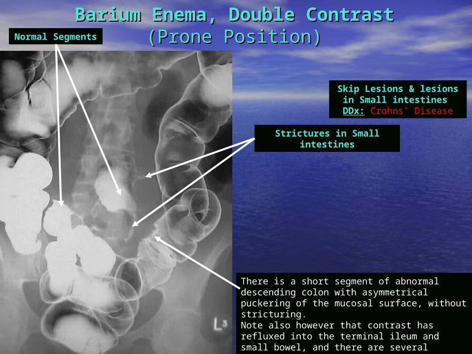
Barium Enema, Double ContrastBarium Enema, Double Contrast(Prone Position)(Prone Position)
There is a short segment of abnormal descending colon with asymmetrical puckering of the mucosal surface, without stricturing.Note also however that contrast has refluxed into the terminal ileum and small bowel, and there are several strictures present within it. One of these lies adjacent to the large bowel abnormality.
Strictures in Small intestines
Normal Segments
Skip Lesions & lesions in Small intestines DDx: Crohns’ Disease
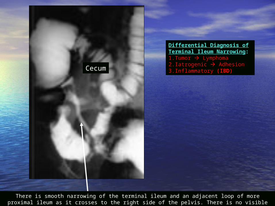
Cecum
Differential Diagnosis of Terminal Ileum Narrowing:1.Tumor Lymphoma2.Iatrogenic Adhesion3.Inflammatory (IBD)
There is smooth narrowing of the terminal ileum and an adjacent loop of more proximal ileum as it crosses to the right side of the pelvis. There is no visible mucosal fold thickening or ulceration.

Cecum
There is abnormal wall thickening, luminal narrowing, and cobblestoning involving a long segment of the distal ileum including the terminal ileum.
Multiple Filling DefectsCobble Stone appearance
DDx: Crohn’s Disease
Barium EnemaBarium Enema
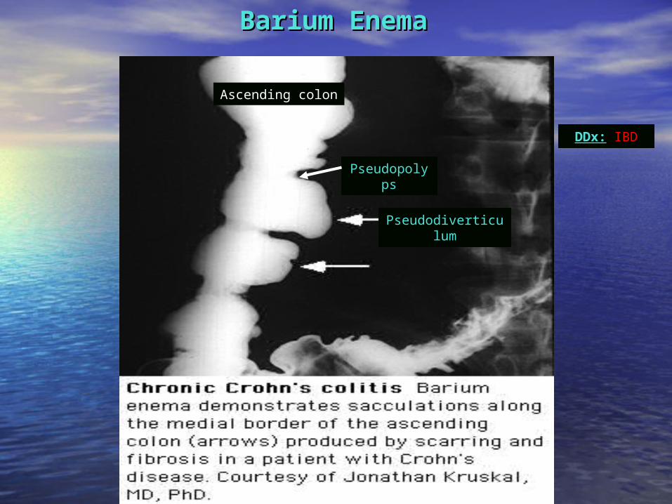
Ascending colon
Pseudopolyps
Barium EnemaBarium Enema
Pseudodiverticulum
DDx: IBD
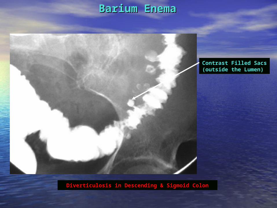
Barium EnemaBarium Enema
Contrast Filled Sacs (outside the Lumen)
Diverticulosis in Descending & Sigmoid Colon
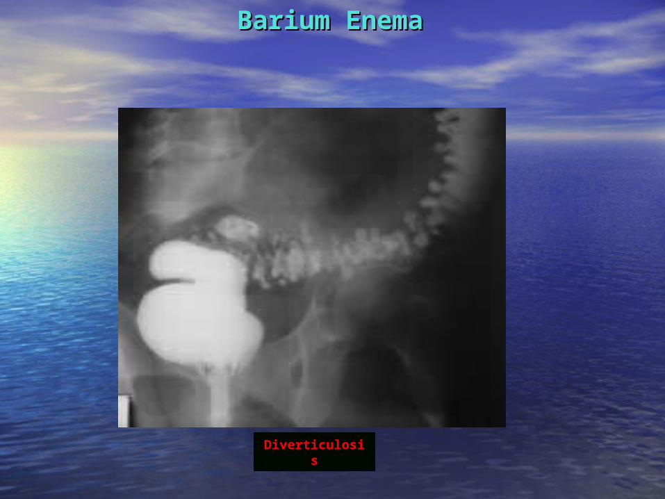
Barium EnemaBarium Enema
Diverticulosis
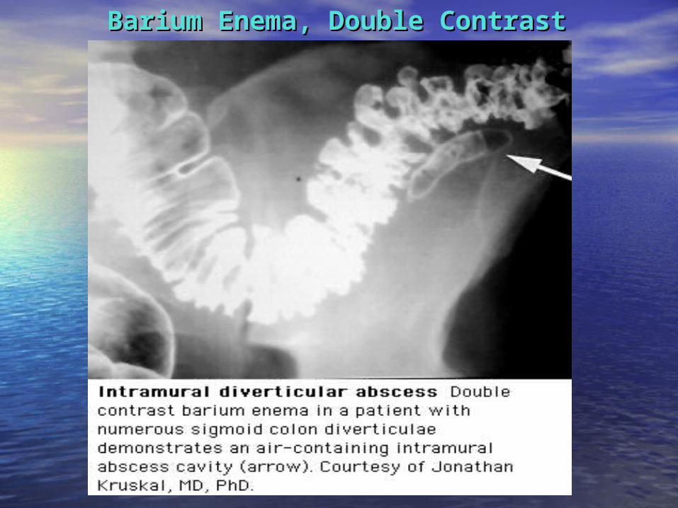
Barium Enema, Double ContrastBarium Enema, Double Contrast
Barium Enema, Double ContrastBarium Enema, Double Contrast
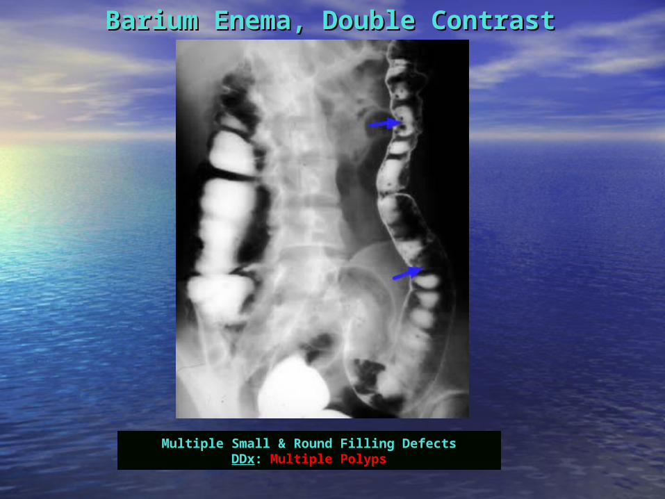
Multiple Small & Round Filling DefectsDDx: Multiple Polyps

Barium Enema, Double ContrastBarium Enema, Double Contrast
Loss of HaustrationsLEAD PIPE SIGN
Terminal Ilium
Cecum
DDx: Ulcerative Colitis
(Pancolitis)
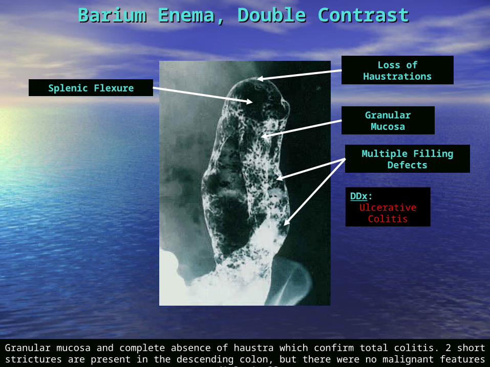
Barium Enema, Double ContrastBarium Enema, Double Contrast
Granular mucosa and complete absence of haustra which confirm total colitis. 2 short strictures are present in the descending colon, but there were no malignant features radiologically
Loss of Haustrations
Multiple Filling Defects
Splenic Flexure
Granular Mucosa
DDx: Ulcerative
Colitis
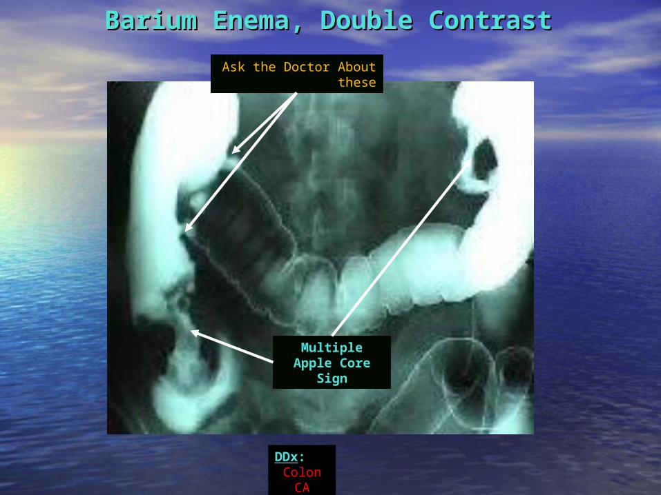
Barium Enema, Double ContrastBarium Enema, Double Contrast
Multiple Apple Core
Sign
Ask the Doctor About these
DDx: Colon
CA

Barium EnemaBarium Enema
DDx: Sigmoid Colon
CA
Apple Core Sign
Stricture
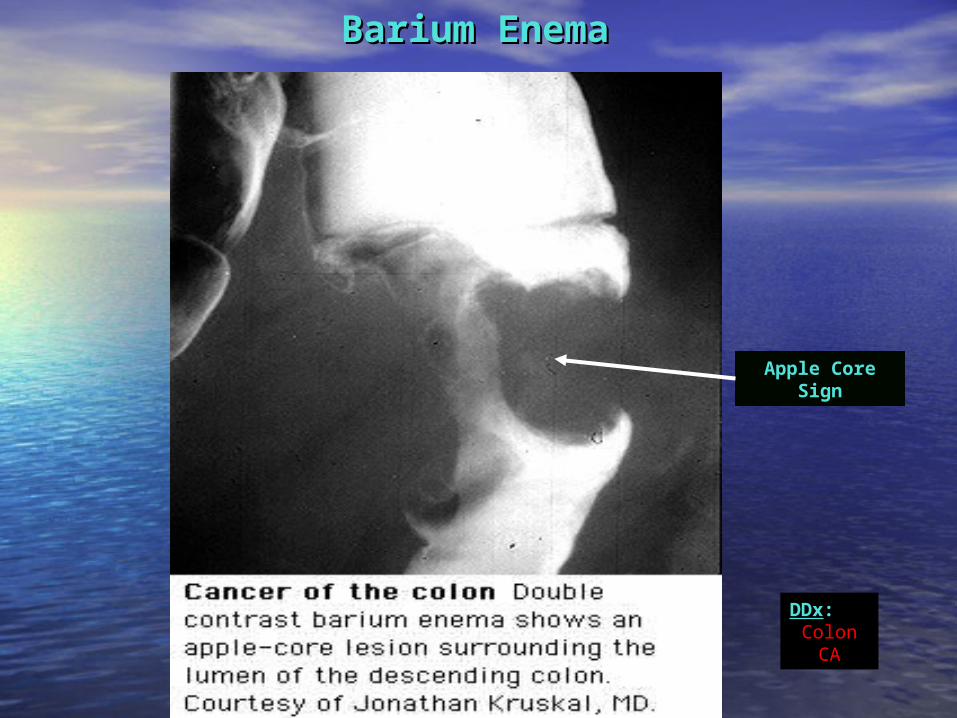
Barium EnemaBarium Enema
Apple Core Sign
DDx: Colon
CA

Barium EnemaBarium Enema
A Sigmoid Stricture is always considered
malignant until proven otherwise

Barium Enema, Double ContrastBarium Enema, Double Contrast
A huge right indirect hernia in the scrotum

Barium Enema, Double ContrastBarium Enema, Double Contrast
A huge mass that has displaced the
intestines(Spleen)

Barium EnemaBarium EnemaMeconiu
mFilling defects & dilated Descending & Sigmoid
ColonTransition
Zone
According to the Transition Zone:Rectum Ultra ShortRectosigmoid ShortTransverse Colon LongBeginning of the Colon Total (microcolon)
DDx: Hirschsprung disease (HD) which is more definitively diagnosed by means of contrast enema examination, which can show the presence of a transition zone, irregular contractions,
mucosal irregularity, and delayed evacuation of contrast material, among other findings
Although the hallmark of the diagnosis is the presence of transition zone but it’s absence exclude the disease
is the term applied to the region in which a marked change in caliber occurs, with the dilated, normal colon above and the
narrowed, aganglionic colon below
For more info visit:http://emedicine.medscape.com/article/409150-
imaging
تتم بنعمته الذي لله الحمدالصالحات
ولكم لي التوفيق الله أسال
Please if you find any mistakes or extra imp notes contact:
[email protected] they will update it