Embed Size (px)
Citation preview
Introduction to 12 Lead ECGs
McHenry Western Lake County EMS System
Objectives
Why 12 Lead ECGs?Critical Concepts in ACSMonitoring vs Diagnostic ECGsAcquisition & Transmission
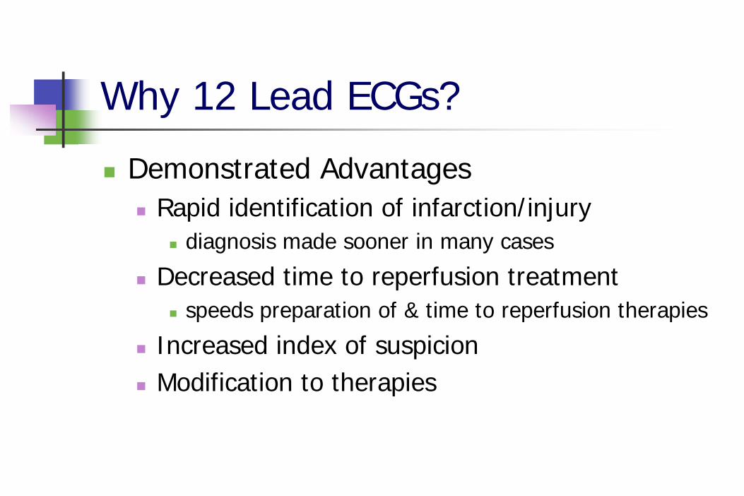
Why 12 Lead ECGs?
Demonstrated AdvantagesRapid identification of infarction/injury
diagnosis made sooner in many cases
Decreased time to reperfusion treatmentspeeds preparation of & time to reperfusion therapies
Increased index of suspicionModification to therapies
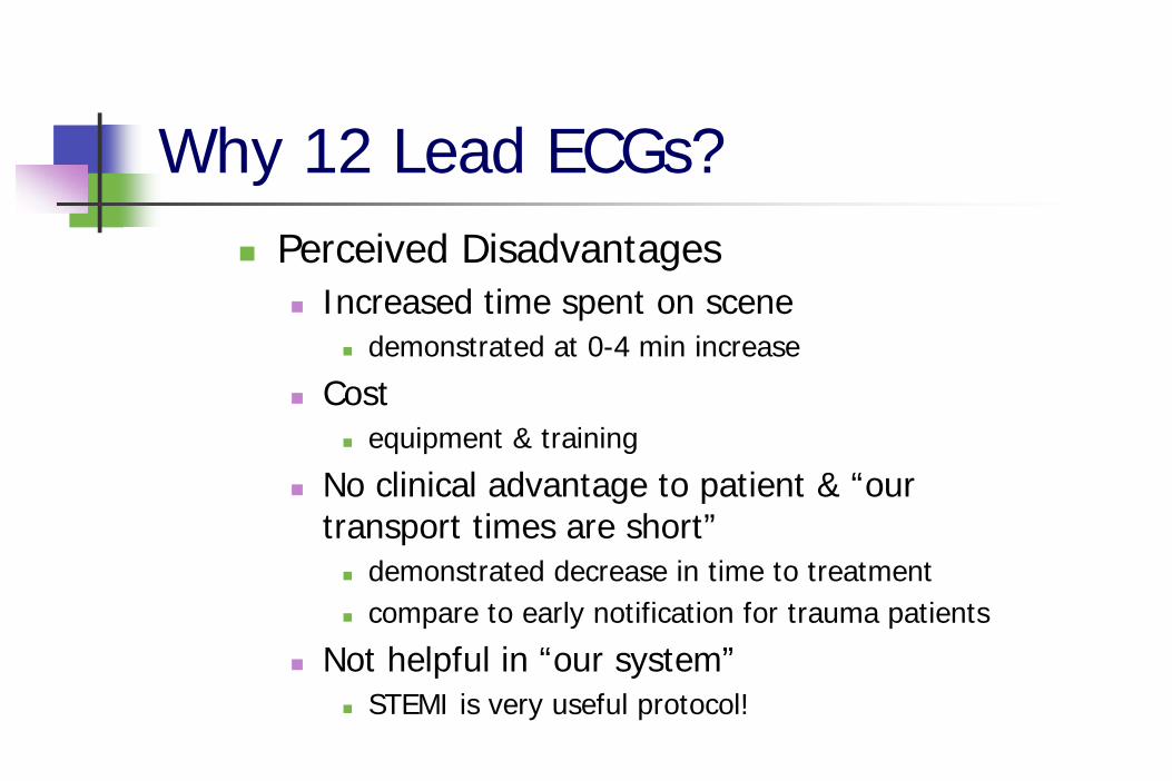
Why 12 Lead ECGs?Perceived Disadvantages
Increased time spent on scenedemonstrated at 0-4 min increase
Costequipment & training
No clinical advantage to patient & “our transport times are short”
demonstrated decrease in time to treatmentcompare to early notification for trauma patients
Not helpful in “our system”STEMI is very useful protocol!
STEMI
STEMI stands for:ST elevated myocardial infarction
The object is to decrease the time of MI to reperfusion by identifying the MI, and getting the patient the reperfusion as fast as possible.
Why 12 Lead ECGs?
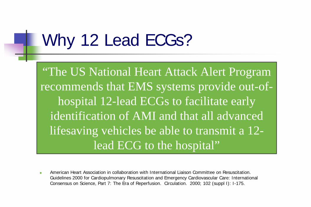
American Heart Association in collaboration with International Liaison Committee on Resuscitation. Guidelines 2000 for Cardiopulmonary Resuscitation and Emergency Cardiovascular Care: International Consensus on Science, Part 7: The Era of Reperfusion. Circulation. 2000; 102 (suppl I): I-175.
“The US National Heart Attack Alert Program recommends that EMS systems provide out-of-
hospital 12-lead ECGs to facilitate early identification of AMI and that all advanced lifesaving vehicles be able to transmit a 12-
lead ECG to the hospital”
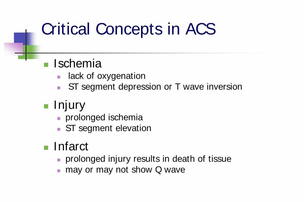
Critical Concepts in ACS
Ischemialack of oxygenation ST segment depression or T wave inversion
Injury prolonged ischemia ST segment elevation
Infarct prolonged injury results in death of tissuemay or may not show Q wave
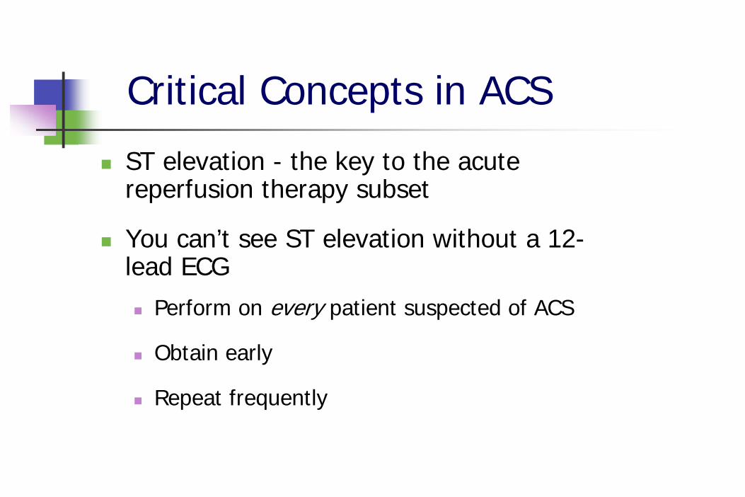
Critical Concepts in ACS
ST elevation - the key to the acute reperfusion therapy subset
You can’t see ST elevation without a 12-lead ECG
Perform on every patient suspected of ACS
Obtain early
Repeat frequently
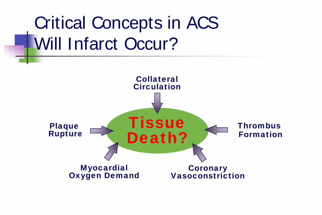
Critical Concepts in ACS Will Infarct Occur?
Tissue Death?
Plaque Rupture
ThrombusFormation
CoronaryVasoconstriction
CollateralCirculation
MyocardialOxygen Demand
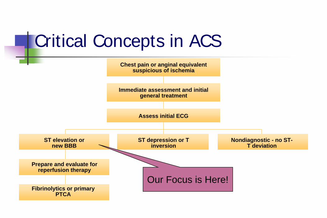
Critical Concepts in ACSChest pain or anginal equivalent
suspicious of ischemia
Immediate assessment and initial general treatment
Assess initial ECG
ST elevation or new BBB
ST depression or T inversion
Nondiagnostic - no ST- T deviation
Prepare and evaluate for reperfusion therapy
Fibrinolytics or primary PTCA
Our Focus is Here!
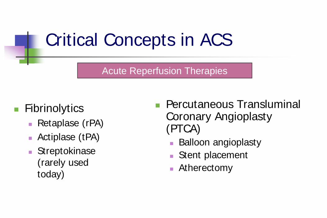
Critical Concepts in ACS
FibrinolyticsRetaplase (rPA)Actiplase (tPA)Streptokinase (rarely used today)
Percutaneous Transluminal Coronary Angioplasty (PTCA)
Balloon angioplastyStent placementAtherectomy
Acute Reperfusion Therapies
Critical Concepts in ACS
Pain is InjuryPain-Free is the GoalTime is MuscleDoor to Reperfusion Therapy Time is the issue
Monitoring vs Diagnostic ECGs
Extra wires3 wires vs 10 wires
Are there other differences?Discuss your departments situation with monitors
Monitoring vs Diagnostic ECGs
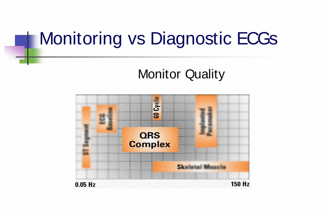
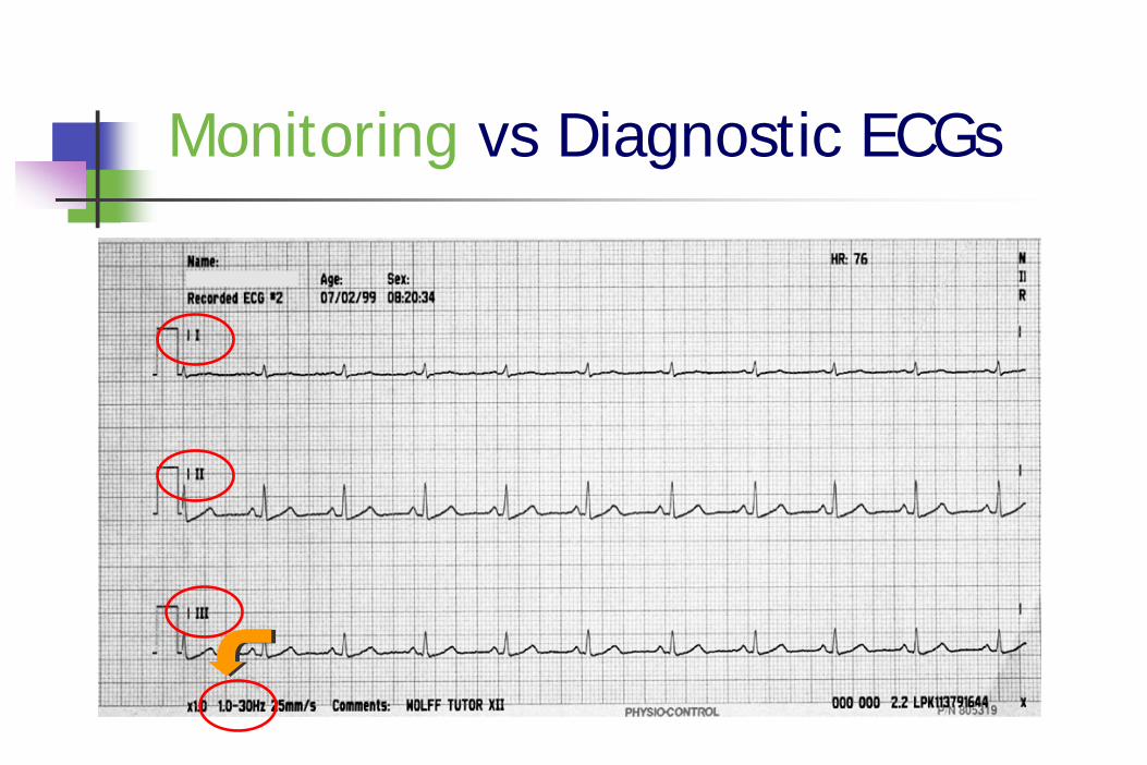
Monitoring Quality ECGDesigned to provide information needed to determine rate and underlying rhythmDesigned to “filter out” artifact
Reduces the amount and degree of electrical activity seen by the ECG monitor
Monitoring vs Diagnostic ECGs
Monitor Quality
Monitoring vs Diagnostic ECGs
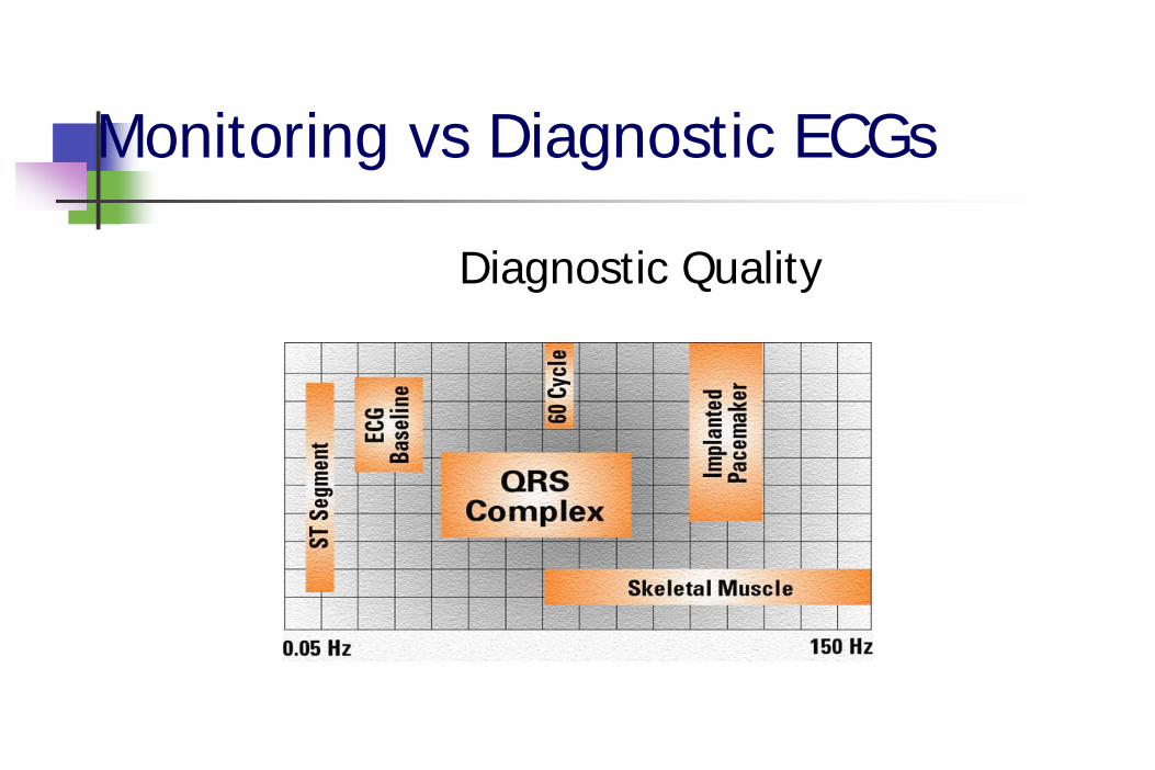
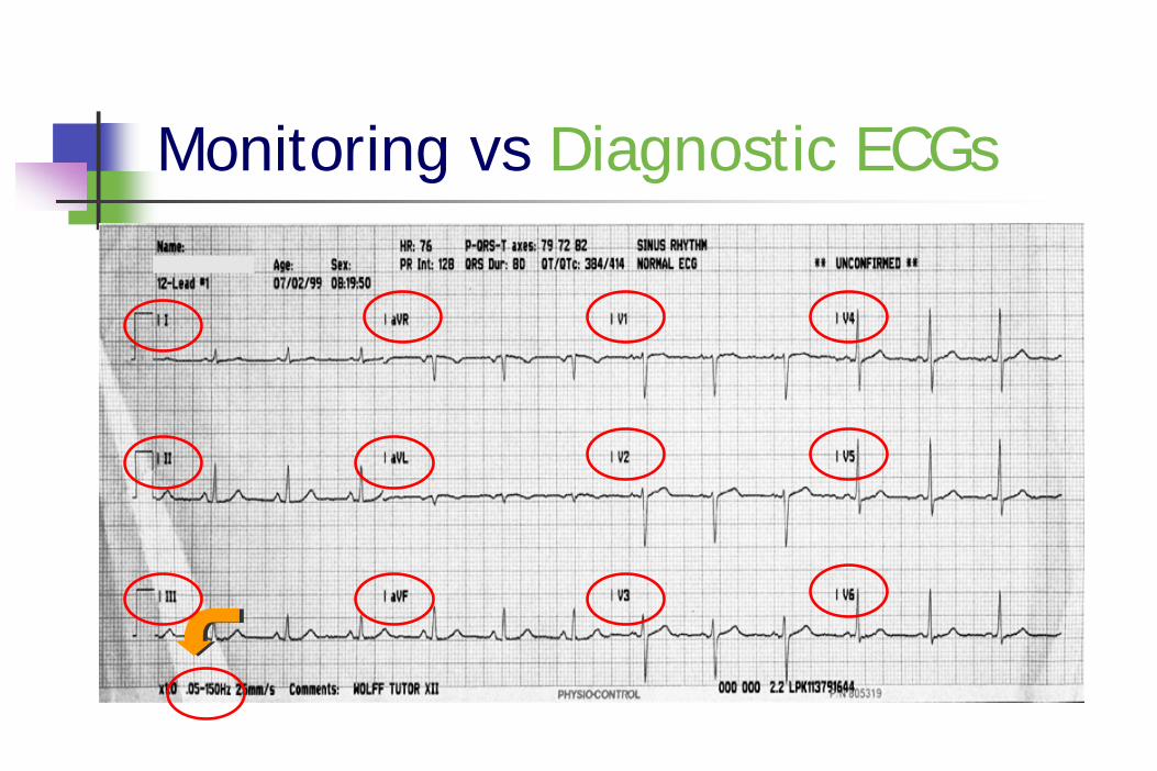
Diagnostic Quality ECGDesigned to accurately reproduce QRS, ST and T waveformsDesigned to look more broadly at the cardiac electrical activityUnfortunately, may result in greater artifact being visible
Monitoring vs Diagnostic ECGs
Diagnostic Quality
Monitoring vs Diagnostic ECGs
Frequency ResponseTerm used to describe the breadth of the electrical spectrum viewed by the ECG monitorDiagnostic quality is usually 0.05 Hz to 150 HzMonitor quality is usually 0.5 Hz to 20-50 HzUsually printed on the ECG recording strip
Monitoring vs Diagnostic ECGs
Monitoring vs Diagnostic ECGs
Acquisition & Transmission
ECG quality begins with skin preparation and electrodes
Hair removalSkin preparationAge & Quality of Electrodes & CablesElectrode Placement
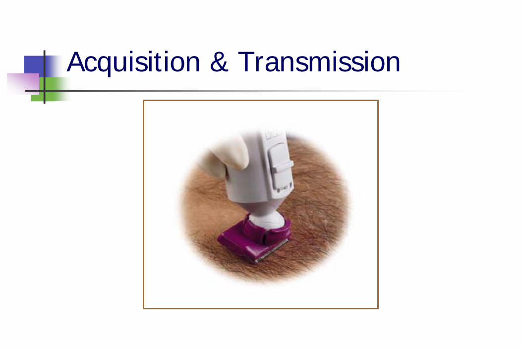
Acquisition & Transmission
Hair RemovalClipper over razor
Lessens risk of cutsQuickerDisposable blade clippers available
Most EMS systems use razors
Acquisition & Transmission
Acquisition & Transmission
Skin PreparationHelps obtain a strong signalWhen measured from skin, heart’s electrical signal about 0.0001 - 0.003 voltsSkin oils reduce adhesion of electrode and hinder penetration of electrode gelDead, dried skin cells do not conduct well
Acquisition & Transmission
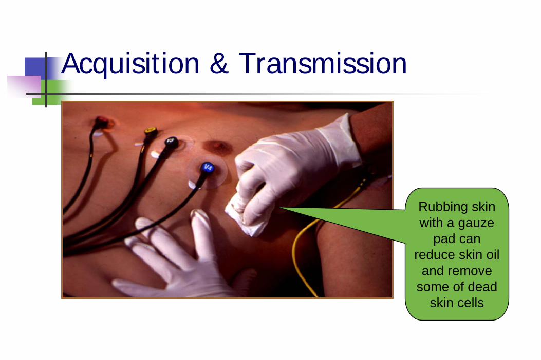
Rubbing skin with a gauze
pad can reduce skin oil
and remove some of dead
skin cells
Acquisition & Transmission
Other causes of artifactPatient movement
Cable movement
Vehicle movement
Electromagnetic Interference (EMI)
Acquisition & Transmission
Patient MovementMake patient as comfortable as possible
Supine preferred (30 degree angle)
Look for subtle movementtoe tapping, shivering
Look for muscle tensionhand grasping rail, head raised to “watch” causes muscle tremors
Acquisition & Transmission
Cable MovementEnough “slack” in cables to avoid tugging on the electrodesMany cables have clip that can attach to patient’s clothes or bed sheet
Acquisition & Transmission
Vehicle MovementAcquisition in a moving vehicle is NOT recommended
May or may not be successful
TipsPull ambulance over for 10-20 seconds during acquisitionAcquire ECG while stopped at traffic light
Acquisition & Transmission
Electromagnetic Interference (EMI)Can interfere with electronic equipment60 cycle interference is a type of EMILook for nearby cell phones, radios or electrical devicesNo contact between cables & power cordsTurn off or move away from AC devicesUse shielded cables; inspect for cracks
Acquisition & Transmission
Things to look forLittle or no artifactSteady baseline
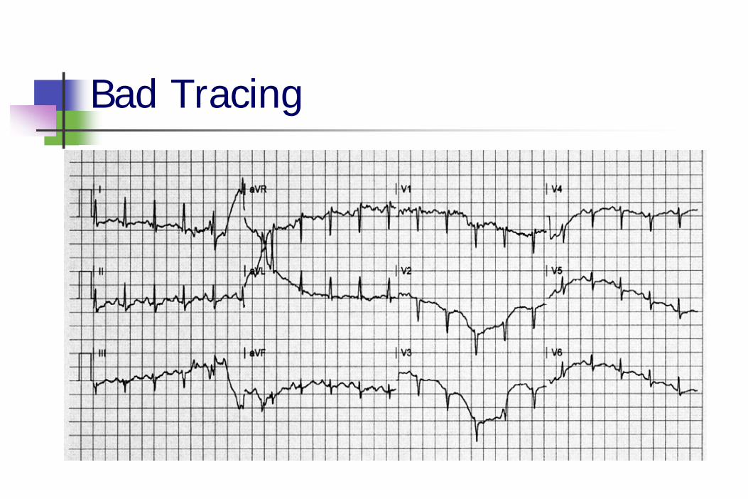
Bad Tracing
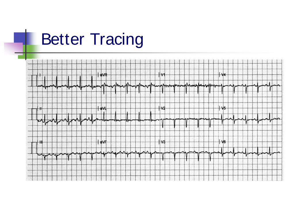
Better Tracing
Acquisition & Transmission
ECG Accuracy depends uponLead placementFrequency responseCalibrationPaper speed
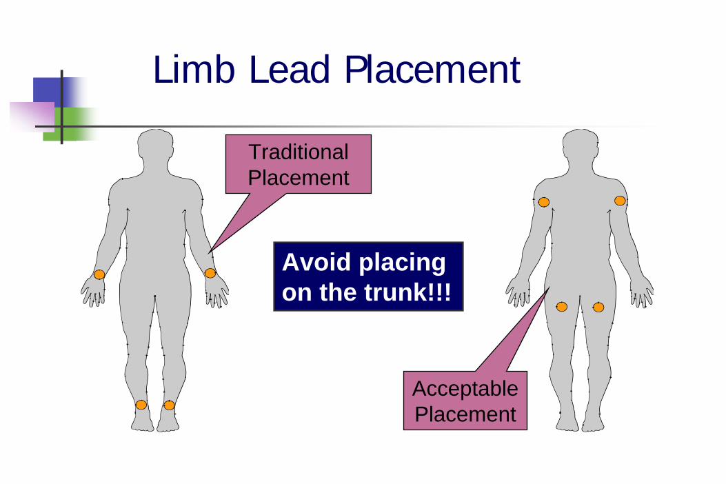
Limb Lead Placement
Traditional Placement
Acceptable Placement
Avoid placing on the trunk!!!
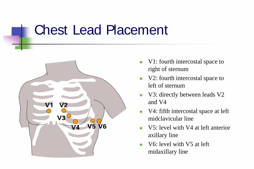
Chest Lead Placement
V1: fourth intercostal space to right of sternumV2: fourth intercostal space to left of sternumV3: directly between leads V2 and V4V4: fifth intercostal space at left midclavicular lineV5: level with V4 at left anterior axillary lineV6: level with V5 at left midaxillary line
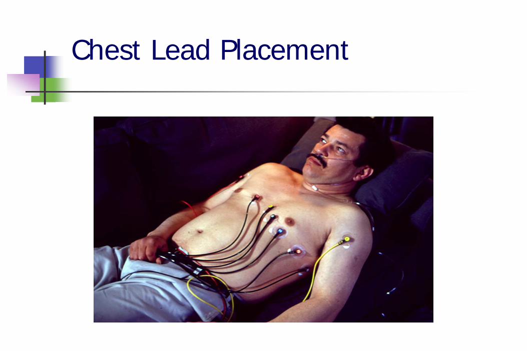
Chest Lead Placement
ECG Accuracy
Look for:Negative aVR
if aVR upright, look for reversed leads
One complete cardiac cycle in each lead Diagnostic frequency responseProper calibrationAppropriate speed
ECG Accuracy
Frequency ResponseDisplay screen is non-diagnosticUse the printed ECG for ST segment analysis
ECG Accuracy
CalibrationVoltage measured verticallyEach 1 mm box = 0.1 mV1 mV = 10 mm
calibration standard
Confirm calibrationcalibration impulse should be 10 mm (2 big boxes tall)stated calibration should be “x 1.0”
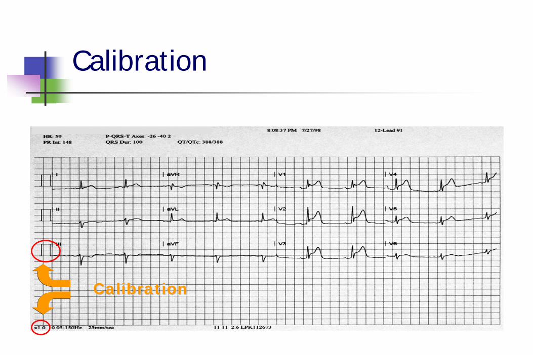
Calibration
CalibrationCalibration
ECG Accuracy
Paper SpeedStandard is 25 mm/sec
Faster paper speed means the rhythm will appear slower and the QRS widerSlower paper speed means the rhythm will appear faster and the QRS narrower
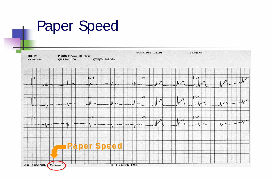
Paper Speed
Paper SpeedPaper Speed
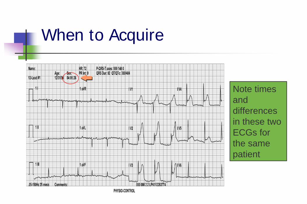
When to Acquire
Note times and differences in these two ECGs for the same patient
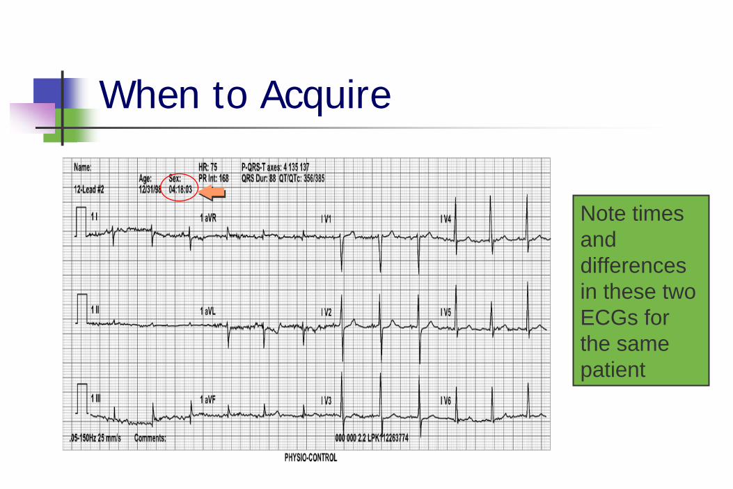
When to Acquire
Note times and differences in these two ECGs for the same patient
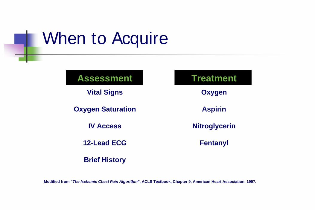
When to Acquire
AssessmentVital Signs
Oxygen Saturation
IV Access
12-Lead ECG
Brief History
TreatmentOxygen
Aspirin
Nitroglycerin
Fentanyl
Modified from “The Ischemic Chest Pain Algorithm”, ACLS Textbook, Chapter 9, American Heart Association, 1997.
Exposing the Chest
Immediately upon suspecting ACS...Remove all clothing above the waist
Or, open shirt/blouse
Replace with gown (if possible)Allows for complete examMinimizes wire entanglementEnhances quick defib if VF occurs
Transmission
Transmit information as soon as possibleCan use patient’s land-lineMany EMS systems use cell phone enroute
Coordinate with EDCorrelate ECG with a specific patientEarly notification of AMI is key!!!Remember STEMI!!!
Special Thanks!
To Acute Coronary Syndrome Consultants, Inc. Tim Phalen, Gary Denton and Assoc.
and Temple College for the use of their materials in this presentation